Phycoerythrin-specific bilin lyase–isomerase controls
blue-green chromatic acclimation in
marine Synechococcus
Animesh Shuklaa, Avijit Biswasb, Nicolas Blotc,d,e, Frédéric Partenskyc,d, Jonathan A. Kartyf, Loubna A. Hammadf, Laurence Garczarekc,d, Andrian Gutua,g, Wendy M. Schluchterb, and David M. Kehoea,h,1
aDepartment of Biology, Indiana University, Bloomington, IN 47405;bDepartment of Biological Sciences, University of New Orleans, New Orleans, LA 70148; cUPMC-Université Paris 06, Station Biologique, 29680 Roscoff, France;dCentre National de la Recherche Scientifique, Unité Mixte de Recherche 7144 Adaptation et Diversité en Milieu Marin, Groupe Plancton Océanique, 29680 Roscoff, France;eClermont Université, Université Blaise Pascal, Unité Mixte de Recherche Centre National de la Recherche Scientifique 6023, Laboratoire Microorganismes: Génome et Environnement, 63000 Clermont-Ferrand, France; fMETACyt Biochemical Analysis Center, Department of Chemistry, Indiana University, Bloomington, IN 47405;gDepartment of Molecular and Cellular Biology, Harvard University, Cambridge, MA 02138; andhIndiana Molecular Biology Institute, Indiana University, Bloomington, IN 47405
Edited by Alexander Namiot Glazer, University of California, Berkeley, CA, and approved October 4, 2012 (received for review July 10, 2012)
The marine cyanobacterium Synechococcus is the second most
abundant phytoplanktonic organism in the world’s oceans. The
ubiquity of this genus is in large part due to its use of a diverse set of photosynthetic light-harvesting pigments called
phycobilipro-teins, which allow it to efficiently exploit a wide range of light
colors. Here we uncover a pivotal molecular mechanism
underpin-ning a widespread response among marine Synechococcus cells
known as“type IV chromatic acclimation” (CA4). During this process,
the pigmentation of the two main phycobiliproteins of this organ-ism, phycoerythrins I and II, is reversibly modified to match changes in the ambient light color so as to maximize photon capture for photosynthesis. CA4 involves the replacement of three molecules of the green light-absorbing chromophore phycoerythrobilin with an equivalent number of the blue light-absorbing chromophore phy-courobilin when cells are shifted from green to blue light, and the
reverse after a shift from blue to green light. We have identified and
characterized MpeZ, an enzyme critical for CA4 in marine
Synecho-coccus. MpeZ attaches phycoerythrobilin to cysteine-83 of the
α-subunit of phycoerythrin II and isomerizes it to phycourobilin.mpeZ
RNA is six times more abundant in blue light, suggesting that its
proper regulation is critical for CA4. Furthermore,mpeZ mutants fail
to normally acclimate in blue light. Thesefindings provide insights
into the molecular mechanisms controlling an ecologically important photosynthetic process and identify a unique class of phycoerythrin lyase/isomerases, which will further expand the already widespread use of phycoerythrin in biotechnology and cell biology applications.
light regulation
|
marine cyanobacteria|
phycobilisomes|
fluorescence|
liquid chromatography-mass spectrometryC
yanobacteria within the Synechococcus spp. are found in marine environments from the equator to the polar circles, and members of this genus contribute significantly to the total phytoplankton biomass and productivity of the oceans (1–3). Their ubiquity is due in part to their wide pigment diversity (4), which arises mainly from differences in the composition of their light-harvesting antennae or phycobilisomes (PBS). PBS consist of a core and six or eight rods radiating from the core that contain the phycobiliprotein phycocyanin (PC) and one or two types of phycoerythrins (PEs), PEI and PEII (5). All phycobiliproteins are α/β heterodimers that are assembled into hexamers by linkers. PEs may bind two different types of chromophores, green light (GL)-absorbing phycoerythrobilin (PEB) and blue light (BL)-ab-sorbing phycourobilin (PUB). These chromophores are ligated to PE by PEB lyases (6, 7) or PEB–lyase–isomerases, which both attach the chromophore and isomerize it to PUB (8). No PE-specific PEB–lyase–isomerase has been described to date. PUB predominates in Synechococcus found in nutrient-poor open ocean waters, vast areas where blue light penetrates deeper than any other color (9).Marine Synechococcus are divided into three major pigment types, with type 1 PBS rods containing only PC, type 2 containing PC and PEI, and type 3 containing PC, PEI, and PEII (4). Type 3 can be further split into four subtypes (3A–D) on the basis of the ratio of the PUB and PEB chromophores bound to PEs. For all pigment types and subtypes, the size and number of PBS may vary with irradiance (10), but only pigment subtype 3d is able to vary its pigmentation in response to changes in ambient light color through a process called type IV chromatic acclimation (hereafter called CA4) (4, 11, 12).
Other CA types, such as CA2 and CA3, have been studied in freshwater cyanobacteria (13, 14). Like CA4, these processes are photoreversible, but they involve very different protein and bilin changes. For example, CA3 inFremyella diplosiphon, which occurs when cells are shifted between red light and GL, involves switching between PC and PE and their corresponding chromophores in the PBS rods (14, 15). In contrast, CA4 occurs when marine Syn-echococcus cells are shifted between GL and BL, and during this process there is no change in the phycobiliprotein composition of the PBS rods (11). Instead, CA4 involves changes in the chro-mophores associated with two different cysteines within the α-PEII subunit (12). In GL, PEB is bound to these sites, whereas in BL, PUB is bound. The mechanism(s) controlling these changes is unknown. Here, we use biochemical and molecular genetic ap-proaches to describe MpeZ, an enzyme involved in the ligation and isomerization of a PEII-linked chromophore, and demon-strate its pivotal role in the poorly understood but globally im-portant process of CA4. The discovery of this class of enzymes has the potential to further expand the current broad use of phyco-erythrin in biotechnology and cell biology applications.
Results
Comparative genomics analysis showed that all sequenced marine Synechococcus strains that undergo CA4 possess a specific gene, calledmpeZ (4). In Synechococcus sp. RS9916 (hereafter 9916), mpeZ is downstream of a gene of unknown function and overlaps a gene putatively encoding a truncated form of the photosystem II core protein PsbA (Fig. 1A). RNA blot analysis demonstrated that mpeZ transcript accumulation was CA4 regulated, being six times more abundant in cells grown in BL than in GL (Fig. 1B). Primary
Author contributions: A.S., A.B., N.B., F.P., J.A.K., L.A.H., L.G., A.G., W.M.S., and D.M.K. designed research; A.S., A.B., N.B., J.A.K., L.A.H., A.G., W.M.S., and D.M.K. performed research; A.S., N.B., J.A.K., L.A.H., and D.M.K. contributed new reagents/analytic tools; A.S., A.B., N.B., F.P., J.A.K., L.A.H., L.G., W.M.S., and D.M.K. analyzed data; and A.S., A.B., F.P., J.A.K., L.A.H., L.G., W.M.S., and D.M.K. wrote the paper.
The authors declare no conflict of interest. This article is a PNAS Direct Submission.
1To whom correspondence should be addressed. E-mail: [email protected].
This article contains supporting information online atwww.pnas.org/lookup/suppl/doi:10. 1073/pnas.1211777109/-/DCSupplemental.
and secondary structure analyses of the encoded protein, MpeZ, revealed a large domain belonging to the PBS lyase HEAT-like repeat family (Fig. S1A), suggesting that this protein could be a bilin lyase–isomerase involved in mediating the shift between PEB- and PUB-enriched PBS rods during CA4.
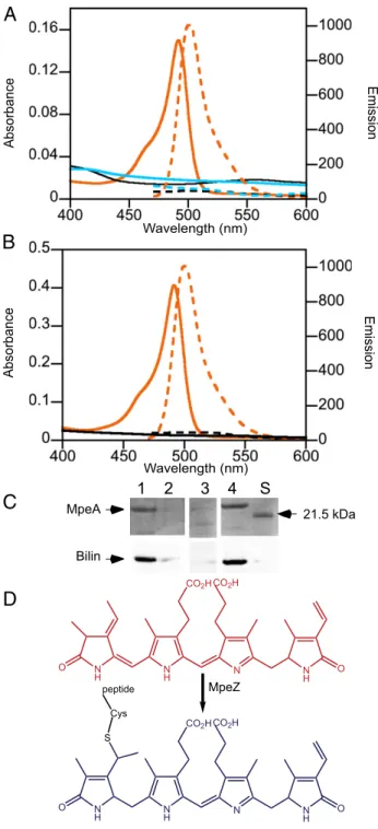
MpeZ was tested for lyase–isomerase activity by producing it in Escherichia coli cells expressing ho1 and pebS, which encode the proteins needed for the synthesis of PEB (16), along with six-his-tidine–tagged (HT) versions of wild-type (WT) and mutant forms of either MpeA (PEII α-subunit) or CpeA (PEI α-subunit). Spectral analyses of purified wild-type HT-MpeA from MpeZ-containing E. coli cells revealed absorbance and fluorescence emission maxima at 495 and 510 nm, respectively (Fig. 2A), which matched the spectral properties of PUB attached to protein (8, 18, 19). HT-MpeA was detectable on protein gels and contained an attached bilin (Fig. 2C). As expected, no absorbance or fluores-cence was detectable from HT-MpeA expressed in cells lacking MpeZ (Fig. 2A and C) because nonchromophorylated recombi-nant PE subunits are generally insoluble inE. coli (6, 20). There are three canonical chromophore-binding cysteines at positions 75, 83, and 140 within MpeA. These cysteines were mutated to alanine in various combinations and expressed in E. coli cells producing MpeZ and PEB. The spectral properties of purified C75A,C140A matched those of HT-MpeA, whereas HT-MpeA-C83A showed no absorbance orfluorescence, indicating that the latter form was nonchromophorylated (Fig. 2B and C) (6, 20). When MpeZ was coexpressed with HT-CpeA in the PEB-producingE. coli strain, the HT-CpeA protein showed no ab-sorbance orfluorescence, indicating that, in this E. coli system, MpeZ does not chromophorylate CpeA (Fig. 2B). From these data we conclude that, when expressed inE. coli, MpeZ functions as a phycobilin lyase–isomerase, attaching PEB at Cys-83 of MpeA and isomerizing it to PUB (Fig. 2D).
To further analyze the role of MpeZ, we created anmpeZ in-sertion mutant in 9916 (Fig. S2) and tested it for its ability to carry out CA4 by recording the Ex495 nm:Ex550 nmfluorescence excitation ratio, with emission set at 580 nm (hereafter Ex495:550), which has been used previously as a proxy for assessing the in vivo PUB:PEB ratio (12). For“control” cultures (cells with normal CA4, carrying the same antibiotic resistance marker as thempeZ insertion mu-tant) acclimated to GL and then switched to BL, the Ex495:550 in-creased from 0.7 to 1.5 over a 6-d period and subsequently remained constant (Fig. 3A). In contrast, this ratio steadily rose from 0.7 to 0.9 formpeZ mutant cultures over the 11-d
experi-unk mpeZ psbA
GL BL mpeZ probe Relativ e ex p ression (%) 0 20 40 60 80 100 120 140 GL BL ribo 3 2 1
A
B
100 bp 0.5Fig. 1. mpeZ genome localization and expression. (A) mpeZ genome con-text:“psbA” denotes a fragment of psbA; “unk” denotes an unknown gene. (B) (Left) Representative RNA blot of transcripts from cells acclimated to GL or BL using mpeZ and 16S rRNA (ribo) probes. Numbers are lengths in kbp. (Right) Relative mean transcript levels of mpeZ in 9916 cells grown in GL or BL. Values expressed as a percentage of transcripts from BL-grown cells after ribo normalization. Data are from three independent RNA blot analyses; error bars show SE.
MpeA Bilin 21.5 kDa
1 2 3 4 S
C
D
Wavelength (nm) Wavelength (nm) Emission Emission Absorbance AbsorbanceA
B
CO2H N H O N H N NH O CO2H MpeZ CO2H N H O N H N NH O CO2H S Cys peptideFig. 2. Analyses of recombinant HT-MpeA and HT-CpeA produced in pres-ence or abspres-ence of MpeZ. (A) Absorbance (solid lines) andfluorescence emission (dashed lines) spectra for (i) HT-MpeA purified from cells contain-ing MpeA, PEB (17), and MpeZ (orange); (ii) HT-MpeA purified from cells containing MpeA and PEB only (no MpeZ; black); and (iii) HT-CpeA purified from cells containing CpeA and PEB and MpeZ (aqua). (B) Absorbance (solid lines) andfluorescence emission (dashed lines) spectra for MpeA with cys-teinyl-binding sites replaced by alanines as (i) HT-MpeA-C75A,C140A purified from cells containing MpeA-C75A,C140A and PEB and MpeZ (orange) and (ii) HT-MpeA-C83 purified from cells containing MpeA-C83A, PEB, and MpeZ (black). (C) (Upper) Coomassie-stained SDS polyacrylamide gel with HT-MpeA purified from cells containing MpeA, PEB with (lane 1) or without (lane 2) MpeZ, from cells with MpeA-C83A, PEB, and MpeZ (lane 3) or from cells containing MpeA-C75A, C140A, PEB, and MpeZ (lane 4). The molecular mass of the standard loaded in lane“S” is indicated on the right. (Lower) Zinc-enhancedfluorescence of bilins within the above gel. (D) The chemical reaction catalyzed by MpeZ is the attachment of PEB (red) to a cysteine residue of a MpeA apoprotein (black) and its isomerization to PUB (blue).
PLANT
mental period. Complementary responses were obtained for con-trol and mpeZ mutant cultures when BL-acclimated cells were shifted to GL (Fig. 3B). Thus, compared with the control, the loss of MpeZ activity resulted in a 75% decrease in the difference between the Ex495:550value in BL versus GL, and this was attrib-utable to the lower Ex495:550value in BL. Control andmpeZ mutant cell growth was measured at three BL irradiances (Fig. 3C). Growth was similar for the two cultures at 15μmol photons m−2·s−1 but was much slower in the mutant at 5μmol photons m−2·s−1. At 1 μmol photons m−2·s−1, the control cells grew slowly whereasmpeZ mutant cells showed virtually no growth. Thus, in BL, the disrup-tion ofmpeZ affected both the fluorescence characteristics of the PBS and growth, especially at low irradiances.
To identify the rod proteins that are acted upon by MpeZ in vivo, the PEI and PEIIα and β subunits (CpeA, CpeB, MpeA and MpeB) were isolated from 9916 wild type andmpeZ mutant cells grown in BL and GL (Fig. S3A and B). The identity of proteins in each of the major peaks was confirmed by mass spectrometry (MS). No difference was observed between the HPLC profiles of phycobiliproteins from wild-type and mpeZ mutant cells. Com-parison of the 280 nm absorbance chromatograms (Fig. S3A and B), did not reveal significant differences in the CpeA:MpeA and CpeB:MpeB ratios in WT andmpeZ mutant cells in either light condition. Spectral analysis of isolated MpeA demonstrated that the absorption spectra were the same for wild-type and thempeZ mutant in GL but differed in BL, where PEB absorbance was detectable in the mutant but not in wild-type cells (Fig. 3D). Similar analyses of isolated CpeA showed that the PUB:PEB absorbance ratios were the same in the wild-type andmpeZ mu-tant cultures in BL and GL (Fig. S3C and D). These data dem-onstrate that MpeZ is involved in the attachment of PUB to MpeA, but not CpeA, in BL-grown wild-type 9916 cells.
Days in BL 0 2 4 6 8 10 12 14 16 18 0.00 0.04 0.08 0.12 0.16 0.20 Days in BL 0 2 4 6 8 10 12 0.8 1.0 1.2 1.4 1.6 control mpeZ Ex 495:550 (a.u.) Days in GL 0 2 4 6 8 10 12 0.6 0.8 1.0 1.2 1.4 1.6 1.8 control mpeZ
A
B
C
Relative absorbance (A 750nm ) Ex 495:550 (a.u.)D
mpeZ mpeZ mpeZ control control controlMpeA from mpeZ
Relative absorbance (a.u.)
WT MpeA
Wavelength (nm) Wavelength (nm)
Fig. 3. Effect of mpeZ disruption on spectral properties and growth of 9916 cells. (A) Ex495:550from control (closed circles) and mpeZ mutant (open cir-cles) cells grown in GL and then shifted to BL at time 0. (B) Ex495:550from the same cell cultures grown in BL and then shifted to GL at time 0. (C) Growth curves for control (closed symbols) and mpeZ mutant (open symbols) cells grown at different BL irradiances: circles, squares, and triangles correspond to 15, 5, and 1μmol photons m−2·s−1, respectively. (D) Absorption spectra of the MpeA protein purified from WT (Left) or mpeZ mutant cells (Right) grown in BL (blue line) and GL (green line).
A
B
C
D
a a a aFig. 4. EICs and UV-VIS absorption spectra for tryptic peptides containing C83 of MpeA isolated from WT 9916 and mpeZ mutant cells grown in BL or GL. (A) EIC for the peptide EKC83KR (M+2H)2+at m/z 625.8 derived from WT cells grown in GL. (Inset) UV-VIS absorption spectrum for the peak at re-tention time 57.5 min (“1” on the chromatogram) indicates PEB on C83. abs., absorbance. (B) EIC for the peptide EKC83KR (M+2H)2+at m/z 625.8 derived from BL-grown WT cells. (Inset) UV-VIS absorption spectrum for the peak at 57.4 min (“2” on the chromatogram) indicates PUB on C83. (C) EIC for the peptide C83KR (M+2H)2+at m/z 496.7 derived from GL-grown mpeZ mutant cells. (Inset) UV-VIS absorption spectrum for the peak at retention time 44.6 min (“3” on the chromatogram) indicates PEB on C83. (D) EIC for the peptide C83K (M+2H)2+at m/z 418.7 derived from BL-grown mpeZ mutant cells. (Inset) UV-VIS absorption spectrum at 50.0 min (“4” on the chromatogram) indicates PEB on C83.
The type of bilins attached to the major PBS rod proteins of wild-type andmpeZ mutant cells grown in BL and GL was de-termined using parallel UV-visible (UV-VIS) spectroscopy and tandem MS of HPLC-purified proteins. The MS peak intensities (extracted ion chromatograms, or EICs) and UV-VIS spectra for MpeA-C83 tryptic peptides are provided in Fig. 4, and those for MpeA-C75 and MpeA-C140 tryptic peptides are provided inFig. S4. Similar data for peptides containing C82 and CpeA-C139 are presented inFigs. S5andS6. The results for the HPLC purification and spectral analysis of CpeB and MpeB from wild type and the mpeZ mutant are shown inFig. S7; the UV-VIS spectra and MS peak intensities for tryptic peptides containing the four chromophore-binding cysteines of CpeB, the two chromo-phore-binding cysteines of MpeB, and the single chromophore-binding cysteine of RpcA, encoding the PCα subunit, are provided
inFig. S8. The data extracted from all of these analyses is
sum-marized in Table 1 and show that CpeA-C139, MpeA-C83, and MpeA-C140 are the three amino acids that undergo changes in bilin composition during CA4. In wild-type cells, each has PEB attached in GL and PUB attached in BL, whereas in thempeZ mutant MpeA-C83 fails to attach PUB in BL. These data confirm that the role of MpeZ in CA4 is to ligate PUB to MpeA-C83 during growth in BL.
Discussion
CA4 is a sophisticated physiological mechanism by which some marine Synechococcus strains can finely tune the absorption properties of their antenna complexes to the ambient light color (4, 11). Here we show that MpeZ, present in allSynechococcus strains sequenced to date that carry out CA4, is a key enzyme in
this process. This enzyme is unique among phycobilin lyase–iso-merases described so far because it chromophorylates phycoery-thrin, specifically PEII (MpeA). The only other such enzymes known are PecE/PecF, which bind phycocyanobilin at C84 of the phycoerythrocyanin α-subunit and isomerize it to a phyco-violobilin (21, 22), and RpcG, which ligates PEB at C84 of a phycocyanin α-subunit and isomerizes it to PUB (8). All three enzymes belong to the E/F clan of phycobilin lyases, characterized by the presence of anα/α-superhelix fold and Armadillo repeat motifs (23–25), although MpeZ is only distantly related to PecE (27–32% identity and 45–47% homology) and the N terminus of RpcG (23–25% identity and 42–43% homology) and matches just a short part of PecF (29% identity and 53–55% homology over 57 residues) (Fig. S1B). It is particularly interesting that the con-served motif of PecF and the PecF-like C terminus of RpcG that is involved in isomerization, NHCQGN (underlined inFig. S1B), is absent in MpeZ (8, 26).
Our results are consistent with MpeZ’s role in the isomerization of PEB to PUB and its attachment at MpeA-C83 in BL. MS analyses revealed that the mpeZ mutant possesses a PEB at MpeA-C83 in both BL and GL, indicating that PEB lyase activity is retained at this position. Although unlikely, we cannot rule out the possibility that thempeZ mutant is producing a form of MpeZ that has kept its PEB ligation activity but lost its isomerase activity. It is also possible that another lyase is adding PEB to MpeA-83 in BL when MpeZ is absent. Recently, it was demonstrated that CpeY was involved in binding a PEB to CpeA-C82 fromFremyella diplosiphon (6) and that this reaction was facilitated by the pres-ence of CpeZ. Because orthologs of CpeY/CpeZ are present in 9916, they very likely catalyze binding of PEB to CpeA-C82. We
Table 1. Chromophores found at different cysteinyl sites for phycobiliproteins examined in WT
andmpeZ cultures grown in BL and GL
Protein PUB:PEB ratio in GL PUB:PEB ratio in BL Cysteine position Bilin in GL Bilin in BL
CpeA-WT 2PEB 1PUB:1PEB 82 PEB* PEB*
139 PEB* PUB*
MpeA-WT 1PUB:2PEB 3PUB 75 PUB* PUB*
83 PEB* PUB*
140 PEB* PUB*
CpeB-WT 1PUB:2PEB 1PUB:2PEB 50, 61 PUB* PUB*
82 PEB PEB*
165 PEB* PEB*
MpeB-WT 1PUB:2PEB 1PUB:2PEB 50, 61 PUB PUB
82 PEB PEB
159 PEB* PEB
RpcA-WT 84 PUB PUB
RpcB-WT 82 PCB PCB
153 PEB PEB
MpeC-WT 1PUB 1PUB 49 PUB PUB
CpeA-mpeZ 2PEB 1PUB:1PEB 82 PEB* PEB*
139 PEB* PUB*
MpeA-mpeZ 1PUB:2PEB 2PUB:1PEB 75 PUB* PUB*
83 PEB* PEB*
140 PEB* PUB*
CpeB-mpeZ 1PUB:2PEB 1PUB:2PEB 50, 61 PUB* PUB*
82 PEB* PEB*
165 PEB* PEB*
MpeB-mpeZ 1PUB:2PEB 1PUB:2PEB 50, 61 PUB* PUB*
82 PEB* PEB*
159 PEB* PEB*
RpcA-mpeZ 84 PUB* PUB
RpcB-mpeZ 82 PCB PCB
153 PEB PEB
MpeC-mpeZ 1PUB 1PUB 49 PUB PUB
The differences between BL and GL resulting from CA4 are highlighted in boldface type, and the only difference between WT and mutant cells is underlined.
*These bilins were confirmed by LC/MS/MS (Fig. 4 andFigs. S4,S5, andS8).
PLANT
hypothesize that CpeY (+CpeZ) may also add PEB to MpeA-C83 in GL while MpeZ adds PUB in BL, and these may be the lyases/ lyase–isomerases that collectively control the CA4-regulated change of chromophorylation at C83 on MpeA.
CA4-mediated changes in chromophorylation at the other two sites, CpeA-C139 and MpeA-C140 (Table 1), are likely mediated by one or two additional lyase/lyase–isomerase pair(s) that have not been identified yet. Alternatively, a separate PUB synthesis pathway could exist that, in concert with the PEB synthesis path-way, increases the PUB:PEB ratio in BL and decreases it in GL. Also, although we cannot exclude the possibility that chro-mophore attachment occurs autocatalytically, we believe that this hypothesis is less likely because such in vivo chromophore at-tachment has not been reported for phycobiliproteins.
An unexpected result from this study is that, although MpeZ appears to be responsible for the chromophorylation of only one of the three sites that change chromophores during CA4, there was a 75% decrease in the difference between the Ex495:550value in BL versus in GL (Fig. 3A and B). This is a more dramatic decrease than might have been expected for a single chromophore change, but may be due in part to the position of the MpeA-C83 chro-mophore in the energy transferflow within a PE hexamer. The structure of R-PE of Polysiphonia urceolata allowed distance measurements between bilins within a PE hexamer and estimates of likely energy transfer pathways (27). PEB at CpeA-C83 played a critical role in transferring energy from the chromophores lo-cated on the outside of the PE hexamer (i.e., β50/61-PUB and α140-PEB in P. urceolata) to the terminal PEB acceptor located at β82 (5, 27). In 9916 cells grown in BL, the two external chromo-phores areβ50/61-PUB and α140-PUB. In the mpeZ mutant, PEB at MpeA-C83 instead of the PUB in wild-type cells (Table 1) may alter relaxation constraints within PEII and/or result in different spectral overlaps with the other bilins present within the hexamer, allowing for dissipation of the excited state by mechanisms other than fluorescence. Quantum yield and fluorescence lifetime measurements for PEII from BL-grown 9916 wild-type andmpeZ mutant cells should resolve this issue.
By allowing marineSynechococcus strains to alter their pigment ratios to match the ambient light color environment, CA4 is likely to confer a fitness advantage over those strains that have fixed pigmentation in habitats where the ratio of blue to green light varies frequently (4). Such an advantage appears to be conferred by CA3, which is beneficial in environments where the red- to green-light ratio varies over time periods longer than the CA3 acclimation time (28). Given the remarkable ubiquity and abun-dance of marineSynechococcus in the world’s oceans, CA4 must be a globally significant light color acclimation process. The dis-covery of thefirst lyase–isomerase controlling CA4 confirms pre-vious proposals that such an enzyme(s) is critical for this response (4, 12). Two other forms of chromatic acclimation that have been analyzed, CA2 and CA3, are complex responses that involve changes in the expression of genes encoding phycobiliprotein and bilin biosynthetic enzymes (14). The fact that MpeZ is a PEII PEB lyase–isomerase, together with data showing that the composition of phycobiliproteins in the rods does not change during CA4 (12), demonstrates that CA4 is fundamentally different from other forms of CA and is likely to be regulated through different light sensing and signal transduction mechanisms (14).
The discovery of MpeZ provides a valuable addition to the array of phycobilin lyases available for producing natural or artificial phycobiliproteins for medical and biological research and industry (29, 30). Because PEB-containing PE conjugated to antibodies or other proteins is currently widely used in bioimaging and cell-sorting applications due to its superior fluorescent properties, MpeZ will be a valuable tool for producing PUB-containing PE for in vivo biotechnological applications.
Materials and Methods
Strains and Growth Conditions. RS9916, isolated from 10 m deep in the Gulf of Aqaba (31), was obtained from the Roscoff Culture Collection (strain no. RCC555) (4). Wild-type or mpeZ mutant Synechococcus RS9916 cells were
grown at 22 °C in PCR-S11 (32) with or without 50μg/mL kanamycin in polycarbonate Nalgene cultureflasks in continuous light using Chroma75 T12fluorescent bulbs (General Electric). Cultures were acclimated for at least 7 d in BL or GL usingfilters (LE716 Mikkel Blue, LE738 Jas Green; LEE Filters) at 15μmol photons m−2·s−1unless noted. Photonflux was measured with a Li-Cor LI-250 light meter connected to a LI109SA quantum sensor. Fluores-cence excitation spectra were measured using a Biotek Synergy-Mx spec-trofluorimeter and used to calculate the Ex495:550.
Plasmid Construction. Plasmids used are listed inTable S1and primers inTable S2. pASmpeZ was made by PCR amplification of an ∼800-nucleotide internal region of mpeZ using primers mpeZ-internal-for and mpeZ-internal-rev, cut-ting with BamHI and insercut-ting into similarly cut pMUT100 (33). The cloning junctions and inserted mpeZ fragment were sequenced. One expression vector used was previously described (16). RS9916 mpeA and RS9916 cpeA were amplified using the corresponding primers listed inTable S2. Amplified frag-ments were cloned in the pCOLA-Duet (Novagen) vector using BamHI-SalI to generate pCOLADuet-RS9916mpeA and into the BamHI and HindIII sites to create pCOLADuet-RS9916cpeA. RS9916 mpeZ was PCR-amplified and cloned into BglII/XhoI-cut pCDF-Duet (Novagen) to create pCDF-RS9916mpeZ. The mpeA and cpeA sequences were inserted into pCOLADuet in frame with the sequence encoding a HT. Single amino acid changes in mpeA were made using fusion PCR amplification and the primers listed inTable S2. All cloning junc-tions and PCR-amplified regions were sequenced.
mpeZ Disruption. pASmpeZ was transformed into E. coli MC1061 (34) con-taining pRK24 (35) and pRL528 (36). Biparental mating of exponentially growing RS9916 and E. coli cells was conducted as described (33), except that 9916 cells were grown in BL and then kept in darkness for 2 d before mating for a minimum of 72 h at 30 °C. Cells were plated as previously described (33), except that plates were kept at 22 °C at 5μmol photons m−2·s−1for the first 3 d and then transferred to 15 μmol photons m−2·s−1. Individual colonies were picked and tested for mpeZ disruption using PCR amplification, nu-cleotide sequencing, and DNA blot analysis using a probe for mpeZ. RNA Analyses. One hundred milliliters of wild-type 9916 cells at a density of ∼108cells mL−1and grown in BL or GL were used for RNA analysis as pre-viously described (37), using 10μg/lane of RNA and a mpeZ probe radio-labeled as for the DNA blot.
Recombinant Protein Expression and Purification. Expression plasmids were cotransformed into E. coli BL21 (DE3) cells and colonies were selected on Luria Bertani (LB) plates with the appropriate antibiotics as described in ref. 6. To produce recombinant proteins, a single colony was inoculated into a 200-mL overnight culture in LB medium with the appropriate antibiotics and shaken at 20 °C at 180× g for 30–48 h until the optical density reached OD600 nm= 0.6. Production of T7 RNA polymerase was induced by the addition of 0.5 mM isopropylβ-D thiogalactoside. Cells were incubated with shaking at 180× g at 20 °C for another 48 h before harvest by centrifugation. Cell pellets were immediately processed for protein pu-rification as previously described (38). The entire purification process was carried out in the dark at 4 °C. Following dialysis to remove imidazole, spectroscopic measurements were taken immediately.
Protein and Bilin Analysis. Polypeptides were resolved by SDS/PAGE (15%, wt/ vol), and polypeptides were visualized by staining with Coomassie Brilliant Blue R-250. Fluorescence from bilins linked to proteins was detected with ex-citation at 488 nm as described in ref. 6.
Fluorescence Emission and Absorbance Spectra of Purified Proteins. Fluores-cence emission and absorbance spectra were recorded as described in ref. 6. HPLC Separation of Phycobiliproteins. PBS were purified as described (39). HPLC was used to separate each phycobiliprotein as described in the legend
forFig. S3. LC/MS/MS analyses were performed on fractions collected from
a C4 column and digested with trypsin as described previously (6). Analysis of Phycobiliproteins by liquid chromatographic, ultraviolet-visible absorption spectroscopy/tandem mass spectrometry. HPLC-separated and trypsin-digested phycobiliprotein samples from WT or mpeZ mutant cells grown in BL or GL were separated by capillary HPLC as described in the legend
forFig. S4. The UV-VIS detector recorded absorption spectra from 250 to 750
nm at 2.5 Hz. Tandem mass spectra were recorded and analyzed as described in the legend forFig. S4.
ACKNOWLEDGMENTS. We thank David Scanlan, Bianca Brahamsha, and Brian Palenik for providing materials and suggesting methods for growing and transforming 9916. This work was supported by grants to F.P. and L.G. from the Agence Nationale de la Recherche program PELICAN (contract ANR-09-GENM-030) and the European program MicroB3 (Seventh Framework
Program contract 287589); an International Projects and Activities Grant from the Office of International Programs at Indiana University (to D.M.K.); and National Science Foundation Grants MCB-1029414 (to D.M.K.) and MCB-0843664 (to W.M.S.). The METACyt Biochemical Analysis Center was supported by a grant from The Lilly Foundation.
1. Olson RJ, Chisholm SW, Zettler ER, Armbrust EV (1990) Pigments, size, and distribution of Synechococcus in the North Atlantic and Pacific Oceans. Limnol Oceanogr 35(1): 45–58.
2. Partensky F, Blanchot J, Vaulot D (1999) Differential distribution and ecology of Pro-chlorococcus and Synechococcus in oceanic waters: A review. Marine Cyanobacteria, eds Charpy L, Larkum A (Musée Océanographique, Monaco), Vol 19, pp 457–475. 3. Zwirglmaier K, et al. (2008) Global phylogeography of marine Synechococcus and
Prochlorococcus reveals a distinct partitioning of lineages among oceanic biomes. Environ Microbiol 10(1):147–161.
4. Six C, et al. (2007) Diversity and evolution of phycobilisomes in marine Synechococcus spp.: A comparative genomics study. Genome Biol 8(12):R259.
5. Ong LJ, Glazer AN (1991) Phycoerythrins of marine unicellular cyanobacteria. I. Bilin types and locations and energy transfer pathways in Synechococcus spp. phycoery-thrins. J Biol Chem 266(15):9515–9527.
6. Biswas A, et al. (2011) Characterization of the activities of the CpeY, CpeZ, and CpeS bilin lyases in phycoerythrin biosynthesis in Fremyella diplosiphon strain UTEX 481. J Biol Chem 286(41):35509–35521.
7. Wiethaus J, et al. (2010) CpeS is a lyase specific for attachment of 3Z-PEB to Cys82 of beta-phycoerythrin from Prochlorococcus marinus MED4. J Biol Chem 285(48):37561–37569. 8. Blot N, et al. (2009) Phycourobilin in trichromatic phycocyanin from oceanic
cyano-bacteria is formed post-translationally by a phycoerythrobilin lyase-isomerase. J Biol Chem 284(14):9290–9298.
9. Morel A, et al. (2007) Optical properties of the“clearest” natural waters. Limnol Oceanogr 52(1):217–229.
10. Six C, Thomas JC, Brahamsha B, Lemoine Y, Partensky F (2004) Photophysiology of the marine cyanobacterium Synechococcus sp. WH8102, a new model organism. Aquat Microb Ecol 35:17–29.
11. Palenik B (2001) Chromatic adaptation in marine Synechococcus strains. Appl Environ Microbiol 67(2):991–994.
12. Everroad C, et al. (2006) Biochemical bases of type IV chromatic adaptation in marine Synechococcus spp. J Bacteriol 188(9):3345–3356.
13. Tandeau de Marsac N (1983) Phycobilisomes and complementary chromatic adapta-tion in cyanobacteria. Bull Inst Pasteur 81:201–254.
14. Gutu A, Kehoe DM (2012) Emerging perspectives on the mechanisms, regulation, and distribution of light color acclimation in cyanobacteria. Mol Plant 5(1):1–13. 15. Grossman AR (2003) A molecular understanding of complementary chromatic
adap-tation. Photosynth Res 76(1–3):207–215.
16. Dammeyer T, Bagby SC, Sullivan MB, Chisholm SW, Frankenberg-Dinkel N (2008) Ef-ficient phage-mediated pigment biosynthesis in oceanic cyanobacteria. Curr Biol 18(6): 442–448.
17. Dammeyer T, Hofmann E, Frankenberg-Dinkel N (2008) Phycoerythrobilin synthase (PebS) of a marine virus. Crystal structures of the biliverdin complex and the sub-strate-free form. J Biol Chem 283(41):27547–27554.
18. Glazer AN, Hixson CS (1977) Subunit structure and chromophore composition of rhodophytan phycoerythrins. Porphyridium cruentum B-phycoerythrin and b-phyco-erythrin. J Biol Chem 252(1):32–42.
19. Bryant DA, Cohen-Bazire G, Glazer AN (1981) Characterization of the biliproteins of Gloeobacter violaceus: Chromophore content of a cyanobacterial phycoerythrin car-rying phycourobilin chromophore. Arch Microbiol 129(3):190–198.
20. Fairchild CD, Glazer AN (1994) Nonenzymatic bilin addition to theα subunit of an apophycoerythrin. J Biol Chem 269(46):28988–28996.
21. Jung LJ, Chan CF, Glazer AN (1995) Candidate genes for the phycoerythrocyanin alpha subunit lyase. Biochemical analysis of pecE and pecF interposon mutants. J Biol Chem 270(21):12877–12884.
22. Storf M, et al. (2001) Chromophore attachment to biliproteins: Specificity of PecE/ PecF, a lyase-isomerase for the photoactive 3(1)-cys-alpha 84-phycoviolobilin chro-mophore of phycoerythrocyanin. Biochemistry 40(41):12444–12456.
23. Andrade MA, Petosa C, O’Donoghue SI, Müller CW, Bork P (2001) Comparison of ARM and HEAT protein repeats. J Mol Biol 309(1):1–18.
24. Morimoto K, Nishio K, Nakai M (2002) Identification of a novel prokaryotic HEAT-repeats-containing protein which interacts with a cyanobacterial IscA homolog. FEBS Lett 519(1–3):123–127.
25. Morimoto K, Sato S, Tabata S, Nakai M (2003) A HEAT-repeats containing protein, IaiH, stabilizes the iron-sulfur cluster bound to the cyanobacterial IscA homologue, IscA2. J Biochem 134(2):211–217.
26. Zhao KH, et al. (2005) Amino acid residues associated with enzymatic activities of the isomerizing phycoviolobilin-lyase PecE/F. Biochemistry 44(22):8126–8137. 27. Jiang T, Zhang JP, Liang DC (1999) Structure and function of chromophores in
R-Phycoerythrin at 1.9 A resolution. Proteins 34(2):224–231.
28. Stomp M, et al. (2008) The timescale of phenotypic plasticity and its impact on competition influctuating environments. Am Nat 172(5):169–185.
29. Sekar S, Chandramohan M (2008) Phycobiliproteins as a commodity: Trends in applied research, patents and commercialization. J Appl Phycol 20(2):113–136.
30. Glazer A (1994) Phycobiliproteins: A family of valuable, widely usedfluorophores. J Appl Phycol 6(2):105–112.
31. Fuller NJ, et al. (2003) Clade-specific 16S ribosomal DNA oligonucleotides reveal the predominance of a single marine Synechococcus clade throughout a stratified water column in the Red Sea. Appl Environ Microbiol 69(5):2430–2443.
32. Rippka R, et al. (2000) Prochlorococcus marinus Chisholm et al. 1992 subsp. pastoris subsp. nov. strain PCC 9511, thefirst axenic chlorophyll a2/b2-containing
cyanobac-terium (Oxyphotobacteria). Intl J Syst Evol Microbiol 50(Pt 5):1833–1847. 33. Brahamsha B (1996) A genetic manipulation system for oceanic cyanobacteria of the
genus Synechococcus. Appl Environ Microbiol 62(5):1747–1751.
34. Casadaban MJ, Cohen SN (1980) Analysis of gene control signals by DNA fusion and cloning in Escherichia coli. J Mol Biol 138(2):179–207.
35. Meyer R, Figurski D, Helinski DR (1977) Physical and genetic studies with restriction endonucleases on the broad host-range plasmid RK2. Mol Gen Genet 152(3):129–135. 36. Elhai J, Wolk CP (1988) Conjugal transfer of DNA to cyanobacteria. Methods Enzymol
167:747–754.
37. Seib LO, Kehoe DM (2002) A turquoise mutant genetically separates expression of genes encoding phycoerythrin and its associated linker peptides. J Bacteriol 184(4):962–970. 38. Shen G, et al. (2006) Identification and characterization of a new class of bilin lyase:
The cpcT gene encodes a bilin lyase responsible for attachment of phycocyanobilin to Cys-153 on the beta-subunit of phycocyanin in Synechococcus sp. PCC 7002. J Biol Chem 281(26):17768–17778.
39. Collier JL, Grossman AR (1992) Chlorosis induced by nutrient deprivation in Synecho-coccus sp. strain PCC 7942: Not all bleaching is the same. J Bacteriol 174(14):4718–4726.
PLANT