HAL Id: tel-00545917
https://tel.archives-ouvertes.fr/tel-00545917
Submitted on 13 Dec 2010HAL is a multi-disciplinary open access archive for the deposit and dissemination of sci-entific research documents, whether they are pub-lished or not. The documents may come from teaching and research institutions in France or abroad, or from public or private research centers.
L’archive ouverte pluridisciplinaire HAL, est destinée au dépôt et à la diffusion de documents scientifiques de niveau recherche, publiés ou non, émanant des établissements d’enseignement et de recherche français ou étrangers, des laboratoires publics ou privés.
Numéro d’ordre 9782
Respective roles of the ferredoxin:NADP-oxidoreductase
isoforms in the cyanobacterium
Synechocystis sp. PCC 6803
Anja Korn
Service de Bioénergétique, Biologie Structurale et Mécanismes (SB2SM) Institut de Biologie et de Technologies de Saclay (IBITEC-S)
CNRS-CEA Saclay
JURY
Soutenue le 19 février 2010 devant le jury composé de :
Présidente du jury Chantal Astier Rapporteurs Caroline Bowsher
Giovanni Finazzi Examinateurs Ghada Ajlani
Laurent Cournac Directeur de thèse Pierre Sétif
Contents
Résumé xi
Abbreviations xvii
Acknowledgements xix
1 Introduction 1
1.1 Photosynthesis and bioenergetics . . . 1
1.1.1 Linear electron transfer and membrane complexes. . . 2
1.1.2 Respiration . . . 8
1.1.3 Alternative electron sinks and cyclic electron transfer . . . 8
1.1.4 Cyanobacteria . . . 11 Synechocystis sp. strain PCC6803 . . . 12 1.2 Light-harvesting antenna . . . 13 1.2.1 Phycobilisome. . . 13 Phycobilisome structure . . . 13 Phycobilisome function . . . 16
1.2.2 Phycobilisome rod mutants . . . 19
1.3 Photosystem I and its electron acceptors . . . 19
1.3.1 Photosystem I . . . 19
1.3.2 Ferredoxin . . . 20
1.3.3 Ferredoxin:NADP oxidoreductase . . . 23
Structure of ferredoxin:NADP oxidoreductase . . . 23
2.1.1 Purification of FNRL-PC . . . 39
2.1.2 FNR quantification in FNRL-PC . . . 43
2.1.3 Reconstitution of PBS-FNRL . . . 44
2.1.4 NADPH oxidase activity. . . 46
Ferricyanide reductase activity . . . 46
Ferredoxin-mediated cytochrome c reductase activity . . . 47
NADP+inhibition of cyt c reduction . . . 50
2.1.5 NADP+reductase activity . . . 53
Single reduction by reduced Fd . . . 54
Catalytic turnover of FNR isoforms during NADP+reduction . . . 59
2.1.6 Catalytic properties of cyanobacterial FNRS . . . 61
NADPH oxidase activities . . . 61
NADP+reductase activities . . . 62
2.1.7 Catalytic properties of plant FNRS . . . 63
NADPH oxidase activities . . . 63
NADP+reductase activity . . . 64
2.2 Conclusion . . . 65
Catalytic properties of FNR isoforms. . . 66
3 In vivo studies 69
3.1 Results and discussion . . . 69
3.1.1 NADP+/NADPH ratio . . . 70
3.1.2 P700 oxidation kinetics using white light . . . 71
3.1.3 P700 oxidation kinetics using far-red light. . . 74
P700 oxidation kinetics in the presence of inhibitors . . . 77
Induction of cyclic ET under low CO2 . . . 79
3.1.4 PQ reduction in the dark. . . 82
3.2 Conclusion . . . 83
4 Conclusions and Perspectives 87 4.1 Conclusions . . . 87
4.2 Perspectives . . . 89
5 Materials and Methods 91 5.1 Bacterial growth conditions . . . 91
5.2 Biochemical techniques. . . 92
5.2.1 Chlorophyll quantification . . . 92
5.2.2 Purification of photosystem I, ferredoxin and short FNR isoform . . . 92
5.2.3 Purification of FNRL-PC . . . 93
Phycobilisome isolation . . . 93
IMAC purification of FNRL-PC . . . 94
Gel filtration of FNRL-PC . . . 94
5.2.4 Quantification of apoprotein and active protein . . . 95
FNR apoprotein quantification . . . 95
FNR holoenzyme quantification . . . 95
5.3 In vitro studies . . . 96
5.3.1 NADPH oxidase activities . . . 96
5.3.2 NADP+reductase activities . . . 97
5.4.4 Monitoring of the transient increase in chlorophyll fluorescence . . . 104
Bibliography 105
List of Figures
1.1 The structure of thylakoid membrane-protein complexes. . . 3
1.2 The structure of photosystem II. . . 3
1.3 The cofactors of photosystem II. . . 4
1.4 The structure of cytochrome-b6f complex and its cofactors. . . 5
1.5 The structure of photosystem I and its cofactors. . . 6
1.6 A composite model for the structure of the chloroplast F-ATPase. . . 7
1.7 Scheme of the thylakoid membrane. . . 8
1.8 Scheme of the thylakoid membrane 2. . . 10
1.9 A Synechocystis 6803 cell. . . 12 1.10 Types of phycobilisomes.. . . 14 1.11 Phycocyanobilin. . . 15 1.12 Phycocyanin hexamer. . . 16 1.13 Phycobilisome representation.. . . 17 1.14 Energy transfer in PBS. . . 18 1.15 Phycobilisome mutant. . . 19 1.16 Cofactors of photosystem I. . . 20
1.17 Kinetics of charge separation in photosystem I. . . 21
1.18 Stromal subunits of photosystem I. . . 21
1.19 Ferredoxin:NADP oxidoreductases. . . 24
1.20 Ferredoxin:NADP oxidoreductase structure. . . 25
1.21 Ferredoxin : ferredoxin:NADP oxidoreductase complex. . . 27
L
2.4 FAD release from the FNRL-PC complex. . . 44
2.5 Reconstitution of PBS-FNRL.. . . 45
2.6 Scheme of ferricyanide reduction.. . . 47
2.7 Ferricyanide reductase activities of FNRSand FNRL-PC. . . 48
2.8 Scheme of Fd-mediated cyt c reduction. . . 49
2.9 Fd-mediated cyt c reductase activities of FNRSand FNRL-PC. . . 50
2.10 Inhibition of cyt c reductase activities of FNRL-PC. . . 52
2.11 Lineweaver-Burk plot of the inhibition of FNRL-PC cyt c reductase activities. . . . 52
2.12 Schemes of single FNR reduction.. . . 54
2.13 Flash titration of FNRS and FNRL-PC in the presence of NADP+ under single reduction conditions. . . 57
2.14 Flash titration of FNRS and FNRL-PC in the absence of NADP+ under single reduction conditions. . . 58
2.15 Flash titration of FNRS and FNRL-PC in the presence of NADP+ under catalytic turnover conditions. . . 60
3.1 Scheme of the situation in vivo. . . 70
3.2 P700 oxidation and reduction kinetics of WT strain under high CO2. . . 72
3.3 Blue-green fluorescence of WT and M55.. . . 73
3.4 P700 oxidation and reduction kinetics in high CO2 of WT, FS1 and MI6 mutant strains. . . 75
3.5 Scheme of P700 oxidation in WT and MI6. . . 76
3.6 Scheme of P700 oxidation and reduction in FS1. . . 76
3.7 P700 oxidation and reduction in the presence of inhibitors. . . 78
3.8 P700 oxidation and reduction kinetics in low CO2 of WT, FS1- and MI6 mutant strains. . . 80
3.9 P700 oxidation kinetics under high and low CO2for WT. . . 80
3.10 P700 oxidation kinetics under high and low CO2of MI6 and FS1 mutant strains. . 81
3.11 PQ reduction for WT and FS1. . . 82
3.12 Scheme of FNRSand FNRL association. . . 84
4.1 Crystal structures of FNRSand the PC hexamer. . . 89
5.1 Differential absorption spectrum for FNR single reduction by Fd. . . 98
5.2 Calibration curve of NADP+quantification. . . 101
5.3 Normalized absorbance spectra of WT. . . 102
5.4 Normalized absorbance spectra of MI6 and FS1. . . 102
5.5 Scheme of the pulse amplitude modulation (PAM) chlorophyll fluorometer . . . . 103
List of Tables
2.1 Quantification of FNRLand FAD in FNRL-PC. . . 43
2.2 Extinction coefficients for cofactors.. . . 43
2.3 Calculated FNRLstoichiometries for CBH- and CBFS PBS. . . 46
2.4 Ferricyanide reductase activity of FNRL-PC and FNRS. . . 48
2.5 Cyt c reductase activity of FNRL-PC and FNRS. . . 50
2.6 Inhibition of cyt c reductase activity for FNRL-PC. . . 51
2.7 Single reduction of FNRL-PC and FNRSby Fdredin the presence of NADP+. . . 59
2.8 Single reduction of FNRL-PC and FNRSby Fdredin the absence of NADP+. . . 59
2.9 Multiple turnover of FNRL-PC and FNRS. . . 61
2.10 NADPH oxidation of the Anabaena FNRS. . . 62
2.11 Multiple turnover of FNRS. . . 63
3.1 Averaged NADP+/NADPH molar ratios for WT, FS1 and MI6. . . 71
3.2 P700 oxidation kinetics under high and low CO2for WT, MI6 and FS1. . . 82
Résumé
Ce travail de thèse concerne la photosynthèse oxygénique, le processus utilisé par les cyanobac-téries, les algues et les plantes pour convertir la lumière solaire en énergie chimique et stocker cette énergie. Lors des étapes initiales dépendant de la lumière, ce processus rejette de l’oxygène et forme de l’ATP et du NADPH, qui sont produits lors d’un flux linéaire d’électrons. Ces deux molécules énergétiques sont utilisées pour réduire le CO2et l’assimiler sous forme de sucres. Des
modes alternatifs de transfert d’électrons, cyclique et pseudo-cyclique, conduisent seulement à la formation d’ATP. Deux flux d’électrons cycliques majeurs ont été proposés: un flux qui dépend de la ferrédoxine et un flux qui dépend du NADPH. Le premier et le deuxième flux sont respec-tivement les flux cycliques majeurs dans les plantes et les cyanobactéries. D’une part, le transfert cyclique implique chez les cyanobactéries le recyclage des excès de NADPH vers le pool des plastoquinones (PQ) dans la membrane thylakoïdale. D’autre part, le transfert pseudo-cyclique implique chez les cyanobactéries la formation de NADPH, dont les électrons ne sont pas recyclés vers le pool de PQ mais "perdus" pour réduire l’oxygène en eau.
Dans les membranes photosynthétiques des cyanobactéries et également à un degré moin-dre des chloroplastes se déroulent des transferts d’électrons respiratoires, producteurs d’ATP à partir du NAD(P)H lui-même issu de la dégradation des sucres. La cyanobactérie étudiée ici - Synechocystis sp. PCC6803 (Synechocystis) - est un organisme non seulement photoautotrophe mais aussi hétérotrophe facultatif.
Le phycobilisome (PBS) est le complexe majeur collecteur de lumière chez les cyanobactéries. Il transfert l’énergie capturée essentiellement vers le photosystème (PS) II. De plus, il constitue jusqu’à 30% des protéines dans la cyanobactérie et peut être dégradé en conditions de carence en nutriments. Le PS I (PSI) est responsable de la formation photosynthétique de NADPH, les électrons étant transportés des accepteurs du PSI vers la ferrédoxine (Fd) et finalement vers le NADP+via la ferrédoxine:NADP oxydoréductase (FNR).
Chez les plantes, différentes isoformes de Fd et de FNR sont présentes dans différents tissus et codées par différents gènes. Ainsi dans les chloroplastes de feuilles, des Fds réduisent la FNR photosynthétique pour former le NADPH. Par contre, dans les plastes des racines, la FNR
ment une des isoformes ont été récemment obtenus. De l’étude de la croissance des mutants en différentes conditions a été formulée l’hypothèse de ce travail que FNRL est impliquée dans
la réduction du NADP+ (transfert d’électrons linéaire) et FNRS dans l’oxydation du NADPH
(transfert d’électrons respiratoire/cyclique).
Ce travail de thèse a fait l’objet de deux approches différentes. D’une part, les activités catalytiques des deux isoformes ont été étudiées in vitro. D’autre part, les mutants de FNR ont été comparés au type sauvage (WT) dans différentes études in vivo.
FNRL étant toujours liée au PBS in vivo et étant protéolysée lorsqu’elle n’est pas liée, nous
avons choisi de purifier un sous-complexe du PBS comprenant FNRL et un hexamère de
phyco-cyanine (PC) du PBS. Ce complexe, appelé FNRL-PC, est stable tout en présentant des propriétés
d’absorption compatibles avec des études de spectroscopie d’absorption. FNRL-PC a été purifié
à partir d’une souche mutée de Synechocystis possédant d’une part un seul hexamère de PC par bâtonnet (au lieu de trois) et d’autre part une étiquette histidine insérée dans le domaine charnière de la FNRL(situé entre le domaine linker et les domaines catalytiques). Puisqu’une à deux FNRL
sont attachées par PBS aussi bien dans le mutant que dans le WT, nous avons multiplié par trois le rapport FNRL/PC dans le matériel de départ. De plus, la présence de l’étiquette histidine a
facilité la purification du complexe par l’utilisation d’une étape de chromatographie d’affinité au nickel, les impuretés restantes étant ensuite éliminées par filtration sur tamis moléculaire.
Grâce à cette approche, nous avons atteint notre premier objectif qui était de purifier FNRL-PC
à l’homogénéité et d’augmenter son rendement de purification. Le complexe FNRL-PC contient
FNRL, un hexamère de PC et un linker bâtonnet-coeur appelé LRC, pour une masse moléculaire
d’environ 330 kDa. Il possède un groupe prosthétique FAD par FNRLet est stable à 4◦C, ce qui
confirme que la liaison à l’hexamère de PC protège FNRLde la protéolyse. Nous avons également
initié des études de reconstitution du PBS et de FNRL, ce qui permet d’envisager la production
de grandes quantités de complexe en vue d’une analyse structurale.
Nous avons ensuite effectué une étude enzymologique détaillée des activités NADP+
-réductase et NADPH-oxydase de FNRL-PC que nous avons comparées à celles de FNRS. Nous
avons caractérisé l’activité d’oxydation du NADPH par des tests classiques d’enzymologie et commencé l’étude de l’inhibition par le produit de la réaction. L’activité NADP+-réductase a été mesurée par spectroscopie d’absorption différentielle résolue en temps en présence de PSI.
Bien que dans l’ensemble assez proches, les mesures d’activité présentent quelques différences majeures entre FNRL-PC et FNRS. La différence la plus importante concerne l’affinité réduite
de la Fd oxydée (Fdox) pour FNRL-PC vs. FNRSlors de l’oxydation du NADPH. L’effet observé
peut s’expliquer par l’encombrement stérique de l’hexamère de PC dans le complexe FNRL-PC.
Comme il est généralement admis que la dissociation de Fdox est cinétiquement limitante lors
de la réduction du NADP+, cet effet pourrait favoriser la réduction du NADP+par le complexe. Ceci est en accord avec les caractéristiques de croissance des mutants de FNR, le mutant expri-mant uniquement FNRLpoussant mieux en photoautotrophie (réduction du NADP+). Cet effet
d’encombrement s’est aussi manifesté pour les cinétiques de réduction de la FNR par Fdred.
L’augmentation d’affinité pour le NADPH de FNRL-PC vs. FNRS a été également observée.
Cette augmentation pourrait ne pas avoir d’effet négatif sur l’oxydation in vivo du NADPH par FNRS, car l’induction de FNRSchez le WT en conditions de stress ou d’hétérotrophie est corrélée
à une augmentation, au moins transitoire, de la concentration de NADPH. Une meilleure affinité pour le substrat NADP+/NADPH pourrait par contre renforcer la réduction du NADP+ par FNRL-PC. Nous avons de plus mis en évidence que dans les conditions de force ionique élevée
que nous utilisons pour garder intact FNRL-PC, la première réduction de la FNR par Fdredest
limitante pour la réduction du NADP+, ce qui n’est pas le cas à faible force ionique.
Les différences que nous avons observées sont en désaccord avec les différences observées chez les isoformes de FNR de plantes (feuilles vs. racines). Les isoformes de Synechocystis correspondent peut-être mieux aux différentes isoformes trouvées dans les feuilles d’un même organisme. D’autres facteurs que les propriétés catalytiques, tels que la disponibilité des substrats (Fdox/Fdredet NADP+/NADPH) et la localisation de la FNR, sont probablement essentiels pour
expliquer les rôles physiologiques respectifs des deux isoformes de la FNR de Synechocystis. Ainsi FNRL est liée au PBS tandis que FNRSest soit soluble, soit liée à la membrane, soit liée à
des complexes membranaires (cytochrome b6f, NADPH déshydrogénase NDH-1).
C’est pourquoi nous avons commencé des études in vivo sur le WT ainsi que sur des mutants appelés MI6 et FS1 qui expriment une seule isoforme, respectivement FNRLet FNRS. Nous avons
effectué trois types de mesure. D’abord nous avons caractérisé l’état rédox du pool de NADP par la mesure du rapport NADP+/NADPH. Ensuite nous avons mesuré la réduction transitoire du pool des PQ de la membrane, à l’obscurité juste après une période d’éclairement, par des mesures de fluorescence chlorophyllienne. Enfin nous avons identifié le(s) mode(s) prédominant(s) de
un signal de réduction plus élevé pour FS1 que pour WT/MI6. Les cinétiques de photooxyda-tion du P700 sont également cohérentes avec les mesures précédentes: la photooxydaphotooxyda-tion est rapide chez WT/MI6 et beaucoup plus lente chez FS1. Ces résultats s’expliquent par un trans-fert d’électrons linéaire (et peut-être pseudo-cyclique) dominant chez WT/MI6 et un transtrans-fert d’électrons respiratoire/cyclique beaucoup plus efficace chez FS1.
Nous avons ensuite répété les mesures de photooxydation du P700 sur les trois souches cultivées à faible CO2(CO2atmosphérique, pas de bicarbonate) car ces conditions sont connues
pour induire le transfert d’électrons cyclique/respiratoire. MI6 et FS1 montrent pas ou peu de différences phénotypiques avec les observations précédentes tandis que le comportement du WT se rapproche de celui du FS1: le P700 est beaucoup plus lentement photooxydé. Nous proposons que cela est dû à l’accumulation de FNRS qui se produit généralement chez le WT en situation
de stress (manque d’azote, excès de lumière). La mesure de MI6 (comportement identique à fort ou faible CO2) renforce cette interprétation car ce mutant est incapable d’exprimer FNRS.
L’accumulation de FNRSdans le WT favorise ainsi un transfert d’électrons respiratoire/cyclique,
en accord avec l’idée que ces modes de transfert d’électrons sont induits à faible CO2, lorsque le
cycle de Calvin est ralenti.
L’implication de FNRLdans le transfert d’électron linéaire/pseudo-cyclique et de FNRSdans le
transfert d’électron respiratoire/cyclique est donc confirmée par nos études in vivo. Pour remplir son rôle, FNRSpourrait se lier à d’autres complexes membranaires comme le cytochrome b6f ou
les complexes NDH-1. Nous favorisons la dernière hypothèse car le mode dominant de transfert cyclique ainsi que la respiration chez les cyanobactéries implique les complexes NDH-1 et qu’un des complexes NDH-1 est fortement induit à faible CO2.
Les résultats in vitro ainsi que les mesures de l’état rédox du pool de NADP sont décrits dans un article publié en 2009 dans "The Journal of Biological Chemistry" joint à la fin du manuscrit.
Ce travail ouvre de nombreuses perspectives pour des études in vitro et in vivo. Les études
d’inhibition de l’oxydation du NADPH par le produit de la réaction (NADP+) permettront de comparer les affinités relatives de FNRS pour le NADP+ et le NADPH. La production massive
d’une FNRLcontenant une étiquette histidine favorisera l’obtention par reconstitution de grandes
quantités de complexes FNRL-PC pour des études structurales ou spectroscopiques. Les études
in vivo suivantes doivent être entreprises pour préciser le rôle de FNRS et ses partenaires dans
la réduction du pool de PQ chez les cyanobactéries hétérotrophes facultatives: accumulation de FNRS dans le WT à faible CO2, étude de l’état rédox du pool de PQ à l’obscurité et de sa
réduction transitoire à fort et faible CO2, mesures en temps réel du NADPH par fluorescence.
L’étude transcriptomique des mutants de FNR permettra d’identifier les régulations induites par l’accumulation d’une seule isoforme. Une étude génétique est en cours afin d’étudier le rôle de la région 5’ non-codante de l’ARN messager du gène petH dans l’accumulation de FNRS.
Abbreviations
A−0 Chlorophyll of PSI, reduced in the primary electron transfer
AL Actinic light
AP Allophycocyanin PB subunit of an phycobiliprotein, PB indicates the protein
ATP Adenosine 5’-triphosphate
αPCand βPC Subunits of phycocyanin
b6f Cytochrome b6f complex
chl Chlorophyll
CP43 Core antenna complex with apparent mass of 43 kDa
CP47 Core antenna complex with apparent mass of 47 kDa
Cys Cysteine cyt c Cytochrome c Da Dalton DBMIB 2,5-Dibromo-3-methyl-6-isopropylbenzoquinone DCMU 3-(3’,4’-Dichlorophenyl)-1,1-dimethylurea DCPIP 2,6-Dichlorophenolindophenol
EDTA Ethylene diaminetetraacetic acid
Em Midpoint redox potential
ET Electron transfer
(FA,FB) (4Fe-4S) Clusters, the terminal acceptors of PSI
Fd Ferredoxin
(4Fe-4S) and (2Fe-2S) Iron sulfur clusters
FNR Ferredoxin-NADP(H)-oxidoreductase
FNRL Large Synechocystis FNR isoform, Synechocystis indicates the cyanobacterium
FNRS Small Synechocystis FNR isoform, Synechocystis indicates the cyanobacterium
FNRsq Singly reduced FNR/semiquinone form
FR Far red
GDH Glucose dehydrogenase
HC high CO2
P680 Primary electron donor of photosystem II
P700 Primary electron donor of photosystem I
PAM Pulse amplitude modulation
PBP Phycobiliprotein
PBS Phycobilisome
Pc Plastocyanin
PC Phycocyanin, an αβ protomer
PCB Phycocyanobilin
PCC Pasteur Culture Collection
PCR Polymerase chain reaction
pdb protein data bank
PQ Plastoquinone
ps picosecond, 1 ps=10−12s
PSI Photosystem I
PSII Photosystem II
QA Primary electron acceptor quinone of photosystem II
QB Secondary electron acceptor quinone of photosystem II
SDS Sodium dodecyl sulphate
SDS-PAGE Polyacrylamide gel electrophoresis in the presence of SDS Synechocystis Synechocystis sp. PCC6803
TCA Trichloroacetic acid
Tricine N-[2-Hydroxy-1,1-bis(hydroxymethyl)ethyl]glycine
Tris Tris hydroxymethyl methylamine
UV-Vis Ultraviolet-visible
YZ Tyrosine residue of photosystem II
WT Wild type
Acknowledgments
First of all, I thank my Supervisor Pierre Sétif from the Laboratoire de Photocatalyse et Bio-hydrogène (LPB) for accepting me as a PhD students and guiding me through the three and half years. I thank Bernard Lagoutte and Véronique Mary from the LPB for providing various purified enzymes for activity tests and for help in biochemical techniques.
I am grateful to a second lab I collaborated strongly with, the Laboratoire de Bioénergétique Membranaire et Stress (LBMS) and especially to Ghada Ajlani. I thank Amin Nasser and Andrew Gall from the LBMS for fruitful collaborations and Bill Rutherford and Bruno Robert for funding.
I am grateful to all the members of the jury and especially to the two reviewers Caroline Bow-sher and Giovanni Finazzi for judging my manuscript. Thanks to Chantal Astier for presiding the jury and to Laurent Cournac for helpful suggestions.
Jean-Claude Thomas, Cindy Bordot and Bettina Ughy are acknowledged for initiating the work on the two FNR isoforms. Francis Haraux is acknowledged for discussions and Alain Boussac for the gift of T. elongatus soluble extracts. Diana Kirilovsky and Anja Liszkay are acknowledged for help during in vivo studies. I thank the topic of my thesis for teaching me diverse techniques and challenging me during the time of my PhD.
I thank the people that supported me, mostly from the building 532 in CEA Saclay, espe-cially Klaus Brettel, Tiona Andrianaivomananjaona, Katya Sybirna, Sameh Herga, Eiri Heyno, Adrienne Gomez de Gracia, Naoko Ishida-Blanc, Thanh-Lan Lai, Clémence Boulay, Thiagu Viru-tachalam, Adjélé Wilson, Sandrine Cot, Arezki Sedoud, Alain Rambourg and Christian Chauvin and many others. Last but not least, I am grateful to my family, friends and my boyfriend Rami Ajaj for support.
Chapter 1
Introduction
The field of photosynthesis research is very broad and comprises research at various levels -from eco-systems to isolated proteins [Messinger et al.,2009]. We will introduce in the following sections photosynthesis and bioenergetics and present various pathways of electron transfer. The antenna complexes and the photosystem I acceptor side are described in detail.
1.1 Photosynthesis and bioenergetics
Photosynthesis is the process that transforms light energy into electrochemical energy following the basic stoichiometry shown in Reaction1.1.1[Kiang et al.,2007]:
CO2+ 2H2A+ hνGGGGGGGGGA
pigments
(CH2O)+ H2O+ 2A (1.1.1)
When H2A is H2O, this reaction is called oxygen-evolving photosynthesis. This reaction is
divided in the photochemical reactions (formerly known as light reactions) and a series of enzy-matic reactions involved in the Calvin-Benson-Bassham cycle (Calvin cycle) for CO2assimilation
(formerly known as dark reactions) [Dubbs and Tabita, 2004]. The different stages of energy storage will be detailed later. In the following, we will describe the photosynthetic organisms.
Autotrophs derive all their cellular carbon from CO2, whereas heterotrophs derive cellular
carbon from organic carbon compounds [Blankenship,2002]. In this way, heterotrophs depend on autotrophs to provide them with the organic carbon compounds. A further distinction can be made concerning the energy source. Phototrophs derive their energy from sunlight, whereas chemotrophs derive energy from different chemical compounds. Organisms that derive their
(plastid) called chloroplast. Chloroplasts originated by endosymbiosis from cyanobacteria. Ini-tially a cyanobacterial-like cell was a symbiont with a protoeukaryotic cell and became a semi-autonomous part of the host cell. This explains the similar mechanism of photosynthesis of cyanobacteria compared to that of photosynthetic eukaryotes.
In the chloroplast as well as in cyanobacteria an extensive internal membrane system called the thylakoid contains chlorophyll and the electron transport chain that carries out initial light energy capture and storage.
1.1.1 Linear electron transfer and membrane complexes
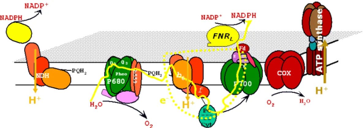
In oxygenic photosynthetic organisms, the major mode of electron transfer (ET) is the linear ET (noncyclic). It involves water oxidation to molecular oxygen and the reduction of NADP+ into NADPH. This is achieved by two sequential photoreactions involving two photosystems (Figure1.1). We will further introduce the four integral membrane protein complexes involved in photosynthetic electron transfer and ATP build-up that are photosystem II (PSII), cytochrome b6f (cyt b6f ), photosystem I (PSI) and ATP-synthase (ATPase).
PSI and PSII contain numerous pigments that harvest light and funnel the excitation to the primary electron donors, which can reduce an electron acceptor and accept electrons from specific electron donors. The cyt b6f complex mediates electron transport between PSII and PSI
and converts redox energy into a high-energy intermediate (protonmotive force; pmf) for ATP formation. The reaction catalyzed by PSII is shown in Equation1.1.2:
2H2O+ 2PQ + 4H+stromaGGGAO2+ 2PQH2+ 4H+lumen (1.1.2)
The reaction involves the reduction of plastoquinone (PQ) into plastoquinol (PQH2) and the
oxidation of water into molecular oxygen. The PSII reaction center is composed of two similar
1.1. Photosynthesis and bioenergetics
Figure 1.1 The structures of the four large membrane-protein complexes in thy-lakoid membranes that drive oxygenic photosynthesis taken from [Nelson and Ben-Shem,2004].
Figure 1.2 The structure of photosystem II from the cyanobacterium Thermosyne-chococcus elongatus [Ferreira et al.,2004].
its PSII binding site.
Figure 1.3 The cofactors of photosystem II published in [Ferreira et al.,2004].
The cyt b6f complex (Figure1.4a) is also called "plastoquinol-plastocyanin oxidoreductase".
The complex operates following the "Q-cycle" [Mitchell,1976,Trumpower,1990] and is similar in structure and function to the cytochrome bc1complex from the respiratory ET chain. It exhibits
two PQ binding sites, the Qosite close to haem bLbinds quinol (lumenal side) and the Qisite close
to haem bHbinds quinone (stromal side). The PQH2looses a first electron, which is transferred
to the Rieske iron-sulfur protein, the cyt f and finally to plastocyanin. The second electron from PQH2 is transferred via two cyt b to the Qi site where another PQ is reduced (Figure1.4b). The
quinone analogue 2,5-dibromo-3-methyl-6-isopropylbenzoquinone (DBMIB) acts as an inhibitor of cyt b6f as it is thought to bind at the Qo site. The presence of a further haem group that is
covalently bound to cytochrome b6is puzzling [Kurisu et al.,2003,Stroebel et al.,2003]. Overall,
two protons will be translocated across the thylakoid membrane for one electron that is passed to PSI via plastocyanin.
Plastocyanin (Pc), a single copper containing protein, transfers its electron to PSI (Figure 1.5a). Controlled by the Cu availability in the growth media, cytochrome c6can replace Pc in its
function in many algae and cyanobacteria. The bulk of the reaction center of PSI is built of two,
1.1. Photosynthesis and bioenergetics
Figure 1.4 The structure of cytochrome-b6f complex (a) and its cofactors (b) from the alga Chlamydomonas reinhardtii [Stroebel et al.,2003].
Figure 1.5 The model of a super-complex containing photosystem I, plastocyanin and ferredoxin (a) and their cofactors (b) from a higher plant (Pisum sativum var. alaska) [Ben-Shem et al.,2003].
homologous, large subunits (PsaA and PsaB) that harbor most of the PSI pigments and all of the cofactors up to FX. Photoexcitation gives rapidly (ps time range) P700+ and A−1. An electron
from Pc (or cyt c6) regenerates the special pair P700. The electron on the phylloquinone A1 is
passed through the FX and FA/FBiron sulfur centers (Figure1.5b). The electrons at the acceptor
side of PSI are transferred to the soluble ferredoxin (Fd) and finally lead to NADP+reduction. This reaction is catalyzed by the enzyme ferredoxin-NADP+oxidoreductase (FNR) which is the subject of this thesis and will be described in section 1.3.3. N,N-dimethyl-4,4’-bipyridinium dichloride (methylviologen; MV) is an efficient electron acceptor, from (FA,FB) and Fd. Reduced
MV reacts very rapidly with O2(contrary to (FA,FB) and Fd which react much more slowly).
A total of six protons are translocated, for 2 PSII charge separations and cyt b6f ET, to the
lumen that lead to an electrochemical-potential gradient (proton motive force; pmf) across the thylakoid membrane. The pmf will be used in the fourth complex, the ATPase, in order to synthesize ATP. During this process, protons flow from the lumen to the stroma through the integral membrane part of the ATPase CF0and ATP is generated at the level of the soluble part
CF1(Figure1.6).
Most of the details shown so far concern the photosynthetic electron transfer in higher plants and algae [Nelson and Ben-Shem,2004]. All the complexes cited above are identical in cyanobac-teria. Photosynthetic and respiratory electron transfers are carried out in the same compartment in cyanobacteria. This phenomenon is also present in chloroplasts and is called chlororespiration [Bennoun,1982,Rumeau et al.,2007].
1.1. Photosynthesis and bioenergetics
Figure 1.6 A composite model for the structure of the chloroplast F-ATPase. This model was created by W. Frasch using available structural data for mitochondrial F-ATPase subcomplexes [Abrahams et al., 1994,Stock et al.,1999, Gibbons et al., 2000].
Figure 1.7 Scheme of the thylakoid membrane. The different electron translocating complexes are shown. Respiratory complexes involve the COX: cytochrome oxidase complex, NDH: NAD(P)H dehydrogenase (NDH-1) complex.
Succinate dehydrogenase (SDH) is the only enzyme of the tricarboxylic acid pathway which is found attached to the membrane. This complex functions in the respiratory ET as complex II. Evidence was found for the implication of SDH in the PQ pool reduction in Synechocystis [Cooley et al.,2000].
In the thylakoids of both cyanobacteria and plastids, distinct NAD(P)H dehydrogenases are found that oxidize NAD(P)H (NDH-1) and are equivalent to complex I involved in respiration. In cyanobacteria, the NDH-1 is extensively studied and is involved in a variety of functions like respiration, cyclic electron flow (introduced later) around PSI and CO2uptake [Battchikova and
Aro,2007].
In addition to that, three respiratory terminal oxidases (RTOs, complex IV) exist in Syne-chocystis: cytochrome c oxidase (CCox), quinol oxidase (Cyd), and alternative RTO (ARTO) [Pils and Schmetterer,2001,Hart et al.,2005]. ET through the respiratory complexes lead to a proton gradient, hence to ATP formation, at the expense of reductants (NADPH and succinate).
1.1.3 Alternative electron sinks and cyclic electron transfer
Light-induced linear electron transfer between the two photosystems generate ATP and reducing equivalents in the form of NADPH. ATP and NADPH are used in a variety of metabolic processes. Under photoautotrophic growth conditions, CO2assimilation in the Calvin cycle constitutes the
major electron sink for NADPH. However, stress conditions may lead to electron redirection
1.1. Photosynthesis and bioenergetics
toward alternative electron sinks. Alternative electron sinks involve essentially the Mehler-reaction, cyclic electron transfer or respiration.
A substantial part of electrons can be transferred from PSI to molecular oxygen, which results in photoreduction of O2 via superoxide anion to H2O2 in chloroplasts, i.e. the Mehler-reaction
[Mehler,1951,Asada,1999]. The produced reactive oxygen species (ROS) are quickly detoxified by the combined action of superoxide dismutase and peroxidases. Thereby, the photoreduction of O2 acts as an electron sink (pseudocyclic ET) under certain conditions, where up to 30% of
the electrons from the light reactions can be directed to oxygen [Hackenberg et al., 2009]. For Synechocystis sp. PCC6803 (Synechocystis), it was shown that O2is reduced directly to water in
one reaction mediated by A-type flavoproteins [Helman et al.,2003]. The genome of Synechocystis encodes four putative A-type flavoproteins, but only two of them, Flv1 and Flv3, are apparently involved in light-dependent O2 reduction activity [Helman et al., 2003]. Recently, a role in
the photoprotection of PSII has been shown for the two other Synechocystis flavoproteins, Flv2 and Flv4 [Zhang et al., 2009]. For pseudocyclic ET, which involves the Mehler-reaction or photorespiration, redox poising was proposed as a plausible function [Allen,2003].
Under CO2-limiting conditions, the Calvin cycle activity is strongly reduced and
photorespi-ration, a Rubisco oxygenase reaction, increases. The so-called photorespiratory 2PG-metabolism helps to avoid depletion of Calvin-cycle intermediates. Due to the efficient inorganic carbon con-centrating mechanism [Badger et al.,2006], it was assumed that cyanobacteria do not possess a photorespiratory 2PG-metabolism. In contrast to this earlier view, it was recently demonstrated that an active photorespiratory 2PG-metabolism involving three different pathways exists in Synechocystis [Eisenhut et al.,2008]. The complete loss of all three pathways leads to a high-CO2
-requiring-phenotype and highlights the essential function of photorespiratory 2PG-metabolism for cyanobacteria despite the carbon concentrating mechanism [Eisenhut et al.,2008]. The con-tribution of the Mehler-reaction may be controlled by inorganic carbon [Badger et al., 2000]. Transcription of one of the A-type flavoproteins, Flv3, essential for photoreduction of O2 in
cyanobacteria, is increased under high-light and low-CO2conditions [Eisenhut et al.,2007]. This
may indicate an increase in pseudocyclic ET under low CO2.
We will focus in the following especially on cyclic electron flow. This is an alternative electron flow that generates exclusively a proton gradient to build up ATP without accumulation of NADPH. It is assumed to involve PSI and the cyt b6f (Figure1.8). In addition to that, several
partners are proposed to catalyze donation of electrons from the acceptor side of PSI (Fd, FNR, NADPH) back into the PQ pool.
The assimilation of CO2in the Calvin cycle requires ATP and NADPH in a 3:2 ratio [Allen,
2002]. The number of protons that are translocated through the membrane per ATP is function-specific [Stock et al.,1999,Seelert et al.,2000]. On one side, it was recently calculated in spinach
Figure 1.8 Scheme of the thylakoid membrane. The different electron and proton translocating complexes are shown. The two major pathways for cycling of electrons are indicated with a blue and a black arrow. They involve the respiratory NDH-1 complex (NDH dependent; from NADPH) and a non-identified Ferredoxin:Quinone reductase (FQR; from Ferredoxin).
Photophosphorylation requires a redox poise - a balance in its input and output of electrons. Hence, photosynthetic systems try to maintain a poised plastoquinone pool. Over-reduction of the plastoquinone pool is expected when the Calvin cycle is unable to use NADPH, and one reason for this is insufficient ATP [Allen,2003]. It was suggested that linear ET alone would not be sufficient to generate the ATP required for CO2fixation and an obligatory role for cyclic ET
was proposed [Golding et al.,2004]. Another role that has been postulated for cyclic ET in plants is to generate a trans-thylakoid pH gradient (∆pH) [Heber and Walker,1992]. The debate about the role of cyclic ET is fuelled by the difficulty of measuring it directly. Two approaches have been commonly used - P700+steady-state measurements and P700+relaxation following far-red illumination.
What is for sure is the fact that cyclic electron transfer is triggered under excess of NADPH - for example under high light or low CO2[Miyake et al.,2005]. High light leads to a fast accumulation
of electrons on the acceptor side of PSI and the utilization of these reducing equivalents can be limited through the Calvin cycle. Under low CO2 conditions, the Calvin cycle is limited by
1.1. Photosynthesis and bioenergetics
substrate availability and NADPH accumulates. In addition to that, pseudocyclic ET involving the Mehler-reaction is triggered under low CO2[Hackenberg et al.,2009].
What is already known about the pathways of cyclic electron flow? According to Joliot [Joliot and Joliot,2005], it is generally believed that the pathway starting from ferredoxin, the ferre-doxin:quinone reductase (FQR) dependent pathway, constitutes the major pathway for cycling of electrons in plants (shown by the blue arrow in Figure 1.8). This pathway was originally found by inhibiting cyclic electron transfer using the cyt bc1specific inhibitor antimycin A and
no biochemical evidence for the FQR pathway was obtained so far. However, Pgr5 and Pgrl1 ([DalCorso et al.,2008] and references therein) were found to participate in this cyclic electron transfer. In plants, there are two partially redundant pathways taken by electrons in PSI cyclic ET [Shikanai,2007,Munekage et al.,2002,2004,2008].
In cyanobacteria, the FQR pathway is generally considered to be a minor pathway for cy-cling of electrons whereas the NDH dependent pathway (Figure1.8black arrow) is believed to constitute the major pathway.
In Synechocystis, a mutant called M55 was constructed, defective in ndhB which is the single gene coding for the subunit NdhB of the NDH-1 complex. Due to this mutant the various functions of NDH-1 complexes were discovered. M55 is characterized by impaired cyclic ET [Mi et al.,1992b, 1994,1995], impaired respiration and presents an impaired CO2 uptake [Ogawa,
1991]. Due to multiple copies of genes ndhD and ndhF, distinct NDH-1 complexes with distinct functions were identified [Ohkawa et al.,2000].
NDH-1 is expressed only in low levels under high CO2 photoautotrophic growth. Two
distinct NDH-1 complexes are implicated in the carbon concentrating mechanism (CCM) and the expression of one of these complexes is induced under low CO2[Battchikova and Aro,2007].
In addition to the major pathways, several other pathways have been proposed. In chloro-plasts, association of FNR to PSI and/or cyt b6f has sometimes been taken as a structural evidence
for different pathways of cyclic ET [DalCorso et al.,2008]. Supercomplex formation - e.g. PSI/cyt b6f/Fd - has also been proposed to support cyclic ET [Joliot and Joliot,2005] but there is no clear
biochemical evidence yet for such supercomplexes in cyanobacteria (see [Peng et al., 2008] for chloroplasts). During our study we wanted to address the issue of FNR involvement in these different cycling routes.
1.1.4 Cyanobacteria
Cyanobacteria constitute a large and diverse group of photosynthetic prokaryotes. They inhabit almost any illuminated environment (freshwater, marine or terrestrial). All cyanobacteria are photoautotrophs and some species can grow as well photoheterotrophically.
freshwater lake in California (Figure1.9) [Zhang,2006].
Figure 1.9 Electron micrograph of a thin section through a Synechocystis 6803 cell. Taken from www.nsf.gov/news/mmg/media/images/.
In addition to the cell envelope, these organisms have an internal system of thylakoid mem-branes (closely spaced memmem-branes in Figure1.9) where the electron transfer reactions of photo-synthesis and respiration occur. The three-dimensional organization of the cytoplasm has been investigated using standard transmission electron microscopy and electron tomography. On one hand, it was shown that the thylakoid membranes are physically discontinuous from the plasma membrane [Liberton et al.,2006]. On the other hand, close connections between thylakoids and cytoplasmic membrane systems were observed [van de Meene et al.,2006]. Therefore, this debate is not closed yet.
We will now introduce the differences between cyanobacteria and chloroplasts that are im-portant for the following study. We will start by a detailed description of the light-harvesting
1.2. Light-harvesting antenna
complex - the phycobilisome - in cyanobacteria followed by an introduction on structure and function of FNR isoforms and the acceptor side of PSI in general.
1.2 Light-harvesting antenna
Prior to charge separation, a photon is absorbed by a pigment-protein (antenna) complex. The resulting excitation energy is transferred using radiationless steps to the chlorophylls involved in primary charge separation. These chlorophylls are generally associated to several hundred pigment molecules serving as light harvesting. This is necessary so that photosynthesis is less light-limited [Blankenship,2002,Glazer,1989].
Light-harvesting complexes can be divided into integral and external membrane complexes. Furthermore, they can be classified into seven families. These families include the core antenna complexes (e.g. CP47 and CP43 for PSII), the proteobacterial antenna complexes (e.g. LH1, LH2 and LH3 of purple bacteria), the eucaryotic LHC superfamily (e.g. LHC1 associated to PSI in algae and plants), the peridinin-Chl a protein (in Dinoflagellate algae), the chlorosome (in green sulfur bacteria and green filamentous bacteria) and the phycobilisome (in cyanobacteria and red algae) [Ughy,2005]. The major difference between cyanobacteria/red-algae and green algae/brown algae/plants is the presence of a giant antenna complex, the phycobilisome (PBS; [Adir,2005]).
1.2.1 Phycobilisome
The phycobilisome is an external membrane complex that is attached to the stromal side of the photosynthetic membrane and constitutes an accessory antenna. It allows species bearing these antennas to harvest light in the spectral gap (500-660 nm) between the major chlorophyll absorbing bands and thus to utilize the entire visible range of sunlight [Glazer,1989,Adir,2005]. In the following, we will further detail some advances in (ultra)structure determination and function of these large macromolecular protein complexes (7 to 15 Megadaltons).
Phycobilisome structure
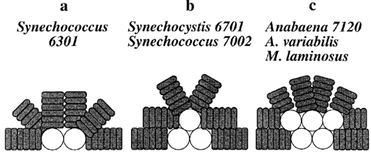
Different types of PBS exist. We will focus on the hemidiscoidal PBS because they are present in Synechocystis. They are composed of a core subdomain and peripheral rods. The hemidiscoidal PBS are divided in three types (shown in Figure1.10). They include bicylindrical, tricylindrical and pentacylindrical PBS (Figure1.10; [MacColl,1998]). In Synechocystis, the common tricylin-drical hemidiscoidal PBS is present (Figure1.10b). In addition to these classical PBS, minor PBS were identified in Synechocystis that do not contain core subunits [Kondo et al.,2005,2007,2009].
Figure 1.10 Schematic representation of the three types of hemidiscoidal PBS. a, bicylindrical; b, tricylindrical; c, pentacylindrical. Taken from [Ducret et al.,1998].
Phycobiliproteins The PBS is composed of phycobiliproteins (PBP) and mostly colorless linker polypeptides. Four major subgroups of PBP are found. They include the allophycocyanin of the core (AP, λmaxA =652 nm), phycocyanin that is always present close (proximal) to the core in the rods (PC, λmaxA =620 nm), phycoerythrin (PE, λmaxA =560 nm) and phycoerythrocyanin (PEC, λmax
A =575 nm) far away (distal) to the core in the rods when present. The PBPs are physically
arranged to favor an energy gradient from PE (or PEC) through PC to AP and finally to the reaction center (λ=670-680 nm).
The smallest PBP unit are α and β (≈ 17 and 18 kDa) subunits that form a heterodimer - the "(αβ) monomer (protomer)". Each subunit contains one or more covalently attached bilins. Bilins are linear tetrapyrrole prosthetic groups. The covalent attachment of the bilins in the amino acid sequence is conserved, α84 in the α monomer, β84 and β155 in the β monomer. An α subunit of PC with the phycocyanobilin (PCB) covalently attached to a cysteinyl residue (ring A) is shown in Figure1.11. In Synechocystis, only AP and PC are present in the core and rod subdomains, respectively. They are both composed of subunits that contain exclusively PCB.
Linker polypeptides In addition to the PBP, a variety of linker polypeptides are present. Except for the LCM, these do not contain any chromophore. Different linkers are specifically responsible
for each level of PBP assembly and function to stabilize the PBS and optimize its absorption and energy transfer characteristics. There exist linkers that anchor the core to the photosynthetic membrane (LCM), linkers that connect the rods to the core (LRC) and linkers that are only present
in the core and the rods: LCand LR, respectively. CpcC2, cpcC1 and cpcD encode the rod linkers
L30R, L33R and L10R (Figure1.13). Two independent genes (cpcG1 and cpcG2) encode the rod-core linker (LRC) [Ughy and Ajlani,2004].
The core-membrane linker LCMis a high molecular weight polypeptide. This major linker is
1.2. Light-harvesting antenna
Figure 1.11 αsubunit of phycocyanin (PC) with its chromophore phycocyanobilin (PCB).
responsible for the assembly of AP discs into cylinders and core formation. LCMalso plays a key
role in anchoring the PBS to the photosynthetic membrane and in tuning the properties of the bound pigment cofactors such that absorbed light is funnelled towards the photosystems. Two copies of this multifunctional polypeptide (mass 75-125 kDa) are present per PBS core [Capuano et al.,1991,Arteni et al.,2009].
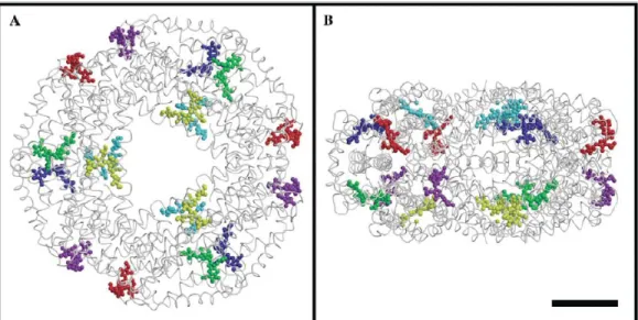
Phycobilisome Assembly PBS substructures are built up from stacked PBP discs made either of trimers (AP) or of hexamers (PC) of PBP subunits. X-ray crystallography was used to determine the structure of PBP discs. The crystal structure of allophycocyanin showed two loosely stacked trimers [Liu et al.,1999]. C-phycocyanin is a hexameric disc of 110Å diameter and 60Å thickness (Figure1.12and [Nield et al.,2003]). In many structures of PC, the two (αβ)3 trimers that form
a hexameric disk (αβ)6 were positioned face to face. The hexamer is easily disassembled in
(αβ) monomer in diluted solutions, indicating that just a limited number of salt-bridges and/or hydrogen bonds are involved in monomer stability. Except in one case [Reuter et al.,1999], the available structures do not contain any linker.
Each of the cylinders in the tri-cylindrical core is composed of four trimeric AP discs. These discs have slightly different compositions. They involve simple AP trimers, LC-containing AP
trimers, trimers containing an alternative AP-B α subunit and trimers that possess a red-shifted βisoform and an α subunit provided by the LCM. As indicated above, LCMtogether with the LC
assemble the AP discs into cylinders and into a core substructure.
The rod-core linkage position is always occupied by a PC hexamer that is attached due to
Figure 1.12 S. elongatus C-PC hexameric cluster. The 3 chromophores of each αβ heterodimer are depicted in the same color. Three αβ heterodimers form a disk around the three-fold axis. Bar represents 25 Å. A, projection parallel to the three-fold axis; B, projection normal to the three-fold axis. Taken from [Nield et al.,2003].
LRC. For each disc at a particular rod location there exists a specific linker. Six rods radiate in
a hemidiscoidal array from the core. In Synechocystis, rods are composed of three stacked PC hexamers. The position of the rod linkers L10
R, L30R, L33R and LRC in the PC hexamers is indicated
in Figure1.13.
Although many isolated components of different PBSs were crystallized, the structure of the entire PBS and its association to PSII is only studied by electron microscopy [Arteni et al.,2009].
Phycobilisome function
Attached primarily to reaction centers of PSII, the PBS can functionally link more than 600 energy-absorbing pigments to a single PSII dimer in addition to the PSII integral antenna subunits CP43 and CP47. On one side, direct measurements of fluorescence recovery after photobleaching [Mullineaux, 2004] indicated that the 10 Megadalton PBS is quite mobile in vivo, much more than the photosystems. On the other side, ultrastructures of the Synechocystis photosynthetic membranes indicated that the width of the stromal space between two membranes matches the PBS height (see Figure1.9). Therefore, it seems difficult to imagine highly mobile PBS between the closely spaced thylakoids.
In addition to light absorption, the PBS can function as a source of nutrients under starvation conditions. There exists a mechanism of ordered PBS disassembly that requires the presence of a number of gene products. Keeping in mind that the PBS can account for up to 30 % of the total protein mass in a cyanobacterial cell, it constitutes a significant reservoir.
1.2. Light-harvesting antenna
Figure 1.13 Representation of a hemi-discoidal PBS, as seen from the side. Kindly provided by Dr. Ajlani.
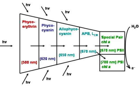
Energy transfer within the PBS The absorbed light energy harvested at the periphery of the PBS is transferred to the PSII reaction center complex by radiationless excitation energy transfer with an efficiency of > 95%. This implies that the energy-transfer mechanism must proceed rapidly in order to avoid energy losses by competing radiative or non-radiative decay processes. Light energy is absorbed mainly by the peripheral rods, where the shortest wavelength absorbing PBP (PE or PEC) are located. The excitation energy is then transferred by a radiationless resonance energy transfer to C-PC and then to AP. Energy is finally transmitted to PSII and partially to PSI reaction centers through the terminal emitters of the PBS (Figure1.14) [Sidler,1994].
The spectroscopic properties of the bilins are modified by the protein in two ways which are critical for their role in light absorption and energy transfer:
1. the bilins in all PBP are held rigidly in extended conformations,
2. the excited state lifetime of the pigment in the protein is long (vs. the isolated pigment).
The extended conformations allows the strong absorption in the visible part of the light spectrum. The long excited-state lifetime avoids the loss of excitation energy by radiationless de-excitation pathways. The absorption and transfer of light energy is performed by chromophores that are called donors. Acceptors can both absorb excitation energy and fluoresce. Thus, the steady state fluorescence emission originates almost exclusively from the acceptors.
In C-PC, the bilins at α84 and β155 are donors and the bilin at β84 is the acceptor. PCB β84 extends into the center of the trimeric disc, whereas those at α84 and β155 lie toward the
Figure 1.14 Energy flow in PBS of cyanobacteria and red algae. Radiationless excitation energy transfer from short-wavelength (PE) to long-wavelength-absorbing pigment-protein complexes (AP). Energy is finally transferred to and distributed between PSII and PSI. Adapted from [Sidler,1994].
periphery. In the face-to-face arrangement of double discs in the rods, the consecutive discs are arranged to favor rapid energy transfer. Picosecond energy transfer measurements showed that the excitation energy absorbed by any bilin at the periphery is rapidly localized on the centrally located acceptor bilins (yellow and light blue chromophores in Figure1.12A) [Glazer,1989].
Directional energy transfer is promoted through the PBS. Between the PC discs, interaction with different linker polypeptides confer distinctive spectroscopic properties to the acceptor bilins. The absorption and emission spectra of the (αPCβPC)6LRCcomplex are shifted towards the
red relative to those of (αPCβPC)6L33R. In consequence, the favored direction of transfer is from the
distal disc to the PC disc proximal to the core.
In summary, the energy absorbed by any of the bilins in the PBS localizes rapidly (< 8 ps) on the four terminal acceptor bilins (APB and LCM) in the core. The emission of these bilins overlaps
precisely the absorption spectrum of the reaction center of PSII. The light-guide function of the PBS is completed when energy is transferred radiationless from the terminal acceptors in the PBS to the reaction center [Glazer,1989].
1.3. Photosystem I and its electron acceptors
1.2.2 Phycobilisome rod mutants
PBS mutants were constructed in three rod-linker-coding genes located in the cpc operon of Synechocystis [Ughy and Ajlani,2004]. CpcC1 and cpcC2 encode L33R and L30R, respectively. L33R and L30
R are linker polypeptides that attach the middle and the distal PC hexamer of the rods (Figure
1.13). During in vitro studies, we used a mutant called CB in which cpcC1 and cpcC2 were deleted. The PBS contained only one PC hexamer per rod (Figure1.15) [Ughy and Ajlani,2004].
Figure 1.15 Representation of the PBS in the CB mutant. WT PBS contains 3 hex-amers of PC per rod, whereas CB contains only 1 hexamer of PC per rod. Kindly provided by Dr. Ajlani.
1.3 Photosystem I and its electron acceptors
We were interested in the acceptor side of PSI. Electrons following the linear electron transfer are designated to reduce NADP+, building up the reducing power NADPH. We will now further introduce PSI, Fd and FNR.
1.3.1 Photosystem I
The three-dimensional structure of cyanobacterial PSI was solved by Jordan et al. in [2001]. It provided atomic details of the 12 subunits and 127 cofactors comprising 96 chlorophylls, two phylloquinones, three [4Fe-4S] clusters, carotenoids and lipids. The cofactors involved in ET are located within the membrane subunits PsaA/PsaB and the stromal subunit PsaC (Figure1.16). It can be seen that the chlorophyll pairs are arranged in two branches labelled A and B. There has been some controversy about whether the two branches work [Brettel and Leibl,2001] and this issue is now settled in favor of the theory that the two branches work. The first of the three [4Fe-4S] cluster FXis located in the middle between PsaA and PsaB. The following two [4Fe-4S]
FA and FB are provided by the stromal subunit PsaC. We will now further introduce the ET
kinetics inside PSI.
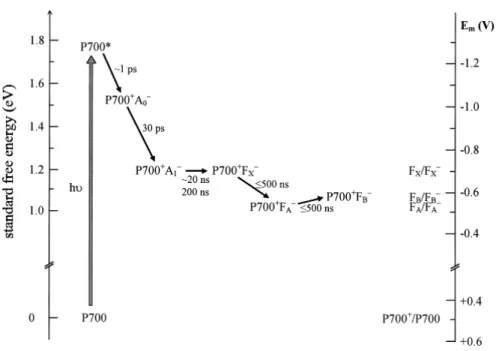
Electron transfer in PSI was reviewed [2001] by Brettel and Leibl. Standard free energy levels and kinetics of charge separation are shown in Figure1.17. Charge recombination between P700+
Figure 1.16 Cofactors of the electron transfer chain (ETC) and of PsaC. View parallel to the membrane plane. The pairs of chlorophylls of the ETC are arranged in two branches A and B. The chlorophylls a (P700 and A0), the phylloquinones (A1) and the [4Fe-4S] clusters (FX, FA and FB according) are labelled to their spectroscopic terms. The center-to-center distances between the cofactors (black lines) are given in Å. Adapted from [Jordan et al.,2001].
and the reduced form of any one of the electron acceptors can be observed when forward electron transfer to the subsequent acceptor is blocked. In addition to that, reduction midpoint potentials are indicated on the right in Figure1.17. The reduction midpoint potentials are very similar for FA and FB and it is generally thought that FA and FB undergo fast (< 1 µs) redox equilibrium
[Setif, 2001]. They are both higher than the midpoint electron potential for FX/FX −. We will
be furthermore interested in the stromal subunits of PSI as they provide the docking site for Fd binding.
The three stromal subunits of PSI are shown in Figure1.18. In addition to providing the two terminal electron acceptors of PSI, they provide the docking side for Fd that is shown as a dashed ellipse in Figure1.18. This complex formation is important for efficient electron transfer. We will now further introduce Fd structure and function in plants and cyanobacteria.
1.3.2 Ferredoxin
Ferredoxin (Fd) is a soluble, low molecular weight protein (ca. 11 kDa) that mediates transfer of one electron from a donor to an acceptor. The redox active center is a [2Fe-2S] cluster with a highly negative redox potential (-350 to -450 mV), making reduced Fd a powerful reductant. The [2Fe-2S] cluster is ligated by four highly conserved Cys residues [Bottin and Lagoutte,1992,
1.3. Photosystem I and its electron acceptors
Figure 1.17 Approximate standard free energy levels and kinetics of charge sepa-ration in PSI. The standard free energy of the dark state (P700) was arbitrarily set to zero. Reduction midpoint potentials (versus NHE) obtained by redox titrations of intact PSI are indicated on the right-hand scale. Taken from Brettel and Leibl [2001].
Figure 1.18 View along the membrane normal from the stromal side showing sub-units PsaC, PsaD and PsaE. They cover some of the loop regions and helices of PsaA and PsaB (light grey). Dashed ellipse: putative docking site of ferredoxin, covering loops of PsaA. Taken from [Jordan et al.,2001].
Fds are present as multiple isoforms in many plants and algae. We will further introduce the isoforms existing in plants and cyanobacteria.
Plant isoforms Higher plants contain distinct leaf and root Fd isoforms with conserved differ-ences, reflecting the different electron donors to Fd in photosynthetic and non-photosynthetic tissues. Functional differences have been demonstrated between leaf and root Fds, with respect to redox potential and activity in assays of NADP+photoreduction and NADPH oxidation. These differences are highly conserved among species: Leaf Fds have a redox potential around 50 mV more negative than root Fds; during NADP+ photoreduction, leaf FNR has an affinity around 10-fold higher for leaf Fds than for root Fds, and during NADPH oxidation root FNR has an affinity around five times higher for root Fds than for leaf Fds.
As the redox potential of leaf Fd is around -420 mV, a 50 mV difference may not appear dramatic, but the flux through photosynthesis is so vast that small changes in efficiency are likely to have a profound physiological impact. It was stated that the concentration of Fd in the chloroplast is of the same order as the concentration of Fd-dependent enzymes, which could therefore be in competition, giving great physiological significance to even small differences in affinity and activity [Gou et al.,2006].
Cyanobacteria isoforms Analogously to plants, cyanobacteria also possess several molecular forms of [2Fe-2S] Fd encoded by distinct genes. The most abundant protein form has been termed Fd1 (Fed1). The fed1 gene (ssl0020 in Synechocystis) was found to be strongly expressed as a light-induced transcript. The other fed-like genes appeared to be silent or moderately expressed. fed1 was found to be critical to Synechocystis viability in spite of fed-like genes slr0150, sll1382 or flavodoxin induction, even after the addition of glucose that compensates for the loss of photosynthesis [Poncelet et al., 1998]. We used during our studies only the major Fd isoform assuming that it is involved in all the metabolic pathways under our conditions. This is in
1.3. Photosystem I and its electron acceptors
contrast to the plant isoforms, where tissue specificity ensures the function of distinct isoforms in distinct metabolic pathways.
Kinetics of ferredoxin reduction After PSI photoexcitation, several fast kinetic components (submicrosecond and microsecond) for Fd reduction have been identified in vitro. The rates of the fast kinetic components did not depend on the concentration of the partners. In addition to that, a slow kinetic component was also identified characterized by a rate that depends linearly on the Fd concentration. The fast and slow kinetic components are thus called first-order and second-first-order phases, respectively. The first-first-order phases are thought to correspond to ET processes which occur within PSI/Fd complexes. The second-order phase corresponds to a diffusion-limited ET which is observable in the fraction of PSI which does not bind Fd before flash excitation [Setif,2001].
At least two or three first-order components were necessary to describe the first-order kinetics of Fd reduction. The spectra of the three phases obtained in Synechocystis were shown to be consistent with Fd reduction from (FA,FB)−. At least 80% of Fd is reduced within the PSI/Fd
complex at pH 8 in Synechocystis. The two slower first-order processes might result from some rate limitation either in ET from FB to Fd or during intramolecular PSI ET. The distal cluster
FB is photoreduced in the submicrosecond time range in PSI. Heterogeneity of ET kinetics is an
intrinsic property of Fd reduction, and was ascribed to different conformations of the PSI/Fd complex.
Fd reduction should compete efficiently with the recombination reaction between P700+and
(FA,FB)−and this would imply a t1/2of several orders of magnitude faster than the recombination
(30-100 ms). Furthermore, a high efficiency of Fd may be required for avoiding reduction of oxygen from (FA,FB)− which is potentially harmful for PSI. It has been speculated that, in vivo,
complex formation is useful, though not critical, for promoting efficient reduction of the soluble acceptors and avoiding reduction of oxygen by (FA,FB)−[Karplus and Faber,2004].
Complex formation was found to occur as well between Fd and the enzyme FNR in plants and cyanobacteria [Hanke et al.,2004b]. As stated above, FNR receives two electrons from two Fds to finally reduce NADP+to NADPH during linear electron flow. We will now introduce the structure and function of FNR that catalyzes the last step of the building up of the NADPH.
1.3.3 Ferredoxin:NADP oxidoreductase Structure of ferredoxin:NADP oxidoreductase
From their primary sequences, the different ferredoxin:NADP oxidoreductases (FNRs) can be grouped in three major branches. The plant-leaf chloroplast FNRs are on one branch, the widely
Figure 1.19 Relationships among various FNR amino-acid sequences. In the un-rooted dendrogram shown, each branch represents 1 of 30 known plastid FNR se-quences. The lengths of the branches are proportional to the level of sequence difference. Taken from [Karplus and Faber,2004].
diverse cyanobacterial FNRs are on a second branch, and the root plastid enzymes together with the enzymes from green algae chloroplasts are on the third branch (Figure 1.19). All of the plastid type FNRs share sequence identities of over 40%. Crystallographic structures have been determined for six different FNRs: four leaf-type enzymes, one root-type enzyme and one cyanobacterial enzyme (Anabaena variabilis). These enzymes all have equivalent structures including two classical structural domains (Figure1.20A). The amino-terminal residues (ca. 150) form the FAD-binding domain (blue in Figure 1.20A) and the carboxy-terminal residues (ca. 150) form the NADP+ binding domain (pink in Figure1.20A). The plant chloroplast enzymes are structurally similar and the root and cyanobacterial enzymes are structurally variable. One major difference in the corn root enzyme structure compared to leaf is that the amino terminus packs in a completely different position. Based on sequence comparisons, this amino-terminal packing appears to be conserved among root-type enzymes. With regard to amino-acid residue conservation, among the ca. 40 known FNR sequences, about 25% of the residues in the protein are conserved. It was stated that this high level of conservation over such long evolutionary distances implies a fairly stringent level of selection [Karplus and Faber,2004].
FAD binding The FAD binding site in FNRs is quite highly conserved for the FMN half of the prosthetic group that contains the isoalloxazine (upper part in Figure1.20A). In contrast, the adenosine portion of FAD (lower part in Figure1.20A) shows significant variation in its position of binding. The isoalloxazine moiety is the best defined portion of the FAD and the adenosine
1.3. Photosystem I and its electron acceptors
Figure 1.20 Ferredoxin:NADP oxidoreductase structure. A: The Cα polypeptide backbone of plant-type ferredoxin:NADP oxidoreductase. FNR is a two-domain flavoprotein. The computer graphic is based on X-ray diffraction data for the spinach enzyme, with the FAD binding domain shown in blue, the NADP(H) binding domain in pink, and the FAD prosthetic group in yellow. Taken from [Carrillo and Ceccarelli, 2003]. B: Geometry of the productive NADPH-FAD Michaelis charge transfer com-plex. A view is shown including all atoms surrounding the locus of hydride transfer, that is the nicotinamide C4- and the FAD N5-atoms. The model is that of the pea FNR Y308S-NADPH complex (pdb entry 1QFZ chain A). Taken from [Karplus and Faber,2004].
crystallographic studies, it was observed that only in a small fraction of the enzyme (15% pea FNR, [Piubelli et al., 2000]) and an even smaller fraction of cyanobacterial enzymes [Hermoso et al., 2002] the C-terminal Tyr swings out of the way so that the NMN half of NADP binds properly. A more complete understanding of nicotinamide binding was finally obtained using a mutant of pea FNR with the C-terminal Tyr converted to Ser [Deng et al.,1999,Piubelli et al., 2000]. A surprise compared to other NAD(P) dependent flavoenzymes was that the nicotinamide was not co-planar with the flavin, but made a 30◦
angle with it [Karplus and Faber,2004].
Roles of active site residues were proposed by Deng et al. [1999] and involved the boat-like conformation of the nicotinamide ring to facilitate hydride transfer. In addition to that, the C-terminal Tyr does not play an active role in hydride transfer, but is primarily a placeholder for the nicotinamide that modulates the binding thermodynamics of NADP and protects the flavin from reaction with oxygen. It was also speculated that reduction of the flavin or Fd binding might promote the movement of Tyr, even if dynamics of Tyr movements may be sufficient to support catalysis [Karplus and Faber,2004].
Ferredoxin binding Protein-protein interaction is an important determinant for electron trans-fer between Fd and FNR. The X-ray crystal structures of complexes formed between Fd and FNR from the cyanobacterium Anabaena 7120 [Morales et al.,2000], maize leaf [Kurisu et al.,2001] and maize root have been solved (see Figure 1.21for the cyanobacterial Fd-FNR complex). In the complex structure of Fd and FNR, Fd binds to a concave region on the FAD-binding domain of FNR, bringing the [2Fe-2S] cluster into close proximity to that of FAD. Different orientations of Fd relative to FNR have been found in cyanobacteria, plant-leaf and plant-root complexes. The relative buried surface areas differ as well, the root complex having a decreased buried surface area [Hanke et al.,2004b].
The complex in general is largely electrostatic in nature. The pattern of interaction between Fd and FNR is composed of a core of hydrophobic interactions surrounding the prosthetic groups, stabilized by a series of interactions between charged side chains and through hydrogen bonds.
1.3. Photosystem I and its electron acceptors
Hydrophobic effects originating from dehydration of water molecules in the protein-protein interface may also give a significant contribution. In Anabaena, a total of ten hydrogen-bonding and ionic-bridge interactions stabilize the complex [Morales et al.,2000]. The side chains involved in intermolecular charge interactions concern mainly acidic Fd residues and basic FNR residues [Hurley et al.,2002].
Figure 1.21 Ribbon drawing of Anabaena Fd : FNR complex (PDB code: 1EWY). One intermolecular salt bridge is shown as ball-and-stick model: 1, FNRLys75-FdGIu94. Taken from [Hanke et al.,2004b].
Redox potentials may change when binding occurs [Batie and Kamin, 1984b]. A negative redox shift in the potential of the [2Fe-2S] cluster of Fd was observed that would be advantageous to ET in the photosynthetic direction [Hanke et al.,2004b]. The more weakly binding complexes seem to yield more rapid ET and catalytic turnover. Thus, it could be that effective in vivo ET involves very short-lived (nearly collisional) complexes, rather than a tight, highly specific complex [Karplus and Faber,2004].
FNR superfamily Given the early origin of photosynthesis, FNRs would be expected to be ancient proteins. Consistent with this, there exists a large and diverse family of oxidoreductases which have as a catalytic core the two-domain FNR-like module [Karplus et al.,1991]. They are diverse enough that some use FAD and others use FMN whereas some use NADP and others use NAD. Structurally known members of the family are sulfite reductase, NO synthase, NADPH: cytochrome P450 reductase, etc..
![Figure 1.2 The structure of photosystem II from the cyanobacterium Thermosyne- Thermosyne-chococcus elongatus [Ferreira et al., 2004].](https://thumb-eu.123doks.com/thumbv2/123doknet/12702525.355708/26.918.233.671.700.926/structure-photosystem-cyanobacterium-thermosyne-thermosyne-chococcus-elongatus-ferreira.webp)