HAL Id: hal-02096824
https://hal.archives-ouvertes.fr/hal-02096824
Submitted on 11 Apr 2019
HAL is a multi-disciplinary open access
archive for the deposit and dissemination of
sci-entific research documents, whether they are
pub-lished or not. The documents may come from
teaching and research institutions in France or
abroad, or from public or private research centers.
L’archive ouverte pluridisciplinaire HAL, est
destinée au dépôt et à la diffusion de documents
scientifiques de niveau recherche, publiés ou non,
émanant des établissements d’enseignement et de
recherche français ou étrangers, des laboratoires
publics ou privés.
Distributed under a Creative Commons Attribution| 4.0 International License
40–100 mg archaeological bone
Helen Fewlass, Thibaut Tuna, Yoann Fagault, J.-J. Hublin, Bernd Kromer,
Edouard Bard, Sahra Talamo
To cite this version:
Helen Fewlass, Thibaut Tuna, Yoann Fagault, J.-J. Hublin, Bernd Kromer, et al.. Pretreatment and
gaseous radiocarbon dating of 40–100 mg archaeological bone. Scientific Reports, Nature Publishing
Group, 2019, 9, 5342 (2019), �10.1038/s41598-019-41557-8�. �hal-02096824�
pretreatment and gaseous
radiocarbon dating of 40–100 mg
archaeological bone
H. Fewlass
1, t. tuna
2, Y. Fagault
2, J.-J. Hublin
1, B. Kromer
1,3, e. Bard
2& s. talamo
1 Radiocarbon dating archaeological bone typically requires 300–1000 mg material using standard protocols. We report the results of reducing sample size at both the pretreatment and 14C measurementstages for eight archaeological bones spanning the radiocarbon timescale at different levels of preservation. We adapted our standard collagen extraction protocol specifically for <100 mg bone material. Collagen was extracted at least twice (from 37–100 mg material) from each bone. Collagen aliquots containing <100 μg carbon were measured in replicate using the gas ion source of the
AixMICADAS. The effect of sample size reduction in the EA-GIS-AMS system was explored by measuring
14C of collagen containing either ca. 30 μg carbon or ca. 90 μg carbon. the gas dates were compared to
standard-sized graphite dates extracted from large amounts (500–700 mg) of bone material pretreated with our standard protocol. the results reported here demonstrate that we are able to reproduce accurate radiocarbon dates from <100 mg archaeological bone material back to 40,000 BP.
Bone is one of the most frequently radiocarbon-dated materials recovered from archaeological sites. However, many precious archaeological bones, such as human remains or Palaeolithic bone tools, are too small or valuable for extensive destructive sampling. The reduction of sample size to enable direct dating of precious bone is there-fore a key concern for the archaeological community.
In the 1960s and 1970s, gas proportional counters required many grams of bone to produce a radiocarbon date1,2. The development and utilisation of Accelerator Mass Spectrometers (AMS) in the 1980s represented a
revolutionary step in the reduction of sample size and time required for dating3. Routine measurements today
typically require 500–1000 micrograms of carbon (μg C) to produce a high precision date. In recent years, sev-eral AMS labs have worked on modifications to the graphitisation and AMS measurement process for smaller samples containing <500 μg C4–13. However, the graphitisation of small sample sizes is often time consuming
and can be prone to large contamination effects14,15. A recent study by Cersoy, et al.16 demonstrated that graphite
targets containing ca. 200 μg C from archaeological bones can be successfully produced and measured using the IonPlus Automated Graphitisation Equipment III (AGE 3)17 and MIni CArbon DAting System (MICADAS)18,19
developed at ETH Zurich. However, the hybrid nature of the MICADAS system offers an alternative solution to the complex process of graphitising small samples. Organic samples containing <100 μg C can be placed into an elemental analyser (EA) directly coupled to the gas ion source of the MICADAS via the gas interface sys-tem (GIS)15,18,20–24. The automated system reduces both sample preparation time and the risk of contamination
through handling, and has been successfully utilised in environmental and climatic applications23,25–28. In our
preliminary study29 we demonstrated that the gas ion source of the AixMICADAS30 is suitable for dating bone
collagen CO2 samples of <100 μg C back to 35,000 BP (uncalibrated radiocarbon years before AD 1950).
However, as sample size is reduced the effect of contamination during pretreatment and measurement increases greatly. Sample pretreatment involves the extraction and purification of carbon endogenous to the orig-inal bone. Any contamination remaining in the sample at the time of dating can lead to erroneous results. The effects become increasingly catastrophic with the increasing age of the sample due to the minute concentrations of residual 14C. For example, in a bone extract ca. 40,000 BP, 1% modern carbon contamination would skew the
resulting 14C age by over 7,000 years.
1Department of Human Evolution, Max Planck Institute for Evolutionary Anthropology, Deutscher platz 6, D-04103,
Leipzig, Germany. 2ceReGe, Aix Marseille Univ, cnRS, iRD, inRA, collège de france, technopôle de l’Arbois, BP
80, 13545, Aix-en-Provence, France. 3Institute of Environmental Physics, University of Heidelberg, INF 229,
D-69120, Heidelberg, Germany. Correspondence and requests for materials should be addressed to H.F. (email: helen_ fewlass@eva.mpg.de)
Received: 8 October 2018 Accepted: 11 March 2019 Published: xx xx xxxx
It is standard practice to extract the proteinaceous portion of bone for 14C measurement, generally referred
to as ‘collagen’31. Although collagen forms around 22% weight of modern bone, degradation following death and
burial makes collagen extraction increasingly challenging with advancing age32. Whilst the minimum threshold
for reliable 14C dating is generally considered to be 1%32, it is common for the collagen portion of Palaeolithic
bone to constitute <10% weight. The lower the level of collagen preservation, the more bone must be pretreated to obtain sufficient material to assess the quality of the extract (i.e. isotopic and elemental analysis) and for 14C
dating. Therefore, 300–1000 mg material is commonly sampled for dating Palaeolithic bones.
The majority of 14C labs follow collagen extraction protocols based on Longin33. This involves
demineral-isation of either powdered bone or bone chunks using hydrochloric acid (HCl) followed by gelatindemineral-isation of the collagen in weakly acidic water and freeze-drying of the final extract. Different labs vary in the strength of reagents used, the duration of treatments and the inclusion of further decontamination steps. Many studies have been published comparing the collagen yields and isotopic values of the various extraction protocols published in the literature34–38 as variations in pretreatment conditions can lead to differences in the quantity and quality of the
final extracts. The addition of an ultrafiltration step, first proposed by Brown, et al.39 has in particular improved
the accuracy of 14C dating of Palaeolithic bones40; gelatinised samples are filtered to concentrate large (>30 kDa)
molecules to produce a ‘cleaner’ collagen extract. The technique is not unanimously agreed upon due to the risk of contamination from the humectant-coated filter41, the effectiveness of the application37 and the loss of collagen
during filtration34. However, stringent cleaning steps have been established42–44 and in many cases the re-dating
of ancient bones with ultrafiltration methods has produced much older dates than previous measurements from non-ultrafiltered extracts40,45,46. The collagen pretreatment protocol routinely applied to Palaeolithic bone at
the Max Planck Institute for Evolutionary Anthropology (MPI-EVA, Leipzig, Germany) is based on a modified Longin plus ultrafiltration protocol36 and has a strong track record of obtaining high yields of high quality
colla-gen from ca. 500 mg samples of Palaeolithic bone47.
The aim of this study was to determine a suitable method to pretreat <100 mg bone material and further investigate if the gas ion source of the AixMICADAS29,30 at CEREGE (Centre de Recherche et d’Enseignement de
Geosciences de l’Environnement, Aix-en-Provence, France) is suitable for measuring small archaeological bone samples with sufficient accuracy and precision. We investigated the effect of sample size reduction at both the pre-treatment and gas measurement stages. Tests were performed on a set of eight archaeological bones ranging from 1% to >10% collagen preservation known to date from >50,000–1,400 BP. Each bone was pretreated multiple times from starting weights of 37–100 mg bone material. Each collagen extract was split and dated multiple times with the gas ion source of the AixMICADAS to test replicability. The gas dates were compared with graphite dates from collagen extracted from >500 mg material of the same bones. We further compared gas dates of ca. 30 μg C and ca. 90 μg C to explore the effect of sample size on the blank level of the EA-GIS-AMS system. The results demonstrate our ability to obtain accurate and moderately precise radiocarbon dates from <100 μg C extracted from 37–100 mg bone material back to 40,000 BP. The methods described will be used to extract and 14C date
collagen from precious archaeological bone artefacts with minimal sample destruction.
Results
Bone pretreatment.
Prior to this study, 500 to 700 mg of each bone had been pretreated using our standard collagen extraction protocol36. The extracts were analysed by EA-IRMS at the MPI-EVA to assess their suitabilityfor dating (C%, N%, C:N, δ13C, δ15N) and were measured at the Klaus-Tschira-AMS lab in Mannheim, Germany
(lab code: MAMS). The same collagen extracts from R-EVA 1489, R-EVA 123 and R-EVA 124 were also dated at the AixMICADAS facility to cross-check the ages29. The results were used as a reference for the preparation of
small (<100 mg) aliquots of bone.
Modifications to our standard pretreatment protocol were carried out for five bones (Fig. 1): three relatively ‘well-preserved’ (>10% collagen preservation) archaeological bones (Fig. 1a,b,e) and two ‘poorly-preserved’ bones (<5% collagen preservation) (Fig. 1c,d). Once we had determined the optimum pretreatment protocol for <100 mg material, we applied this to three more archaeological samples: R-EVA 1489, R-EVA 1905 and R-EVA 1860 (two extracts per bone) (pretreatment information shown in Supplementary Dataset S1).
The standard practice in our lab is to extract large bone aliquots (ca. 500 mg material) as a solid piece. Although this method requires a large time investment (demineralisation can take up to four weeks with the HCl 0.5 M changed twice per week), we observe much higher collagen yields using this technique compared to powdered extracts of equal starting weight. Small aliquots (<100 mg) of the test bones were initially pretreated as both fine powder and as solid chunks. For solid pieces of bone, in most cases the collagen yield from small extracts (<100 mg) equalled or exceeded the collagen yields of large extracts (500–700 mg material) and no dif-ference was observed between aliquots of 50 mg bone compared to 70 mg or 100 mg bone material (Fig. 1). In contrast, the powdered aliquots of well-preserved bones generally yielded around half the amount of collagen compared to solid pieces, in line with our observation for large starting weights of bone. Powdered aliquots from the poorly preserved bones either yielded nothing or small amounts (<1 mg) of crumbly yellow material. Due to the poor results from the pretreatment of powdered samples, our protocol for small amounts of bone is based on the extraction of solid pieces as per our standard protocol for larger aliquots. The pretreatment information for powdered extracts is included in the supplementary information.
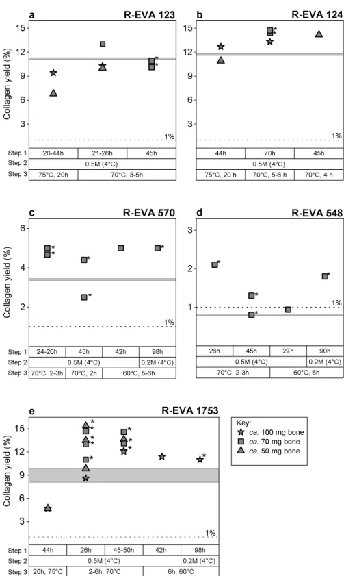
We initially applied our standard collagen extraction protocol to <100 mg bone material of the well-preserved bones. Three steps of the pretreatment protocol were then modified to see what effect this had on the collagen yield and quality of extracts from small bone aliquots (Fig. 1): step (1) the duration of the demineralisation stage; step (2) the strength of HCl during the demineralisation stage; step (3) the temperature and duration of the gelatinisation stage. Bone collagen yields along with elemental (C%, N% and C:N) and stable isotopic data (δ13C and δ15N) were used to evaluate the extracts from the different methods. In addition, Fourier Transform
Figure 1. Graphs showing the collagen yields from small aliquots of bone according to variations in pretreatment conditions: (a) R-EVA 123, (b) R-EVA 124 (c) R-EVA 570, (d) R-EVA 548 and (e) R-EVA 1753. Step 1: duration of the demineralisation stage. Step 2: strength of HCl during demineralisation. Step 3: duration and temperature of the gelatinisation stage (HCl pH3). In (a–d) the horizontal grey line shows the collagen yield from a large aliquot (>500 mg material) of the same bone. A higher number of data points are present for R-EVA 1753 (e) as an aliquot of this bone was extracted alongside each batch of samples. The horizontal grey band in e shows the range in collagen yield of repeated large extractions from the background bone. The dashed lines at 1% show the guideline minimum requirement for reliable 14C dating. Asterisks mark extracts which
the presence of possible carbon contaminants31,48,49. Detailed pretreatment information for all extracts can be
seen in Supplementary Dataset S1.
For the poorly preserved bones (Fig. 1c: R-EVA 570 and Fig. 1d: R-EVA 548) the pretreatment was softened in order to minimise collagen loss during the extraction. The weaker HCl (0.2 M) (step 2) and lower gelatinisa-tion temperature (60 °C) (step 3) required a greater time investment and did not necessarily increase the yield of collagen compared to using stronger acid (HCl 0.5 M) during demineralisation and higher temperatures (70 °C) during gelatinisation. For the poorly preserved samples, demineralisation in HCl 0.5 M generally occurred after one day (4 °C). As Schoeninger, et al.50 observed that one disadvantage of extracting collagen from solid chunks
was the tendency for incomplete demineralisation, several extracts were demineralised in HCl 0.5 M for two days. This resulted in lower collagen yields for the poorly preserved bones and in the case of R-EVA 548, the yield of these extracts was so low that the extracts were affected by C contamination to a large extent.
During the gelatinisation stage (step 3), the collagen yield was higher from aliquots which were removed from the heater block as soon as solubilisation had occurred compared to those left on the heater block for 20 h as per our standard protocol for >500 mg. For all bone samples >30,000 BP, solubilisation occurred in <6 h (Fig. 1), whereas R-EVA 1489 and R-EVA 1905 required up to 27 h for full solubilisation (Supplementary Dataset S1).
Of the extracts dated, two (R-EVA 548.13 and R-EVA 548.14) fell close to or under the minimum threshold (1%) for reliable 14C dating (Supplementary Dataset S1). There were small variations in elemental values between
pretreatments of the same bone but all values (Supplementary Dataset S1) fell within the accepted ranges of ‘well-preserved’ collagen32. The stable isotopic values were in keeping with the palaeodietary expectations for
each animal and were consistent between extracts. Analysis with FTIR was performed for all collagen extracts; each extract dated had a spectra characteristic of well-preserved collagen when compared to library spectra (see Figure 2. Summary of bone pretreatment protocols used at the MPI-EVA for large (left) and small (right) bone samples.
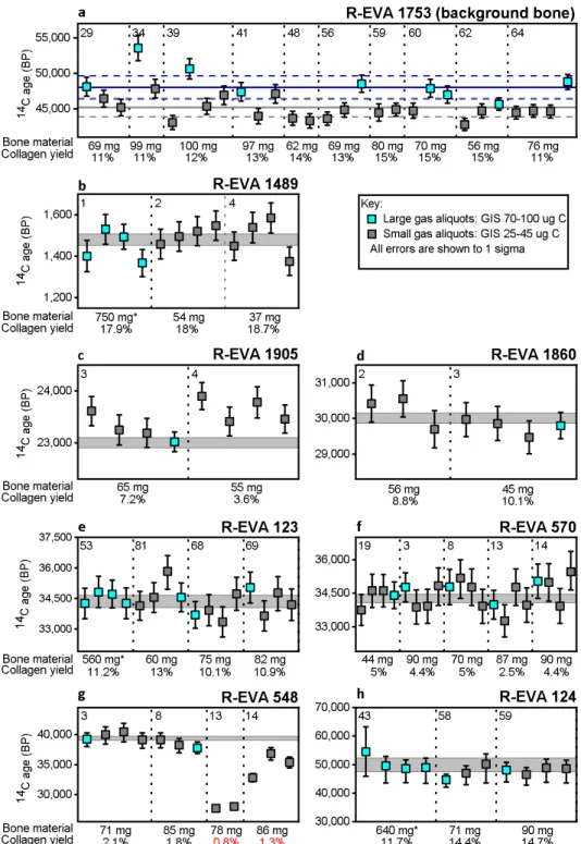
Figure 3. 14C gas measurements of small (25–40 μg C) and large (70–100 μg C) aliquots of collagen extracted from
eight bones (a–h) spanning the 14C time range. Each data point shows the 14C age (BP) and 1σ error (years) of a single
EA-GIS-AMS measurement. a) Shows the uncorrected measurements of background bone R-EVA 1753 (>50,000 BP). An aliquot of this bone was prepared alongside every batch of samples from sampling to measurement to monitor contamination introduced during sample preparation. These measurements were used in the age calculation of the other archaeological samples (b–h), according to session, size (small or large) and type (solid bone extract). The arithmetic mean and associated SD of system blank (IAEA-C1/phthalic anhydrite) measurements are shown as a solid horizontal blue line and dashed blue lines respectively for large 80–100 μg C measurements and as a solid horizontal grey line and dashed grey line for small 25–40 μg C measurements. For all gas measurements in graphs b-h: the absolute error of the blank has been set to 0.001 and an external error of 3.5‰ has been added to all measurements based on the long term standard deviation of standards. Dates >15,000 BP have been rounded to the nearest 10 years. Asymmetrical errors are shown where F14C ≤ 1σ*10. Grey shaded bands show the 1σ range of graphite dates measured from large extracts of the same bone. In a-h, the vertical dotted lines separate different collagen extracts of the same bone with the bone starting weight and collagen yield shown below. The number in the top left of each section is the preparation number of the bone, corresponding to Supplementary Dataset S1. Asterisks mark collagen extracts dated with the gas ion source reported in Fewlass, et al.29.
Supplementary Fig. S3). Considering the collagen yields and 14C measurements, the optimum pretreatment
pro-tocol for small aliquots of bone (<100 mg) is shown in Fig. 2.
14
C dating.
For each of the bones, several collagen extracts (bone weight ranging from 37–100 mg, marked with asterisks in Fig. 1) were dated using the EA-GIS-AixMICADAS (Fig. 3). Each collagen extract was split and measured multiple times. Between two and four replicates were measured containing ca. 30–40 μg C, run for the duration of one titanium (Ti) target (ca. 12 minutes) and for each bone >20,000 BP, a single aliquot containing ca. 80–90 μg C was measured over the duration of three targets to increase precision (see Supplementary Dataset S2). The gas ages obtained were compared to one or more graphite dates measured from collagen extracted from 500–700 mg bone material (Supplementary Dataset S2). Discussed here are measurements made from collagen extracted from solid pieces of bone. Details of measurements made from powdered aliquots (lower collagen yields) are included in the supplementary information.Figure 3 shows the ages obtained for each bone. The accuracy of the dates generated by the gas ion source is clearly seen in comparison with the graphite dates. Of the 74 new measurements made with the EA-GIS-AMS system shown in Fig. 3b–h, 69 measurements agree within the 95% confidence limit (2σ) of the corresponding graphite dates and 57 agree within 1σ. There are five measurements outside 2σ: four are measurements of the two collagen extracts (R-EVA 548.13; R-EVA 548.14) which fell at or below the minimum threshold of preservation suitable for 14C dating (Fig. 3g), and the last (R-EVA 1905.4.1; Aix-12023.2.1) is slightly older than the other
rep-licates of the same extract (Fig. 3c).
Chi-squared tests (χ2)51 were performed using the R_Combine feature in OxCal 4.252 using the F14C and
associated error for gas replicates of each collagen extract individually and for all replicates per bone. The replicate measurements are statistically indistinguishable for R-EVA 1489, R-EVA 1905, R-EVA 1860, R-EVA 123, R-EVA 570 and R-EVA 124 (output of all statistical tests are included in Supplementary Dataset S2), demonstrating the reproducibility of the measurements and consistency between different pretreatment batches across the range of the 14C timescale. In addition, all of the measurements of R-EVA 1489, R-EVA 123 and R-EVA 124 from this
study agree with the EA-GIS-AMS measurements made in 2016 reported in Fewlass, et al.29 (Supplementary
Dataset S2).
The exception is the roughly 40,000 year old bone R-EVA 548, which at ca. 1% collagen preservation repre-sents the limits of C14 dating. The gas dates obtained from the two low yield extracts (R-EVA 548.13 and R-EVA
548.14) were much younger than the other extracts of this bone (Fig. 3g), showing they had been affected by contamination from modern carbon. Due to the low yield, under normal circumstances R-EVA 548.13 would not have been passed for dating following pretreatment. Excluding these two extracts, the replicates from R-EVA 548.3 and R-EVA 548.8 are consistent with the graphite date for this bone.
For background bone R-EVA 1753 (>50,000 BP), the dates from the collagen extracts (Supplementary Dataset S3) were on par with the blank standards (IAEA-C1/phthalic anhydride) of equal size (Supplementary Dataset S4). As expected, the blank level in the EA-GIS system was affected by the reduction in sample size from 90 μg C to 30 μg C (Fig. 3a). The ages of the seven <50,000 BP samples were corrected with background collagen measurements of the same size (ca. 30 μg C or ca. 90 μg C) and type (solid/powder) measured during the same session.
Discussion
Using a slightly modified version of our standard pretreatment protocol the collagen yield from <100 mg bone material was of equally high quality as extracts from ‘large’ (>500 mg) bone samples. Decreasing sample size from ca. 100 mg to <50 mg bone material also had no detrimental effect on collagen yield. The agreement in age between multiple collagen extracts from different starting weights of bone (Fig. 3) indicates firstly that we obtain reproducible results with the pretreatment protocol and secondly, that the reduction in material during pretreatment did not detrimentally affect the results of 14C dating. In particular, the results indicate that the
clean-ing steps used for the ultrafilters are sufficient as any C remainclean-ing in the filters after cleanclean-ing would have a more pronounced effect on reduced sample sizes.
The main alteration to our standard protocol involved reduction in the duration of the gelatinisation stage, with samples removed from the heater block as soon as they had gelatinised (see Fig. 2). Different gelatinisation conditions have been well documented to affect the final extract quality and yield38,39,53,54. The higher collagen
yields from these extracts supports observations that gelatinised collagen is degraded by prolonged exposure to higher temperatures and acidity39,53.
R-EVA 548 represents a very challenging prospect for collagen extraction and radiocarbon dating due to the exceptionally low levels of preservation (<1% weight collagen) and old age (ca. 39,400 BP), even working with larger sample sizes. The harshest demineralisation (HCl 0.5 M, 2 days, 4 °C) applied to small aliquots of this bone (R-EVA 548.13; R-EVA 548.14) resulted in very low yields of ≤1 mg collagen, likely due to the solubilisation of collagen during the longer demineralisation stage. The resultant underestimated dates clearly show that these ali-quots were massively affected by modern carbon contamination. Prior to dating, the consideration of the quality of the extract is crucial in order to obtain reliable dates. Given the low yield of collagen (≤1%) following pretreat-ment, under normal circumstances these extracts would not been dated or would have been treated with caution. This bone demonstrates the difficulty of pretreatment of poorly preserved bones at the limit of the 14C method.
At such small sample sizes, the consideration of the background correction is crucial. The gas measure-ments of R-EVA 1489, R-EVA 1905, R-EVA 1860, R-EVA 123, R-EVA 570, R-EVA 548 and R-EVA 124 were all corrected with gas measurements of background bone collagen (R-EVA 1753) of equal size (ca. 30 µg C or ca. 90 µg C) prepared alongside every batch of samples and measured during the same measurement session to account for any C added during sample preparation and measurement. Figure 3a shows the ages obtained for the
background bone containing ca. 25–40 μg C (small) and ca. 80–100 μg C (large). The large measurements (mean F14C = 0.0024, SD = 0.0006, n = 9, equivalent to 48,600 BP) are on par with the system blank (either IAEA-C1 or
phthalic anhydride) measurements of equal size (mean F14C = 0.0026, SD = 0.0006, n = 7, equivalent to 48,000
BP) (Supplementary Datasets S3 and S4), indicating that no carbon contamination was introduced during sam-ple preparation. An increased sensitivity to modern 14C is to be expected at lower levels of carbon and it is clear
that the smaller background collagen measurements are generally younger. The 25–40 μg C background collagen samples (mean F14C = 0.0039, SD = 0.0007, n = 22, equivalent to 44,530 BP) are likewise equal to the system blank
measurements of equal size (mean F14C = 0.0036, SD = 0.0006, n = 5, equivalent to 45,180 BP) (Supplementary
Datasets S3 and S4). These values are lower than previously published values for blank IAEA-C1 samples meas-ured at CEREGE reported in Bard, et al.30 (F14C = 0.02 for sample sizes around 30 µg C and F14C = 0.005 for
samples of 80–100 µg C) and to phthalic anhydride blanks measured at ETH Zurich reported in McIntyre, et al.24
(mean F14C = 0.0046 ± 0.0012, n = 6, size range 84–100 μg C). The results indicate the lower limit of 14C detection
with the gas ion source to be around F14C = 0.004. As demonstrated by R-EVA 124, beyond this limit the minute
levels of 14C can be measured but the uncertainty of the background correction dominates accuracy and precision.
The system blank of the EA-GIS-AMS is affected by the carbon content of the silver cups, cross-talk of the zeo-lite trap and the cleanliness of the ion source at the time of the measurement24. The mass (M
c) and F14C (F14Cc) of
the constant contamination of the EA + GIS system was deduced by least square regression of modern carbonate and blanks (IAEA-C1) with sample weights ranging between 3 and 100 µg C to be Mc = 0.55 ± 0.05 ug C and
F14C
c = 0.12 ± 0.0355. The silver cups (5 × 3 mm from Elementar; cleaned at 800 °C, 2 h) had a consistent carbon
contribution of 0.049 ± 0.02 µg C. The zeolite trap was heated (450 °C) and the system was flushed with helium between samples to minimize cross-contamination. However, small amounts of C may reside in the zeolite trap after flushing which has been demonstrated to have a large influence on samples <20 µg carbon23,55. With this
in mind, even our ‘small’ samples were kept >20 μg carbon. To further alleviate problems of cross-talk, samples were run in order of increasing activity (oldest to youngest) according to the standard practice55. Background
corrections of samples were applied according to sample size and an external error was added during the age calculation of all samples based on the long term standard deviation of standards and blanks (error 2 described in Fewlass, et al.29).
In a real life situation, if a small bone sample yielded a high amount of collagen (for example, the mammoth bone R-EVA 123 or the Medieval human bone R-EVA 1489 included in this study), dating with graphite targets would be preferentially undertaken as the precision achieved is much higher and measurements can be made routinely. However, the results of this study demonstrate that the gas ion source can produce an accurate radiocar-bon date at low precision from as little as 30 µg C. The precision of the date can be improved when larger sample sizes (up to 100 µg C) are available for measurement over several targets (as demonstrated in Fig. 3). In order to assess variability in handling and blank contribution, in this study we compared multiple measurements of ca. 30 µg C with larger aliquots containing ca. 90 µg C. When taking the weighted mean and error of the three small aliquots the precision achieved is higher compared to the single large measurement of a roughly equal amount of carbon. However, as the likelihood of contamination being introduced via handling, the EA-GIS or the silver cup is increased for the smaller sample sizes, the preferred method for measuring larger samples would be to measure several targets from a single syringe, rather than splitting a sample into smaller aliquots. Although the measure-ment of gas samples requires more supervision than graphite targets, the direct coupling of the EA with the GIS significantly reduces sample preparation time by cutting out the graphitisation step which poses a large risk of contamination at such small sample sizes. Therefore in situations where sample size is limited the gas ion source offers an attractive solution for archaeological, as well as environmental, applications.
Even working with the assumption of 1% collagen preservation, in theory sufficient collagen could be extracted from less than 10 mg bone material to obtain a 14C date using the EA-GIS-AMS. However in order to
assess the quality of the extract prior to dating and obtain high-resolution stable isotopic data for palaeodietary reconstruction, collagen should also be analysed with an EA-IRMS. At 1%, around 40 mg bone material would supply enough collagen for dating and isotopic analysis. For any sample >1% preservation, excess collagen would be available for further analyses and/or multiple aliquots could be measured with the gas ion source to achieve better counting statistics and thus increase precision. Bearing this in mind, when dating highly precious bone it would be useful to assess the preservation of the artefact prior to sampling or have an understanding of collagen preservation at the archaeological site (for example if other fauna has been sampled for isotopic or 14C dating
purposes). Bones of high patrimonial value could be sampled strategically – i.e. for older samples expected to have less than 10% collagen preservation 40 mg bone material could be sampled, whereas for well-preserved Holocene bone much smaller samples could be taken. The case of R-EVA 548 demonstrates that for very old sam-ples (>35,000 BP) with very poor levels of preservation (1–2%), yields falling below 1 mg collagen can be subject to severe contamination issues.
The results presented here provide further confirmation that 14C measurements using the gas ion source of the
MICADAS are stable, reproducible and accurate, reaching a level of precision suitable for dating archaeological samples particularly for Palaeolithic samples back to 40,000 BP. In this respect this technique will be highly useful for directly dating precious archaeological bone where limited material is available.
Methods
sample selection.
Eight bones were selected to span the 14C timescale (back to 50,000 BP) at a range ofpres-ervation typical for archaeological bones. Collagen extracts from bones R-EVA 124, R-EVA 123 and R-EVA 1489 were previously dated using both graphite targets and the gas ion source in Fewlass, et al.29. R-EVA 124 was
pre-viously labelled as a bison bone but recent aDNA analysis has identified it as belonging to a woolly rhinoceros56.
R-EVA 548 and R-EVA 570 are two faunal long bones from Teixoneres, Spain. R-EVA 1860 is a faunal long bone excavated from the site of Ranis, Germany and R-EVA 1905 is a predominantly trabecular fragment of horse bone
excavated from Pietraszyn, Poland. R-EVA 1753 is a well-preserved cave bear rib known to date beyond the 14C
timescale based on repeated measurements. As standard practice, an aliquot of this bone is extracted and dated alongside every batch of samples to monitor contamination introduced during sample preparation and is used in the age correction of the unknown samples. This is the referred to in the text as the ‘background bone’.
Collagen extraction.
For each bone, large aliquots (500–700 mg material) were pretreated using our stand-ard acid-base-acid + gelatinisation + ultrafiltration protocol (see Fig. 2) based on Talamo and Richards36 topro-duce collagen for dating with graphite targets.
In order to optimise our standard protocol for sample sizes <100 mg, small aliquots of each bone were pre-treated multiple times to compare collagen yields and sample quality. Firstly, the outer surface of bone was removed using a sandblaster and aliquots were taken using a rotary drill. Fine diamond grit disc drill pieces were used to remove solid pieces of bone. Fine powder was drilled using round tungsten carbide burs (2.3 mm diameter). Aliquots were weighed via a microbalance into cleaned glass tubes. Solid samples were demineralised in HCl at 4 °C with regular visual and mechanical checks and monitoring of CO2 effervescence. For powdered
samples, HCl was added and samples were monitored at room temperature (RT) until CO2 effervescence had
stopped. Following demineralisation, samples were rinsed with ultra-pure Milli-Q water to a neutral pH. Samples were treated with NaOH (0.1 M) at RT for 10 min to remove humic acid contamination and re-acidified with HCl (0.5 M). If a considerable colour change was observed, NaOH was changed and left for another 10 min. Samples were then gelatinised in weak HCl (pH 3) on a heater block set to 60 °C, 70 °C or 75 °C. Samples were either left for 20 h (as per our standard pretreatment), or regularly monitored and removed from the heater block when the sample had fully solubilised. The resultant gelatin was filtered to remove large particles >80 µm (Ezee filters, Elkay labs, UK) and ultrafiltered with Sartorius VivaSpin Turbo 15 (30 kDa MWCO) ultrafilters precleaned according to Brock, et al.43 to separate the high molecular weight fraction (>30kD) for freeze drying (48 h). For details of
acid strength, duration of treatment and temperature during pretreatment of samples <100 mg, see Fig. 1 and Supplementary Dataset S1.
Collagen quality assessment.
To assess the quality of the collagen, all extracts were analysed via EA-IRMS to obtain elemental (C%, N%, C:N) and stable isotopic data (δ13C and δ15N). Collagen (ca. 400 μg) was weighedinto tin cups using a microbalance and measured on a ThermoFinnigan Flash EA coupled to a Thermo Delta plus XP isotope ratio mass spectrometer (IRMS). Stable carbon isotope ratios were expressed relative to VPDB (Vienna PeeDee Belemnite) and stable nitrogen isotope ratios were measured relative to AIR (atmospheric N2),
using the delta notation (δ) in parts per thousand (‰). Repeated analysis of both internal and international stand-ards indicates an analytical error of 0.2‰ (1σ) for δ13C and δ15N. Where sufficient material was available, collagen
(ca. 300 μg) was homogenized and mixed with ∼40 mg of IR grade KBr powder in an agate mortar and pestle, pressed into a pellet using a manual hydraulic press (Wasserman) and analysed with an Agilent Technologies Cary FTIR Spectrometer with a DTGS detector. Spectra were recorded in transmission mode at 4 cm−1 resolution
with averaging of 34 scans between 4000 and 400 cm−1 using Resolution Pro software (Agilent Technologies). The
spectra were evaluated and compared to library spectra of well-preserved collagen and bone to look for evidence of incomplete demineralisation, degraded collagen or the presence of any exogenous material in the extracts.
AMS graphite measurements.
Each bone was pretreated as per our standard protocol fromapproxi-mately 500 mg material. From theses extracts, approxiapproxi-mately 3–5 mg collagen was weighed into pre-cleaned tin cups at the MPI-EVA and sent to the Curt-Engelhorn-Centre for Archaeometry Klaus-Tschira-AMS facility in Mannheim, Germany (lab code: MAMS) for graphite dating. The samples were combusted in an EA and the sample CO2 was converted catalytically to graphite. The samples were dated using the MICADAS-AMS57. Age
and error calculation of unknown samples was performed using BATS software58, using background collagen
samples and standards measured in the same batch, with an added external error of 1‰ as per their standard practice. Collagen samples measured at CEREGE were weighed into tin cups (ca. 2 mg), combusted in a vario MICRO cube EA (Elementar Analysensysteme GmbH, Germany), graphitized using the AGE 3 and dated using the AixMICADAS. Oxalic acid standards and background collagen samples measured in the same session were used to calculate the age of the samples. An external error of 1‰ was also propagated in the error calculation.
AMS gas ion source measurements.
Small aliquots (<100 mg) of the same bones were pretreated topurify the collagen. Three or four aliquots of each collagen extract (containing ca. 25–40 μg C and a single aliquot per bone containing ca. 80–100 μg C) were measured via a microbalance into pre-cleaned silver cups (800 °C, 2 h). These were placed into the auto-sampler of a vario MICRO cube EA which was directly coupled to the gas ion source of the AixMICADAS via the GIS20,22. Following combustion, sample CO
2 was adsorbed on a zeolite
trap and subsequently expanded to the syringe of the GIS where it was mixed with He (5% CO2) and introduced
to the gas ion source at a flow rate of ca. 2 µg C/min. The EA-GIS system was flushed with helium between sam-ples. Pre-cleaned titanium (Ti) gas targets were pre-sputtered for approximately two minutes in the ion source to remove any remaining surface contamination before the sample CO2 injection. Around 30–40 µg C was
con-sumed by the AMS over the duration of one Ti target21,55. For the large aliquots containing ca. 80–90 μg C
meas-urements were performed over multiple targets (which can be changed during measurement). Each step was fully controlled via the gas-interface handling software.
The gas measurements in this study were made over two measurement sessions six months apart, both carried out shortly after the ion source had been cleaned. Each measurement session commenced with two oxalic acid II NIST standards (from a gas canister) to normalize and correct samples for fractionation. Blank (14C-free) CO
2
samples (also from a gas canister) were then measured to purge the system and reach a stable operational level (F14C < 0.004) (these measurements were not used in age calculation). In the first session, carbonate reference
material (IAEA-C1) were run prior to the collagen samples to check the background level of the instrument and begin the measurement of old samples under optimal conditions. In the second measurement session, phthalic anhydride was run for the same purpose. In order to alleviate problems of memory effect, the GIS system was flushed with helium between samples and samples were measured in order of increasing activity as per standard procedure (for further discussion, see Tuna, et al.55). Low energy ion currents for the gas analyses were in the
range of 10–15 μA. BATS58 was used for data reduction. The uncorrected collagen background (R-EVA 1753)
measurements of the corresponding type (piece/powder) and equal size were used to correct the archaeological samples measured in the same session (i.e. ‘small’ sample aliquots were corrected only with ‘small’ background collagen samples). For all samples, the long term standard deviation of blanks (F14C = 0.001) was used as the
absolute blank error and an external error of 3.5‰ was added to take into account the long-term variability of standards (‘error 2’ described in Fewlass, et al.29).
Data Availability
All data generated or analysed during this study are included in this article and the accompanying supplementary information files.
References
1. Oakley, K. P. Dating Skeletal Material. Science 140, 488, https://doi.org/10.1126/science.140.3566.488 (1963).
2. Berger, R., Horney, A. G. & Libby, W. F. Radiocarbon Dating of Bone and Shell from Their Organic Components. Science 144, 995–1001, https://doi.org/10.1126/science.144.3621.995 (1964).
3. Wood, R. From revolution to convention: the past, present and future of radiocarbon dating. Journal of Archaeological Science 56, 61–72, https://doi.org/10.1016/j.jas.2015.02.019 (2015).
4. Pearson, A. Microscale AMS 14C Measurement at NOSAMS. Radiocarbon 40, 61–75 (1998).
5. Hua, Q., Zoppi, U., Williams, A. A. & Smith, A. M. Small-mass AMS radiocarbon analysis at ANTARES. Nuclear Instruments and
Methods in Physics Research Section B: Beam Interactions with Materials and Atoms 223, 284–292, https://doi.org/10.1016/j. nimb.2004.04.057 (2004).
6. Santos, G. M., Southon, J. R., Griffin, S., Beaupre, S. R. & Druffel, E. R. M. Ultra small-mass AMS 14C sample preparation and analyses at KCCAMS/UCI Facility. Nuclear Instruments and Methods in Physics Research Section B: Beam Interactions with Materials
and Atoms 259, 293–302, https://doi.org/10.1016/j.nimb.2007.01.172 (2007).
7. Smith, A. M., Petrenko, V. V., Hua, Q., Southon, J. & Brailsford, G. The Effect of N2O, Catalyst, and Means of Water Vapor Removal on the Graphitization of Small CO2 Samples. Radiocarbon 49, 245–254 (2007).
8. Genberg, J., Stenstrom, K., Elfman, M. & Olsson, M. Development of graphitization of μg-sized samples at Lund University.
Radiocarbon 52, 1270–1276 (2010).
9. Delqué-Količ, E. et al. Advances in Handling Small Radiocarbon Samples at the Laboratoire de Mesure du Carbone 14 in Saclay, France. Radiocarbon 55, 648–656, https://doi.org/10.1017/S0033822200057805 (2013).
10. Liebl, J. et al. Carbon background and ionization yield of an AMS system during C-14 measurements of microgram-size graphite samples. Nuclear Instruments and Methods in Physics Research Section B: Beam Interactions with Materials and Atoms 294, 335–339, https://doi.org/10.1016/j.nimb.2012.06.015 (2013).
11. Walter, S. R. S. et al. Ultra-Small Graphitization Reactors for Ultra-Microscale C-14 Analysis at the National Ocean Sciences Accelerator Mass Spectrometry (NOSAMS) Facility. Radiocarbon 57, 109–122, https://doi.org/10.2458/azu_rc.57.18118 (2015). 12. Freeman, E., Skinner, L. C., Reimer, R., Scrivner, A. & Fallon, S. Graphitization of Small Carbonate Samples for Paleoceanographic
Research at the Godwin Radiocarbon Laboratory, University of Cambridge. Radiocarbon 58, 89–97, https://doi.org/10.1017/ RDC.2015.8 (2016).
13. Steier, P., Liebl, J., Kutschera, W., Wild, E. M. & Golser, R. Preparation Methods of μg Carbon Samples for 14C Measurements.
Radiocarbon 59, 803–814, https://doi.org/10.1017/RDC.2016.94 (2016).
14. Ertun, T. et al. Progress in AMS target production of sub-milligram samples at the NERC radiocarbon laboratory. Radiocarbon 47, 453–464 (2005).
15. Ruff, M. et al. Gaseous radiocarbon measurements of small samples. Nuclear Instruments and Methods in Physics Research Section B:
Beam Interactions with Materials and Atoms 268, 790–794, https://doi.org/10.1016/j.nimb.2009.10.032 (2010).
16. Cersoy, S. et al. Radiocarbon dating minute amounts of bone (3–60 mg) with ECHoMICADAS. Scientific Reports 7, 7141, https:// doi.org/10.1038/s41598-017-07645-3 (2017).
17. Wacker, L., Němec, M. & Bourquin, J. A revolutionary graphitisation system: Fully automated, compact and simple. Nuclear
Instruments and Methods in Physics Research Section B: Beam Interactions with Materials and Atoms 268, 931–934, https://doi. org/10.1016/j.nimb.2009.10.067 (2010).
18. Ruff, M. et al. A gas ion source for radiocarbon measurements at 200 kV. Radiocarbon 49, 307–314 (2007). 19. Wacker, L. et al. MICADAS: Routine and High-Precision Radiocarbon Dating. Radiocarbon 52, 252–262 (2010).
20. Ruff, M. et al. On-line radiocarbon measurements of small samples using elemental analyzer and MICADAS gas ion source.
Radiocarbon 52, 1645–1656 (2010).
21. Fahrni, S. M., Wacker, L., Synal, H. A. & Szidat, S. Improving a gas ion source for 14C AMS. Nuclear Instruments and Methods in
Physics Research Section B: Beam Interactions with Materials and Atoms 294, 320–327, https://doi.org/10.1016/j.nimb.2012.03.037 (2013).
22. Wacker, L. et al. A versatile gas interface for routine radiocarbon analysis with a gas ion source. Nuclear Instruments and Methods in
Physics Research Section B: Beam Interactions with Materials and Atoms 294, 315–319, https://doi.org/10.1016/j.nimb.2012.02.009 (2013).
23. Wacker, L., Lippold, J., Molnár, M. & Schulz, H. Towards radiocarbon dating of single foraminifera with a gas ion source. Nuclear
Instruments and Methods in Physics Research Section B: Beam Interactions with Materials and Atoms 294, 307–310, https://doi. org/10.1016/j.nimb.2012.08.038 (2013).
24. McIntyre, C. P. et al. Online 13C and 14C Gas Measurements by EA-IRMS–AMS at ETH Zürich. Radiocarbon 59, 893–903, https:// doi.org/10.1017/RDC.2016.68 (2016).
25. Bonvalot, L. et al. Estimating contributions from biomass burning and fossil fuel combustion by means of radiocarbon analysis of carbonaceous aerosols: application to the Valley of Chamonix. Atmos. Chem. Phys. 16, 13753–13772, https://doi.org/10.5194/acp-2016-351 (2016).
26. Hoffmann, H. M. Micro radiocarbon dating of the particulate organic carbon fraction in Alpine glacier ice: method refinement, critical
evaluation and dating applications, Universität Heidelberg (2016).
27. Gottschalk, J. et al. Radiocarbon Measurements of Small-Size Foraminiferal Samples with the Mini Carbon Dating System (Micadas) at the University of Bern: Implications for Paleoclimate Reconstructions. Radiocarbon 60, 469–491, https://doi.org/10.1017/ Rdc.2018.3 (2018).
28. Haghipour, N. et al. Compound-Specific Radiocarbon Analysis by Elemental Analyzer–Accelerator Mass Spectrometry: Precision and Limitations. Analytical Chemistry 91, 2042–2049, https://doi.org/10.1021/acs.analchem.8b04491 (2019).
29. Fewlass, H. et al. Size Matters: Radiocarbon Dates of <200 µg Ancient Collagen Samples with AixMICADAS and Its Gas Ion Source.
Radiocarbon 60, 425–439, https://doi.org/10.1017/rdc.2017.98 (2017).
30. Bard, E. et al. AixMICADAS, the accelerator mass spectrometer dedicated to 14C recently installed in Aix-en-Provence, France.
Nuclear Instruments and Methods in Physics Research Section B: Beam Interactions with Materials and Atoms 361, 80–86, https://doi. org/10.1016/j.nimb.2015.01.075 (2015).
31. DeNiro, M. J. & Weiner, S. Chemical, enzymatic and spectroscopic characterization of “collagen” and other organic fractions from prehistoric bones. Geochimica et Cosmochimica Acta 52, 2197–2206, https://doi.org/10.1016/0016-7037(88)90122-6 (1988). 32. van Klinken, G. J. Bone Collagen Quality Indicators for Palaeodietary and Radiocarbon Measurements. Journal of Archaeological
Science 26, 687–695 (1999).
33. Longin, R. New Method of Collagen Extraction for radiocarbon Dating. Nature 231, 241–242 (1971).
34. Jørkov, M. L. S., Heinemeier, J. & Lynnerup, N. Evaluating bone collagen extraction methods for stable isotope analysis in dietary studies. Journal of Archaeological Science 34, 1824–1829, https://doi.org/10.1016/j.jas.2006.12.020 (2007).
35. Pestle, W. J. Chemical, elemental, and isotopic effects of acid concentration and treatment duration on ancient bone collagen: an exploratory study. Journal of Archaeological Science 37, 3124–3128, https://doi.org/10.1016/j.jas.2010.07.013 (2010).
36. Talamo, S. & Richards, M. A comparison of bone pretreatment methods for AMS dating of samples >30,000 BP. Radiocarbon 53, 443–449 (2011).
37. Fulop, R.-H., Heinze, S., John, S. & Rethemeyer, J. Ultrafiltration of bone samples is neither the problem nor the solution.
Radiocarbon 55, 491–500 (2013).
38. Cersoy, S., Zazzo, A., Lebon, M., Rofes, J. & Zirah, S. Collagen Extraction and Stable Isotope Analysis of Small Vertebrate Bones: A Comparative Approach. Radiocarbon 59, 679–694, https://doi.org/10.1017/rdc.2016.82 (2016).
39. Brown, T. A., Nelson, D. E., Vogel, J. S. & Southon, J. R. Improved collagen extraction by modified longin method. Radiocarbon 30, 171–177 (1988).
40. Higham, T. European Middle and Upper Palaeolithic radiocarbon dates are often older than they look: problems with previous dates and some remedies. Antiquity 85, 235–249 (2011).
41. Hüls, C. M., Grootes, P. M. & Nadeau, M. J. Ultrafiltration: Boon or Bane? Radiocarbon 51, 613–625, https://doi.org/10.1017/ S003382220005596X (2009).
42. Bronk Ramsey, C., Higham, T., Bowles, A. & Hedges, R. Improvements to the pretreatment of bone at Oxford. Radiocarbon 46, 155–164 (2004).
43. Brock, F., Bronk Ramsey, C. & Higham, T. Quality assurance of ultrafiltered bone dating. Radiocarbon 49, 187–192 (2007). 44. Brock, F., Higham, T., Ditchfield, P. & Bronk Ramsey, C. Current pretreatment methods for AMS radiocarbon dating at the Oxford
Radiocarbon Accelerator Unit (ORAU). Radiocarbon 52, 103–112 (2010).
45. Higham, T. F. G., Jacobi, R. M. & Ramsey, C. B. AMS radiocarbon dating of ancient bone using ultrafiltration. Radiocarbon 48, 179–195 (2006).
46. Wood, R. E. et al. Radiocarbon dating casts doubt on the late chronology of the Middle to Upper Palaeolithic transition in southern Iberia. Proceedings of the National Academy of Sciences 110, 2781–2786 (2013).
47. Hublin, J. J. et al. Radiocarbon dates from the Grotte du Renne and Saint-Cesaire support a Neandertal origin for the Chatelperronian. Proceedings of the National Academy of Sciences 109, 18743–18748, https://doi.org/10.1073/pnas.1212924109 (2012).
48. Yizhaq, M. et al. Quality Controlled Radiocarbon Dating of Bones and Charcoal from the Early Pre-Pottery Neolithic B (PPNB) of Motza (Israel). Radiocarbon 47, 193–206, https://doi.org/10.1017/S003382220001969X (2005).
49. D’Elia, M. et al. Evaluation of possible contamination sources in the 14C analysis of bone samples by FTIR spectroscopy.
Radiocarbon 49, 201–210 (2007).
50. Schoeninger, M. J., Moore, K. M., Murray, M. L. & Kingston, J. D. Detection of bone preservation in archaeological and fossil samples. Applied Geochemistry 4, 281–292, https://doi.org/10.1016/0883-2927(89)90030-9 (1989).
51. Ward, G. K. & Wilson, S. R. Procedures for Comparing and Combining Radiocarbon Age Determinations: A Critique. Archaeometry
20, 19–31, https://doi.org/10.1111/j.1475-4754.1978.tb00208.x (1978).
52. Bronk Ramsey, C. Bayesian Analysis of Radiocarbon Dates. Radiocarbon 51, 337–360 (2009).
53. Semal, P. & Orban, R. Collagen Extraction from Recent and Fossil Bones: Quantitative and Qualitative Aspects. Journal of
Archaeological Science 22, 463–467, https://doi.org/10.1006/jasc.1995.0045 (1995).
54. Brock, F., Geoghegan, V., Thomas, B., Jurkschat, K. & Higham, T. F. G. Analysis of Bone “Collagen” Extraction Products for Radiocarbon Dating. Radiocarbon 55, 445–463, https://doi.org/10.1017/S0033822200057581 (2013).
55. Tuna, T., Fagault, Y., Bonvalot, L., Capano, M. & Bard, E. Development of small CO2 gas measurements with AixMICADAS. Nuclear
Instruments and Methods in Physics Research Section B: Beam Interactions with Materials and Atoms 437, 93–97, https://doi. org/10.1016/j.nimb.2018.09.012 (2018).
56. Korlević, P., Talamo, S. & Meyer, M. A combined method for DNA analysis and radiocarbon dating from a single sample. Scientific
Reports 8, 4127, https://doi.org/10.1038/s41598-018-22472-w (2018).
57. Kromer, B., Lindauer, S., Synal, H. A. & Wacker, L. MAMS - A new AMS facility at the Curt-Engelhorn-Centre for Achaeometry, Mannheim, Germany. Nuclear Instruments and Methods in Physics Research Section B: Beam Interactions with Materials and Atoms
294, 11–13, https://doi.org/10.1016/j.nimb.2012.01.015 (2013).
58. Wacker, L., Christl, M. & Synal, H. A. Bats: A new tool for AMS data reduction. Nuclear Instruments and Methods in Physics Research
Section B: Beam Interactions with Materials and Atoms 268, 976–979, https://doi.org/10.1016/j.nimb.2009.10.078 (2010).
Acknowledgements
We would like to thank the Max Planck Society for funding this research. The AixMICADAS was acquired and is operated in the framework of the EQUIPEX project ASTER-CEREGE (PI E. Bard) with additional matching funds from the Collège de France, which also supports the salaries of the authors from CEREGE. We are indebted to Dr. Raquel Silva Maria and Sven Steinbrenner of the Department of Human Evolution at the MPI-EVA for technical assistance. We give kind thanks to Caterina Pangrazzi, Jordi Rosell, Ruth Blasco, Marcel Weiss and Andrzej Wisniewski for access to samples during the course of the study.
Author Contributions
H.F., S.T., B.K., J.-J.H. and E.B. devised the study; H.F. carried out sample pretreatment, FTIR and EA-IRMS analyses under the supervision of S.T.; T.T. and Y.F. performed EA-GIS-AMS measurements; T.T., B.K. and H.F. performed data reduction; H.F. wrote the paper with input from all authors.
Additional Information
Supplementary information accompanies this paper at https://doi.org/10.1038/s41598-019-41557-8. Competing Interests: The authors declare no competing interests.
Publisher’s note: Springer Nature remains neutral with regard to jurisdictional claims in published maps and institutional affiliations.
Open Access This article is licensed under a Creative Commons Attribution 4.0 International License, which permits use, sharing, adaptation, distribution and reproduction in any medium or format, as long as you give appropriate credit to the original author(s) and the source, provide a link to the Cre-ative Commons license, and indicate if changes were made. The images or other third party material in this article are included in the article’s Creative Commons license, unless indicated otherwise in a credit line to the material. If material is not included in the article’s Creative Commons license and your intended use is not per-mitted by statutory regulation or exceeds the perper-mitted use, you will need to obtain permission directly from the copyright holder. To view a copy of this license, visit http://creativecommons.org/licenses/by/4.0/.