HAL Id: hal-01718887
https://hal.archives-ouvertes.fr/hal-01718887
Submitted on 28 Mar 2018HAL is a multi-disciplinary open access
archive for the deposit and dissemination of sci-entific research documents, whether they are pub-lished or not. The documents may come from teaching and research institutions in France or abroad, or from public or private research centers.
L’archive ouverte pluridisciplinaire HAL, est destinée au dépôt et à la diffusion de documents scientifiques de niveau recherche, publiés ou non, émanant des établissements d’enseignement et de recherche français ou étrangers, des laboratoires publics ou privés.
Structural and magnetic properties of He+ irradiated
Co2MnSi Heusler alloys
Iman Abdallah, Nicolas Ratel-Ramond, C. Magen, Béatrice Pécassou, Robin
Cours, Alexandre Arnoult, Marc Respaud, Jean-François Bobo, Gérard
Benassayag, Etienne Snoeck, et al.
To cite this version:
Iman Abdallah, Nicolas Ratel-Ramond, C. Magen, Béatrice Pécassou, Robin Cours, et al.. Structural and magnetic properties of He+ irradiated Co2MnSi Heusler alloys. Materials Research Express, IOP Publishing Ltd, 2016, 3 (4), pp.046101. �10.1088/2053-1591/3/4/046101�. �hal-01718887�
1
Structural and magnetic properties of He
+irradiated
Co
2MnSi heusler alloys
I. Abdallah
1, N. Ratel-Ramond
1, C. Magen
2,3, B. Pecassou
1, R. Cours
1, A. Arnoult
4, M.
Respaud
5, J.F. Bobo
1, G. BenAssayag
1, E. Snoeck
1and N. Biziere
1.
1. CEMES-CNRS, UPR 8011, Université de Toulouse, 29 rue Jeanne Marvig, BP 94347,
31055 Toulouse Cedex 04, France.
2. LMA, INA-
ARAID, and Departamento de Fısica de la Materia Condensada, Universidad
de Zaragoza, 50018 Zaragoza, Spain.
3. 3. TALEM, CEMES-INA, CNRS-Universidad de Zaragoza, 31055 Toulouse Cedex 04,
France
4. CNRS, LAAS, F-31000 Toulouse, France.
6. Laboratoire de Physique et Chimie des Nano-Objets, LPCNO, UMR 5215
INSA-UPS-CNRS, Université de Toulouse; 135 avenue de Rangueil, 31077 Toulouse, France.
Contact Author :
nicolas.biziere@cemes.fr
Keywords : Heusler Alloys, Structure, X-ray diffraction, HAADF-STEM,
Abstract: We have investigated the atomic disorder induced by a 150 keV He+ ion irradiation in a 40 nm thick Co2MnSi Heusler alloy. Disorder parameters on each atomic site are deduced from normal and anomalous X-ray diffraction measurements with Co and Cu Kα sources. While the film grows mainly in the L21 phase with inclusion of B2 grains, we observe an increase of both the Mn-Si and Co-Mn exchanges with the ion fluence. HAADF-STEM analysis demonstrates that the increase in Mn-Si disorder corresponds to a growing size of the B2 grains while the Co-Mn exchange is accounted for a D03 disorder type in the L21 matrix. These structural modifications are shown to decrease the
2
average magnetization of the alloy, which is due to D03 disorder and local defects induced by irradiation.
I. Introduction
In the last ten years, full Heusler compounds with general formula X2YZ have become a major topic in spintronics, especially for spin torque devices requiring low damping and high spin polarization. Among them, Co2MnSi (CMS) is a very promising candidate. It is predicted to be half metallic, with a Curie temperature well above the room temperature [1-4] and a very low Gilbert damping coefficient as compared to other ferromagnetic metals [5,6].
In all Heusler alloys, the magnetic properties and the atomic order are intimately related. For example, half metallicity has been predicted in CMS for the L21 or B2 structure only [7]. The L21 structure is the most ordered phase and corresponds to 8 body centered cubic (bcc) sub-lattices having the Co atoms at the corners of the bcc cells and the center sites occupied either by the Mn or Si atoms. A random distribution between Mn and Si or between Mn and Co atoms corresponds to the B2 or D03 order respectively while a random distribution of the Co, Mn and Si atoms between the different atomic sites leads to the disordered A2 phase.
Ion irradiation with light ions is an efficient technique to improve the local chemical order in different magnetic alloys such as FePt [8] or more recently in Heusler alloy [9]. Indeed, Gaier et al. demonstrated that He+ ion irradiation at 30 keV increases the long range order parameter in CMS grown in the B2 phase. Therefore, ion irradiation appears as a very interesting complementary or alternative technique to high temperature annealing, incompatible with microelectronic processes. Before this, further studies about the structural modification induced by irradiation are needed. In this work, we offer to study the structural modification in both the L21 and B2 order in Co2MnSi irradiated with 150 keV He+ ions.
Several techniques can be used to characterize the crystal structure such as neutron diffraction, nuclear magnetic resonance, X-ray absorption and circular magnetic dichroism (XAS/XMCD), photoemission spectroscopy (HAXPES) or HAADF-STEM techniques [10-13]. However, X-ray diffraction is an accessible and commonly used technique for macroscopic characterization of the crystal order [14-16]. Quantitative information on the presence of the different phases can be extracted from the measurements of the intensity of different diffraction peaks. For example, superlattice (h,k,l) diffraction peaks for which with h, k and l are odd numbers (e.g. 111) only appear when the L21 and/or D03 phases are present while diffraction peaks for which h+k+l = 4n+2 (e.g. 002) appear for L21, D03 and B2 phases. Finally, the fundamental peaks whose h+k+l = 4n (e.g. 022) appear for all crystal phases.
3
One of the major issues encountered in X-ray diffraction experiments to discriminate between the various ordered phases in CMS is that the Co and Mn scattering factors are very close at the Cu Kα edge, making it almost impossible to distinguish between D03 and L21 phases. This issue can however be overpassed using a Co-Kα source because, due to anomalous diffraction of the Co at the K-edge, the Co and Mn scattering factors becomes very different and Co and Mn atoms can be differentiated.
Thus, the different disorder parameters in CMS can be obtained combining diffraction measurements using Cu and Co Kα sources. The method, based on a model proposed by Niculescu et al. [17], has recently been applied by Takamura et al. [18] for the structural characterization of Co2FeSi. In this model α, β and γ are three disorder parameters. α corresponds to the number of Mn atoms located on Si sites and then represents the Mn/Si substitution per CMS unit. Similarly, β and γ corresponds to the number of Co atoms on Si and Mn sites respectively. Then, the structure factors for the different peak of interest are expressed as:
F111∝ (1 − 2α − β)(fMn− fSi) + (γ − β)(fCo− fMn) (1)
F002∝ (1 − 2β)(fCo− fSi) + (1 − 2γ)(fCo− fMn) (2)
F022∝ 2fCo+ fMn+ fSi (3)
where fCo, fMn and fSi are the scattering factors calculated from Ref [19]. We can see from eq. 1 and 2 that if fCo and fMn are close, as for Cu Kα source, the intensity of the diffraction peaks I111 (∞ │F111│2) and I002 (∞│F002│2) are not sensitive to D03 order while it is for Co-Kα source in anomalous conditions.
I. Experimental details.
In this study CMS have been grown by magnetron sputtering on MgO (001) single crystals in a Plassys MPU 600 S ultrahigh vacuum (UHV) chamber. Details about the deposition conditions are presented in Ref [20]. The thickness of the CMS layer is 42 +/- 1 nm and a 10 nm MgO capping layer is deposited to avoid oxidation. This reference sample is then cut into four pieces, one as a reference and three for He+ irradiation at 150 keV performed with a 200A2 Varian ion implanter. The irradiation is performed at room temperature and the fluences for the three samples are 1x10¹⁵, 5x10¹⁵ and 1x10¹⁶ ions per cm² respectively. The high kinetic energy of the ions prevents from implantation of He+ in the CMS film as they stop several hundreds of nm deep in the substrate.
X-ray diffraction experiments were performed on a Bruker D8-Discover (Da-Vinci) diffractometer equipped with a Cu Kα1 source (λ=0.154 nm) to measure the (002) and (004) peaks while a Panalytical Empyrean diffractometer equipped with a Co Kα1 source (λ=0.179 nm) has been used to measure the (111), (002) and (022) diffraction peaks. Note that the (004) and (022) peaks
4
show similar structure factor and are both ‘fundamental’ peaks. Figure 1 presents examples of different diffraction peaks obtained either with Co or Cu Kα1 sources.
II. Results and discussion.
According to eq 2 and 3, comparing the experimental I002/I004 ratio to the calculated one allows the extraction of the β values. Then α and γ are deduced from the measurements of the I002/I022 and I111/I022 ratios.
Figure 1. (color online) (002) (a) and (111) (b) diffraction peak at Co Kα1 edge for reference (black) and 1016 ions/cm² irradiated (red) samples. (c) (002) and (004) diffraction peaks at Cu Kα1 edge. (d) Disorder parameters obtained from X-ray diffraction experiments.
The results for the deduced α, β, γ are presented in Figure 1.d. For all the samples, we observe that the β parameter, i.e. Co/Si disorder, remains almost constant (0.04 +/- 0.02) whatever the ion fluence. Additionally α increases from 0.14 +/- 0.01 to 0.22 +/- 0.01 for the reference sample and the 1016 irradiated one respectively. While we already observe a small increase of α for 1x10¹⁵ ions/cm², there is a clear step at 5x10¹⁵. Similarly the γ parameter, i.e. Co/Mn disorder, increases significantly for fluences above 5x10¹⁵. For lower fluences, the Co-Mn substitution (γ) is not detectable within the uncertainty of the measurements, meaning it is less than 0.02.
Using these disorder parameters one can calculate the probability of presence of the different atoms on their original site considering the L21 phase as starting structure (see details in Ref 18). We
5
found values of 98, 86 and 83 % for the Co, Mn and Si respectively for the reference sample. These probabilities fall down to 93, 71 and 75% at 1016 ions/cm². Considering that α = 0.5 for full B2 order, we can estimate that our sample grows as mixture of L21 and B2 phases, with ratios of about 25% of B2 and 75% of L21 order (neglecting the small amount of Co/Si disorder).
We also observe structural modifications of the alloy for fluences above 5.1015 /cm². The 002 and 004 diffraction peaks of the irradiated sample in Figure 1 are slight shifted toward low angle compared to the peak position of the reference sample, indicating an increase of the out-of-plane lattice parameter from 5.67 to 5.69 Ǻ. In contrary the (111) reflection remains at the same Bragg angle. As the (111) peak is related to the L21 and/or D03 phases only, we may assume that only regions of the thin film presenting the B2 order have their out-of-plane lattice parameter increasing with the irradiation.
While X-ray diffraction gives macroscopic information about the structural order, it does not explain the way the different atomic exchanges get organized in the material. For example, it is difficult to know if the B2 and D03 disorders are diluted in the initial matrix or if grains of a particular ordered phase grow from the L21 starting structure. To answer this question we performed HAADF-STEM experiments which provide information on the local ordering at the atomic scale. Measurements have been performed at 300 kV on an FEI Titan 60-300 microscope, equipped with a spherical aberration corrector for the probe. Two lamellas have been prepared by FIB, one extracted from the reference sample and the other one prepared from the sample irradiated at 1016 ions/cm². HAADF-STEM studies were realized in the [-110] zone axis. In this orientation and for L21 order, the intensity of each atomic column, which increases with Z, corresponds to only one type of atoms.
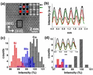
Figure 2.a shows an example of a HAADF-STEM image obtained on the reference sample. The insert shows a zoom on few atomic columns. Figure 2.b shows the intensity profiles of STEM image taken along different lines reported in Figure2.a. The profiles clearly shows the alternation of high and low peak intensity corresponding to Mn (Z = 25) and Si (Z = 14) columns, demonstrating the L21 order. We also note that the difference of intensity between Co and Mn columns is weak as expected from the Co and Mn atomic numbers (ZCo = 27). Similarly to classical X-ray diffraction using a Cu Kα source, HAADF-STEM is not very sensitive to D03 disorder.
6
Figure 2. (color online) (a) HAADF-STEM image of CMS. The insert at top right is a zoom over 6x6 atomic columns. (b) Intensity profile of the lines denoted by the colored arrows in (a). (c) Statistical analysis of intensity profile obtained from (a) on a region of 11 x 23 atomic columns. (d) statistical analysis of the area denoted by the black box in (a). In insert the intensity profile of the three central lines in the black box.
In order to demonstrate that the L21 order is the main phase of the sample we performed a statistical analysis of maximum peak intensities of the HAADF-STEM images for the reference sample (Figure 2) and for the sample irradiated at 1016 ions/cm² (Figure 3). For the first one, three distinct intensity distributions corresponding to the Co, Mn and Si atomic columns are observed (Figure 2.c). The values are normalized by the value of the Co intensity at the center of the Co distribution. The appearance of three different intensities in the HAADF-STEM image is in good agreement with the L21 order even if some spreading of the intensity distribution is observed. One source of spreading comes from the slight change of thickness across the lamella prepared for STEM experiment. This effect prevents any statistics over very large areas.
In some particular regions of the film other intensity distribution are observed, as the one reported in Figure 2.d which corresponds to statistical analysis performed in the black box in Figure 2.a. The inset in Figure 2.d shows that STEM intensities corresponding to the Mn and Si columns are very similar. Even if the statistical analysis is performed on a small number of atomic columns, it clearly shows that the STEM intensities corresponding to Mn and Si atomic columns converge to a single value, which could be associated to the appearance of the B2 order. This result indicates that very small grains with B2 order are distributed in the L21 matrix.
While the B2 order in the reference sample is only observed in small regions of similar size as the black box in Figure 2.a, it can be observed in more extended areas for a fluence of 1016 ions/cm². This is shown in Figure 3.a. The B2 order can be observed either from the intensity profile of the different
7
lines (Figure 3.b) or statistically over the selected area (Figure 3.c). Similar values for the STEM intensity of the Mn and Si columns are measured while the intensity corresponding to the Co columns remains higher. On other part of the sample we still observe the L21 order (Figure 3.d). We then suggest that ion irradiation induces a L21 to B2 transformation, which occurs around the initial B2 grains.
Figure 3. (color online) (a) HAADF-STEM image of CMS irradiated at 1016 ions/cm². The black boxes correspond to a zone with B2 or L21 character. (b) Intensity profile of the lines denoted by red and black lines in the B2 or L21 boxes in (a). Statistical analysis of the B2 (c) and L21 (d) regions in (a).
We also observe for the irradiated sample that the relative STEM intensity of the different atomic columns spread over slightly larger values as compared to the reference sample. The most probable explanation relies on the difference between the two prepared samples, especially the thicknesses which modify the absolute STEM intensity. Another possibility relies on local defects such as vacancies induced by the irradiation.
As already explained, it is very challenging to state about any Co/Mn exchange from HAADF-STEM experiments. However one can argue that if Co/Mn substitutions arise in the B2 phase, one should observe a spreading of the STEM intensity associated to the Mn columns that range between Si and Co ones. This is however not what is observed. Moreover, the relative intensity of the Mn columns compared to the Co ones in the L21 phase shows similar values (as for the reference sample). Therefore we assume that the D03 order most probably arises in the L21 matrix.
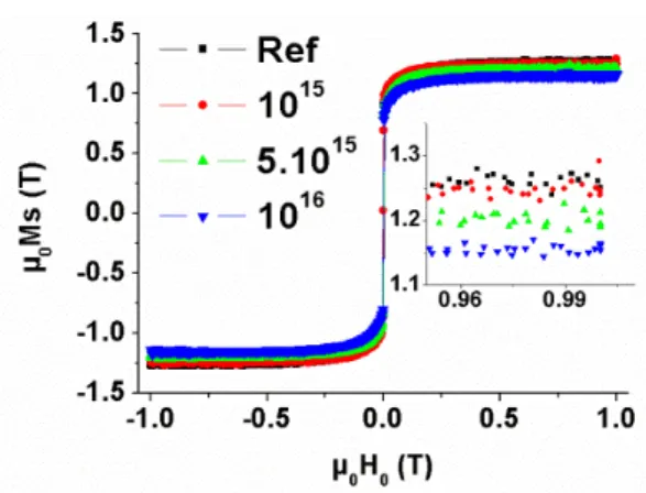
Finally, we measured the magnetization of the four samples (Figure 4) with a QUANTUM design PPMS VSM magnetometer at 300 K. We clearly observe a decrease of the magnetization versus the ion fluence. As demonstrated in Ref [2] the B2 and L21 phase have the same magnetic moment (µ0Ms ≈ 1.25 T) while it decreases by 10% in the D03 phase. Therefore we argue the decrease of the
8
magnetization can be accounted for the local Co-Mn exchange in the L21 matrix and vacancies induced by the irradiation.
Figure 4. (color online) Evolution of the CMS average magnetization at 300 K as a function of the He+ fluence.
III. Conclusions.
In conclusion we demonstrated that He+ ion irradiation at 150 keV induces Mn/Si exchange and then favors the B2 structure at the cost of the L21 phase. However, for fluences above 5x10¹⁵ ions/cm² , the out of plane lattice parameter of the B2 phase increase and might have an impact upon half metallicity. In addition, for this threshold value, Co/Mn substitution occurs in the L21 structure leading to a decreased magnetization and so half metallicity. Below this threshold, no significant improvement of L21 order has been observed. Therefore our study demonstrates that ion irradiation is an interesting alternative to annealing only for the B2 order as long as fluence is kept under a threshold value.
Acknowledgments
The authors thank LPCNO for X-ray diffraction facilities. This work has been supported by the French Agence Nationale pour la Recherche (ANR NASSICS 12-JS10-008 01) and the "French RENATECH network".
References.
9
[2] I. Galanakis, P. H. Dederichs and N. Papanikolaou, Phys. Rev. B 66, 174429, (2002). [3] I. Galanakis, P. H. Dederichs and N. Papanikolaou, Phys. Rev. B 66, 174429, (2002). [4] S. Ishida, S. Fujii, S. Kashiwagi, and S. Asano, J. Phys. Soc. Jpn., 64, 2152, (1995).
[5] R. Yilgin, Y. Sakuraba, M. Oogane, S. Mizukami, Y. Ando, and T. Miyazaki, Jap. J. of Appl. Phys, 46, 205, (2007).
[6] M. Oogane, T. Kubota, H. Naganuma and Y. Ando, J. Phys. D: Appl. Phys. 48, 164012, (2015). [7] S. Picozzi, A. Continenza, A. J. Freeman, Phys. Rev. B 69, 094423, (2004).
[8] J Fassbender, D Ravelosona and Y Samson, J. Phys. D: Appl. Phys. 37, R179, (2004).
[9] O. Gaier, J. Hamrle, B. Hillebrands, M. Kallmayer, P. Porsch, G. Schonhense, H. J. Elmers, J. Fassbender, A. Gloskovskii, C. A. Jenkins, G. H. Fecher, C. Felser, E. Ikenaga, Y. Sakuraba, S. Tsunegi, M. Oogane, and Y. Ando, Appl. Phys. Lett., 94, 152508, (2009).
[10] P. J. WEBSTER, J. Phys. Chem. Solids, 32, 1221, (1971).
[11] T. Miyajima, M. Oogane, Y. Kotaka, T. Yamazaki, M. Tsukada, Y. Kataoka, H. Naganuma, and Y. Ando, Applied Physics Express 2, 093001, (2009)
[12] S. Rodan, A. Alfonsov, M. Belesi, F. Ferraro, J. T. Kohlhepp, H. J. M. Swagten, B. Koopmans, Y. Sakuraba, S. Bosu, K. Takanashi, B. Büchner, and S. Wurmehl, Appl. Phys. Lett., 102, 242404 (2013).
[13] B. Ravel, M.P. Raphael, V.G. Harris, and Q. Huang, Phys. Rev. B, 65, 184431, (2002). [14] B. Balke, S. Wurmehl, G. H. Fecher, C. Felsera, M. C. M. Alves, F. Bernardi, and J. Morais, Appl. Phys. Lett. 90, 172501, (2007).
[15] B. Ravel, J. O. Cross, M. P. Raphael, V. G. Harris, R. Ramesh, and L. V. Saraf, Appl. Phys. Let., 81, 2812, (2002).
[16] B. A. Collins, Y. Zhong, Y. S. Chu, L. He and F. Tsui, J. Vac. Sci. Technol. B, 25, 999, (2007). [17] V. Niculescu, K. Raj, T. J. Burch, and J. I. Budnick, Phys. Rev. B, 13, 3167, (1976).
[18] Y. Takamura, R. Nakane, and S. Sugahara, J. Appl. Phys. 107, 09B111, (2010). [19] Caroline H. MacGillavry & Gerard D. Rieck ( Eds ), International Tables for X-ray
Crystallography, vol 3 ( Physical & Chemical Tables ), International Union of Crystallography / Kynoch Press 1968.
[20] G. Ortiz, A. Garcıa, N. Biziere, F. Boust, J. F. Bobo, and E. Snoeck, J. Appl. Phys. 113, 043921 (2013).

![Crivellari, Daniele, Marcas autoriales de segmentación en las comedias autógrafas de Lope de Vega: estudio y análisis [Reseña]](data:image/gif;base64,R0lGODlhAQABAIAAAP///wAAACH5BAEAAAAALAAAAAABAAEAAAICRAEAOw==)