HAL Id: cea-01692824
https://hal-cea.archives-ouvertes.fr/cea-01692824
Submitted on 25 Jan 2018
HAL is a multi-disciplinary open access
archive for the deposit and dissemination of
sci-entific research documents, whether they are
pub-lished or not. The documents may come from
teaching and research institutions in France or
abroad, or from public or private research centers.
L’archive ouverte pluridisciplinaire HAL, est
destinée au dépôt et à la diffusion de documents
scientifiques de niveau recherche, publiés ou non,
émanant des établissements d’enseignement et de
recherche français ou étrangers, des laboratoires
publics ou privés.
Luminescence study of defects in silica glasses under
near-UV excitation
Jérôme Neauport, Jessica Fournier, P. Grua, Evelyne Fargin, Veronique
Jubera, D. Talaga, S. Jouannigot
To cite this version:
Jérôme Neauport, Jessica Fournier, P. Grua, Evelyne Fargin, Veronique Jubera, et al.. Luminescence
study of defects in silica glasses under near-UV excitation. Physics Procedia, Elsevier, 2010, 8,
pp.39-43. �10.1016/j.phpro.2010.10.009�. �cea-01692824�
Physics Procedia 00 (2010) 000–000
www.elsevier.com/locate/procedia
VI Encuentro Franco-Español de Química y Física del Estado Sólido
VI
èmeRencontre Franco-Espagnole sur la Chimie et la Physique de l’État Solide
Luminescence study of defects in silica glasses under near-UV
excitation.
J. Fournier
a, b*, J. Néauport
a, P. Grua
a, V. Jubera
b, E. Fargin
b, D. Talaga
c,and S.
Jouannigot
da CEA CESTA, 12 Avenue des sablières, 33114 LE BARP (France) b ICMCB-CNRS, 87 Avenue du Docteur Albert Schweitzer,
33608 PESSAC Cedex (France)
c ISM, Groupe spectroscopie moléculaire, 351 Cours de la libération,
33405 TALENCE Cedex (France)
d LCTS, 3 Avenue de la Boetie, 33600 PESSAC (France)
Abstract
In this paper, we report the results of photo-luminescence experiments on high purity silica glass. Two different types of defects located on optical pieces are investigated: indented sites and laser damage craters. Visible luminescence excited at the wavelength of 325 nm (3.81 eV) is observed using confocal microscopy. Each type of defect produces qualitatively different luminescence spectra. In the case of indented sites, the well known Oxygen Deficient Center (ODC) band peaking at 2.75 eV is clearly observed, while for laser damage areas, another classical luminescent object is found: the Non Bridging Oxygen Hole Center (NBOHC) which peaks at 1.9 eV. On both types of defects, the luminescence spectra yield a strong peak around 2.2 eV but its interpretation is still controversory.
Keywords : Photoluminescence, Silica, Defects, Indentations, Laser damage.
1. Introduction
The Laser MegaJoule (LMJ) Facility is under construction at the CEA-CESTA in France. This facility will be used for inertial fusion studies. It consists in 176 laser beams focused at 355nm (3Ȧ) on a few millimeter target. High purity silica optics is used to convey the laser beam in the UV. Fused silica optics have a propensity for surface laser damage in the typical fluence range starting around 10 J/cm² for 3 ns pulse duration, well below the intrinsic bulk damage threshold which is beyond 100 J/cm² for this pulse length. In this context, getting a better understanding of defect of silica is of much importance. In silica glasses, two luminescent defects are well known:
* Jessica Fournier. Tel.: +33 5 40 00 26 50; fax: +33 5 40 00 27 61.
E-mail address: [email protected].
c 2010 Published by Elsevier Ltd. Physics Procedia 8 (2010) 39–43 www.elsevier.com/locate/procedia 1875-3892 c 2010 Published by Elsevier Ltd. doi:10.1016/j.phpro.2010.10.009
Open access under CC BY-NC-ND license.
J. Fournier / Physics Procedia 00 (2010) 000–000
the ODC (Oxygen Deficient Center) with an emission band at 2.75eV (450 nm) and the NBOHC (Non Bridging Oxygen Hole Center) with an emission band at 1.9 eV (650 nm) [1]. These two luminescent objects have been identified in our experiments, but such fact must be considered as anomalous, since the absorption spectra of ODC’s and NBOHC’s do not match our excitation wavelength of 325 nm [1]. Such defects are unlikely to trigger damage in the fluence range we consider. Other known defects can be absorbent but are not luminescent at this excitation wavelength.
In order to understand laser damage initiation; we study different types of defects: indentations and laser damage. The main feature of this experimental study is the appearance of a strong luminescence peak at 2.2 eV, not present for pristine material.
2. Sample preparation
Samples used in this study are high purity silica glasses (CORNING 7980) polished by SESO, of 50 mm diameter and 5 mm thick.
2.1. Indentations
The indentations are made with spheroconical diamond tip of 10 μm radius. Many parameters are adjustable: the
final load Ff corresponding to the maximum load, the loading rate (vl) which is the speed when loading from Fi to Ff,
and the time tFmax during which we maintain the final load on the sample. Our experiments are performed with
parameters given in table 1.
Table 1: Experimental parameters for indentations
Indenter radius Ff vl tFmax
10 μm 1.5 N 2000 mN/s 60 s
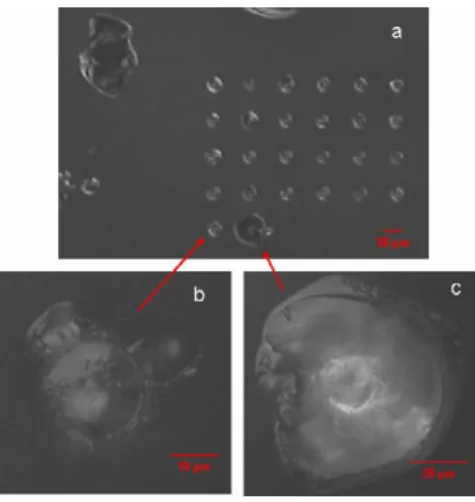
Despite the fact that our experimental parameters are constant, resulting indentation sites are not equivalent from site to site as shown on Figure 1. We chose indentations shown on Figures 1b and 1c for our luminescence study. They are referred to as indentation 1 and indentation 2.
Figure 1. (a) Image of indentations matrix, (b) and (c) enlargement of the two analysed indentations. 40 J. Fournier et al. / Physics Procedia 8 (2010) 39–43
2.2. Laser damage:
We create laser damage in our samples with a Nd:YAG laser, at 351 nm wavelength, 2.7 ns pulse duration, and a 6mm beam diameter. The laser fluence is about 40 J/cm². For each damage site, we use a multi-shot technique (one to seven shots per site) in order to obtain different sizes of damages, as seen on Figure 2.
Figure 2. Image of laser damage matrix.
3. Experiments
Photoluminescence spectra are made on a LABRAM HR-800 spectrometer, a high resolution Raman spectrometer, permitting luminescence confocal microscopy. Luminescence is excited by a continuous laser at 325 nm (3.81 eV) at room temperature. The spatial resolution is about 1μm and the signal is collected with a CCD camera. We use a laser of 100 μW power, except for our reference sample: a silica sample without artificial surface defect, for which we operate at 1mW. The beam waste is controlled with a pinhole opened at 300μm. For all measurements, the obtained spectra are corrected by the equipment function.
4. Results
4.1. Indentations
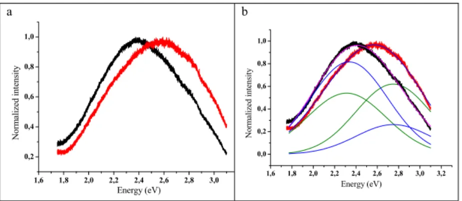
The luminescence spectra (Figure 3a), obtained for the indentation 1 (Figure 1b) and the indentation 2 (Figure 1c), show different characteristics. The spectrum is shifted to lower energies when the indentation induces more fractures. In order to identify the defects, the spectra (Figure 3b) are decomposed using Gaussian curves. The parameters of the fit are given in Table 2.
Table 2: Parameters used for fitting luminescence spectra of the indentations
Indentation 1 Indentation 2 Position (eV) Width (eV) Position (eV) Width (eV) 1st band 2.33 0.72 2.31 0.74 2nd band 2.75 0.70 2.75 0.70 1 mm
J. Fournier / Physics Procedia 00 (2010) 000–000 a 1,6 1,8 2,0 2,2 2,4 2,6 2,8 3,0 0,2 0,4 0,6 0,8 1,0 N o rm ali zed intensit y Energy (eV) b 1,6 1,8 2,0 2,2 2,4 2,6 2,8 3,0 3,2 0,0 0,2 0,4 0,6 0,8 1,0 N o rm alized intensity Energy (eV)
Figure 3. a) Comparison of photo luminescence spectra excited at 325 nm for indentations: indentation 1 (red) and indentation 2 (black); b) Comparison of fitted spectra for the two indentations: indentation 1 (green) and indentation 2 (blue).
The parameters used are in good agreement with those found in literature [2-3]. The spectra fits show that two defects are present in the indentations. The first one around the 2.3 eV band is not attributed to a known defect. The second band at 2.75 eV is attributed to the Oxygen Deficient Center [1].
4.2. Laser damage
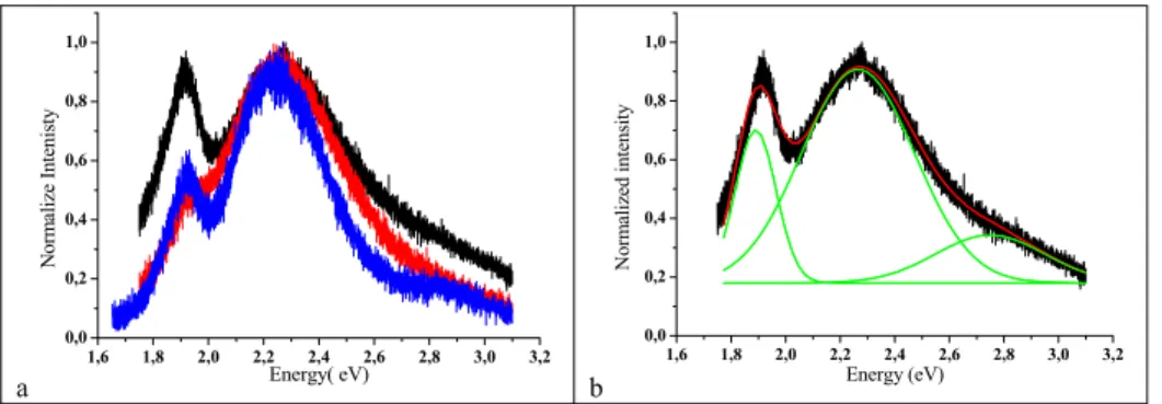
First results obtained on laser damage seem to indicate that the luminescence behaviour of defects is strongly correlated to the nature of damages (figure 4a). Strong differences are observed in the relative intensity of the emission bands. In all the cases, the emission bands can be fitted by three Gaussian curves (Figure 4b: only the Gaussian fit for the two laser shots is shown), parameters are given in Table 3. The corresponding curves are in good agreement with parameters found in literature [2-3]. Three luminescence bands are present in laser damages in different ratios. The first band at 1.89 eV corresponds to the NBOHC defect [1-5]. The second band at 2.25 eV is, to our knowledge, not related to a known defect. Finally, the third defect at 2.75 eV corresponds to ODC defect [1,2,4,5].
Table 3: Parameters used for fitting luminescence spectra of the laser damage craters
2 laser shots 4 laser shots 5 laser shots
Position (eV) Width (eV) Position (eV) Width (eV) Position (eV) Width (eV)
1stband 1.89 0.15 1.89 0.15 1.89 0.15
2ndband 2.26 0.41 2.25 0.42 2.24 0.34
3rdband 2.75 0.40 2.75 0.40 2.75 0.40
a 1,6 1,8 2,0 2,2 2,4 2,6 2,8 3,0 3,2 0,0 0,2 0,4 0,6 0,8 1,0 N o rm alize In tenisty Energy( eV) b 1,6 1,8 2,0 2,2 2,4 2,6 2,8 3,0 3,2 0,0 0,2 0,4 0,6 0,8 1,0 Norm ali zed int ensity Energy (eV)
Figure 4. a) Comparison of luminescence spectra for 2 laser shots (black), 4 laser shots (red), and 5 laser shots (blue); b) Fitted luminescence spectra for two laser shots (black: experimental data, green: partial fits, red: final fit).
5. Discussion
We show that indentations and laser damage craters give rise to different spectra. In indentations, only two defects are present: ODC and an unknown defect emitting around 2.2 eV. The luminescence bands are also larger than for laser damage. In laser damage, there are three luminescence bands which are ODC, NBOHC and the unknown defect emitting at 2.2 eV. Concerning this defect, two hypotheses are mainly proposed. The first one concerns the Self Trapped Exciton [2,6]. This defect is strongly luminescent under 150 K but at room temperature it may still has
a weak luminescence. The second hypothesis involves an E’Gcenter located in small silicon clusters [3-5], since a
correlation between the Electron Paramagnetic Resonance signal of E’Gcenter and the luminescence band around
2.2-2.3 eV was evidenced from the study of Ȗ-irradiated amorphous silicon. 6. Conclusion
This study shows that a luminescence is found in both cases studied (indentations and laser damage). In the two cases, ODC defect is found at 2.75 eV and an unknown defect around 2.2 eV. In laser damage a third defect is found at 1.89 eV and attributed to NBOHC defect. The 2.2 eV is not clearly ascribed now. New investigations are required in order to interpret accurately this 2.2-2.3 eV luminescence band.
Acknowledgements
We would like to thank H. Bercegol for active contribution to these results, T. Cardinal and A. Garcia for fruitful discussions concerning luminescence spectroscopy, and T. Donval for assistance with laser damage accomplishment.
References
[1] L. Skuja, J. Non-Cryst. Solids 239 (1998) 16-48
[2] M. A. Stevens Kalceff, Phys. Rev. B, Vol 57 Num 10 (1998) 5674-5683 [3] H. Nishikawa et al, J. Appl. Phys. 80-6 (1996) 3513-3517
[4] Demos et al, in Laser Applications in Microelectronic and Optoelectronic Manufacturing V, Proceedings of SPIE Vol. 3933 (2000) 316-320
[5] Kozlowski et al, in Laser-Induced Damage in Optical Materials, Proceedings of SPIE Vol. 3902 (2000) 138-144 [6] A. N. Trukhin et al, J. Non-Cryst. Solids 353 (2007) 530-536