Received: 16 May 2002 Revised: 15 July 2002 Accepted: 6 August 2002
Published online: 19 October 2002 © Springer-Verlag 2002
Abstract Hepatic peliosis is a rare entity that represents focal, multifo-cal, segmental, or diffuse dilatation of liver sinusoids. Hepatic peliosis is often associated with chronic wasting diseases but also has been reported in association with ana-bolic, contraceptive, or other hor-monal treatment, and even in con-text with HIV-related bacterial in-fections. Hepatic peliosis is usually clinically unapparent and mostly found only during autopsy, but oc-casionally it may lead to diagnostic problems if detected radiologically since the imaging findings in hepat-ic peliosis are quite variable accord-ing to the variety of its possible his-tologic features as well as the possi-bility of additional hemorrhage. We present a case of hepatic peliosis
as-sociated with bronchial carcinoma that showed moderate centripetal enhancement during the portal-nous phase on CT, pronounced ve-nous pooling on contrast enhanced T1-weighted images acquired dur-ing the hepatic-venous phase, and bright signal on T2-weighted imag-es, thus mimicking in some way a capillary hemangioma. We also dis-cuss some not yet described CT and MR features of this rare entity which should be included into the differential diagnosis of atypical liver lesions in patients with the above-mentioned conditions. Keywords Focal liver lesions · Hepatic peliosis · Computed tomography · Magnetic resonance tomography Eur Radiol (2003) 13:1916–1919 DOI 10.1007/s00330-002-1675-9 H E PAT O B I L I A R Y – PA N C R E A S K. Steinke L. Terraciano W. Wiesner
Unusual cross-sectional imaging findings
in hepatic peliosis
Introduction
Hepatic peliosis represents focally or segmentally dilated liver sinusoids with or without macroscopically visible blood-filled cysts ranging from <1 mm to several centime-ters in size with no preferential location in the liver [1]. He-patic peliosis is often associated with chronic wasting dis-eases, such as tuberculosis and malignancies, but it has also been reported in patients treated long-term with anabolic steroids, oral contraceptives, hormones, or azathioprine with generally observed regression of the liver lesions after discontinuation of treatment with these agents [2, 3, 4, 5, 6]. Recently, even a bacterial causative agent (Rochalimea
hensela) has been reported as a cause of HIV-related
hepat-ic peliosis whhepat-ich typhepat-ically also shows regression after ap-propriate antibiotic treatment with erythromycin [7, 8, 9].
In the past, hepatic peliosis has been considered a rare entity and most of the published articles on hepatic peli-osis were only case reports which supported the opinion that this entity usually represents an incidental autopsy finding in asymptomatic patients; however, presently he-patic peliosis is considered to be a more common finding which may present with hepatic dysfunction, shock from hepatic failure, or even hemorrhage with or without he-moperitoneum from liver rupture and which occasionally may also lead to differential diagnostic problems if de-tected by CT or MR in a patient with the above-men-tioned conditions [1, 2, 3, 4, 5, 6, 7, 8, 9, 10].
The purpose of this case report is to remind the reader of the rare differential diagnosis of hepatic peliosis and to discuss its variable and, therefore, also potentially misleading, CT and MRI findings.
K. Steinke · W. Wiesner (
✉
) Department of Diagnostic Radiology, University Hospital Basel, Petersgraben 4, 4031 Basel, Switzerland e-mail: wwiesner@uhbs.ch Tel.: +41-61-2652525 Fax: +41-61-2654354 L. Terraciano Department of Pathology,University Hospital Basel, Petersgraben 4, 4031 Basel, Switzerland
1917
Case report
A 59-year-old man presented with productive cough, fe-ver, chills, and nonspecific abdominal fullness at our emergency department. Physical examination revealed crackles over the right lung base and a slight hepatomeg-aly. Laboratory findings showed an elevated CRP and
leukocytosis of 20,000 cells/mm3but hepatic parameters
were within normal ranges.
Chest CT revealed a bronchogenic carcinoma with a post-stenotic pneumonic infiltration in the right lower lobe and bronchoscopic fine-needle aspiration confirmed a non-small cell lung cancer; however, as an incidental finding two large hypodense liver lesions were detected on chest CT leading to a dual-phase abdominal CT scan (Fig. 1) and subsequently also to MRI of the liver (Fig. 2) for further lesion characterization.
Fig. 1 a Arterial-phase CT scan of the liver reveals two large
hy-poattenuating and ill-defined liver lesions in liver segment 3/4a in a subcapsular location and in liver segment 7, respectively (arrows). Both lesions show no mass effect and both lesions have a more pronounced hypoattenuating center surrounded by a less hypoatten-uating aura. b In the portal-venous phase the lesion in liver seg-ment 7 disappears completely due to its moderate but relatively ho-mogeneous contrast enhancement, whereas the lesion in segment 3/4a shows a moderate and diffuse peripheral enhancement only of its above-mentioned aura during the portal-venous phase which subsequently becomes isodense to the surrounding liver in this phase (arrows). Contrarily, the center of the lesion in segment 3/4a is still unenhanced during the portal-venous phase and subsequent-ly still visible as hypoattenuating lesion (arrowheads in Fig. 2b)
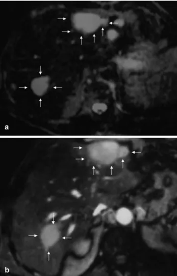
Fig. 2a, b On T2-weighted images the central portions of both
he-patic lesions are highly hyperintense (arrows in a) and on T1-weighted postcontrast images obtained at a late hepatic-venous phase both lesions show a pronounced and homogeneous contrast enhancement of their central portions due to pooling of contrast in these regions (arrows in b), whereas the surrounding and above-mentioned aura of both lesions that was less hypodense on arteri-al-phase CT (compare with Fig. 1a) and that became isodense to the surrounding liver parenchyma on portal-venous-phase CT scans (compare to Fig. 1b) is isointense to the surrounding liver parenchyma on both, the T2-weighted and the contrast-enhanced T1-weighted MR images
1918
Helical arterial-phase CT scan of the liver performed on a Somatom 4 Plus CT scanner (Siemens, Erlangen, Germany) with a time delay of 40 s after injection of 120 ml of non-ionic contrast material (injection rate 2 ml/s) revealed two large hypoattenuating and ill-de-fined liver lesions in liver segment 3/4a in a subcapsular location and in liver segment 7, respectively (arrows in Fig. 1a). Both lesions showed no mass effect and both le-sions presented with a more pronounced hypoattenuating center surrounded by a less hypoattenuating aura at this arterial phase. In the portal-venous phase, which was started 70 s after intravenous contrast injection, the le-sion in liver segment 7 disappeared completely due to its moderate but relatively homogeneous contrast enhance-ment. The lesion in segment 3/4a showed only a moder-ate enhancement of its aura during the portal-venous phase which resulted in disappearance of only this aura while the center of the lesion in segment 3/4a was unen-hanced and subsequently still visible as hypoattenuating lesion (arrows in Fig. 2b).
On MRI, performed on a 1.5-T scanner (Symphony, Siemens, Erlangen, Germany), both lesions showed a hy-pointense signal on T1-weighted precontrast images [2D fast low-angle shot (FLASH), breath hold, TR 160 ms, TE 4.53 ms], whereas on T2-weighted images (turbo spin echo, TR 4720 ms, TE 120 ms) the central portions of both hepatic lesions were highly hyperintense (arrows in Fig. 2a). On T1-weighted post-contrast images ob-tained at a hepatic-venous phase (2D FLASH, breath hold, fat suppression, TR 211 ms, TE 4.38 ms) both le-sions showed a pronounced and homogeneous contrast
enhancement of their central portions due to pooling of contrast in these regions, whereas the surrounding aura of both lesions (which had been less hypodense on arte-rial-phase CT scans and which became isodense to the surrounding liver parenchyma on portal-venous-phase CT scans) was isointense to the surrounding liver paren-chyma on both, T2-weighted and contrast-enhanced T1-weighted images (Fig. 2).
According to their appearance on CT and especially on MRI, both lesions were initially misinterpreted as large and mainly capillary hemangiomas, but on autopsy – the patient died of severe pneumonia several days later – the lesions turned out to represent multifocal hepatic peliosis and there was no hemangioma found in the liver during autopsy (Fig. 3).
Discussion
Imaging findings in peliosis hepatis are variable and de-pend on the variety of possible histopathologic features of the disease and often concomitant liver steatosis. The CT and MRI findings may therefore differ according to the size of the lesions, the extent of communication with the sinusoids, the presence or absence of thrombi within the cavities, and the presence or absence of hemorrhage. Therefore, its appearance on CT may range from invisi-ble [1] to small hypodense lesions <1 cm, remaining hy-podense after intravenous contrast injection [6] or show-ing a slow enhancement durshow-ing the portal-venous phase, to primarily hyperattenuating lesions on unenhanced CT due to hemorrhage. However, a typical finding of hepatic peliosis is the lack of mass effect on hepatic vessels [4].
On MRI the lesions are typically of low signal intensity on T1-weighted images and of high signal on T2-weight-ed images, with late and slow but intense enhancement on contrast-enhanced T1-weighted images. This bright signal on T2-weighted images together with a slow, delayed cen-tripetal enhancement of the lesions may in some way mimic a purely or mainly capillary hemangioma, but the lack of any nodular peripheral contrast enhancement as observed in our case would be very atypical for a heman-gioma, which, according to its size, should show at least some cavernous areas causing the typical peripheral nodu-lar enhancement. Other differential diagnoses that must be included in liver lesions that show such a bright signal on T2-weighted images are highly vascularized liver tumors or liver metastases, e.g., of neuroendocrine origin; howev-er, although these tumors may be very bright on T2, they typically would show a rapid and early enhancement, oc-curring typically during the arterial or early portal-venous phase as well as an early washout due to intratumoral an-gioneogenesis and a pronounced capillary bed, and this stays in clear contrast to the late enhancement and to the venous pooling of our two lesions that was due to dilated venous structures (sinusoids).
Fig. 3 Medium-power photomicrograph (hematoxylin–eosin
stain, original magnification, ×160) of hepatic peliosis shows large blood-filled spaces with incomplete endothelial lining in the liver which are surrounded by normal liver parenchyma. These large blood-filled cavities were responsible for the typically bright sig-nal intensities of hepatic peliosis on T2-weighted images and also for the delayed and prolonged contrast enhancement and pooling that was observed in both our lesions lesions on CT and MR
1919
1. Jamadar DA, D'Souza SP, Thomas EA, Giles TE (1994) Case report: radiologi-cal appearances in peliosis hepatis. Br J Radiol 67:102–104
2. Zak FG (1950) Peliosis hepatis. Am J Pathol 26:1–15
3. Bagheri SA, Bojer JL (1974) Peliosis hepatis associated with androgenic– anabolic steroid therapy: a severe form of hepatic injury. Ann Intern Med 81:610–618
4. Gouya H, Vignaux O, Legmann P, de Pigneux G, Bonnin A (2001) Pelio-sis hepatis: triphasic helical CT and dy-namic MRI findings. Abdom Imaging 26:507–509
5. Van Erpecum KJ, Janssens AR, Kreuning J, Ruiter DJ, Kroon HM, Grond AJ (1988) Generalized peliosis hepatis and cirrhosis after long-term use of oral contraceptives. Am J Gastroenterol 83:572–575
6. Maves CK, Caron KH, Bisset GS III, Agarwal R (1992) Splenic and hepatic peliosis: MR findings. Am J Roent-genol 158:75–76
7. Quint L, Jaccard A, Mainguene C et al. (1993) Rochalimea henselae infection. Febrile pancytopenia and hepatic pelio-sis in a patient with HIV infection. Presse Med 22:532–534
8. Hayward SR, Lucas CE, Ledgerwood AM (1991) Peliosis hepatis: old dis-ease, new case. Gastroenterology 101:864–866
9. Rousseaou MC, Brouqui P, Raoult D (1994) New bacterial infections in the course of AIDS. Rev Prat
44:1333–1338
10. Vignaux O, Legmann P, de Pinieux G, Chaussade S, Spaulding C, Couturi-er D, Bonnin A (1999) Hemorrhagic necrosis due to peliosis hepatis: imag-ing findimag-ings and pathological correla-tion. Eur Radiol 9:454–456
Nevertheless, it must be mentioned that occasionally even quite atypical signal intensities may be encountered in hepatic peliosis due to the presence of hemorrhage in different stages, and that in such cases hepatic peliosis may become indistinguishable from partially necrotic hypervascularized hepatic malignancies or metastases by CT or MRI [6].
A recent case report described a large peliotic liver le-sion with low attenuation on unenhanced CT, showing a predominantly central enhancement during the arterial phase and slow centrifugal progression of enhancement during the portal-venous and delayed phases (target sign) [4]. Our case showed the exact opposite enhancement pattern, from the periphery to the center – a CT and MR appearance of hepatic peliosis which, to our knowledge, has not been reported previously; however, the CT and MRI findings in our case are quite typical and easy to explain since the centers of both lesions corresponded to those regions with extended dilatation of sinusoids, whereas the surrounding and less hypoattenuation aura that was visible only during the arterial phase on CT ob-viously represented a borderline zone of only mildly di-lated sinusoids which connected the central areas with the normal surrounding liver parenchyma.
This explains why on arterial-phase CT scans both re-gions, the central portion as well as the surrounding aura, were hypodense, and why the aura became isodense to the surrounding liver parenchyma on CT in the portal-venous phase, similarly as it was isointense to the sur-rounding normal liver parenchyma on MRI. The delayed centripetal fill-in phenomenon was clearly visible in both lesions on contrast-enhanced T1-weighted images which were acquired at a hepatic-venous phase and, therefore, later than the portal-venous-phase CT scans; however, together with the CT scans these T1-weighted contrast-enhanced images nicely showed the delayed contrast en-hancement of hepatic peliosis and the prolonged pooling of contrast within the central portions of both lesions in
our patient. This corresponds well to the high signal in-tensities that these central portions of hepatic peliosis showed on T2-weighted images and which may be ex-plained by the low flow or even the stasis of blood with-in the dilated swith-inusoids.
The fact that the lesion in segment 7 disappeared total-ly on CT in the portal-venous phase while the central por-tion of the lesion in segment 3/4a remained still hypo-dense on CT during the portal-venous phase may be ex-plained by the size of the lesions: the lesion in segment 7 and especially also its central portion were smaller than the one in segment 3/4a, and, therefore most probably un-derwent faster contrast filling, whereas the larger lesion in segment 7 was probably just not yet completely opaci-fied in its central portions in the portal-venous phase. This might also explain why both lesions showed a dif-ferent appearance on contrast-enhanced CT acquired dur-ing the portal-venous phase compared with the contrast-enhanced T1-weighted MRI sequences which were ac-quired later during a hepatic-venous phase.
Conclusion
It is concluded that hepatic peliosis is an entity more of-ten encountered than previously thought, and that hepatic peliosis may have a variety of etiologies and presents with a broad spectrum of potentially misleading imaging findings; therefore, and according to its chameleon-like features, hepatic peliosis should be added to the differen-tial diagnosis of an atypical liver lesion in patients with the above-mentioned conditions. If related to a specific medication, it may regress after its discontinuation, and if related to a bacterial etiology, it may be treated with antibiotics; however, most commonly it is observed as an incidental finding in patients with wasting conditions and in such cases it requires a specific therapy only if larger subcapsular hemorrhagic lesions rupture [10].