RETINAL DISORDERS
Intravitreal ranibizumab (Lucentis®) for the treatment
of myopic choroidal neovascularization
Lazaros Konstantinidis&Irmela Mantel&
Jean-Antoine C. Pournaras&Leonidas Zografos&
Aude Ambresin
Received: 4 August 2008 / Revised: 11 October 2008 / Accepted: 29 October 2008 / Published online: 29 November 2008 # Springer-Verlag 2008
Abstract
Background Macular choroidal neovascularization (CNV) is one of the most vision-threatening complications of myopia, which can lead to severe vision loss. The purpose of this study was to evaluate the safety and efficacy of intravitreal ranibizumab in the treatment of myopic CNV. Methods We conducted a prospective, consecutive, inter-ventional study of patients with subfoveal or juxtafoveal CNV secondary to pathologic myopia (PM) treated with intravitreal injection of ranibizumab in the Jules Gonin University Eye Hospital from June 2006 to February 2008. Best-corrected visual acuity (BCVA), optical coherence tomography (OCT), and fluorescein angiography (FA) were performed at baseline and monthly for all patients. Indications for retreatment were loss in BCVA associated either with persistent leakage from CNV shown on FA, and/ or evidence of CNV activity on OCT.
Results The study included 14 eyes of 14 patients. The mean spherical equivalent refractive error was −12.5 (range, −8.0 D to −16.0 D). Mean time of follow-up was 8.4 months (range from 3 to 16 months, SD: 3). The mean number of intravitreal injections administered for each patient was 2.36 (SD 1.5). The mean initial visual acuity (VA) was 0.19 decimal equivalent (logMAR: 0.71, SD: 0.3). A statistically significant improvement to a mean VA of 0.48 decimal equivalent (log-MAR:0.32, SD: 0.25) was demonstrated at the final follow-up. VA improved by a mean of 3.86 (SD 2.74) lines. Nine patients (64%)
demonstrated a gain of 3 or more lines. Mean central macular thickness (CMT) measured with OCT was 304μm (SD: 39) at the baseline, and was reduced significantly at the final follow-up to 153μm (SD: 23). Average CMT reduction was 170 μm (SD: 57). No injection complications or drug-related side effects were noted during the follow-up period. Conclusions In this small series of eyes with limited follow-up, intravitreal ranibizumab was a safe and effective treatment for CNV secondary to PM, resulting in functional and anatomic improvements.
Keywords Choroidal neovascularization . Myopia . Ranibizumab
Introduction
Macular choroidal neovascularization (CNV) is one of the most vision-threatening complications of myopia [1–3]. It is also the most common cause of CNV in young individuals, accounting for almost 60% of CNV in patients younger than 50 years of age [4]. CNV as a complication of pathologic myopia (PM) can be devastating, leading to severe vision loss [1].
Photodynamic therapy (PDT) with verteporfin (Visu-dyne; Novartis AG, Basel, Switzerland) is currently the only approved treatment by the European Agency for the Evaluation of Medicinal Products and the US Food and Drug Administration (FDA) for subfoveal CNV related to PM that has shown stabilization of vision compared with placebo. However, the primary outcome was not statisti-cally significantly in favor of verteporfin therapy at 2-year follow-up [5].
Several other treatments have been assessed, including thermal laser photocoagulation [6], macular translocation Financial Support: None
L. Konstantinidis
:
I. Mantel:
J.-A. C. Pournaras:
L. Zografos:
A. Ambresin (*)Hôpital Ophtalmique Jules Gonin, University of Lausanne, 15 Av. de France,
CH-1004 Lausanne, Switzerland e-mail: aude.ambresin@ophtal.vd.ch
[7, 8], surgical removal of CNV [9], radiotherapy [10], indocyanine green mediated photothrombosis [11,12], and combined PDT and intravitreal triamcinolone injection [13, 14]. However, the beneficial effects of these treatments are questionable, primarily because of the risk of severe complications or poor long-term results.
The factors that stimulate pathologic neovascularization are incompletely understood, but vascular endothelial growth factor-A (VEGF-A), which is a diffusible cytokine that promotes angiogenesis and vascular permeability, has been implicated as an important factor promoting neo-vascularization [15,16]. Two isoforms have been detected in choroidal neovascular lesions [15].
Ranibizumab (Lucentis®, Novartis, Basel, Switzerland) is a specific, affinity-mature fragment of a recombinant, humanized IgG1 monoclonal antibody that neutralizes all active forms of VEGF-A. It has been found to be effective in the treatment of exudative age-related macular degener-ation (AMD), demonstrating encouraging signs of biologic activity, with acceptable safety [17].
Several studies have reported promising short-term results with off-label use of the intravitreal anti-VEGF drug bevacizumab (Avastin™, Genentech, South San Francisco, CA, USA) for the treatment of CNV in PM [18–25]. There are no published reports regarding the use of ranibizumab for the treatment of CNV in PM.
Ranibizumab has several advantages over bevacizumab. In addition to its smaller size, ranibizumab is genetically engineered to have a greater affinity for VEGF, and is formulated for intraocular use; more of the drug may therefore penetrate all layers of the retina [26,27]. It has been hypothesized that the efficacy of intravitreal ranibizu-mab in PM might be better than the results achieved with intravitreal bevacizumab [28].
This study evaluated the safety and efficacy of intravitreal ranibizumab (Lucentis®) as treatment for CNV due to PM.
Patients and methods Patient selection
This was a prospective, consecutive, interventional study of patients with subfoveal or juxtafoveal CNV secondary to PM treated with intravitreal injection of ranibizumab in the Jules Gonin University Eye Hospital from June 2006 to February 2008.
Inclusion criteria were (1) a minimum spherical equiv-alent refractive error of 6.0 diopters (D) and retinal signs of PM, (2) a minimum of 3 months of follow-up, (3) best-corrected visual acuity (BCVA) of 0.05 or better (decimal equivalent), and (4) evidence of an active CNV on the basis of the presence of leakage on fluorescein angiography (FA)
and/or intraretinal or subretinal fluid on optical coherence tomography (OCT).
Patients were excluded from the study if they presented features of any condition other than PM associated with choroidal neovascularization that could suggest a different etiology such as age-related macular degeneration. Further-more, patients were excluded who presented any significant ocular disease (other than CNV) that could compromise vision in the studied eye. Previous PDT treatment was not an exclusion criterion, on condition that it had been administered at least 6 months before Lucentis® treatment. Examination
Patient age, sex, affected eye, spherical equivalent refrac-tion, and any previous treatment administered were recorded.
The patients were followed every month. Both initial ophthalmic examination and each follow-up included: (1) evaluation of best-corrected distant and near VA using an EDTRS chart and a nearpoint Snellen acuity card at 40 cm respectively, (2) a fundus exam with dilated pupils, (3) fundus photography, (4) digital FA, and (5) an evaluation of retinal architecture and measurement of foveal thickness using the OCT Stratus (Carl Zeiss Meditec, Inc.). Any ocular or systemic adverse events were also recorded.
Indications for retreatment were persistent leakage from CNV shown on FA and/or presence of intraretinal or subretinal fluid shown on OCT. An informed consent was obtained from all patients after a thorough discussion before each injection.
Surgical procedure
All injections were performed by the same surgeon (LK) using the same technique. Ranibizumab 0.5 mg (0.05 ml) was administered by intravitreal injection under sterile conditions in the operating theatre. Before injection, tetracaine 0.5% was applied topically. Povidone iodine was applied to eyelid margins, eyelashes, and conjunctival surface, and a lid speculum was placed. An additional drop of povidone iodine was applied to site of injection. Using a 30-gauge needle, 0.05 ml ranibizumab was injected 3.5– 4.0 mm posterior to the corneal limbus into the vitreous cavity. The injection site was compressed with a cotton swab to avoid reflux when the needle was removed. Postoperatively, a topical antibiotic (ofloxacin) was admin-istered three times daily for 7 days.
Statistical analysis
The statistical analysis focused on the evaluation of the change in BCVA and the foveal thickness measured by
OCT before treatment and at the final follow-up. Addition-ally, the absence or persistence of leakage on FA at the final follow-up was evaluated. BCVA values were converted to logarithm of the minimum angle of resolution (log-MAR) for statistical analysis. To determine the equivalent mean decimal, log-MAR values were converted back to decimal notation [29].
Serial changes in BCVA, OCT, and central focal thickness (CFT) were compared using the Wilcoxon signed-rank test and two-tailed t-test. A p≤0.05 was considered to be statistically significant.
These parameters were analyzed using SPSS statistical software (version 16 for Mac OS X, SPSS Inc.).
Results
The study included 14 eyes of 14 patients. There were five (36%) male and nine (64%) female patients. The mean spherical equivalent refractive error was−12.5 (range, −8.0 D to−16.0 D). All eyes had retinal abnormalities consistent with pathologic myopia, such as lacquer cracks. The mean age of the patients was 59 years (range from 37 to 87 years); 50% of the patients were older than 61 years, and 50% were younger than 58 years. Mean time of follow-up was 8.4 months (range from 3 to 16 months, SD: 3). Angiographic features in all cases demonstrated classic CNV. Seven eyes (50%) had previously received PDT. CNV was subfoveal in 11 patients (79%) and juxtafoveal in three (21%). The mean number of intravitreal injections administered for each patient was 2.36 (SD 1.5). The mean number of intravitreal injections administered concerning the 11 patients that had a follow-up of at least 6 months (mean 9.7 months, SD: 3) was 2.73 (SD 1.5). The mean number of intravitreal injections administered concerning the seven patients that had a follow-up of at least 9 months (mean 11.4 months, SD: 2.4) was 2.71 (SD 1.7).
Visual acuity
The mean initial VA was 0.19 (decimal equivalent), with a range from 0.06 to 0.5. Mean logMAR initial VA was 0.71 (SD: 0.3). A statistically significant improvement to a mean
VA of 0.48 decimal equivalent (logMAR 0.32, SD: 0.25) was demonstrated at the final follow-up (p=0.001, Wil-coxon signed-rank test). The VA improved by a mean of 3.86 (SD 2.74) lines. Nine patients (64%) demonstrated a gain of 3 or more lines. Thirteen patients (93%) have shown improvement of VA, while VA remained unchanged in one patient. Table1 shows the changes in mean BCVA during the study.
Optical coherence tomography and fluorescein angiography results
Mean CMT measured with OCT was 304 μm (SD: 39) at baseline, and decreased significantly at the final follow-up to 153 μm (SD: 23) (p<0.001, two-tailed t-test). Average CMT reduction was 170μm (SD: 57) (Fig.1).
Table 1 shows the changes in mean OCT CMT during this study. All eyes exhibited diminution or absence of leakage on FA at the final follow-up (Figs.2 and3).
Patient age in this study appeared to have no influence in a statistically significant way on VA improvement or changes in mean OCT CMT.
Complications
No injection complications or drug-related side effects were noted during the follow-up period.
Discussion
It has been shown that VEGF has an important role in regulating angiogenesis and cell proliferation [30], and therefore is related to the formation and regression of CNV [31]. Tong et al. demonstrated that the levels of VEGF were found to be significantly increased in the aqueous humor of eyes with active CNV related to AMD and with CNV related to PM [32], indicating that VEGF plays an important role in the pathogenesis of CNV related to these clinical conditions.
Ranibizumab is a recombinant humanized monoclonal antibody fragment that inhibits the biologic activity of all isoforms of human VEGF-A, and consequently leads to
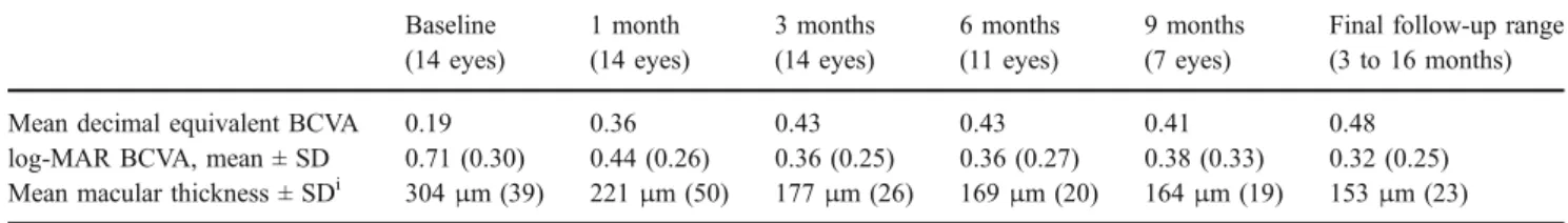
Table 1 Best-corrected visual acuity (BCVA) and optical coherence tomographic parameters at baseline and each follow-up Baseline (14 eyes) 1 month (14 eyes) 3 months (14 eyes) 6 months (11 eyes) 9 months (7 eyes)
Final follow-up range (3 to 16 months) Mean decimal equivalent BCVA 0.19 0.36 0.43 0.43 0.41 0.48
log-MAR BCVA, mean ± SD 0.71 (0.30) 0.44 (0.26) 0.36 (0.25) 0.36 (0.27) 0.38 (0.33) 0.32 (0.25) Mean macular thickness ± SDi 304μm (39) 221μm (50) 177μm (26) 169μm (20) 164μm (19) 153μm (23) ilog-MAR: logarithm of the minimum angle of resolution
reductions in cell proliferation, vascular leakage, and formation of new blood vessels [33]. Extensive, stringent, multicenter, randomized clinical trials have shown that ranibizumab provides significant VA benefit to patients with all angiographic subtypes of CNV related to AMD (i.e., minimally or predominantly classic or occult with no classic component), with a low incidence of serious ocular and non-ocular adverse events and an acceptable rate of non-serious adverse events [17, 34, 35]. Furthermore, ranibizumab was found to be superior to verteporfin for treating predominantly classic CNV related to AMD [35]. On the evidence of these trials, the FDA approved ranibizumab for the treatment of macular degeneration on June 30 2006, after a priority review stating that ranibizu-mab is the first treatment that can maintain the vision of more than 90% of patients with wet AMD [36].
However, for subfoveal CNV related to PM, PDT with verteporfin remains the only approved treatment. It is nevertheless worth mentioning that the 2-year outcomes of PDT in cases of subfoveal CNV associated with PM failed to reveal a statistically significant treatment effect in its primary outcome, the proportion of eyes with fewer than 8 letters of VA loss. In the trial, 36% of verteporfin-treated eyes compared with 51% placebo-treated eyes had at least an 8-letter loss in VA (p=0.11) [5].
Although there are no published studies regarding the use of ranibizumab for the treatment of CNV in PM, several studies have reported promising short-term results for the treatment of CNV in PM with off-label use of the intravitreal anti-VEGF drug bevacizumab; bevacizumab is approved by the FDA for the treatment of colorectal cancer [18–25].
Chan et al. treated 22 eyes for CNV secondary to PM with intravitreal bevacizumab, reporting a visual mean improvement of 2.6 lines and anatomic improvements during 6 months of follow-up [18]. Hernandez-Rojas et al. reported, in a study of 14 patients treated with intravitreal bevacizumab for CNV secondary to PM, that foveal thickness improved from 385.43±125.83 μm to 194.54± 54.35μm after 3 months [19]. Mandal et al., in a case series of 12 eyes, suggested that bevacizumab is a safe and effective treatment for CNV secondary to PM, reporting an average reduction of 174.25 μm of foveal thickness at 6 months. Yamamoto et al. treated 11 eyes with intravitreal bevacizumab for CNV due to PM, achieving an average reduction of 103 μm of foveal thickness and a mean VA improvement of 3.5 lines after an average follow-up of 153 days.
In our study, a mean VA improvement of 3.86 lines (SD 2.74) was demonstrated after a mean follow-up of Fig. 1 Baseline OCTs (a, c) showing hyper-reflectivity (1,2) under
the fovea due to the neovascular complex (1) with diffuse intraretinal exudation (3,4). Last follow-up OCTs (b, d) showed a reduced
thickness of the neovascular complex with residual intraretinal cystic exudation (5,6)
8.4 months. Furthermore, an average reduction of 170μm (SD: 57) of foveal thickness was documented, and the mean CNV size on FA was decreased significantly.
It is difficult to compare the different studies, consider-ing the variability of the methodology, the follow-up, and the re-treatment criteria of each study. Furthermore, the
visual results are much dependent on patient characteristics like the duration of symptoms, age, CNV size and the pre-treatment VA.
Ranibizumab, which is specifically formulated for intraocular use [26], has several advantages over bevacizu-mab. The primary molecular difference between these two Fig. 2 a, b Baseline and last follow-up visit; color fundus
photo-graphs. c, d Early phase on baseline FA shows a well-defined neovascular complex (1) in lacquer cracks, with significant exudation
(2) in the late phase. e, f On the last follow-up FA, there was a marked reduction in size and exudation (3) of the neovascular membrane
drugs is their molecular weight: ranibizumab is a 48-kD Fab fragment, whereas bevacizumab is a complete 149-kD antibody [37]. It has been demonstrated that ranibizumab fully penetrates the retinal layers to the outer retina and inner choroids, the pathophysiologic site of neovascular AMD [38]. In an animal model, it was shown that a
full-length antibody [37] did not penetrate the inner limiting membrane of the retina, in contrast to the Fab antibody fragment that diffused through the neural retina to the retinal pigment epithelial layer. Furthermore, ranibizumab penetrates the retina faster than bevacizumab [27]. Fre-quently, CNV due to PM is not associated with consider-Fig. 3 g, h Baseline fundus photograph shows a subretinal
hemor-rhage (4) at the margin of the neovascular complex, which has disappeared by the last follow-up. j Last follow-up FA shows a
marked reduction in size of the neovascular complex (5). k, l Late phase of both initial and last follow-up FA showed mild leakage (6,7)
able retinal edema; thus, retinal barriers might be less disturbed in comparison to AMD cases, and thus a possible higher penetration ability of the smaller molecule of ranibizumab might be a significant advantage. Furthermore, the ranibizumab active binding site reportedly has a greater affinity to VEGF-A than bevacizumab, through a process of affinity maturation [39]. In addition, ranibizumab could theoretically be associated with fewer potential systemic risks, given findings with systemic pharmacokinetics suggesting that it is cleared much more rapidly from the serum than bevacizumab [40].
One potential risk that should be considered in the treatment of myopic CNVs with anti-VEGF, which has been revealed in a recent study, is the possible formation of marginal crack lines after treatment-related contraction of the myopic CNVs, which was considered an indication of early damage of retinal pigment epithelium that might lead to expanding macular chorioretinal atrophy [41].
In conclusion, in this small series of eyes with limited follow-up, intravitreal ranibizumab seems to be a safe and effective treatment for CNV secondary to PM, resulting in functional and anatomic improvements. The limitations of this study include its small number of eyes, the relatively short follow-up, and the absence of a control group. One should consider that many treatments including PDT with verteporfin, even if they have shown a beneficial effect in the short term, failed to show significantly favourable results in the long term. Various reasons can compromise the long-term results, including progressive chorioretinal atrophy or treatment-related cumulative, damage to the photoreceptors and underlying retinal pigment epithelium, as hypothesized in the VIP study [5].
In view of the encouraging results from this prospective study, we consider that randomized, long-term clinical trials are needed to determine more accurately the potential clinical benefit of intravitreal ranibizumab and its safety.
References
1. Hampton GR, Kohen D, Bird AC (1983) Visual prognosis of disciform degeneration in myopia. Ophthalmology 90:923–926 2. Miller DG, Singerman LJ (2001) Natural history of choroidal
neovascularization in high myopia. Curr Opin Ophthalmol 12:222–224. doi:10.1097/00055735-200106000-00014
3. Avila MP, Weiter JJ, Jalkh AE, Trempe CL, Pruett RC, Schepens CL (1984) Natural history of choroidal neovascularization in degenera-tive myopia. Ophthalmology 91:1573–1581
4. Cohen SY, Laroche A, Leguen Y, Soubrane G, Coscas GJ (1996) Etiology of choroidal neovascularization in young patients. Ophthalmology 103:1241–1244
5. Blinder KJ, Blumenkranz MS, Bressler NM, Bressler SB, Donato G, Lewis H, Lim JI, Menchini U, Miller JW, Mones JM, Potter MJ, Pournaras C, Reaves A, Rosenfeld P, Schachat AP, Schmidt-Erfurth U, Sickenberg M, Singerman LJ, Slakter JS, Strong HA, Virgili G, Williams GA (2003) Verteporfin therapy of
subfoveal choroidal neovascularization in pathologic myopia: 2-year results of a randomized clinical trial–VIP report no. 3. Ophthalmol-ogy 110:667–673. doi:10.1016/S0161-6420(02)01998-X
6. Secretan M, Kuhn D, Soubrane G, Coscas G (1997) Long-term visual outcome of choroidal neovascularization in pathologic myopia: natural history and laser treatment. Eur J Ophthalmol 7:307–316
7. Mateo C, Moreno J, Rosales G, Lechuga M, Castillo R, Vaz F, Corcostegui B (2004) Two-year results of macular translocation with scleral infolding in myopic choroidal neovascularisation. Semin Ophthalmol 19:29–42. doi:10.1080/08820530490520013
8. Fujii GY, Humayun MS, Pieramici DJ, Schachat AP, Au Eong KG, de Juan E Jr (2001) Initial experience of inferior limited macular translocation for subfoveal choroidal neovascularization resulting from causes other than age-related macular degeneration. Am J Ophthalmol 131:90–100. doi:10.1016/S0002-9394(00)00769-8
9. Ruiz-Moreno JM, de la Vega C (2001) Surgical removal of subfoveal choroidal neovascularisation in highly myopic patients. Br J Ophthalmol 85:1041–1043. doi:10.1136/bjo.85.9.1041
10. Kobayashi H, Kobayashi K (2000) Radiotherapy for subfoveal neovascularisation associated with pathological myopia: a pilot study. Br J Ophthalmol 84:761–766. doi:10.1136/bjo.84.7.761
11. Liu DT, Lam DS, Chan WM (2004) Selective occlusion of subfoveal choroidal neovascularization in pathologic myopia using a new technique of ingrowth site treatment. Am J Ophthalmol 137:383 author reply 383-386. doi:10.1016/j.ajo.2003.10.015
12. Costa RA, Calucci D, Teixeira LF, Cardillo JA, Bonomo PP (2003) Selective occlusion of subfoveal choroidal neovasculariza-tion in pathologic myopia using a new technique of ingrowth site treatment. Am J Ophthalmol 135:857–866. doi: 10.1016/S0002-9394(02)02257-2
13. Montero JA, Ruiz-Moreno JM (2007) Combined photodynamic therapy and intravitreal triamcinolone injection for the treatment of choroidal neovascularisation secondary to pathological myopia: a pilot study. Br J Ophthalmol 91:131–133. doi:10.1136/bjo. 2006.106526
14. Chan WM, Lai TY, Wong AL, Liu DT, Lam DS (2007) Combined photodynamic therapy and intravitreal triamcinolone injection for the treatment of choroidal neovascularisation secondary to pathological myopia: a pilot study. Br J Ophthalmol 91:174– 179. doi:10.1136/bjo.2006.103606
15. Rakic JM, Lambert V, Devy L, Luttun A, Carmeliet P, Claes C, Nguyen L, Foidart JM, Noel A, Munaut C (2003) Placental growth factor, a member of the VEGF family, contributes to the development of choroidal neovascularization. Invest Ophthalmol Vis Sci 44:3186–3193. doi:10.1167/iovs.02-1092
16. Otani A, Takagi H, Oh H, Koyama S, Ogura Y, Matumura M, Honda Y (2002) Vascular endothelial growth factor family and receptor expression in human choroidal neovascular membranes. Microvasc Res 64:162–169. doi:10.1006/mvre.2002.2407
17. Rosenfeld PJ, Brown DM, Heier JS, Boyer DS, Kaiser PK, Chung CY, Kim RY (2006) Ranibizumab for neovascular age-related macular degeneration. N Engl J Med 355:1419–1431.
doi:10. 1056/NEJMoa054481
18. Chan WM, Lai TY, Liu DT, Lam DS (2007) Intravitreal bevacizumab (Avastin) for myopic choroidal neovascularization: six-month results of a prospective pilot study. Ophthalmology 114 (12):2190–2196. doi:10.1016/j.ophtha.2007.03.043
19. Hernandez-Rojas ML, Quiroz-Mercado H, Dalma-Weiszhausz J, Fromow-Guerra J, Amaya-Espinosa A, Solis-Vivanco A, Reyna-Castelan E, Abraham-Marin M, Martinez-Castellanos MA, Aiello LP (2007) Short-term effects of intravitreal bevacizumab for subfoveal choroidal neovascularization in pathologic myopia. Retina 27:707–712
20. Laud K, Spaide RF, Freund KB, Slakter J, Klancnik JM Jr (2006) Treatment of choroidal neovascularization in pathologic myopia
with intravitreal bevacizumab. Retina 26:960–963. doi:10.1097/ 01.iae.0000240121.28034.c3
21. Mandal S, Venkatesh P, Sampangi R, Garg S (2007) Intravitreal bevacizumab (Avastin) as primary treatment for myopic choroidal neovascularization. Eur J Ophthalmol 17:620–626
22. Nguyen QD, Shah S, Tatlipinar S, Do DV, Anden EV, Campochiaro PA (2005) Bevacizumab suppresses choroidal neo-vascularisation caused by pathological myopia. Br J Ophthalmol 89:1368–1370. doi:10.1136/bjo.2005.066431
23. Sakaguchi H, Ikuno Y, Gomi F, Kamei M, Sawa M, Tsujikawa M, Oshima Y, Kusaka S, Tano Y (2007) Intravitreal injection of bevacizumab for choroidal neovascularisation associated with pathological myopia. Br J Ophthalmol 91:161–165. doi:10.1136/ bjo.2006.099887
24. Tewari A, Dhalla MS, Apte RS (2006) Intravitreal bevacizumab for treatment of choroidal neovascularization in pathologic myopia. Retina 26:1093–1094. doi:10.1097/01.iae.0000254896. 78766.74
25. Yamamoto I, Rogers AH, Reichel E, Yates PA, Duker JS (2007) Intravitreal bevacizumab (Avastin) as treatment for subfoveal choroidal neovascularisation secondary to pathological myopia. Br J Ophthalmol 91:157–160. doi:10.1136/bjo.2006.096776
26. Steinbrook R (2006) The price of sight–ranibizumab, bevacizu-mab, and the treatment of macular degeneration. N Engl J Med 355:1409–1412. doi:10.1056/NEJMp068185
27. Bakri SJ, Snyder MR, Reid JM, Pulido JS, Ezzat MK, Singh RJ (2007) Pharmacokinetics of intravitreal ranibizumab (Lucentis). Ophthalmology 114:2179–2182. doi:10.1016/j.ophtha.2007.09.012
28. Rosenfeld PJ (2007) Intravitreal Avastin for choroidal neovascu-larisation in pathological myopia: the controversy continues. Br J Ophthalmol 91:128–130. doi:10.1136/bjo.2006.101337
29. Holladay JT (1997) Proper method for calculating average visual acuity. J Refract Surg 13:388–391
30. Ohno-Matsui K, Yoshida T, Uetama T, Mochizuki M, Morita I (2003) Vascular endothelial growth factor upregulates pigment epithelium-derived factor expression via VEGFR-1 in human retinal pigment epithelial cells. Biochem Biophys Res Commun 303:962–967. doi:10.1016/S0006-291X(03)00446-7
31. Ohno-Matsui K, Morita I, Tombran-Tink J, Mrazek D, Onodera M, Uetama T, Hayano M, Murota SI, Mochizuki M (2001) Novel mechanism for age-related macular degeneration: an equilibrium shift between the angiogenesis factors VEGF and PEDF. J Cell Physiol 189:323–333. doi:10.1002/jcp.10026
32. Tong JP, Chan WM, Liu DT, Lai TY, Choy KW, Pang CP, Lam DS (2006) Aqueous humor levels of vascular endothelial growth factor and pigment epithelium-derived factor in polypoidal choroidal vasculopathy and choroidal neovascularization. Am J Ophthalmol 141:456–462. doi:10.1016/j.ajo.2005.10.012
33. Kourlas H, Abrams P (2007) Ranibizumab for the treatment of neovascular age-related macular degeneration: a review. Clin Ther 29:1850–1861. doi:10.1016/j.clinthera.2007.09.008
34. Regillo CD, Brown DM, Abraham P, Yue H, Ianchulev T, Schneider S, Shams N (2008) Randomized, double-masked, sham-controlled trial of Ranibizumab for neovascular age-related macular degeneration: PIER study year 1. Am J Ophthalmol 145:239–248. doi:10.1016/j.ajo.2007.10.004
35. Brown DM, Kaiser PK, Michels M, Soubrane G, Heier JS, Kim RY, Sy JP, Schneider S (2006) Ranibizumab versus verteporfin for neovascular age-related macular degeneration. N Engl J Med 355:1432–1444. doi:10.1056/NEJMoa062655
36. Nagpal M, Nagpal K, Nagpal PN (2007) A comparative debate on the various anti-vascular endothelial growth factor drugs: pegap-tanib sodium (Macugen), ranibizumab (Lucentis) and bevacizu-mab (Avastin). Indian J Ophthalmol 55:437–439
37. Mordenti J, Cuthbertson RA, Ferrara N, Thomsen K, Berleau L, Licko V, Allen PC, Valverde CR, Meng YG, Fei DT, Fourre KM, Ryan AM (1999) Comparisons of the intraocular tissue distribution, pharmacokinetics, and safety of 125I-labeled full-length and Fab antibodies in rhesus monkeys following intravitreal administration. Toxicol Pathol 27:536–544. doi:10.1177/019262339902700507
38. Gaudreault J, Fei D, Beyer JC, Ryan A, Rangell L, Shiu V, Damico LA (2007) Pharmacokinetics and retinal distribution of ranibizumab, a humanized antibody fragment directed against VEGF-A, following intravitreal administration in rabbits. Retina 27:1260–1266
39. Ferrara N, Damico L, Shams N, Lowman H, Kim R (2006) Development of ranibizumab, an anti-vascular endothelial growth factor antigen binding fragment, as therapy for neovascular age-related macular degeneration. Retina 26:859–870. doi:10.1097/01. iae.0000242842.14624.e7
40. Olsen TW (2007) Treatment of exudative age-related macular degeneration: many factors to consider. Am J Ophthalmol 144:281–283. doi:10.1016/j.ajo.2007.05.005
41. Sayanagi K, Ikuno Y, Soga K, Wakabayashi T, Tano Y (2008) Marginal crack after intravitreal bevacizumab for myopic choroi-dal neovascularization. Acta Ophthalmol (Copenh) 85:50–54