Removal of Cells from Body Fluids by Magnetic Separation in
Batch and Continuous Mode: In
fluence of Bead Size, Concentration,
and Contact Time
Nils Bohmer,
†,∥Nino Demarmels,
†,∥Elena Tsolaki,
‡Lukas Gerken,
†Kerda Keevend,
†Sergio Bertazzo,
‡Marco Lattuada,
§and Inge K. Herrmann
*
,††
Materials Meet Life Department, Swiss Federal Laboratories for Materials Science and Technology (Empa), Lerchenfeldstrasse 5,
CH-9014, St. Gallen, Switzerland
‡
Department of Medical Physics and Biomedical Engineering, University College London (UCL), Malet Place Engineering Building,
London, WC1E 6BT, United Kingdom
§
Department of Chemistry, University of Fribourg, Chemin du Muse
́e 9, CH-1700, Fribourg, Switzerland
*
S Supporting InformationABSTRACT:
The magnetic separation of pathogenic compounds
from body
fluids is an appealing therapeutic concept. Recently,
removal of a diverse array of pathogens has been demonstrated using
extracorporeal dialysis-type devices. The contact time between the
fluid and the magnetic beads in such devices is limited to a few
minutes. This poses challenges, particularly if large compounds such as
bacteria or cells need to be removed. Here, we report on the feasibility
to remove cells from body
fluids in a continuous dialysis type of
setting. We assessed tumor cell removal e
fficiencies from physiological
fluids with or without white blood cells using a range of different
magnetic bead sizes (50
−4000 nm), concentrations, and contact times. We show that tumor cells can be quantitatively removed
from body
fluids within acceptable times (1−2 min) and bead concentrations (0.2 mg per mL). We further present a
mathematical model to describe the minimal bead number concentration needed to remove a certain number of cells, in the
presence of competing nonspeci
fic uptake. The present study paves the way for investigational studies to assess the therapeutic
potential of cell removal by magnetic blood puri
fication in a dialysis-like setting.
KEYWORDS:
blood cells, blood puri
fication, magnetic particles, mathematical modeling, nanoparticles, tumor cells
■
INTRODUCTION
Magnetic separation o
ffers a direct way for the removal of
disease-causing factors from body
fluids. Magnetic beads
func-tionalized by a capturing moiety are employed to selectively
bind to target compounds and are then subsequently removed
by magnetic separation.
1−3A variety of beads, mostly based on
iron oxide, iron, and iron carbide, have successfully been
applied to capture target compounds. Removal of a wide variety
of different substances, including metal ions,
4,5small molecule
drugs,
6,7and proteins
6has been demonstrated in various in
vitro and in vivo models. Recently, the concept has been
extended to remove bacteria from body
fluids in diagnostic,
8therapeutic,
9and theranostic
10settings. A similar concept
based on micron-sized magnetic beads has been employed for
magnetic cell isolation in cell culture and diagnostics for
decades already.
11−13Although micrometer-sized beads work
well for diagnostic applications, Xu and colleagues have
highlighted significant drawbacks of microparticles, such as
low surface area (and binding capacity) and slow binding.
14In the same report, they also have demonstrated promising
separation e
fficiencies of tumor cells using 30-nm iron oxide
beads in a batch type setting. The removal of tumor cells
from body
fluid holds some promise
15and various studies have
been conducted on the isolation of circulating tumor cells
(CTCs).
1−3,16−20For example, McDonald and colleagues have
demonstrated in an early study that ovarian cancer progression in
mice is 10-fold lower when migratory tumor cells are removed by
magnetic
filtration from intraperitoneal fluids.
21Most of the
reported results in the literature aim at the diagnosis
1,16,17,20and
monitoring of cancer via isolation of CTCs from the blood of
patients by micro
fluidic techniques.
1,3,16−18,20However, rapid
therapeutic removal of cells by magnetic separation in a
con-tinuous process has not yet been investigated systematically, and
intrinsic process limitations remain unclear.
In a continuous extracorporeal process, similar to a dialysis
setting, the contact time between the beads and the
fluid
con-taining the target cells is limited to a few minutes.
22For e
fficient
binding, long contact times would be bene
ficial, but the
http://doc.rero.ch
Published in "ACS Applied Materials & Interfaces 9 (35): 29571–29579, 2017"
which should be cited to refer to this work.
receptor binding process is in competition with the uptake of
particles into phagocytic cells. Additionally, long contact times
compromise the overall process throughput. The e
ffective
removal of tumor cells relies on high particle
−tumor cells
collision frequencies and speci
fic binding of magnetic clusters
to tumor cells in the presence of proteins and other cells, such as
white blood cells. Here, we investigated the technical feasibility
and limitations for the therapeutic removal of cells from body
fluids using a magnetic blood purification process in batch and
continuous mode. We assessed the in
fluence of bead size and
con-centration, and bead
−body fluid contact times on tumor cell
sepa-ration e
fficiency in the presence of white blood cells, employing a
combination of experimental and theoretical methods.
■
MATERIALS AND METHODS
Magnetic Beads and Antibodies. Protein A/G coated Bio-Adembeads from Ademtech (04631, Lot 15G022-1 and Lot 15L012-1) were used. For size- and time-dependence experiments, beads from Creative Diagnostic Absolute Mag were used in addition to the Adembeads. For all bead functionalizations, the EpCAM anti-body from Abcam (ab187372 (VU-1D9]) was used. EpCAM-FITC (ab112067 (VU-1D9)) was used for analysis of the EpCAM epitope on Caco-2 cells. Transmission electron micrographs of beads were collected using a JEOL 100 Plus TEM. Hydrodynamic sizes of the particles were measured using a dynamic light scattering (DLS) instrument (ZetaSizer, 90° configuration).
Bead Functionalization. Bio-Adembeads were functionalized according to the manufacturer’s protocol for IgG cross-linking. The incubation time was increased to 1 h. Bead pelleting was done in a 500-μL Eppendorf tube with a cubic Neodymium magnet, 4.6 × 3× 1 cm, acquired from supermagnete.ch. Creative Diagnostics beads with a bead concentration of 10 mg per mL were functionalized by adding, e.g., 1μg of antibody to 10 μL of beads. The solution was then incubated for 1 h at 1000 rpm on the Heidolph Titramax 101 shaker. Subsequently, the tube was placed on the magnet until a bead pellet formed. The supernatant was discarded and the pellet was resus-pended in PBS. The washing was repeated twice.
Cell Lines. For all separation experiments the Caco-2 cell line (ACC169), acquired from Deutsche Sammlung von Mikroorganismen and Zellkulturen GmbH was used. Undifferentiated Caco-2 cells were cultured in cell culture medium (Minimum Essential Medium Eagle (MEM), Sigma-Aldrich, M2279) containing 10% fetal calf serum (FCS, Sigma-Aldrich, F9665), 1% non-essential amino acids (Sigma-Aldrich, M7145), 1% penicillin−streptomycin−neomycin (Sigma-Aldrich, P4083), and 1%L-glutamine (Sigma-Aldrich, G7513). Cells were maintained at
37°C and 5% CO2in humidified atmosphere and routinely subcultured
twice a week at 70−80% confluence by treatment with 0.5% trypsin-EDTA (Sigma-Aldrich, T3924). THP-1 cells (TIB-202) were acquired from American Type Culture Collection. THP-1 monocytic cells were cultured in suspension in RPMI-1640 medium (Sigma-Aldrich, R0883) supplemented with 10% FCS, 1% penicillin−streptomycin−neomycin, and 1%L-glutamine and routinely subcultured once a week.
AnnexinV/PI Assay. To exclude acute cytotoxicity of the nano-particles, apoptosis and necrosis induced were assessed. For detection of apoptotic and necrotic cells the FITC Annexin V Apoptosis Detection Kit I (556547, BD Biosciences) was used. Caco-2 cells were seeded in 24-well plates (50 000 cells/well or 2680 cells/cm2) and
grown to 50−60% confluency before adding stimuli or nano-particles. Cells were incubated in duplicates for 24 h with EpCAM-functionalized magnetic beads in a concentration of 0.4 mg per mL (0.11 mg/cm2) and 0.04 mg per mL (0.01 mg/cm2). For short
incubation, mimicking the therapeutic time scale, magnetic beads were spiked directly into the cell suspension during the staining procedure. Silica nanoparticles (Aerosil200, Evonik) in the same concentrations served as a positive control for nanoparticle toxicity. Cadmium sulfate (100μM) was added for 3 h to untreated cells as a control for necrosis while staurosporine (10μM) was added for 4 h as a control for the induction of apoptosis. After incubation with stimuli, cells were
harvested by treatment with Trypsin/EDTA and stained according to the manufacturer’s instructions. Cells were assessed with the Gallios flow cytometer (10 000 events gated in forward/side scatter or 2 min max. acquisition time) and the fluorescence signals of PI and AnnexinV-FITC were detected in FL4 or FL1, respectively.
Blood. Blood was donated in-house by healthy volunteers (n = 5). Every donor signed a written consent approved by the ethical com-mission of St. Gallen (EKSG 12/111). After having obtained written informed consent, 2−5 mL of citrated blood was withdrawn using a 20G needle. All donations were collected in a Vacutainer buffered with sodium citrate at 0.109 M (Belliver Industrial Estate, Plymouth, UK, Lot 40340010). Only volunteers who had not taken any drugs known to affect platelet function for 2 weeks prior to donation were included.
Cell Staining. Specific cell staining was performed to allow discrimi-nation between the different cell types after separation experiments in flow cytometry. The THP-1 cells were stained using Hoechst 33342, dissolved in PBS at a concentration of 0.5μg per mL, and incubated for 20 min at 4 °C. Caco-2 cells were stained with CellTracker Green CMFDA (Thermo Fisher Scientific, C2925), dissolved in prewarmed cell culture medium at a concentration of 60 μM, and incubated for 15 min at 37°C. After incubation the cells were centrifuged at 200g for 5 min and resuspended in prewarmed MEM cell culture medium.
Cell Separation Experiments. Separation experiments were done in prewarmed MEM with 10% fetal calf serum, 1% non-essential amino acids, 1% penicillin−streptomycin−neomycin, and 1%
L-glutamine. Beads were functionalized according to the
above-described procedure. Cells were resuspended in culture medium at a concentration of 100 000 cells per mL. For experiments with human leukocytes, blood was collected and part of the blood was incubated with VersaLyse buffer to lyse the red blood cells. Leukocytes were counted and used immediately or, when fixed leukocytes were required, the pellet was fixed in 4% PFA. The leukocytes were resuspended in cell culture medium, which contained 100 000 stained Caco-2 cells per mL. Then, 25μL of cell culture medium or 25 μL of beads was added to the samples and incubated at 37 °C on a linear shaker. Magnetic separation was achieved by holding a cubic neodymium magnet next to the 1.5-mL Eppendorf for 1 min. The entire supernatant was then aspirated. The pellet was resuspended in cell culture medium. This allowed analysis of the total number of cells, on the one hand the separated residual cells and on the other hand the unseparated supernatant. Counting beads (CountBright-Absolute Counting Beads from Molecular Probes) were then added and samples were analyzed in aflow cytometer.
Flow Cytometric Analysis. A Beckmann & Coulter Gallios TM Flow Cytometer was used in combination with Kaluza Analysis Software. Samples that contained CellTracker Green stained cells were excited at 488 nm and measured with a 525/540 nm bandpass. Samples containing Hoechst 33342 stained cells were excited at 405 nm and measured with a 540/550 nm bandpass. Counting beads were excited at 635 nm and measured at 755 nm. All samples were measured in Micronic 1.4-mL tubes.
To ensure comparable cell counts in every sample, data acquisition was stopped after 5000 counting beads gated influorescence channel 7 (FL7). Cells were gated according to their specific fluorescent label in complex mixtures or depending on their forward and side scatter signal when only one cell type was involved.
TEM Sample Preparation and Imaging. For electron microscopy, cells (white blood cells (WBC) and tumor cells (TC), incubated with particles for 1 min and 5 h) were gently washed with prewarmed PBS and fixed with 4% methanol-free paraformaldehyde (PFA) overnight in the fridge. Pellets were then washed with ddH2O
(3x) and cacodylate buffer (0.1 M) (2x) and stained with 2% osmium tetroxide and 1.5% potassium ferricyanide for 1 h. Pellets were washed with ddH2O and then gradually dehydrated using an ethanol gradient
(40%, 50%, 60%, 70%, 80%, 90%, 95%, 100% (3x)). Cell pellets were then embedded in epoxy resin (EPON 812), according to procedures described in the manufacturer’s protocol. Resin blocks were cured in the oven for 72 h, trimmed with a razor blade and then sectioned in 100-nm sections using an ultramicrotome. The thin sections were imaged in a JEOL 100 Plus TEM at 80 keV.
Continuous Experiment. A blood purification device was assembled at a scale of 1:10 of a previously constructed magnetic blood purification device.22
Two pumps (Ismatech Pump ISM 833C Type 335193) were used. Flow rates were set to 1.5 mL per min for the main tube and beads were added at a 1:10 dilution. Functionalized beads were pumped in a PharMedBPT orange/yellow tube into the main silicon TR60 2/4MM tube to reach afinal bead concentration of 0.4 mg per mL. The silicon tube was replaced for each new sample. For experiments including a mixer, a pearl chain mixer (Fluidic 658 from Microfluidic chipshop) was connected to the tubing. The volume of the entire device was 5 mL. The volume between mixer and magnets was 3 mL, resulting in a contact time of 2 min. The beads were separated from thefluid flow by using two neodymium magnets, assembled using an in-house 3D-printed scaffold. Final cell con-centrations were analyzed inflow cytometry by mixing 300 μL of the cell suspension after separation with 25 μL of the counting beads. Acquisition was stopped after 5000 counting beads and the residual cell number was determined for every sample.
Mathematical Model. To quantify the time required for magnetic particles to bind to cells, and consequently gain information about the time evolution of the fraction of cells with a given number magnetic particles bound to their surface, a mathematical model based on popu-lation balance equations has been used. This model, which is similar to the one used in our previous work,10 relies on the solution of mass balances for the overall concentration of free magnetic particles, as well as for the concentration of cells carrying an arbitrary number i of magnetic particles bound to them. Let us call N the number concentration of unbound magnetic particles, and Ci the concentration of cells with i
particles adsorbed. The mass balance for magnetic particles reads
∑
= − · · = − N t N K C d d i M i i 0 1 (1) where M is the maximum number of particles that can be attached on a cell. Equation 1 states that the adsorption of particles onto cells is described as a bimolecular event, proportional to the concentration of free particles and to the concentration of cells onto which particles are going to attach. The“rate constant Ki”, corresponding to the rate ofparticle attaching on a cell with having already i particles bound to its surface, is given by the following expression, valid for diffusion-limited events: η = + ⎛ + ⎜ − ⎟ ⎝ ⎜ ⎞ ⎠ ⎟⎛⎝ ⎞⎠ K k T R R R R i M 2 3 ( ) 1 1 1 i b C MP C MP (2)
InEquation 2, RCis the radius of the cell, RMPis the radius of the
magnetic particles, T is the absolute temperature, kBis the Boltzmann
constant, andη is the viscosity of the medium in which particles and cells move. The last term in the equation is the correction accounting for the fact that a cell with i particles bound to it has only a portion of its surface available for further particle attachment. When M particles are attached, no more particles can be attached, since the model does not foresee the possibility of multiple layers of particles attached to the surface of a cell. The mass balance equations for cells are
= − · · = · · − · · = − · · − − − − C t K N C C t K N C K N C C t K N C d d d d d d i i i i i M M M 0 0 0 1 1 1 1 (3)
We can write two conservation equations. Thefirst is the conservation of cells
∑
= = C C i M i T 0 (4)where CT is the total number of cells, which is equal to the initial
number of cells with zero particles adsorbed on them.
The second is the conservation of particles
∑
· + = = i C N N i M i 1 0 (5) where N0 is the initial and total number concentration of magneticparticles.
It is possible tofind a solution in closed form ofEquations 1−5, as shown in theSupporting Information. The closed form solution makes the calculations of all relevant quantities (such as the time evolution of the average number of particles bound to a cell) very easy.
The model can be further extended to the case of unspecific binding. In this case, it is assumed that there are two populations of cells: those to which magnetic particles are supposed to bind, and the second population, to which particles bind unspecifically, and with a considerably lower rate.Equation 3still holds for the cells to which magnetic particles are supposed to bind. A similar set of equations can be written in the case of cells with unspecific binding. Overall, the balance equations for the two cell populations are
= − · · = · · − · · = − · · = − · · = · · − · · = − · · − − − − − − − − C t K N C C t K N C K N C C t K N C C t K N C C t K N C K N C C t K N C d d d d d d d d d d d d i i i i i M M M i i i i i M M M 1,0 1,0 1,0 1, 1, 1 1, 1 1, 1, 1, 1, 1 1, 1 2,0 2,0 2,0 2, 2, 1 2, 1 2, 2, 2, 2, 1 2, 1 (6)
where the symbols with subscript 1 and 2 refer to cells belonging to population 1 (specific) and 2 (unspecific), respectively. The corre-sponding balance for the particles becomes
∑
∑
= − · · + · = − = − N t N K C K C d d (i ) M i i i M i i 0 1 1, 1, 0 1 2, 2, 1 2 (7) And the rate constants are given byη η = + + − = + + − ⎛ ⎝ ⎜⎜ ⎞⎠⎟⎟⎛⎝⎜ ⎞ ⎠ ⎟ ⎛ ⎝ ⎜⎜ ⎞ ⎠ ⎟⎟⎛⎝⎜ ⎞ ⎠ ⎟ K k T R R R R i M K k T W R R R R i M 2 3 ( ) 1 1 1 2 3 ( ) 1 1 1 i b C MP C MP i b C MP C MP 1, 1, 1, 1 2, 2, 2, 2 (8)
where M1and M2are the maximum number of particles that can bind
to the two populations of cells. If the sizes of the two cells are identical, then M1= M2. The only difference between the two rate constants is
the presence of the W factor in the denominator of the secondeq 8. This factor, assumed to be W≫ 1, corrects for unspecific binding, which is assumed to be much slower than the specific binding. 1/W represents the probability that a collision between a magnetic particle and a cell leads to an unspecific binding event. The total cells and particles conservation equations read
∑
∑
∑
∑
= = · + · + = = = = = C C C C i C i C N N i M i T i M i T i M i i M i 0 1, 1, 0 2, 2, 1 1, 1 2, 0 1 2 1 2 (9)http://doc.rero.ch
The solution ofequations 6−9has been done partially analytically, partially numerically, as described in theSupporting Information.
■
RESULTS AND DISCUSSION
To investigate the receptor-mediated speci
fic binding of
mag-netic particles to cells and the e
fficiency of the magnetic
separation process, EpCAM-positive tumor cells were chosen as
a model cell line. EpCAM is a surface marker that is widely
expressed by di
fferent tumor cells and has been used to
sepa-rate tumor cells in diagnostic settings for many years already.
17,23EpCAM antibody functionalized beads were assembled using
Protein A (or Protein A/G hybrid)-coated magnetic clusters
with cluster diameters of 50 nm, 200 nm, 300 nm, 1
μm, and
4
μm (according to the manufacturer;
Figure 1
a). Transmission
electron micrographs of the 300-nm clusters show a uniform
size and shape of the particles (
Figure 1
b). Dynamic light
scattering (DLS) measurements show relatively narrow size
distributions and average hydrodynamic sizes of 106(
±0.7) nm,
264(
±5) nm, 367(±1) nm, 432(±5) nm, and 3209(±460) nm
in water (
Figure 1
b). Hydrodynamic sizes in cell culture medium
were comparable to the ones in water (see
SI Figure S1
). These
DLS sizes di
ffer considerably from the expected size provided
by the supplier for 1 and 4
μm particles. However, this may be
explained by anisotropic particle shapes and inaccuracies in the
estimate of the refractive index.
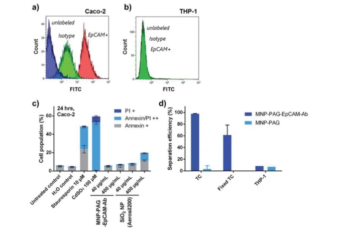
An EpCAM-positive and an EpCAM-negative cell line were
selected for initial bead
−cell interaction studies. The EpCAM
positivity of Caco-2 cells and the low EpCAM expression on
THP-1 cells were con
firmed by flow cytometry (
Figure 2
a,b).
Then, 300-nm beads with and without EpCAM-antibody attached
were added to a suspension of Caco-2 cells (100 000 cells
per mL) at a concentration of 0.4 mg per mL. The
com-patibility of the beads for at least the duration of a typical
experiment and up to 24 h was demonstrated in a membrane
integrity assay (
Figure 2
c). No signi
ficant effect on cell viability
was observed and viability remained
≥90% for at least 24 h of
Figure 2. Cell characterization and interactions between magnetic beads and cells. EpCAM expression on (a) EpCAM-positive Caco-2 cells and (b) EpCAM-negative THP-1 cells. (c) Flow cytometry analysis of PI and Annexin stained Caco-2 cells following exposure to magnetic beads (300 nm) for 24 h and comparison to silica nanoparticles (Aerosil200) (n = 2). (d) Tumor cell separation efficiency using magnetic beads functionalized with EpCAM and beads without antibodies and an incubation time of 10 min (n = 2).
Figure 1. (a) Protein A-coated magnetic nanoparticles (or Protein A/G hybrid-coated 300-nm nanoparticles) where functionalized with EpCAM antibodies. (b) Transmission electron micrographs of 300-nm protein A/G hybrid-coated nanoparticles. (c) Dynamic light scattering measurements of nanoparticle clusters of different size.
incubation time. The nonspeci
fic interaction of protein A/G
coated beads (without EpCAM-antibody) with Caco-2 cells
was investigated (
Figure 2
d). Less than 5% of tumor cells were
separated when beads without EpCAM-antibody were used.
For EpCAM functionalized beads, separation e
fficiencies of
>95% were reached using bead concentrations of 0.4 mg
per mL and contact times of 10 min. These results show that
there is very little nonspeci
fic interaction between cells and
beads without antibody for incubation times up to 10 min.
Additionally, an analogous experiment with (EpCAM-negative)
THP-1 cells shows that <10% of THP-1 cells are removed
by the EpCAM-antibody functionalized magnetic beads.
To di
fferentiate between the competitive receptor binding
and endocytosis processes, separation e
fficiencies of fixed and
un
fixed tumor cells were compared. When PFA-fixed Caco-2
cells were used, separation e
fficiencies were slightly reduced
and reached 60% for EpCAM-antibody functionalized beads.
Less than 1% of PFA-
fixed cells were removed when beads
without antibody were employed.
Separation e
fficiencies of EpCAM-positive Caco-2 cells by
EpCAM-antibody-functionalized beads were then assessed
in the presence of EpCAM-negative cells. First, a human
monocyte cell line (THP-1) with comparatively low phagocytic
activity was used as a model system. Binding of EpCAM-coated
magnetic beads to EpCAM-negative THP-1 cells was found to
be <10% and separation e
fficiencies of Caco-2 cells in the
presence of THP-1 cells were >95%.
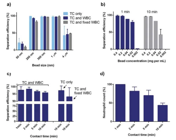
To assess the in
fluence of magnetic bead size on tumor cell
binding and uptake into phagocytic cells, di
fferent bead sizes
(50 nm, 200 nm, 300 nm, 1
μm, and 4 μm) were compared
at the same mass concentration in presence and absence of
EpCAM-negative white blood cells (WBC,
Figure 3
a). Because
undi
fferentiated THP-1 cells have a relatively low phagocytic
activity, native white blood cells isolated from fresh human
whole blood were employed to mimic conditions in human
blood more realistically. At a concentration of 0.4 mg per mL, it
becomes apparent that beads with sizes between 200 nm and
1
μm are most efficient. This is in agreement with a study
on bacteria removal from Kang et al.
24The 50-nm beads are
incompletely separated from the suspension by magnetic
separation due to their weaker magnetic moment. Also, smaller
beads are readily taken up by both WBC and tumor cells (TC),
which possibly explains the slightly reduced separation e
fficacy
in the presence of WBC for 50 and 200 nm. On the other hand,
the number of 4-
μm beads is too low to reach complete
sepa-ration. This is in agreement with
findings from Xu and colleagues,
who report slow and ine
fficient binding of tumor cells by 3−5 μm
beads.
14Next, the in
fluence of bead concentration was investigated
using 300-nm beads (
Figure 3
b). Experiments show that beads
are in excess and that bead concentration of down to 0.2 mg
per mL leads to separation efficiencies of >98%. However,
in the presence of white blood cells, with increasing incubation
time, the minimum number of required beads to achieve
Figure 3. Study of the magnetic bead−cell interactions. Tumor cell separation efficiency as a function of (a) bead size in the presence of tumor cells only (TC), tumor cells and white blood cells (TC and WBC), and tumor cells and fixed white blood cells (TC and fixed white blood cells). (b) Separation efficiency using 300-nm magnetic beads as a function of bead concentration and incubation time (1 and 10 min). (c) The influence of bead−fluid contact time for particle concentrations of 0.2 mg per mL for tumor cells in the presence of white blood cells. (d) The neutrophil count in the supernatant as a function of incubation time with 300-nm magnetic beads at a concentration of 0.2 mg per mL (n = 3).
quantitative separation is signi
ficantly higher, indicative of a
competitive process of bead phagocytosis and binding of beads
to tumor cells.
The in
fluence of contact time between the beads and the
cells has shown little e
ffect of the contact time on TC
sepa-ration e
fficiencies for bead concentration of >0.2 mg per mL
(
Figure 3
c). However, a striking decrease in WBC, and
partic-ularly neutrophil counts, is observed with increasing contact
times (
Figure 3
d). After 10 min of contact time, the neutrophil
count drops by 50% of the initial count (1 million per mL).
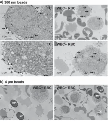
Transmission electron micrographs con
firm uptake of magnetic
nanoparticles, with a preferential uptake of smaller particle
(i.e., 300-nm) compared to 4-
μm particles (
Figure 4
a,b).
Occasionally, also 4
−μm particles are found inside of the cells
(
Figure 4
b). These
findings indicate that blood−bead contact
times should be limited to a few minutes in order to avoid
uptake of magnetic particles into white blood cells.
Finally, we demonstrate e
fficacious removal of
EpCAM-positive cells in a continuous process. We assembled a dialysis
like system in analogy to the ones reported in previous
studies.
9,22,25The volume of the device was 5 mL and
fluid was
pumped at a
flow rate of 1.5 mL per min. Beads were added to
the
fluid stream at a final bead concentration of 0.4 mg per mL.
In the continuous process, single pass tumor separation e
fficiency
was found to be reduced (78.8%) compared to the batch setting
(>95%). To improve contact between beads and
fluid, a
micro-fluidic pearl chain mixer was introduced (
Figure 5
a,b). Using
the mixer, separation e
fficiency was increased from 78.8% to
>92%, even in the presence of physiological numbers of WBC
(
Figure 5
c). The mixing of the beads with the cells is indeed a
critical factor a
ffecting separation performance.
Notably, we have used comparatively high number
concen-trations of tumor cells. The reason for that is mostly of
technical nature, since such high concentrations allow reliable
Figure 4. Particle uptake. Transmission electron micrographs of tumor cells (TC) and white blood cells (WBC) (and red blood cells (RBC)) incubated with 300-nm and 4-μm particles for 1 min or 5 h, respectively. (a) The 300-nm particles are localized primarily on the membrane of tumor cells after short incubation times. Some 300-nm particles can be found inside both TC and WBC. Smaller particles are more readily internalized (black arrows). (b) Most of the 4-μm particles are located extracellularly, however, occasionally, also 4-μm particles (MP) can be found inside of cells (termed as intracellular microparticles (iMP)).
Figure 5. Study of the magnetic bead fluid contact. A device for continuous operation was assembled. (a) Two different geometries were investigated. Geometry A consisted of a silicon tube with a total volume of 5 mL and a volume of 3 mL between the bead injection point and the magnetic separator. Geometry B consisted of the same type of silicon tubing but the tubing was connected to a pearl chain microfluidic chip mixer. The volume between the injection point and the separator again was 3 mL (a, b). (c) The tumor cell separation efficiencies compared for the two geometries (n≥ 2).
cell counting by
flow cytometry. The target cell concentration
in certain clinical scenarios may be much lower, especially for
circulating tumor cells where concentrations of
≤10 cells per
mL are reported,
23,26and this may a
ffect separation
perform-ance. To address how target cell concentration will a
ffect the
removal e
fficiency, we calculated the estimated binding times
based on a mathematical model. We used an estimated target
cell size of 10
μm × 10 μm × 10 μm and a bead concentration
of 5
× 10
9beads per mL. The number of target cells was varied
from 10 cells per mL to 100 000 cells per mL. The modeling
results show that beads are added in such great excess that the
cell concentration only marginally a
ffects the removal efficiency
(
Figure 6
a,b). The number of beads per cell increases
irrespec-tive of the cell number in that case (
Figure 6
c,d). In particular,
Figure 6
d shows the time evolution of cells with at least 10
magnetic beads. It is estimated that this is the requirement
necessary to achieve good separation.
24One can see how in a
couple of minutes all the cells contain at least 10 beads. Because
particles are much smaller than cells in that case, the relative
movements of particles are much faster, and cells can almost be
considered as stationary phase.
27The rate-determining step
in the binding process is the movement and the binding
of nanoparticles to the cell surface, and the cell concentration
thus has very little e
ffect on the separation efficiency. However,
nonspeci
fic uptake may of course significantly affect the
separation performance. We therefore adapted the model to
take into account nonspeci
fic uptake. To do so, we assumed
that particles can also attach to nontarget cells, at a rate which is
α times smaller than the rate of binding to target cells. We have
performed a sensitivity analysis varying several parameters of
the model in order to assess the e
ffect of unspecific binding on
the outcome of the separation. The results of such analyses are
shown in
SI Figures S2 and S3
. The results are presented in
terms of average number of cells bearing at least 10 particles as
a function of the
α value, for two extremely different ratios
of concentrations of specific versus unspecific cells, C
1/C
2.
Figure S2
shows four cases, corresponding to four different
magnetic particles concentrations. One can observe that, for
su
fficiently low values of the parameter α, the separation
between speci
fic and unspecific cells is almost complete.
Not surprisingly, at
α values sufficiently close to 1, the
separa-tion becomes progressively poorer, until speci
ficity is completely
lost. As the particle concentration decreases, it appears that the
number of cells with at least 10 particles that are present after
10 min decreases considerably. As a rule of thumb, in order
to achieve an analogous performance, a decrease in particles
concentration by about an order of magnitude can be
com-pensated by an increase in contact time by 1 order of
magni-tude, as
Figure S3
shows. In fact the fraction of cells with
speci
fic binding shown in
Figures S2c and S3a
is quite similar.
In the former case, a concentration of particles of 5
× 10
7per
mL has been used for 10 min, while in the latter case a particle
concentration of 5
× 10
6per mL for a time of 100 min has been
used. Clearly the fact that the
final results are not identical
is due to the nonidentical consumption of particles, which
becomes a problem when their concentration becomes su
ffi-ciently small, as
Figure S2b
shows, where a particle
concen-tration of 5
× 10
6per mL for a time of 1000 min has been used.
The almost complete consumption of particles in the latter case
can be seen in
Figure S3d
. Taken together, this model enables
Figure 6. Mathematical modeling results for magnetic beads with a size of 300 nm and a number concentration of 5 × 10⁹beads per mL. (a) Change
in magnetic nanoparticle concentration as a function of cell number (10−105per mL) concentration and time. (b) The fraction of cells with no
magnetic particles attached as a function of cell number concentration and time. (c) The average number of magnetic particles attached to a cell and (d) the time evolution of the fraction of cells carrying at least 10 particles.
estimation of the minimal particle concentration required to
remove a certain number of cells in a given time as a function of
nonspeci
fic interactions.
■
CONCLUSIONS
The present article demonstrates the conceptual feasibility of
cell removal from body
fluids in a continuous extracorporeal
dialysis-type of setting. The contact between magnetic beads
and target substances is critical, and can be enhanced by
introducing a pearl chain mixer. Contact times higher than
2 min should be avoided in order to prevent nonspeci
fic uptake
into blood cells. Also, particle surface chemistry may be further
optimized to improve speci
fic cell binding and limit nonspecific
interactions. The current setting could be easily scaled up to
increase the throughput to 50
−150 mL per min, similar to that
of currently used blood puri
fication devices. This study paves
the way to investigate the therapeutic bene
fit of magnetic cell
removal in conditions such as metastatic cancer or leukemia,
where circulating tumor cells could be removed, or in
auto-immune diseases.
■
ASSOCIATED CONTENT
*
S Supporting InformationThe Supporting Information is available free of charge on the
ACS Publications website
at DOI:
10.1021/acsami.7b10140
.
Figure S1. Hydrodynamic size of magnetic beads in water
and cell culture medium. Further description of the
mathematical model. Figures S2 and S3. Mathematical
modeling results for magnetic beads with a size of
300 nm (
)
■
AUTHOR INFORMATION
Corresponding Author*
[email protected]
.
ORCIDSergio Bertazzo:
0000-0003-4889-8190Marco Lattuada:
0000-0001-7058-9509Inge K. Herrmann:
0000-0002-3018-6796 Author Contributions∥
N.B. and N.D. contributed equally as
first authors.
Author ContributionsN.D. and N.B. performed the experimental work on cell
removal, N.B. supervised the
flow cytometry work, performed
cytotoxicity assays, and helped write the manuscript, E.T.
analyzed samples in TEM, L.G. performed dynamic light
scattering measurements, K.K. helped with nanoparticle and
TEM sample preparation, S.B. supervised TEM studies, M.L.
performed the mathematical modeling, I.K.H. supervised the
study and wrote the manuscript. All authors contributed to
discussions and edited the manuscript.
Notes
The authors declare no competing
financial interest.
■
ACKNOWLEDGMENTS
We acknowledge support from the Novartis Foundation for
Medical-Biological Research. M.L. acknowledges
financial
support from the Swiss National Science Foundation (grant
PP00P2_159258). N.B. acknowledges funding from the
NanoScreen Materials Challenge cofunded by the Competence
Centre for Materials Science and Technology (CCMX). We
thank Ursina Tobler for assistance with blood collection from
healthy volunteers.
■
REFERENCES
(1) Issadore, D.; Chung, J.; Shao, H.; Liong, M.; Ghazani, A. A.; Castro, C. M.; Weissleder, R.; Lee, H. Ultrasensitive Clinical Enumeration of rare Cells ex vivo using aμ-Hall Detector. Sci. Transl. Med. 2012, 4 (141), 141ra92−141ra92.
(2) Mohamadi, R. M.; Besant, J. D.; Mepham, A.; Green, B.; Mahmoudian, L.; Gibbs, T.; Ivanov, I.; Malvea, A.; Stojcic, J.; Allan, A. L.; Lowes, L. E.; Sargent, E. H.; Nam, R. K.; Kelley, S. O. Nanoparticle-Mediated Binning and Profiling of Heterogeneous Circulating Tumor Cell Subpopulations. Angew. Chem. 2015, 127 (1), 141−145.
(3) Poudineh, M.; Aldridge, P. M.; Ahmed, S.; Green, B. J.; Kermanshah, L.; Nguyen, V.; Tu, C.; Mohamadi, R. M.; Nam, R. K.; Hansen, A.; Sridhar, S. S.; Finelli, A.; Fleshner, N. E.; Joshua, A. M.; Sargent, E. H.; Kelley, S. O. Tracking the Dynamics of Circulating Tumour Cell Phenotypes using Nanoparticle-mediated Magnetic Ranking. Nat. Nanotechnol. 2016, 12 (3), 274−281.
(4) Wang, L.; Yang, Z.; Gao, J.; Xu, K.; Gu, H.; Zhang, B.; Zhang, X.; Xu, B. A Biocompatible Method of Decorporation: Bisphosphonate-Modified Magnetite Nanoparticles to Remove Uranyl Ions from Blood. J. Am. Chem. Soc. 2006, 128 (41), 13358−13359.
(5) Lee, H. Y.; Bae, D. R.; Park, J. C.; Song, H.; Han, W. S.; Jung, J. H. A Selective Fluoroionophore Based on BODIPY-functionalized Magnetic Silica Nanoparticles: Removal of Pb2+ from Human Blood. Angew. Chem., Int. Ed. 2009, 48 (7), 1239−1243.
(6) Herrmann, I. K.; Urner, M.; Koehler, F. M.; Hasler, M.; Roth-Z’Graggen, B.; Grass, R. N.; Ziegler, U.; Beck-Schimmer, B.; Stark, W. J. Blood Purification using Functionalized Core/Shell Nanomagnets. Small 2010, 6 (13), 1388−1392.
(7) Cai, K.; Li, J.; Luo, Z.; Hu, Y.; Hou, Y.; Ding, X. [small beta]-Cyclodextrin conjugated Magnetic Nanoparticles for Diazepam Removal from Blood. Chem. Commun. 2011, 47 (27), 7719−7721.
(8) Shen, H.; Wang, J.; Liu, H.; Li, Z.; Jiang, F.; Wang, F.-B.; Yuan, Q. Rapid and Selective Detection of Pathogenic Bacteria in Bloodstream Infections with Aptamer-Based Recognition. ACS Appl. Mater. Interfaces 2016, 8 (30), 19371−19378.
(9) Kang, J. H.; Super, M.; Yung, C. W.; Cooper, R. M.; Domansky, K.; Graveline, A. R.; Mammoto, T.; Berthet, J. B.; Tobin, H.; Cartwright, M. J.; Watters, A. L.; Rottman, M.; Waterhouse, A.; Mammoto, A.; Gamini, N.; Rodas, M. J.; Kole, A.; Jiang, A.; Valentin, T. M.; Diaz, A.; Takahashi, K.; Ingber, D. E. An extracorporeal Blood-cleansing Device for Sepsis Therapy. Nat. Med. 2014, 20 (10), 1211− 1216.
(10) Lattuada, M.; Ren, Q.; Zuber, F.; Galli, M.; Bohmer, N.; Matter, M. T.; Wichser, A.; Bertazzo, S.; Pier, G. B.; Herrmann, I. K. Theranostic Body Fluid Cleansing: rationally designed Magnetic Particles enable Capturing and Detection of Bacterial Pathogens. J. Mater. Chem. B 2016, 4 (44), 7080−7086.
(11) Molday, R. S.; Yen, S. P. S.; Rembaum, A. Application of Magnetic Microspheres in Labelling and Separation of Cells. Nature 1977, 268 (5619), 437−438.
(12) Miltenyi, S.; Müller, W.; Weichel, W.; Radbruch, A. High gradient Magnetic Cell Separation with MACS. Cytometry 1990, 11 (2), 231−238.
(13) Molday, R. S.; Mackenzie, D. Immunospecific Ferromagnetic Iron-dextran Reagents for the Labeling and Magnetic Separation of Cells. J. Immunol. Methods 1982, 52 (3), 353−367.
(14) Xu, H.; Aguilar, Z. P.; Yang, L.; Kuang, M.; Duan, H.; Xiong, Y.; Wei, H.; Wang, A. Antibody conjugated Magnetic Iron Oxide Nanoparticles for Cancer Cell Separation in fresh Whole Blood. Biomaterials 2011, 32 (36), 9758−9765.
(15) Green, B. J.; Saberi Safaei, T.; Mepham, A.; Labib, M.; Mohamadi, R. M.; Kelley, S. O. Beyond the Capture of Circulating Tumor Cells: Next-Generation Devices and Materials. Angew. Chem., Int. Ed. 2016, 55 (4), 1252−1265.
(16) Khoo, B. L.; Warkiani, M. E.; Tan, D. S.-W.; Bhagat, A. A. S.; Irwin, D.; Lau, D. P.; Lim, A. S. T.; Lim, K. H.; Krisna, S. S.; Lim, W.-T.; Yap, Y. S.; Lee, S. C.; Soo, R. A.; Han, J.; Lim, C. T. Clinical Validation of an Ultra High-Throughput Spiral Microfluidics for the Detection and Enrichment of Viable Circulating Tumor Cells. PLoS One 2014, 9 (7), e99409.
(17) Nagrath, S.; Sequist, L. V.; Maheswaran, S.; Bell, D. W.; Irimia, D.; Ulkus, L.; Smith, M. R.; Kwak, E. L.; Digumarthy, S.; Muzikansky, A.; Ryan, P.; Balis, U. J.; Tompkins, R. G.; Haber, D. A.; Toner, M. Isolation of Rare Circulating Tumour Cells in Cancer Patients by Microchip Technology. Nature 2007, 450 (7173), 1235−1239.
(18) Stott, S. L.; Hsu, C.-H.; Tsukrov, D. I.; Yu, M.; Miyamoto, D. T.; Waltman, B. A.; Rothenberg, S. M.; Shah, A. M.; Smas, M. E.; Korir, G. K.; Floyd, F. P.; Gilman, A. J.; Lord, J. B.; Winokur, D.; Springer, S.; Irimia, D.; Nagrath, S.; Sequist, L. V.; Lee, R. J.; Isselbacher, K. J.; Maheswaran, S.; Haber, D. A.; Toner, M. Isolation of Circulating Tumor Cells using a Microvortex-generating Herringbone-chip. Proc. Natl. Acad. Sci. U. S. A. 2010, 107 (43), 18392−18397.
(19) Yamamoto, S.; Shimizu, K.; Fei, J.; Iwata, H.; Okochi, M.; Nakanishi, H.; Honda, H. Ex vivo Culture of Circulating Tumor Cells using Magnetic Force-based Coculture on a Fibroblast Feeder Layer. Biotechnol. J. 2016, 11 (11), 1433−1442.
(20) Yoon, Y.; Cho, S.; Kim, S.; Choi, E.; Kim, R. K.; Lee, S. J.; Sul, O.; Lee, S. B. Separation and Capture of Circulating Tumor Cells from Whole Blood using a Bypass integrated Microfluidic Trap Array. In 36th Annual International Conference of the IEEE Engineering in Medicine and Biology Society, 26−30 Aug. 2014; 2014; pp 4431−4434. (21) Scarberry, K. E.; Mezencev, R.; McDonald, J. F. Targeted Removal of Migratory Tumor Cells by Functionalized Magnetic Nanoparticles impedes Metastasis and Tumor Progression. Nano-medicine 2011, 6 (1), 69−78.
(22) Herrmann, I. K.; Bernabei, R. E.; Urner, M.; Grass, R. N.; Beck-Schimmer, B.; Stark, W. J. Device for Continuous Extracorporeal Blood Purification using Target-specific Metal Nanomagnets. Nephrol., Dial., Transplant. 2011, 26 (9), 2948−U1516.
(23) Allard, W. J.; Matera, J.; Miller, M. C.; Repollet, M.; Connelly, M. C.; Rao, C.; Tibbe, A. G. J.; Uhr, J. W.; Terstappen, L. W. M. M. Tumor Cells Circulate in the Peripheral Blood of All Major Carcinomas but not in Healthy Subjects or Patients With Non-malignant Diseases. Clin. Cancer Res. 2004, 10 (20), 6897−6904.
(24) Kang, J. H.; Um, E.; Diaz, A.; Driscoll, H.; Rodas, M. J.; Domansky, K.; Watters, A. L.; Super, M.; Stone, H. A.; Ingber, D. E. Optimization of Pathogen Capture in Flowing Fluids with Magnetic Nanoparticles. Small 2015, 11 (42), 5657−5666.
(25) Herrmann, I. K.; Schlegel, A.; Graf, R.; Schumacher, C. M.; Senn, N.; Hasler, M.; Gschwind, S.; Hirt, A.-M.; Guenther, D.; Clavien, P.-A.; Stark, W. J.; Beck-Schimmer, B. Nanomagnet-based Removal of Lead and Digoxin from Living Rats. Nanoscale 2013, 5 (18), 8718−8723.
(26) Yu, M.; Stott, S.; Toner, M.; Maheswaran, S.; Haber, D. A. Circulating Tumor Cells: Approaches to Isolation and Character-ization. J. Cell Biol. 2011, 192 (3), 373−382.
(27) Rossier, M.; Koehler, F. M.; Athanassiou, E. K.; Grass, R. N.; Waelle, M.; Birbaum, K.; Günther, D.; Stark, W. J. Energy-Efficient Noble Metal Recovery by the Use of Acid-Stable Nanomagnets. Ind. Eng. Chem. Res. 2010, 49 (19), 9355−9362.