PAEDIATRICS
Impact of depth of propofol anaesthesia on functional residual
capacity and ventilation distribution in healthy preschool children
B. S. von Ungern-Sternberg
1*, F. J. Frei
1, J. Hammer
2, A. Schibler
3, R. Doerig
1and T. O. Erb
11
Division of Anaesthesia and 2Division of Pulmonology and Intensive Care, University of Basel Children’s Hospital, Roemergasse 8, CH-4005 Basel, Switzerland.3Division of Paediatric Intensive Care, Mater
Misericordiae Hospital, South Brisbane, QLD 4101, Australia
*Corresponding author: Division of Anaesthesia, University of Basel Children’s Hospital, Roemergasse 8, CH-4005 Basel, Switzerland. E-mail:[email protected]
Background. Propofol is commonly used in children undergoing diagnostic interventions under anaesthesia or deep sedation. Because hypoxaemia is the most common cause of critical deterioration during anaesthesia and sedation, improved understanding of the effects of anaes-thetics on pulmonary function is essential. The aim of this study was to determine the effect of different levels of propofol anaesthesia on functional residual capacity (FRC) and ventilation distribution.
Methods. In 20 children without cardiopulmonary disease mean age (SD) 49.75 (13.3) months
and mean weight (SD) 17.5 (3.9) kg, anaesthesia was induced by a bolus of i.v. propofol
2 mg kg21 followed by an infusion of propofol 120 mg kg21min21 (level I). Then, a bolus of propofol 1 mg kg21was given followed by a propofol infusion at 240 mg kg21min21 (level II). FRC and lung clearance index (LCI) were calculated at each level of anaesthesia using multi-breath analysis.
Results. The FRC mean (SD) decreased from 20.7 (3.3) ml kg21 at anaesthesia level I to 17.7
(3.9) ml kg21at level II (P,0.0001). At the same time, mean (SD) LCI increased from 10.4 (1.1)
to 11.9 (2.2) (P¼0.0038), whereas bispectral index score values decreased from mean (SD)
57.5 (7.2) to 35.5 (5.9) (P,0.0001).
Conclusions. Propofol elicited a deeper level of anaesthesia that led to a significant decrease of the FRC whereas at the same time the LCI, an index for ventilation distribution, increased indicating an increased vulnerability to hypoxaemia.
Br J Anaesth 2007; 98: 503–8
Keywords: anaesthetics i.v., propofol; anaesthesia, paediatric; lung, clearance index; respiratory function, functional residual capacity; sedation; ventilation, distribution; ventilation, homogeneity
Accepted for publication: December 19, 2006
Anaesthesia or deep sedation are frequently required in children because of their lack of cooperation with diagnostic procedures or minor interventions.1 On the basis of the invasiveness of the intervention (e.g. magnetic
resonance imaging scanning, bronchoscopy), varying
levels of anaesthesia are required to perform such pro-cedures successfully.
In children undergoing anaesthesia, hypoxaemia is the most common and clinically relevant cause of critical
deterioration.2 Children have lower oxygen reserves
because they breathe at tidal volumes overlapping closing
capacity resulting in airway closure and areas of
ventilation – perfusion mismatch. The balance between chest and lung recoil pressure determines the static resting volume of the lung. The highly compliant chest wall of young children results in equilibrium at a lower lung volume compared with adults.3 4 Additionally, children are particularly vulnerable to hypoxaemia because of their higher oxygen demand per kilo body weight. Thus, because anaesthetic drugs are known to decrease functional residual
capacity (FRC), optimizing FRC can be critical in children undergoing anaesthesia or deep sedation.5
For deep sedation in children, propofol is a commonly used sedative agent. Increasing doses of propofol can jeopardize upper airway patency by increasing airway collapsibility;6 7 however, the impact of different doses of propofol on pulmon-ary function is unknown in the paediatric population. The aim of this study was to determine the effect of two depths of pro-pofol anaesthesia on FRC and ventilation distribution and to test the hypothesis that FRC and ventilation homogeneity decrease with an increased dose of propofol.
Methods
After approval from the local Ethics Committee (Basel, Switzerland) and after obtaining written parental informed consent, 20 healthy children, 2 – 6 yr of age and classified as American Society of Anaesthesiologists physical status I, were included in this study. Exclusion criteria included clinical evidence of cardiopulmonary diseases (including respiratory tract infection during the 2 weeks before surgery) or thoracic deformities.
One hour before sedation, a eutetic mixture of local anaesthetic patches was applied to the dorsal side of each hand. Premedication consisted of midazolam 0.3 mg kg21 administered orally or rectally 15 min before induction of anaesthesia. In uncooperative patients, nitrous oxide 70% in oxygen was administered to facilitate the insertion of an i.v. line. In all cases, nitrous oxide was immediately stopped when venous access was achieved. Thereafter, a mixture of oxygen and air resulting in a FIO
2of 0.5 was administered to all children for the remainder of the study. Anaesthesia was induced with a bolus of i.v. propofol 2 mg kg21followed by an infusion of i.v. propofol at 120 mg kg21min21 (level I). After the first measurement, a bolus of i.v.
pro-pofol 1 mg kg21 was given followed by i.v. propofol
240 mg kg21min21(level II). According to Kataria’s model,8 this regime reaches pseudo-steady-state-conditions 5 min after application of the bolus with a calculated effect site con-centration of 1.8 mg ml21(level I) and 3.1 mg ml21(level II).
For the FRC measurements, an ultrasonic transit-time airflow meter (Exhalyzer D with ICU insert, Eco Medics, Duernten, Switzerland) that simultaneously measures flow and the molar mass of the breathing gas was placed between the close fitting face mask (Vital sign, size 2, Totowa, NJ, USA) and the fresh gas outlet. This equip-ment has been described in detail previously.9Briefly, this airflow meter combines accurate flow measurements with instantaneous mainstream gas analysis of molecular mass in a single sensor. This analysis is based on an ultrasonic transit time detection measured at a high sampling fre-quency (400 Hz) with piezoelectric sensors that demon-strate a high linearity over a wide amplitude range.
The introduction of sulphur hexafluoride (SF6,
molecu-lar mass 146 g mol21) as a tracer gas into the inspiratory part of the breathing system increases the total molecular
mass of the breathing gas until a steady state is reached. After the discontinuation of sulphur hexafluoride, the mol-ecular mass decreases breath by breath until a steady state is reached when the sulphur hexafluoride has been washed out of the lungs (multibreath washout technique). Analysis of the washout curve allows for calculation of the FRC, physiological dead space volume, lung clearance index (LCI), and mean dilution number (MDN). LCI and MDN are commonly used to measure the degree of ventilation distribution and are sensitive indicators of peripheral airway collapse.10 11 The LCI is the cumulative expired volume needed to lower the end-tidal tracer gas (sulphur hexafluoride) concentration to 1/40 of the starting concen-tration divided by the FRC; that is, the number of lung volume turnovers needed to clear the lungs of the marker gas.11 The MDN is the ratio between the first and the zeroth moments of the washout curve. The number of volume turnovers was calculated using the cumulative expired alveolar volume.10 12 The dead space was calcu-lated with the Fowler method using the molar mass signal. FRC, dead space volume, MDN, and LCI calculations were performed using Spiroware software (Version 1.5.2, ndd Medizintechnik AG, Zurich, Switzerland).
Patient head position was standardized with a sniffing position pillow causing lateral stabilization of the head and a slight atlanto-occipital extension. A close fit of the face mask was achieved by rubber straps fitted around the head of the patient and the mask.13 In order to maintain a patient’s airway, chin lift was done using a tape fixed to the chin of the patient and a frame.14 In order to assure good positioning and seal of the face mask, measurements were only performed when minimal leakage was confirmed by auscultation at the rim of the mask and by equal inspiratory and expiratory volumes as measured by spirometry.
FRC measurements at level I were taken 5 min after the start of anaesthesia and measurements at level II 5 min after increasing the propofol dose. FRC and LCI assess-ments were performed in duplicate at each level of anaes-thesia and the mean values were used for all calculations. At each level of propofol anaesthesia, a venous blood sample was taken between the two FRC measurements to
measure the propofol plasma concentration (HPLC
method, clinical chemistry laboratory, University of Basel Hospital, Basel, Switzerland) and the University of Michigan Sedation Score was determined and documen-ted.15For each study condition, two measurements of FRC and LCI were performed and the mean values were used for all calculations.
Statistical analysis
Sample size calculations were performed using nQuery Advisor 4.0 software (Statistical Solutions Ltd, Boston, MA, USA). On the basis of separate pilot data, a sample size of 20 patients was required in order to detect a FRC difference in means of 1 ml kg21(5%, decrease from 20 to
19 ml kg21) assuming a standard deviation of differences of 1.3 ml kg21after deepening the level of sedation for a two-sided t-test with an a error of 0.05 and a b error of 0.1.
The distribution of data was tested using a Shapiro – Wilk test. Normally distributed data are expressed as mean
(SD) and data not having a normal distribution are
expressed as median (inter-quartile range). For statistical analysis, a paired t-test was used to compare the variables between the repeated measurements and P,0.05 was con-sidered significant. Results were analysed using the StatView for Windows (SAS Institute Inc., Cary, NC, USA, Version 5.0.1).
Results
All 20 children were successfully studied. Patient charac-teristics are shown in Table 1. Each child had a SpO2 of .97% throughout the entire study period. None of the children showed overt clinical signs of upper airway obstruction (e.g. stridor or retraction) at any time. The group mean FRC significantly decreased with a deeper level of anaesthesia whereas LCI and MDN increased sim-ultaneously (Table 2 and Figure 1AandB). Ventilatory
fre-quency, dead space volume, and the tidal volume to FRC ratio remained constant between the two levels of sedation, whereas tidal volume and bispectral index score values significantly decreased (Table 2). The propofol plasma concentration as well as the University of Michigan Sedation Scale increased at the deeper level of anaesthesia.
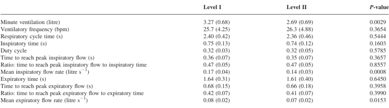
Respiratory cycle parameters except for the mean
inspiratory and expiratory flows remained stable through-out the study period (Table 3).
Discussion
This study examined the effects of two levels of propofol anaesthesia on FRC and ventilation distribution in sedated, preschool children. A deeper level of anaesthesia substan-tially decreased FRC and ventilation distribution indicating a higher susceptibility for hypoxaemia.
FRC
General anaesthetics attenuate airway muscle activity in
a dose-dependent manner.16 17 Propofol blocks the
central part of the motor system and sodium channels in skeletal muscle resulting in myorelaxation.18 19 Propofol affects the sensitive balance between chest wall compli-ance, elastic lung recoil, and tension of the diaphragm leading to a decrease in lung volumes and ventilation distribution.18 – 20
In children, these effects on FRC and lung compliance can be more pronounced because their chest walls are more compliant than those of adults.21 Chest wall compli-ance is increased by drugs exhibiting muscle relaxant properties that impair the natural dynamic elevation of the resting lung volume, which is normally maintained in small children by diaphragmatic activity during the expiratory phase. Because the balance between chest and lung recoil pressure determines the static resting volume of the lung, a child reaches equilibrium at a relatively lower lung volume compared with adults.3Although it is generally assumed that by the end of the first year of
life,3 active elevation of the end-expiratory volume
decreases and the rib cage contribution to tidal breathing approaches adult levels, this mechanism might still account for some of the observed decrease in FRC in the present study population.
Several studies provide FRC reference values for infants and children. Recent studies report smaller lung volumes
Table 1Patient characteristics (n¼20). Mean (SD) or number (n) when appropriate Male:Female 15:5 Age (months) 49.75 (13.3) Height (cm) 104.6 (9.6) Weight (kg) 17.5 (3.9) Smoke exposure (n) 2
Family history of asthma (n) 5 Family history of severe allergic reactions (n) 1
Table 2 Measured variables at the two levels of propofol anaesthesia. Values are mean (SD) for all parameters except the University of Michigan Sedation score which is given as median [IQR]. Statistical significance determined by a paired t-test or Wilcoxon signed rank test as appropriate. *University of Michigan Sedation Scale: 0¼awake and alert, 1¼minimally sedated: tired/sleepy, appropriate response to verbal conversation and/or sound, 2¼moderately sedated: somnolent/sleeping, easily aroused with light tactile stimulation or a simple verbal command, 3¼deeply sedated: deep sleep, rousable only with significant physical stimulation, 4¼unrousable
Level I Level II P-value
FRC (ml) 364 (108) 310 (100) ,0.0001 FRC (ml kg21) 20.7 (3.3) 17.7 (3.9) ,0.0001 LCI 10.4 (1.1) 11.9 (2.2) 0.0038 MDN 2.80 (0.3) 3.28 (0.7) 0.0074 Dead space (ml kg21) 2.79 (0.6) 2.7 (0.6) 0.4902 Tidal volume (ml) 128 (21.6) 104 (25.5) 0.0002 Tidal volume (ml kg21) 7.41 (1.08) 6.03 (1.40) 0.0006
Tidal volume to functional residual capacity ratio 0.36 (0.06) 0.35 (0.11) 0.62
Bispectral index score 57.5 (7.2) 35.5 (5.9) ,0.0001
Propofol plasma concentration (mg ml21) 1.47 (0.4) 3.16 (0.9) ,0.0001 University of Michigan Sedation Scale* 3 [3 – 3] 4 [3 – 4] 0.0011
than those observed in earlier studies, although this could reflect the changes in technology.22Our FRC values are in
the same range as reported values mean (SD) 19.6
(3.4) ml kg21 measured by plethysmographic methods in spontaneously breathing infants sedated with chloralhy-drate, an hypnotic agent that also exhibits muscle relaxant properties.22 23 FRC values mean (SD) 18 (2) ml kg21
obtained with the same SF6multibreath washout technique
measured in unsedated, spontaneously breathing healthy infants are similar to ours.10 However, recent reference data for FRC in preschool children are not available for direct comparison with our data. Moreover, measurements of FRC and ventilation distribution are difficult to obtain
in preschool children because of their lack of cooperation. Sedation, often used in uncooperative children, probably affects FRC, but the extent of this effect is unknown.
Ventilation distribution
Ventilation distribution can be calculated by analysing the washout curve of an insoluble tracer gas (e.g. SF6).24
Several indices have been described.24 Among them, the LCI and MDN are sensitive parameters of ventilation homogeneity with the advantage of being less affected by changes in tidal and dead space volumes.24Regions of the
lung that are less well ventilated than others will be cleared more slowly than those receiving a higher portion of the tidal volume. Although both indices are very sensi-tive in detecting peripheral airway collapse,25 the LCI does not give any insight into the mechanism causing changes in the ventilation distribution. However, it is a good index for ventilation efficiency to further quantify the impact of different doses of propofol anaesthesia. Obstructive airway disorders result in asymmetrical nar-rowing of the intrapulmonary airways leading to a reduced ventilation distribution as reflected by increased LCI and MDN.
The present study is the first to analyse the effect of different doses of propofol anaesthesia on ventilation dis-tribution in children. The reduced ventilation disdis-tribution at a deeper level of anaesthesia is best explained by partial airway collapse in the dependent regions of the lungs while in the supine position.26 One way of correcting for measured dead space is to apply the alveolar-based MDN. Interestingly, neither the dead space volume nor the tidal volume to FRC ratio changed throughout the study period.
Depth of anaesthesia
Depth of anaesthesia was assessed by the University of Michigan Sedation Scale15 and by bispectral index moni-toring. Propofol plasma concentrations were measured at both levels of anaesthesia. The propofol dose that was administered does not have a high predictive value for the clinically observed depth of anaesthesia reflected by the 10 Level I P<0.0001 FRC (ml kg –1 ) Level II 15 20 25 A B Level I 8 12 16 20 LCI P<0.0038 Level II
Fig 1 (A) FRC (ml kg21) at two levels of propofol anaesthesia. (B) LCI
at two levels of propofol anaesthesia.
Table 3 Respiratory cycle variables at two levels of propofol anaesthesia. Data are given as mean (SD)
Level I Level II P-value
Minute ventilation (litre) 3.27 (0.68) 2.69 (0.69) 0.0029
Ventilatory frequency (bpm) 25.7 (4.25) 26.3 (4.88) 0.3654
Respiratory cycle time (s) 2.40 (0.42) 2.36 (0.46) 0.5444
Inspiratory time (s) 0.75 (0.13) 0.74 (0.12) 0.1603
Duty cycle 0.32 (0.03) 0.32 (0.05) 0.5785
Time to reach peak inspiratory flow (s) 0.36 (0.07) 0.35 (0.07) 0.3657 Ratio: time to reach peak inspiratory flow to inspiratory time 0.47 (0.05) 0.47 (0.05) 0.8557 Mean inspiratory flow rate (litre s21) 0.17 (0.04) 0.14 (0.03) 0.0008
Expiratory time (s) 1.64 (0.31) 1.61 (0.40) 0.6450
Time to reach peak expiratory flow (s) 0.68 (0.15) 0.66 (0.18) 0.3958 Ratio: time to reach peak expiratory flow to expiratory time 0.42 (0.07) 0.41 (0.07) 0.3990 Mean expiratory flow rate (litre s21) 0.08 (0.02) 0.07 (0.02) 0.0153
wide inter-individual range of responses, although the bi-spectral index score values for one level of sedation were comparable between the children. However, because, each patient served as his own control for the two measure-ments, inter-individual response variations are less import-ant. In our study, all patients had significantly lower bispectral index score values at the second level of anaes-thesia compared with the first level and their propofol plasma concentrations were higher.
Upper airway patency
Upper airway obstruction can influence respiratory func-tion under spontaneous breathing. In particular, increasing levels of propofol anaesthesia progressively jeopardize upper airway patency.6 7 In order to minimize this factor, great care was taken to optimize upper airway patency by standardized head and neck positioning. Furthermore, chin lift was consistently applied throughout the entire study period. During the assessments, no overt clinical signs of upper airway obstruction were detected.
The times to reach peak inspiratory and expiratory flow are measures that would detect inspiratory or expiratory airways obstruction. These remained similar between the two levels of propofol sedation strongly suggesting that the reduction of minute ventilation by a reduction of tidal volume is a consequence of decreased respiratory drive and not because of airway obstruction. This is also reflected in the decrease in mean inspiratory flows. All parameters of the respiratory cycle were within the normal ranges for healthy children.27 28
Limitations
Although there was no control group for the children exam-ined in this study, it is unlikely that the decrease in FRC was related to sedation time and not to sedation depth. Moreover, in a similar design, FRC and LCI in spon-taneously breathing preschool children under propofol anaesthesia (FIO
2¼0.5) remained constant during the 30 min period;9the time between measurements at each anaesthesia level in the present study was much shorter (5–10 min) and thus time or FIO
2were unlikely to have been the confound-ing factors for changes in FRC, LCI, MDN, and tidal volume observed in this study. Additionally, FRC decreases immediately after induction of anaesthesia in spontaneously breathing adults but then remains stable for at least 1 h.29
The additional administration of midazolam might have
influenced FRC because of its myorelaxant effect.30
However, the comparison between the two levels of propo-fol was likely to be unaffected because the impact of midazolam was similar in both assessments.
In conclusion, a deeper level of propofol sedation reduced FRC and ventilation distribution in preschool-aged children, potentially indicating an increasing vulner-ability to hypoxaemia with increasing doses of propofol.
Acknowledgements
The work should be attributed to the Division of Anaesthesia, University of Basel Children’s Hospital, Basel, Switzerland. Funding was received from the Division of Anaesthesia, University of Basel, Switzerland. Andreas Schibler is supported by Preston James Research Fund and the Golden Casket Research Fund (Australia). The authors thank J. Etlinger, BA, Department of Anaesthesia, University of Basel, Switzerland, for editorial assistance and Michel Pelligrini, MD, Department of Anaesthesia, University of Geneva, Switzerland, for his assistance with the calculations of the propofol pharmacokinetics.
References
1 Krauss B, Green SM. Sedation and analgesia for procedures in children. N Engl J Med 2000; 342: 938 – 45
2 Cote CJ, Notterman DA, Karl HW, Weinberg JA, McCloskey C. Adverse sedation events in pediatrics: a critical incident analysis of contributing factors. Pediatrics 2000; 105: 805 – 14
3 Bancalari E, Clausen J. Pathophysiology of changes in absolute lung volumes. Eur Respir J 1998; 12: 248 – 58
4 Nunn JF, ed. Elastic forces and lung volumes. In: Applied Respiratory Physiology. London: Butterworth, 1987; 23 – 45 5 Dobbinson TL, Nisbet HI, Pelton DA. Functional residual capacity
(FRC) and compliance in anaesthetized paralysed children. I. In vitro tests with the helium dilution method of measuring FRC. Can Anaesth Soc J 1973; 20: 310 – 21
6 Eastwood PR, Platt PR, Shepherd K, Maddison K, Hillman DR. Collapsibility of the upper airway at different concentrations of propofol anesthesia. Anesthesiology 2005; 103: 470 – 7
7 Evans RG, Crawford MW, Noseworthy MD, Yoo SJ. Effect of increasing depth of propofol anesthesia on upper airway configur-ation in children. Anesthesiology 2003; 99: 596 – 602
8 Kataria BK, Ved SA, Nicodemus HF, et al. The pharmacokinetics of propofol in children using three different data analysis approaches. Anesthesiology 1994; 80: 104 – 22
9 von Ungern-Sternberg BS, Regli A, Frei FJ, et al. The effect of caudal block on functional residual capacity and ventilation homogeneity in healthy children. Anaesthesia 2006; 61: 758 – 63 10 Schibler A, Hall GL, Businger F, et al. Measurement of lung
volume and ventilation distribution with an ultrasonic flow meter in healthy infants. Eur Respir J 2002; 20: 912 – 8
11 Gustafsson PM, Kallman S, Ljungberg H, Lindblad A. Method for assessment of volume of trapped gas in infants during multiple-breath inert gas washout. Pediatr Pulmonol 2003; 35: 42– 9 12 Larsson A, Linnarsson D, Jonmarker C, et al. Measurement of
lung volume by sulfur hexafluoride washout during spontaneous and controlled ventilation: further development of a method. Anesthesiology 1987; 67: 543 – 50
13 Shorten GD, Armstrong DC, Roy WI, Brown L. Assessment of the effect of head and neck position on upper airway anatomy in sedated paediatric patients using magnetic resonance imaging. Paediatr Anaesth 1995; 5: 243 – 8
14 Reber A, Wetzel SG, Schnabel K, Bongartz G, Frei FJ. Effect of combined mouth closure and chin lift on upper airway dimen-sions during routine magnetic resonance imaging in pediatric patients sedated with propofol. Anesthesiology 1999; 90: 1617 – 23 15 Malviya S, Voepel-Lewis T, Tait AR, et al. Depth of sedation in
children undergoing computed tomography: validity and reliability of the University of Michigan Sedation Scale (UMSS). Br J Anaesth 2002; 88: 241 – 5
16 Drummond GB. Influence of thiopentone on upper airway muscles. Br J Anaesth 1989; 63: 12 – 21
17 Grounds RM, Maxwell DL, Taylor MB, Aber V, Royston D. Acute ventilatory changes during i.v. induction of anaesthesia with thio-pentone or propofol in man. Br J Anaesth 1987; 59: 1098 – 102
18 Dueck MH, Oberthuer A, Wedekind C, Paul M, Boerner U. Propofol impairs the central but not the peripheral part of the motor system. Anesth Analg 2003; 96: 449 – 55
19 Haeseler G, Stormer M, Bufler J, et al. Propofol blocks human skeletal muscle sodium channels in a voltage-dependent manner. Anesth Analg 2001; 92: 1192 – 8
20 Macklem PT, Murphy B. The forces applied to the lung in health and disease. Am J Med 1974; 57: 371 – 7
21 Papastamelos C, Panitch HB, England SE, Allen JL. Developmental changes in chest wall compliance in infancy and early childhood. J Appl Physiol 1995; 78: 179 – 84
22 Hulskamp G, Hoo AF, Ljungberg H, et al. Progressive decline in plethysmographic lung volumes in infants: physiology or technol-ogy? Am J Respir Crit Care Med 2003; 168: 1003 – 9
23 Hershenson M, Brouillette RT, Olsen E, Hunt CE. The effect of chloral hydrate on genioglossus and diaphragmatic activity. Pediatr Res 1984; 18: 516 – 9
24 Larsson A, Jonmarker C, Werner O. Ventilation inhomogeneity during controlled ventilation. Which index should be used? J Appl Physiol 1988; 65: 2030 – 9
25 Gustafsson PM, Aurora P, Lindblad A. Evaluation of ventilation maldistribution as an early indicator of lung disease in children with cystic fibrosis. Eur Respir J 2003; 22: 972 – 9
26 West JB, ed. Structure and function—how the architecture of the lung subserves its function. In: Respiratory Physiology, The Essentials. Baltimore: Lippincott Williams & Wilkins, 1995; 1 – 9
27 van der Ent CK, Brackel HJ, van der Laag J, Bogaard JM. Tidal breathing analysis as a measure of airway obstruction in children three years of age and older. Am J Respir Crit Care Med 1996; 153: 1253 – 8
28 Carlsen KH, Lodrup Carlsen KC. Tidal breathing analysis and respone to salbutamol in awake young children with and without asthma. Eur Respir J 1994; 7: 2154 – 9
29 Don HF, Wahba WM, Craig DB. Airway closure, gas trapping, and the functional residual capacity during anesthesia. Anesthesiology 1972; 36: 533 – 9
30 Prato FS, Knill RL. Diazepam sedation reduces functional residual capacity and alters the distribution of ventilation in man. Can Anaesth Soc J 1983; 30: 493 – 500
![Table 2 Measured variables at the two levels of propofol anaesthesia. Values are mean ( SD ) for all parameters except the University of Michigan Sedation score which is given as median [IQR]](https://thumb-eu.123doks.com/thumbv2/123doknet/14889414.648500/3.918.112.834.900.1095/measured-variables-propofol-anaesthesia-parameters-university-michigan-sedation.webp)