EDITORIAL
Vascular function of the peripheral and coronary
circulation: Worthwhile to assess their relation?
Ines Valenta, MD,
aUlf Landmesser, MD,
band Thomas H. Schindler, MD, PhD
aSee related article, pp. 238–246
In this issue of the journal, Scholtens et al1report of an absence of any correlation (r = 0.13, SEE = 54; P = .36) between hyperemic myocardial and peripheral blood flows during pharmacological vasodilation with adenosine in a heterogenous group of patients and healthy volunteers as determined with 13N-ammonia PET. The study is unique in that it concurrently mea-sured the blood flow increase or vasomotor response during pharmacologically induced vasodilation of the arteriolar resistance vessels in the myocardium and upper limb muscle. The current investigation agrees with earlier observations from Bottcher et al2 but extends them now also to the same stimulus to induce flow increases in the coronary and peripheral circulation. Bottcher et al2were first to describe that the peripheral arterial flow responses to transient forearm ischemia did not correlate with dipyridamole-induced hyperemic myocardial blood flow increases. Thus, the current and previous investigations2 strongly suggest different reg-ulatory mechanisms of the coronary and peripheral microcirculations in the diseased and normal vascular states. Extrapolations between findings in the two vas-cular beds therefore may not necessarily apply. At the first sight, the results from Scholtens1 and those from Bottcher et al2may indeed contrast the reported asso-ciation between vascular function of the brachial and epicardial artery from a previous investigation conducted by Anderson et al.3 In the latter study, the stimuli to provoke the vasomotor response in the
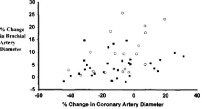
peripheral and coronary circulation were different and a different vascular bed was examined, i.e. conductance arteries. Alterations of epicardial artery diameter in response to intracoronary acetylcholine infusion were determined with quantitative coronary angiography (QCA), while the change in brachial artery diameter in response to reactive hyperemia in the peripheral circu-lation was determined with vascular ultrasound. Thus, endothelial function of the epicardial artery was spe-cifically tested with acetylcholine stimulation of the muscarinergic receptor, whereas flow-mediated brachial artery response was determined in response to hyper-emic flow increases. In both the instances, the endothelial vasoreactivity of the conduit vessels of the periphery and coronary circulation was tested. This may explain the observed statistically significant but rather weak correlation between endothelium-dependent vasomotor responses at the site of conduit vessel of the peripheral and coronary circulation in patients with and without angiographically determined CAD (P = .36, P\ .01) (Figure1)3. Interestingly, this weak correlation appeared to be driven by patients without evidence of structural CAD. Conceptually, if these patients without evidence of structural CAD were taken out of the analysis, no association between peripheral and coro-nary endothelial function would probably exist. Thus, the results from Anderson et al3 suggest that CAD-related advanced structural alterations may actually dissolve the described association of endothelial func-tion between the peripheral and coronary circulafunc-tion.
The prognostic value of the assessment of endo-thelial or vascular dysfunction of the peripheral and coronary circulation is well established.4-6In particular, the power of peripheral and coronary endothelial dys-function in response to various stimuli in the prediction of cardiovascular events appears to be comparable.5 Cardiovascular events therefore may occur remotely from the site of endothelial dysfunction identified. These observations strongly suggest a systemic nature of vas-cular dysfunction and its central role in predicting future cardiovascular events. Vascular dysfunction has been appreciated as a useful integrating index of the overall stress burden by various cardiovascular risk factors on the arterial wall, taking into account the cumulative risk of cardiovascular risk factors and as yet unknown vari-ables and genetic predispositions.4,5,7Despite this, it is
From the Department of Internal Medicine, Division of Cardiology, Nuclear Cardiology,a University Hospitals of Geneva, Geneva, Switzerland; and Department of Cardiology,bCardiovascular Cen-ter, University Hospital Zurich, Zurich, Switzerland.
Reprint requests: Thomas H. Schindler, MD, PhD, Department of Internal Medicine, Division of Cardiology, Nuclear Cardiology, University Hospitals of Geneva, 6th Floor, Rue Gabrielle-Perret-Gentil 4, 1211 Geneva, Switzerland; [email protected]. J Nucl Cardiol 2011;18:201–3.
1071-3581/$34.00
CopyrightÓ 2011 American Society of Nuclear Cardiology. doi:10.1007/s12350-011-9357-0
important to keep in mind that forearm conduit and resistance vessels do not develop atherosclerotic dis-ease.7Currently, no systematic investigations of both the forearm and the coronary circulation have been per-formed, and therefore the relation between vasomotor function in the forearm and coronary circulation remains poorly understood. In this direction, the observations from Scholtens et al1shed some new light in that they did not observe an association between vasomotor function of the resistance vessels in the periphery and the coronary circulation when assessed concurrently with adenosine-induced hyperemic flow increases. Conversely, their study does not provide any mecha-nistic insight about the mechanisms underlying the vasoreactivity in the upper limb microcirculation in response to adenosine stimulation. Intravenous adeno-sine infusion caused a much lower flow increase in the upper limb than in the myocardium, which may be related to effectively lower adenosine arriving in the peripheripheral circulation and, at least in part, different mechanisms underlying peripheral vasoreactivity.1 As regards the hyperemic myocardial blood flow increase during pharmacologically induced vasodilation of the arteriolar vessels, vascular smooth muscle-relaxing substances like adenosine, dipyridamole, or, more recently, adenosine receptor agonists decrease resistance to flow at the site of the coronary arteriolar resistance vessels and, thereby, cause a maximal or submaximal hyperemic myocardial blood flow increase.8,9 The resulting hyperemic coronary flow increase is consid-ered to represent predominantly an endothelium-independent flow response as the aforementioned sub-stances increase hyperemic flow increases through vascular smooth muscle cell relaxation at the site of the coronary arteriolar vessels.8,10 Blocking the endothelial nitric oxide synthase (eNOS) by intravenous infusion of NG-monomethyl-L-arginine, however, results into a
significant loss of adenosine-induced MBF increases by 20%-25% as measured with PET.9,11,12 It may be con-cluded that shear-sensitive components of the coronary endothelium contribute in part through a flow-mediated and, thus, nitric oxide-mediated coronary vasodilation to the overall hyperemic MBF increase during pharmaco-logic vasodilation.7,8,13Such a myocardial flow response has also been appreciated as total integrated coronary circulatory function.8,9,14 The complexity of the mech-anisms underlying hyperemic MBF increases during pharmacologic vasodilation may also explain, at least in part, the absence of any correlation between hyperemic myocardial and peripheral blood flows during pharma-cological vasodilation with adenosine as determined with 13N-ammonia PET.1 Overall, the investigation by Scholtens et al1 add further to the consideration that vascular (dys)function in the peripheral and coronary circulation may indeed reflect different features and stages of vascular disease. Given that the forearm cir-culation does not develop atherosclerotic disease,7 systemic comparative investigations of endothelial, or circulatory dysfunction of both the forearm and the coronary circulation and its response to pharmaceutical intervention15-19 could possibly contribute to better identify and characterize pathophysiological mecha-nisms favoring the initiation and progression of the CAD process and/or possible protective responses within the arterial wall aiming to counterbalance the adverse effects of various cardiovascular risk factors. Such an emerging concept certainly deserves further investigations.
References
1. Scholtens AM, Tio RA, Willemsen A, Dierckx RAJO, Boersma HH, Zeebregts CJ, et al. Myocardial perfusion reserve compared with peripheral perfusion reserve: A [13N]ammonia PET study. J Nucl Cardiol 2011. doi:10.1007/s12350-011-9339-2.
2. Bottcher M, Madsen MM, Refsgaard J, Buus NH, Dorup I, Nielsen TT, et al. Peripheral flow response to transient arterial forearm occlusion does not reflect myocardial perfusion reserve. Circula-tion 2001;103:1109-14.
3. Anderson TJ, Uehata A, Gerhard MD, Meredith IT, Knab S, Delagrange D, et al. Close relation of endothelial function in the human coronary and peripheral circulations. J Am Coll Cardiol 1995;26:1235-41.
4. Bonetti PO, Lerman LO, Lerman A. Endothelial dysfunction: A marker of atherosclerotic risk. Arterioscler Thromb Vasc Biol 2003;23:168-75.
5. Lerman A, Zeiher AM. Endothelial function: Cardiac events. Circulation 2005;111:363-8.
6. Rubinshtein R, Kuvin JT, Soffler M, Lennon RJ, Lavi S, Nelson RE, et al. Assessment of endothelial function by non-invasive peripheral arterial tonometry predicts late cardiovascular adverse events. Eur Heart J. 2010;31:1142-8.
7. Drexler H. Endothelial dysfunction: Clinical implications. Prog Cardiovasc Dis 1997;39:24-287.
Figure 1. Brachial dilator response to reactive hyperemia as a function of the coronary response to acetylcholine in patients without (circles) and with (squares) coronary artery disease (r = 0.36, P = .01) (with kind permission from Anderson et al3).
202 Valenta et al Journal of Nuclear Cardiology
8. Schindler TH, Schelbert HR, Quercioli A, Dilsizian V. Cardiac PET imaging for the detection and monitoring of coronary artery disease and microvascular health. JACC Cardiovasc Imaging 2010;3:623-40.
9. Schindler TH, Zhang XL, Vincenti G, Mhiri L, Lerch R, Schelbert HR. Role of PET in the evaluation and understanding of coronary physiology. J Nucl Cardiol 2007;14:589-603.
10. Valenta I, Quercioli A, Vincenti G, Nkoulou R, Dewarrat S, Rager O, et al. Structural epicardial disease and microvascular function are determinants of an abnormal longitudinal myocardial blood flow difference in cardiovascular risk individuals as determined with PET/CT. J Nucl Cardiol 2010;17:1023-33.
11. Buus NH, Bottcher M, Hermansen F, Sander M, Nielsen TT, Mulvany MJ. Influence of nitric oxide synthase and adrenergic inhibition on adenosine-induced myocardial hyperemia. Circula-tion 2001;104:2305-10.
12. Tawakol A, Forgione MA, Stuehlinger M, Alpert NM, Cooke JP, Loscalzo J, et al. Homocysteine impairs coronary microvascular dilator function in humans. J Am Coll Cardiol 2002;40:1051-8. 13. Drexler H, Zeiher AM, Wollschlager H, Meinertz T, Just H,
Bonzel T. Flow-dependent coronary artery dilatation in humans. Circulation 1989;80:466-74.
14. Schelbert HR. Anatomy and physiology of coronary blood flow. J Nucl Cardiol 2010;17:545-54.
15. Rubinshtein R, Yang EH, Rihal CS, Prasad A, Lennon RJ, Best PJ, et al. Coronary microcirculatory vasodilator function in relation to risk factors among patients without obstructive coronary disease and low to intermediate Framingham score. Eur Heart J 2010;31:936-42.
16. Munzel T, Sinning C, Post F, Warnholtz A, Schulz E. Patho-physiology, diagnosis and prognostic implications of endothelial dysfunction. Ann Med 2008;40:180-96.
17. Schindler TH, Nitzsche EU, Munzel T, Olschewski M, Brink I, Jeserich M, et al. Coronary vasoregulation in patients with various risk factors in response to cold pressor testing: Contrasting myo-cardial blood flow responses to short- and long-term vitamin C administration. J Am Coll Cardiol 2003;42:814-22.
18. Schindler TH, Cadenas J, Facta AD, Li Y, Olschewski M, Sayre J, et al. Improvement in coronary endothelial function is indepen-dently associated with a slowed progression of coronary artery calcification in type 2 diabetes mellitus. Eur Heart J 2009;30:3064-73.
19. Schindler TH, Campisi R, Dorsey D, Prior JO, Olschewski M, Sayre J, et al. Effect of hormone replacement therapy on vaso-motor function of the coronary microcirculation in post-menopausal women with medically treated cardiovascular risk factors. Eur Heart J 2009;30:978-86.
Journal of Nuclear Cardiology Valenta et al 203