HAL Id: tel-01249546
https://tel.archives-ouvertes.fr/tel-01249546
Submitted on 4 Jan 2016HAL is a multi-disciplinary open access archive for the deposit and dissemination of sci-entific research documents, whether they are pub-lished or not. The documents may come from teaching and research institutions in France or abroad, or from public or private research centers.
L’archive ouverte pluridisciplinaire HAL, est destinée au dépôt et à la diffusion de documents scientifiques de niveau recherche, publiés ou non, émanant des établissements d’enseignement et de recherche français ou étrangers, des laboratoires publics ou privés.
Drosophila melanogaster
Vincent Barbier
To cite this version:
Vincent Barbier. Pastrel, a restriction factor for picornalike-viruses in Drosophila melanogaster. Vi-rology. Université de Strasbourg, 2013. English. �NNT : 2013STRAJ114�. �tel-01249546�
École Doctorale des Sciences de la Vie et de la Santé de Strasbourg
UPR-9022 Réponse immunitaire et développement chez les insectes
THÈSE
présentée par :
Vincent BARBIER
soutenue le : 10 décembre 2013pour obtenir le grade de :
Docteur de l’université de Strasbourg
Discipline/ Spécialité
: Immunologie
Pastrel, a restriction factor for
picorna-like viruses in Drosophila melanogaster
THÈSE dirigée par :
M. IMLER Jean-Luc Professeur des universités, université de Strasbourg RAPPORTEURS :
Mme. SALEH Maria-Carla Docteur, Institut Pasteur, Paris M. KOHL Alain Docteur, université de Glasgow
AUTRES MEMBRES DU JURY :
Mme. SCHUSTER Catherine Directeur de recherches, université de Strasbourg M. JIGGINS Francis Professeur des universités, université de Cambridge
I would like to express my sincere gratitude to the people who helped me to get this thesis to this stage.
First of all, I would like to thank Pr. Jean-Marc REICHHART, director of the UPR9022 (Réponse Immunitaire et Développement chez les insectes), for giving me the opportunity to work in this laboratory and making the facilities available for carrying out research in the best conditions.
I would like to acknowledge my PhD supervisor, Pr. Jean-Luc IMLER, for all his support and his guidance of my work over the last three years. I learned a lot and got the chance to gain a lot of experience with you. Thanks for the corrections of this manuscript.
To the members of my jury, my reviewers Dr. Maria-Carla SALEH and Dr. Alain KOHL and my examiner Dr. Catherine SCHUSTER, thank you for engaging in my work and discussing my project.
I would like to express my deep gratitude to Pr. Francis JIGGINS for his invaluable help on Pastrel project and for the support received through the collaborative work undertaken with him and his PhD student, Chuan. Thanks for exchange of results and ideas.
I am thankful to Dr. Dominique FERRANDON and his team, for the collaborative work on Nora virus project, and to Dr. Sébastien PFEFFER and Ali for their precious help on small RNA library construction. I am also indebted to Dr. Stéphane NOSELLI for providing us Pastrel antibodies and UAS-Pst-GFP fly line.
I extend my sincere thanks and gratitude to my present and former laboratory colleagues for their enormous help during the course of this PhD thesis. I would like to express my thanks to Akira GOTO who contributed immensely to this work. Thanks for your inspiring scientific enthusiasm and infinite advice. I really enjoyed our scientific and non-scientific discussions during lunchtime. I am also very grateful to Laurent, Estelle, Carine, Simona, Olivier, Karim, Alice and Bill for their support and advice in experiments. Thanks to former members of the team, Steffi, Stan, Dele, Najate, Melissandre and Evelyne for their help and for nice time spent inside and outside the laboratory. Thanks to sportsmen Miriam and Adrien for their support and encouragements. I am also thankful to François for critically
indirectly contributed to this thesis.
I would like to grateful all my friends from Paris and elsewhere for their encouragements and the enjoying weekends spent with them: Divya, Erin, Sébastien, Luc, Fred and Noémie.
Finally, my acknowledgements would never be complete without the special mention of my family, especially my father Jean-François, Brigitte and my grand-parents who were always supportive and have sacrificed a lot for my studies.
All this work would not have been possible without my loving, encouraging and understanding girlfriend Sarah. You brought me a daily faithful support all the way through this PhD thesis, even if the distance separated each other during these three years. I thank you for your valuable suggestions and help to write this manuscript, and also for your cooking skills that contributed to maintain my good mood during the writing process. You are wonderful.
I also take this opportunity to express my deepest gratitude to my mother Christiane who always believed in me and would have been proud to see me get a PhD degree. This thesis is dedicated to you.
Table of contents
TABLE OF CONTENTS ... 4 LIST OF FIGURES ... 7 LIST OF TABLES ... 10 LIST OF ABBREVIATIONS ... 11 RÉSUMÉ DE THÈSE EN FRANÇAIS ... 17 PART I : INTRODUCTION ... 23 CHAPTER 1 ‐ MODELS OF DROSOPHILA VIRAL INFECTIONS ... 251.1. DROSOPHILA VIRUSES: NATURAL PATHOGENS OF DROSOPHILA ... 26
1.2. OTHER VIRUSES INFECTING DROSOPHILA ... 32
CHAPTER 2 ‐ INNATE ANTIVIRAL IMMUNITY IN DROSOPHILA MELANOGASTER ... 37
2.1. THE RNA INTERFERENCE PATHWAY: A BROAD ANTIVIRAL DEFENSE ... 39
2.2. THE INDUCIBLE RESPONSE: A VIRUS‐SPECIFIC ANTIVIRAL DEFENSE ... 47
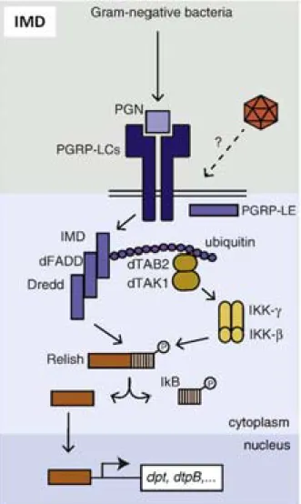
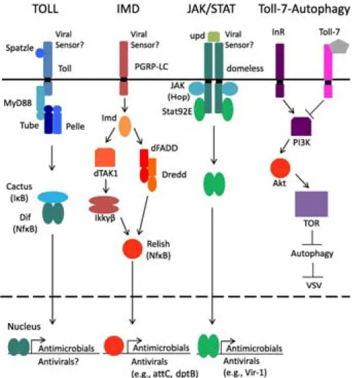
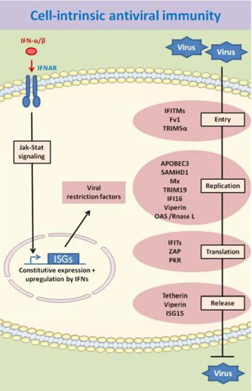
2.2.a. The Toll pathway ... 48 2.2.b. The IMD/TNF‐R pathway ... 51 2.2.c. AMPs ... 53 2.2.d. The Jak‐STAT pathway ... 55 2.2.e. Autophagy ... 57 2.3. PARALLEL WITH INNATE ANTIVIRAL IMMUNITY IN MAMMALS ... 58 2.3.a. Nucleic acid sensors: pattern recognition receptors ... 62 2.3.b. Type I interferon pathway ... 64 CHAPTER 3 ‐ INTRINSIC ANTIVIRAL IMMUNITY ... 66 3.1. VIRAL RESTRICTION FACTORS IN MAMMALS ... 68 3.1.a. Inhibition of viral entry ... 68 3.1.b. Inhibition of viral replication ... 71 3.1.c. Inhibition of viral translation ... 77 3.1.d. Inhibition of viral release ... 80 3.1.e. Multiple step antiviral function ... 83 3.1.f. OAS/RNase L: Intrinsic antiviral pathway ... 84 3.2. VIRAL RESTRICTION FACTORS IN DROSOPHILA ? ... 87 AIM OF THE STUDY ... 90 PART II : MATERIAL AND METHODS ... 91 II.1. FLY STRAINS AND INFECTION ... 92 II.2. DECHORIONATION OF EGGS ... 92 II.3. CELL LINES AND VIRUS INFECTION ... 93 II.4. VIRUS BINDING AND ENTRY ASSAYS ... 94
II.5. TRANSFECTION, LUCIFERASE ASSAY, SECRETION ASSAY ... 94
II.6. GENE SILENCING: DSRNA SYNTHESIS AND TREATMENT ... 94
II.7. VIRUS TITRATION ... 96
II.8. CLONING BY GATEWAY SYSTEM ... 96
II.9. DNA ANALYSIS ... 98
II.10.1. DNA extraction ... 98
II.10.2. Preparation for pastrel gene sequencing ... 98
II.10.3. Southern blot ... 99
II.10.4. PCR genotyping ... 99
II.11. RNA ANALYSIS ... 100
II.11.1. RNA extraction ... 100 II.11.2. cDNA synthesis ... 100 II.11.3. Quantitative real time PCR ... 101 II.12. PROTEIN ANALYSIS ... 102 II.12.1. Protein extraction ... 102 II.12.2. Immnoprecipitation ... 102 II.12.3. Western blot ... 103 II.12.4. Immunostaining ... 103 II.13. STATISTICAL ANALYSIS ... 104 PART III : RESULTS ... 105 CHAPTER 4 ‐ PASTREL: A RESTRICTION FACTOR FOR PICORNA‐LIKE VIRUSES IN DROSOPHILA ... 106
4.1. RESEARCH OF THE CAUSE OF SUSCEPTIBILITY OF FLIES TO DCV INFECTION: IDENTIFICATION OF PASTREL GENE. ... 107 4.1.a. Wide variability in the susceptibility of laboratory control flies to DCV, but not CrPV and FHV, infection. ... 107 4.1.b. Wide variability in the susceptibility of two Ore‐R stocks to picorna‐like viruses DCV and CrPV, and DNA virus IIV‐6 infection, but not FHV, VSV and SINV infection. ... 110 4.1.c. No DCV fragments integrated in the genome of laboratory control flies. ... 114 4.1.d. The susceptibility of flies to DCV infection is genetically transmitted ... 116 4.1.e. Correlation between the polymorphism in pst gene and DCV susceptibility ... 119
4.2. KNOCKDOWN OF PST GENE INCREASES THE SUSCEPTIBILITY OF FLIES TO DCV AND CRPV INFECTION ... 123
4.3. POLYMORPHISMS IN PASTREL GENE DO NOT AFFECT PROTEIN STABILITY ... 129
4.4. DOES PASTREL OVEREXPRESSION RESTRICT DCV INFECTION IN FLIES ? ... 130
4.5. STUDY OF PASTREL GENE IN VITRO: DROSOPHILA S2 CELLS ... 132
4.5.a. Knockdown of pastrel gene increases DCV and CrPV susceptibility in Drosophila cells .... 132 4.5.b. Overexpression of Pastrel protein restricts DCV and CrPV infection ... 136 4.5.c. Pst antiviral function acts at early steps of DCV infectious cycle ... 150 4.5.d. Pst does not affect CrPV IRES translation ... 154 4.5.e. Pst is not involved in protein secretion ... 155 4.5.f. Study of Pst topology by a Biotin‐Streptavidin revelation system ... 156 4.5.g. The C‐terminal region of Pst is required for its antiviral function ... 159 4.6. STUDY OF PST LOCALIZATION IN CELLS... 163 4.6.a. Pst co‐localizes with lipid droplets in non‐infected cells ... 165 4.6.b. Enrichement of COP‐I vesicles staining in the areas where Pastrel and DCV localize ... 166
4.6.c. Pst co‐localizes with DCV capsid protein during infection ... 169
4.6.d. Does DCV colocalize with lipid droplets ? ... 170
4.6.e. DCV infection was not affected by the knockdown of genes involved in lipid metabolism174 CHAPTER 5 ‐ EFFECT OF NORA VIRUS ON THE SUSCEPTIBILITY OF DROSOPHILA TO PATHOGENS . 176 5.1. CORRELATION BETWEEN THE PRESENCE OF NORA VIRUS AND THE SUSCEPTIBILITY OF FLIES TO DCV INFECTION ... 177
5.2. DOES NORA VIRUS PERSISTENT INFECTION CONTRIBUTE TO THE SUSCEPTIBILITY OF FLIES TO DCV INFECTION ? ... 178
5.3. DOES PASTREL AFFECT NORA VIRUS PERSISTENT INFECTION ? ... 181
CHAPTER 6 ‐ RNAI PATHWAY CONTROLS INFECTION BY A DNA VIRUS IN DROSOPHILA ... 183
6.1. BROAD RNA INTERFERENCE‐MEDIATED ANTIVIRAL IMMUNITY IN DROSOPHILA (KEMP ET AL, 2013) ... 184
PART IV : DISCUSSION ... 194
IV.1. PASTREL, A NEW GENE RESTRICTING INFECTION BY PICORNA‐LIKE VIRUSES IN DROSOPHILA ... 195
IV.1.a. Polymorphisms in pastrel gene are correlated with the susceptibility to DCV infection . 195 IV.1.b. Pastrel genotyping is required before conducting experiments with DCV ... 197
IV.1.c. Pastrel gene: duplications and deletions ... 200
IV.1.d. Pastrel gene controls Dicistroviridae infection ... 200
IV.1.e. Towards the characterization of Pastrel antiviral activity ... 203
IV.2. DOES PASTREL AFFECT NORA VIRUS INFECTION? ... 208
IV.3. THE SIRNA PATHWAY CONTROLS IIV‐6 INFECTION ... 209
CONCLUDING REMARKS ... 210
List of Figures
Figure 1. The genomic organisation of dicistroviriruses shares similarities with picornaviruses and
iflaviruses. ... 27 Figure 2. Representation of the Nora virus genome. ... 31 Figure 3. Overview of insect antiviral innate immunity. ... 38 Figure 4. The siRNA pathway in Drosophila. ... 41 Figure 5. The Toll pathway in Drosophila. ... 49 Figure 6. The IMD pathway in Drosophila. ... 52 Figure 7. The Jak‐STAT pathway in Drosophila. ... 56 Figure 8. Inducible antiviral pathways in Drosophila. ... 58 Figure 9. The type I interferon response. ... 65 Figure 10. Cell‐intrinsic antiviral immunity in mammals. ... 68 Figure 11. Wide variability in the susceptibility to DCV infection, but not to CrPV and FHV infection, between laboratory control flies. ... 109 Figure 12. Wide variability in the susceptibility of two Ore‐R stocks to DCV and CrPV infection, but not FHV infection. ... 111 Figure 13. Variability in the susceptibility of two Ore‐R flies to IIV‐6 infection, but not VSV and SINV infections. ... 113 Figure 14. TOLL DNA fragments, but no DCV fragments, were detected by southern blot in DD1 cnbw, Ore‐RJLI and wA5001 flies. ... 115
Figure 15. The susceptibility of two Ore‐R stocks flies to DCV infection is genetically transmitted, and the resistant allele is autosomal‐dominant. ... 117
Figure 16. The susceptibility of flies to DCV infection is genetically transmitted, and the resistant allele is autosomal‐dominant. ... 118
Figure 17. Susceptibility of flies to DCV infection is correlated with polymorphisms in pastrel gene. ... 120
Figure 18. Identification of a deleted version of pastrel gene present in the genome of some flies and cells. ... 121
Figure 19. SNPs found exclusively in pastrel gene of Ore‐RDF flies may correlate with higher sensitivity
to DCV and CrPV infection. ... 122
Figure 20. Allele specific PCR assay for fast genotyping of the sensitive and resistant alleles of pastrel gene. ... 123
Figure 21. Knockdown of pastrel gene in whole flies increases their susceptibility to DCV and CrPV infection. ... 125 Figure 22. pastrel gene expression in organs and tissues from adult Canton‐S flies. ... 126 Figure 23. Specific knockdown of pastrel gene in the fat body increases the susceptibility of flies to DCV and CrPV infection. ... 127 Figure 24. Specific knockdown of pastrel gene in the intestinal epithelium of flies does not affect the resistance to DCV infection. ... 128
Figure 25. Flies expressing PstS and PstR forms show similar levels of Pst protein expression. ... 129
Figure 26. Overexpression of Pst‐GFP in flies does not increase resistance to DCV infection. ... 131
Figure 27. DCV replicates in Drosophila S2 cells and pastrel gene expression is increased after infection. ... 132 Figure 28. Knockdown of pastrel gene in S2 cells increases DCV infection. ... 134 Figure 29. Knockdown of pastrel gene in S2 cells increases DCV infection. ... 135 Figure 30. Knockdown of pastrel gene in S2 cells increases CrPV infection. ... 136 Figure 31. Transient overexpression of Pastrel full lentgh slightly reduces DCV infection in Drosophila S2 cells. ... 137 Figure 32. Pst overexpression decreases DCV RNA level after 16h and 48h of infection. ... 139
Figure 33. PstS and PstR overexpression restricts DCV infection. ... 141
Figure 34. Cells overexpressing Pst are better protected from DCV infection compared to cells with endogenous Pst expression. ... 143
Figure 35. PstS and PstR overexpression decreases CrPV RNA level after 16h and 48h of infection... 144
Figure 36. Overexpression of PstS and PstR decreases the number of cells infected by DCV. ... 146
Figure 37. Overexpression of PstS and PstR decreases the number of cells infected by CrPV. ... 147
Figure 38. Overexpression of PstS and PstR does not affect the number of cells infected by FHV. .... 148
Figure 39. Overexpression of PstS and PstR does not affect the number of cells infected by VSV. .... 149
Figure 40. PstS and PstR overexpression does not affect binding of DCV on cells. ... 150
Figure 42. PstS and PstR overexpression affects DCV and CrPV early after infection. ... 153
Figure 43. PstS and PstR overexpression does not affect CrPV 5’ IRES and CrPV IGR IRES translation. ... 154 Figure 44. Pst is not involved in protein secretion. ... 155 Figure 45. Prediction of 6 putative TM domains in Pastrel protein. ... 156 Figure 46. Topology of N and C‐ter regions of Toll and Pst. ... 158 Figure 47. Overexpression of Pst deleted in C‐ter does not affect DCV RNA level. ... 160 Figure 48. Overexpression of Pst deleted in C‐ter does not restrict DCV infection. ... 162
Figure 49. Endogenous Pst and Pst fusion proteins exhibit a vesicular pattern in the cytoplasm of Drosophila cells. ... 163
Figure 50. Deletion of C‐terminal region of Pst modifies its intracellular localization. ... 164
Figure 51. Pst colocalizes with lipid droplets stained by Nile Red. ... 166
Figure 52. beta‐COP staining is enriched in areas of Pst fusion aggregates. ... 167
Figure 53. Colocalization between DCV capsid and beta‐COP. ... 168
Figure 54. Pst colocalizes with DCV capsid staining in Drosophila S2 cells and in the fat body of infected flies. ... 169 Figure 55. Pst colocalizes with CrPV capsid staining in S2 cells and in Drosophila fat body. ... 170 Figure 56. DCV capsid staining does not colocalize with the surface of lipid droplets in Drosophila S2 cells. ... 171 Figure 57. DCV capsid and Pastrel protein colocalize with some dots of Nile Red staining, but not with large lipid droplets in the fat body of DCV‐infected flies. ... 172 Figure 58. DCV infection affects lipid droplets morphology in the fat body of DCV‐infected flies. .... 173
Figure 59. DCV seems to induce the degradation of large lipid droplets in the fat body of DCV‐ infected flies. ... 174
Figure 60. Knockdown of genes involved in lipid metabolism has no effect on DCV infection in Drosophila S2 cells. ... 175
Figure 61. Small RNAs matching with Nora Virus genome sequence were detected in yw flies. ... 177
Figure 62. Nora Virus RNA genome was detected in Ore‐RDF samples but not in Ore‐RJLI and Ore‐RDF bleached samples. ... 179
Figure 63. Ore‐RDF bleached flies, cured from Nora Virus infection, are still highly sensitive to DCV infection, comparable to Nora Virus persistently infected Ore‐RDF flies. ... 180
Figure 64. Ore‐RJLI flies, contaminated by Nora Virus, are still resistant to DCV infection, comparable
to Ore‐RDF flies. ... 181
Figure 65. Ore‐RDF flies are more sensitive to Nora virus infection compared to Ore‐RJLI flies. ... 182
Figure 66. Pastrel is responsible of the sensitivity of imdsdk and Tab2glr3 mutant flies to DCV infection. ... 198 Figure 67. The transcription factor Dif, operating in the Toll pathway, is involved in the control of DCV infection. ... 199 Figure 68. Speculative model for the antiviral action of Pst on DCV replication. ... 208
List of tables
Table 1. Overview of viruses naturally infecting Drosophila melanogaster. ... 32 Table 2. Overview of viruses experimentally able to infect Drosophila melanogaster. ... 36 Table 3. List of primers used for dsRNA synthesis. ... 96 Table 4. List of primers used for molecular cloning. ... 98 Table 5. List of primers used for PCR genotyping. ... 100 Table 2. List of primers used for qPCR. ... 102 Table 7. The presence of Nora Virus in flies correlates with the susceptibility to DCV infection ... 178
List of Abbreviations
(-) ssRNA negative single-stranded RNA(+) ssRNA positive single-stranded RNA
A Adenine
AA Amino acid
Ago Argonaute
AGS Aicardi-Goutières Syndrome
AIM2 Absent in melanoma 2
AMPs Antimicrobial peptides
ANV American Nodavirus
AP-1 Activator protein-1
APOBEC3 Apolipoprotein B mRNA-editing catalytic polypeptide 3
Ars2 Arsenic resistance protein 2
ATPase Adenosine triphosphatase
BAP Biotin Acceptor Peptide
BCA2 Breast cancer-associated gene 2
BHK Baby hamster kidney
BMP2 Bone morphogenetic protein 2
bp Base pair
C Cytosine
CARD Caspase activation and recruitment domain
CARDIF CARD adapter inducing interferon-β
Cct Cytidylyltransferase
CD Cluster of differentiation
cDNA complementary DNA
cGAMP cyclic-di-GMP-AMP
cGAS cGAMP synthetase
CHIKV Chikungunya virus
CIV Chilo Iridescent virus
COPI Coat protein I
CrPV Cricket Paralysis virus
cxVago Culex Vago
CypA Cyclophilin A
DAI DNA-dependent activator of IFN-regulatory factors
DaPKC Drosophila atypical protein kinase C DAP-PGN Diaminopimelic acid peptidoglycan
DAV Drosophila A virus
Dcr-2 Dicer-2
DCV Drosophila C virus
DDX DExD/H-box helicases
DENV Dengue virus
DFV Drosophila F virus
dPIAS Drosophila protein inhibitor of activated STAT dGTP deoxyguanosine triphosphate
DIF Dorsal-related immunity factor
dMyd88 drosophila myeloid differentiation factor 88
dN deoxynucleoside
DNA Desoxyribonucleic acid
dNTPs deoxynucleoside triphosphates
DPV Drosophila P virus
dsRNA double stranded RNA
dTak1 drosophila TGF-beta activated kinase 1
DTrV Drosophila Tetravirus
DTV Drosophila Totivirus
DUF283 Domain of unknown function 283
DVRF Dengue virus restriction factors
DXV Drosophila X virus
E.Coli Escherichia Coli
EBOV Ebola virus
EFP Estrogen-responsive finger protein
EIAV Equine Infectious Anemia Virus
eIF eukariotic Initiation Factor
EMCV Encephalomyocarditis virus
ER Endoplasmic reticulum
ERV-L Endogenous retrovirus-like elements
ESCRT Endosomal sorting complex required for transport
FACS Fluorescence-Activated Cell Sorting
FADD Fas-associated-death domain
FHV Flock House Virus
FIV Feline immunodeficiency virus
FPPS Farnesyl Diphosphate Synthase
Fv1 Friend-virus susceptibility gene 1
G Guanine
Gag Group-specic antigen
GNBP1 Gram-negative binding proteins
GPI Glycosylphosphatidylinositol
GTPases Guanosine triphosphatase
HBV Hepatitis B virus
HCMV Human cytomegalovirus
HCV Hepatitis C Virus
HD Histidine–aspartic
Herc5 HECT domain and RCC1-like domain containing protein 5
HFV Human Foamy Virus
HIV-1 Human immunodeficiency virus-1
Hop Hopscotch
HPV Human papillomavirus
HRP HorseRadish Peroxidase
HSP Heat shock protein
HSV-1 Herpes Simplex Virus type 1
HTLV-1 Human T-cell leukemia virus type I IAV Influenza A virus
IFI16 gamma-interferon-inductible protein 16
IFITMs Interferon-inducible transmembrane proteins
IFITs IFN-induced protein with tetratricopeptide repeats
IFN Interferon
IGR Intergenic region
IIV-6 Invertebrate Iridescent Virus 6
IKK IκB kinase
IL Interleukin
IL-1R Interleukin-1 receptor
IMD Immune deficiency
IPS-1 IFN-β promoter stimulator-1
IRES Internal Ribosome Entry Site
IRF-9 IFN-regulatory factor 9
ISGs Interferon-stimulated genes
ISGF-3 IFN-stimulated gene factor-3
IκB Inhibitor of κB
Jak Janus kinase
Jak-STAT Janus kinase -Signal Transducer and Activator of Transcription
JEV Japanese encephalitis virus
JNK c-Jun N-terminal kinase
kDa kilodalton
KSHV Kaposi's sarcoma-associated herpesvirus
LACV La Crosse virus
LCMV Lymphocytic choriomeningitis virus
LGP2 Laboratory of genetics and physiology-2
LGTV Langat virus
Loqs-PD Loquacious isoform PD
LPS Lipopolysaccharide
Lv1 Lentivirus susceptibility factor 1
MAPKKK Mitogen-activated protein kinase kinase kinase
MARV Marburg virus
MAVS Mitochondrial antiviral-signaling protein
MDA5 Melanoma differentiation-associated gene 5
mESCs mouse embryonic stem cells
MHV Murine GammaHerpes Virus
MHV Mouse hepatitis virus
miRNA microRNA
MLV Murine leukemia virus
MOI Multiplicity of infection
mRNA Messengers RNA
MV Measle Virus
Mx Myxovirus resistance
NBs Nuclear bodies
Nef Negative Regulatory Factor
NF-κB Nuclear factor–κB
NLRs NOD-like receptors
NOD2 Nucleotide binding oligomerization domain 2
NoV Nodamura virus
NRAMP Natural Resistance-Associated Macrophage Protein
NS1 Non-structural
Nts Nucleotides
OAS 2’-5’-Oligoadenylate synthetase
OASL OAS-like gene
ONNV O’nyong-nyong virus
ORF Open reading frame
OSS Ovary Somatic Sheet
PAMPs Pathogen associated molecular patterns (PAMPs)
PAZ Piwi/Argonaute/Zwille
pDCs plasmacytoid dendritic cells
PDE Phosphodiesterase
PFU Particles Forming Unit
PGRPs Peptidoglycan receptors proteins
PI3K-Akt-TOR Phosphatidylinositol 3-kinase-Akt-Target of rapamycine
piRNA Piwi-associated interfering RNA
PKR Protein kinase R
PML Promyelocytic leukaemia
RdRp RNA-dependent RNA polymerase
Ref(2)P Refractory for Sigma P virus
REF1 Resistance factor 1
RIG-I Retinoid acid-inducible gene
RING Really Interesting New Gene
RIP Receptor interacting protein
RISC RNA-induced silencing complex
RLC RISC loading complex
RLR RIG-I-like receptors
RNA Ribonucleic acid
RNAi RNA interference
RNase Ribonuclease
RNPs Ribonucleoproteins
RpS6 Ribosomal protein S6
RRE Rev response element
rRNAs Ribosomal RNAs
RRV Ross River Virus
Rsad2 Radical S-adenosyl methionine domain-containing protein 2
RVFV Rift Valley fever virus
S2 Schneider 2
SAM Sterile alpha motif
SAM S-adenosylmethionine
SAMHD1 SAM domain HD domain-containing protein 1
SARS-CoV Severe Acute Respiratory Syndrome-Coronavirus
sfRNA subgenomic flavivirus RNA
SFV Semliki Forest virus
SIGMAV Sigma virus
SINV Sindbis Virus
SIV Simian immunodeficiency virus
SMUG1 Single-Strand-Selective Monofunctional Uracil-DNA Glycosylase 1
SNP Single Nucleotide Polymorphism
SREBP Sterol Regulatory Element Binding Protein
Staf-50 Stimulated Trans-Acting Factor of 50 kDa
STAT1 Signal Transducer and Activator of Transcription 1
STING Stimulator of IFN genes
SUMO-1 Small Ubiquitin-like MOdifier
SOCS Suppressor of cytokine signaling
TAP1 Transporter associated with antigen processing 1 gene
Tas Transactivator
TBEV Tick-borne encephalitis virus
TEP Thiol-ester protein
TIR Toll-IL-1 receptor
TLRs Toll-like receptors
TMs Transmembranes
TNFR Tumour-necrosis factor-receptor
TotM Turandot M
TPRs Tetratrico peptide repeats
TRIM5α Tripartite motif protein isoform 5 alpha
TRIMCyp TRIM5α-Cyclophilin A
U Uracil
UbcH8 Ubiquitin-conjugating Enzyme H8
UBE1L Ubiquitin Activating Enzyme E1 Like Protein
UBP43 Ubiquitin protease 43
UNG Uracil-N glycosylase
Upd Unpaired
upd unpaired
VA1 Viral associated
VAP-A or hVAP-33
Vesicle-associated membrane protein-associated protein subtype A
VEEV Venezuelan Equine Encephalitis Virus
Vif Virion infectivity factor
Viperin Virus-inhibitory protein, endoplasmic reticulum–associated, IFN-inducible
Vir-1 Virus-induced RNA-1
VISA Virus-induced signaling adapter
VLPs Virus-like particles
VPg Viral Protein genome-linked
v-piRNAs Virus-derived piRNAs
Vpu Viral protein U
vRNPs Viral ribonucleoproteins
v-siRNA virus-derived small interfering RNAs
VSR Viral suppressors of RNAi
VSV Vesicular Stomatitis Virus
VSV-G G glycoprotein of the vesicular stomatitis virus WNV West Nile virus
YFV Yellow Fever Virus
Résumé de thèse en français
IntroductionLes maladies infectieuses d’origine virale sont responsables d’une mortalité importante chez toutes les espèces. La drosophile est un excellent modèle pour l’étude des mécanismes moléculaires de l’immunité innée, y compris les virus. Elle a permis la caractérisation de mécanismes de défense immunitaire conservés au cours de l’évolution, tel que les voies Toll et IMD qui régulent l’expression des peptides antimicrobiens induits en réponse aux infections fongiques et bactériennes. Un certain nombre de maladies virales ou parasitaires infectant l’homme ou le bétail sont en outre transmises par des insectes hématophages, ce qui représente une motivation supplémentaire pour étudier les mécanismes de l’immunité innée chez les insectes. L’objectif de ma thèse est de comprendre les bases de l’immunité antivirale chez la drosophile.
Problématique
Deux types de réponse sont impliqués dans le contrôle des infections virales chez la drosophile. Une réponse inductible et l’ARN interférence qui est un mécanisme global de défense contre les virus à ARN, dont le virus C de la Drosophile (DCV). Le virus DCV est un virus modèle de la famille des Dicistroviridae, apparenté aux Picornaviridae. Il est couramment utilisé pour étudier les réponses immunitaires chez la drosophile en particulier dans notre laboratoire. Nous avons mis en évidence une différence de sensibilité (charge virale et mortalité accrues) à l’infection par ce virus entre différentes lignées utilisées comme témoins de fond génétique. Une différence de susceptibilité au DCV a également été observée entre deux stocks d’une même lignée sauvage (Oregon-R) maintenus dans deux équipes de notre unité. Plusieurs lignées sensibles au DCV étaient infectées de façon persistante par un virus apparenté aux Picornaviridae, le virus Nora, suggérant qu’il était la cause de la susceptibilité à DCV. L’ensemble de ces observations m’ont incité à m’intéresser à trois aspects au cours de ma thèse :
(1) Quelle est la cause de la sensibilité de certaines lignées de Drosophile à l’infection par le virus DCV ?
(2) La présence du virus Nora persistant chez la Drosophile influence-t-elle la susceptibilité des mouches à d’autres infections virales ou bactériennes ?
(3) Quelle est la contribution de l’ARN interférence dans la défense contre un virus à ADN ?
Résultats (1)
Les mouches contrôles présentent une importante variabilité dans la résistance à l’infection par le virus DCV.
De façon surprenante, nous avons observé une grande variabilité dans la susceptibilité à l’infection par le virus DCV entre les mouches contrôles utilisées au laboratoire. Par exemple, les mouches yw et wA5001 présentent une charge virale et une mortalité accrue à
l’infection par le virus DCV comparé aux mouches DD1 cnbw et Canton-S. Cette différence de sensibilité ne concerne pas tous les virus puisque les mouches yw, wA5001, DD1 cnbw et
Canton-S présentent la même sensibilité au virus FHV. Nous avons également observé une différence de sensibilité spécifique aux virus DCV et CrPV entre deux stocks d’une même lignée de drosophile (Ore-RDF et Ore-RJLI).
La susceptibilité à l’infection par le virus DCV est dépendante du fond génétique.
J’ai observé que lorsque l’on croise un mâle d’une lignée de drosophile sensible à l’infection par le virus DCV avec une femelle d’une lignée de drosophile résistante, la progéniture est résistante à l’infection par le virus DCV. Cette résistance à l’infection par le virus DCV est donc transmise génétiquement de façon dominante à la descendance. De plus, le croisement réciproque d’une femelle sensible avec un mâle résistant à DCV n’affecte pas la résistance de la progéniture au virus DCV. Ceci indique que la résistance au virus DCV n’est pas portée par le chromosome X. Il était ensuite nécessaire de déterminer quel(s) gène(s) du chromosome 2, 3 ou 4 peut être responsable de cette différence de susceptibilité à l’infection par le virus DCV.
La susceptibilité à l’infection par le virus DCV est corrélée au polymorphisme dans le gène pst.
Parallèlement à mes observations, le laboratoire du professeur Francis Jiggins (Cambridge), a associé une région génomique du chromosome 3, comprenant le gène pastrel
(pst), avec des phénotypes de sensibilité associés à l’infection par le virus DCV. J’ai donc
séquencé le gène pst des différentes lignées contrôles du laboratoire et j’ai trouvé trois polymorphismes nucléotidiques simples (SNPs) dont un est présent dans l’exon 6 et induit un changement d’acide aminé. Ce SNP corrèle systématiquement avec la sensibilité ou la résistance au virus DCV. Les deux autres SNPs, présents dans des introns, corrèlent avec la susceptibilité à l’infection par le virus DCV des lignées contrôles à l’exception de la lignée Ore-RJLI. Il était ensuite nécessaire de valider ce gène candidat.
L’expression du gène pst limite l’infection par les virus picorna-like DCV et CrPV in vivo et in vitro.
L’atténuation de l’expression du gène pst par ARN interférence in vivo accroit la charge virale et la mortalité des mouches infectées par le virus DCV comparé aux mouches contrôles. J’ai effectué les mêmes observations avec le virus de la paralysie du criquet (CrPV), qui comme le virus DCV appartient à la famille des Dicistroviridae. De façon consistante, l’atténuation de l’expression du gène pst in vitro augmente la charge virale dans les cellules S2 infectées par les virus DCV et CrPV. Afin de tester si une surexpression du gène pst limite l’infection par le virus DCV, j’ai établi des lignées stables qui surexpriment sous contrôle du promoteur actine la forme sensible ou résistante de la protéine Pst, couplée en N ou C-terminal avec le fluorochrome RFP. La surexepression de la forme sensible ou résistante de la protéine Pst réduit considérablement la charge virale après infection par les virus DCV et CrPV par rapport à la lignée cellulaire contrôle. De façon consistante, dans le cas d’une infection par les virus DCV et CrPV, mais pas FHV et VSV, le nombre de cellules positives pour le virus est réduit dans les lignées stables surexprimant la forme sensible ou résistante de la protéine Pst par rapport à la lignée contrôle. Cette restriction virale apparait dans les premières heures de l’infection par le virus DCV in vitro, sans affecter la fixation du virus sur les cellules. La traduction IRES-dépendante, nécessaire à la synthèse polyprotéique des virus DCV et CrPV, n’est pas affectée par la surexpression de la protéine Pst. Des expériences sont en cours pour tester si la protéine Pst affecte l’entrée ou la réplication du virus.
La région C-terminale de la protéine Pst porte l’activité antivirale.
Afin de déterminer si la région portant le polymorphisme identifié in vivo (associé à la sensibilité ou résistance à l’infection par le virus DCV) confère l’activité antivirale à la protéine Pst, j’ai établi des lignées stables surexprimant une forme tronquée de la protéine Pst, depuis le dernier domaine transmembranaire prédit et précédent le polymorphisme. La surexpression de cette forme tronquée de la protéine Pst n’affecte pas l’infection par le virus DCV, indiquant que la région C-terminale de la protéine est nécessaire pour son activité antivirale. L’effet d’autres délétions est en cours d’analyse. De plus, la délétion de la région C-terminale modifie la localisation de la protéine de fusion.
La protéine Pst colocalise avec les gouttelettes lipidiques révélées par le Rouge de Nil, ainsi qu’avec les protéines de capside des virus DCV et CrPV au cours de l’infection.
L’immunomarquage des cellules révèle que la protéine Pst est localisée dans des structures ponctuelles, concentrées dans une zone juxtanucléaire. Le marquage des gouttelettes lipidiques par le Rouge de Nil colocalise avec le marquage de la protéine Pst endogène dans les cellules S2 in vitro. Au cours de l’infection, la protéine Pst colocalise avec les protéines de capside des virus DCV et CrPV in vitro, mais aussi in vivo dans les cellules du corps gras, un tissu analogue au foie chez les mammifères. Cette colocalisation est cohérente avec une activité antivirale de la protéine Pst sur ces virus.
L’extrémité N et C-terminale de la protéine Pst est exposée du côté cytosolique.
Puisque la protéine Pst et les protéines de capside colocalisent dans le cytoplasme, il est probable que la protéine Pst et les particules virales se rencontrent dans le cytosol, permettant à la protéine Pst d’exercer son activité antivirale portée par la région C-terminale. Pour répondre à cette hypothèse, j’ai mis au point une nouvelle méthode basée sur la spécificité d’interaction entre les protéines Biotine et Streptavidine pour déterminer la topologie des régions N et C-terminale de la protéine Pst. J’ai ajouté des sites de biotinylation en N ou C-terminal de la protéine Pst et exprimé ces protéines de fusion dans des cellules exprimant l’enzyme BirA. Si le site de biotinylation est exposé et accessible dans le cytosol, ce site est biotinylé par l’enzyme BirA. La biotinylation est ensuite révélée par western blot
avec la protéine Streptavidine-HRP. La validité de cette technique a été confirmée en utilisant la protéine Toll, dont la topologie est connue, comme contrôle. J’ai observé que les extrémités N et C-terminal de la protéine Pst sont exposées dans le cytosol.
Résultats (2)
L’infection persistante par le virus Nora n’a pas d’effet sur la susceptibilité des mouches à l’infection par le virus DCV, mais affecte la susceptibilité aux infections bactériennes.
La présence du virus persistant Nora a été détectée dans certaines lignées de laboratoire. Le virus Nora appartient à une nouvelle famille des virus de type picorna. J’ai ainsi vérifié si la persistance de ce virus pouvait contribuer à la susceptibilité au virus DCV. En effet, ce virus est détecté par PCR dans toutes les lignées sensibles à DCV alors que les lignées résistantes à DCV ne sont pas infectées par le virus Nora (à l’exception toutefois de la lignée DD1 cnbw). La déchorionnation des œufs de la lignée Ore-RDF infectée permet l’élimination du virus Nora, qui n’est pas transmis par la lignée germinale. Cependant, cette lignée présente toujours une sensibilité à l’infection par le virus DCV identique à celle de la lignée Ore-RDF non traitée. Par ailleurs, la contamination de la lignée Ore-RJLI, non infectée
par le virus Nora, avec les excréments des mouches de la lignée Ore-RDF infectée, n’induit pas de sensibilité accrue à l’infection par le virus DCV. L’ensemble de ces résultats m’ont permis de conclure que la présence de ce virus persistant n’était pas responsable de la susceptibilité de certaines lignées de drosophiles à l’infection par le virus DCV. Cependant, en collaboration avec l’équipe de Dominique Ferrandon, nous avons observé que la lignée Ore-RDF débarrassée du virus Nora est moins sensible à l’infection par P. aeruginosa et S.
marcescens. Par opposition, la lignée Ore-RJLI contaminée par ce virus devient plus sensible à
ces deux infections bactériennes. Le virus Nora affecte la susceptibilité des mouches aux infections bactériennes. De plus, les mouches infectées subissent un renouvèlement important de l’épithélium intestinal.
Résultats (3)
Nous avons montré que la voie de l’ARN interférence est une voie générale de défense antivirale, puisqu’elle permet, en plus des virus à ARN, de contrôler l’infection par un virus à ADN. Les mouches mutantes pour le gène dicer-2 (dcr-2R416X) infectées par le virus à ADN
IIV-6 présentent une mortalité et une charge virale accrue par rapport aux mouches contrôles
yw. J’ai également observé une mortalité plus importante chez les mouches mutantes pour un
second allèle nul de dicer-2 (dcr-2L811fsx), le phénotype de sensibilité au virus IIV-6 étant
restauré chez ces mutants après insertion d’un transgène correspondant à la région génomique de dicer-2 sauvage. Les mouches mutantes pour les gènes R2D2 et AGO2, deux composants majeurs de cette voie, présentent également une sensibilité accrue au virus IIV-6. J’ai construit une banque de petits ARNs à partir de mouches et de cellules infectées par le virus IIV-6 et identifié la présence de petits ARN, majoritairement de 21 nucléotides, s’alignant avec la séquence du génome viral IIV-6. Ainsi, Dicer-2 produit des ARN interférents contre le génome viral IIV-6. De façon surprenante, à la différence des virus à ARN, ces petits ARN interférents sont produits à partir de régions spécifiques du génome viral puisque leur distribution n’est pas uniforme le long du génome viral. Ces régions correspondent à des régions ou la transcription s’effectue sur les deux brins, conduisant potentiellement à la formation d’ARNdb, substrats de Dicer-2.
Conclusion
J’ai mis en évidence que le gène pst est impliqué dans la susceptibilité à l’infection par les virus de type picorna DCV et CrPV. Les expériences de perte-de-fonction et gain-de-fonction indiquent que la protéine Pst est un facteur de restriction antiviral. Son mécanisme d’action reste à éclaircir. Mon étude de l’effet de la présence du virus Nora sur la susceptibilité des mouches aux infections a permis de révéler que cette infection persistante n’affecte pas la sensibilité des mouches à DCV. Cependant, la présence du virus Nora facilite les infections bactériennes et perturbe le renouvellement des cellules épithéliales de l’intestin. Enfin, j’ai également démontré que l’ARN interférence, en plus des virus à ARN, permet de contrôler l’infection par un virus à ADN. Ces travaux ont été publiés dans le Journal of
Part I
Every species have to face multiple pathogens during their life. In particular, viral infectious diseases are responsible for high lethality in all species. The fruit fly Drosophila
melanogaster is an excellent model to study the molecular mechanisms of innate immunity in
insects (Schneider, 2000). This model allowed the characterization of the evolutionarily conserved Toll and IMD pathways that regulate the expression of antimicrobial peptides in response to fungal and bacterial infections (Hoffmann, 2003). This discovery had a major impact on the understanding of innate immunity in mammals. The necessity to study insect innate immunity is also reinforced by the emergence of human viral diseases transmitted by hematophagous insects, including ticks and mosquitoes (Weaver and Reisen, 2010).
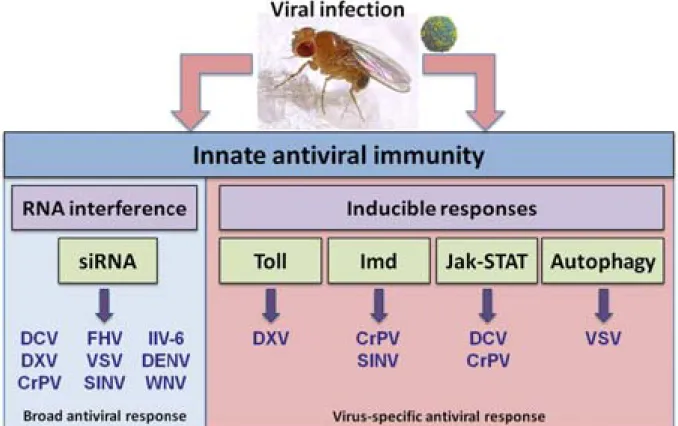
The first part of this introduction will review the natural and non-natural Drosophila viruses used in this study. The second part will address the different innate immune pathways mounted in response to pathogens, with a particular focus on Drosophila antiviral responses. After a brief comparison with the innate antiviral response orchestrated in mammals, the innate intrinsic immunity will be addressed in mammals and Drosophila, with a description of characterized restriction factors.
Chapter 1
Models of Drosophila viral infections
Viruses commonly used in research laboratories to study the mechanisms of antiviral immunity in Drosophila can be divided in two classes: first, the natural pathogens of
Drosophila, which include Drosophila C virus (Dicistroviridae), Sigma virus
(Rhabdoviridae) and Nora Virus (unclassified); secondly, the non-Drosophila viruses that were isolated from other hosts but were able to infect Drosophila flies and cells in experimental conditions. They include Cricket Paralysis virus (Dicistroviridae), Flock House virus (Nodaviridae), Invertebrate Iridescent virus 6 (Iridoviridae), Sindbis virus (Togaviridae) and Vesicular Stomatitis virus (Rhabdoviridae). Both classes cover a wide range of virus families with genomes of different nature and polarity, thus enhancing the robustness of the
Drosophila model to study innate antiviral responses.
1.1. Drosophila viruses: natural pathogens of Drosophila
Drosophila C virus (DCV) was firstly identified in 1972 from the Charolles strain of
Drosophila melanogaster. This laboratory stock exhibited an unusual high mortality rate
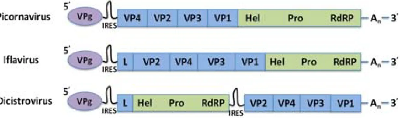
(Jousset et al., 1972). DCV is widely spread in Drosophila, infecting about one third of natural and laboratory populations of Drosophila (Plus et al., 1975a). Viral particles are non-enveloped, icosahedric, with 30 nm of diameter and share physical and chemical characteristics with the Picornaviridae family (Jousset et al., 1977). However, the full sequencing of DCV genome in 1998 revealed clear differences with several Picornaviridae, imposing the creation of the new Dicistroviridae family to classify this Drosophila picorna-like virus (Johnson and Christian, 1998). DCV genome is a positive single stranded ribonucleic acid ((+) ssRNA) of 9,264 nucleotides (nts) length, polyadenylated at the 3’ end. The small viral protein genome-linked (VPg) is attached at the 5’ extremity of the genome (King and Moore, 1988). The genome consists of two open reading frames (ORF): the 5’ ORF encodes non-structural proteins (a viral suppressor of RNA interference (RNAi) DCV-1A, a helicase, a protease, the VPg protein and the viral RNA-dependent RNA polymerase (RdRp)) and the 3’ ORF encodes structural proteins (capsid proteins VP1-4). Each ORF is preceded by an internal ribosome entry site (IRES) that initiates translation of non-structural (5’ IRES) or structural (Intergenic region (IGR) IRES) polyprotein precursors. Final proteins are obtained after cleavage of the polyproteins by the viral protease. This contrasts with the viral genome from Picornaviridae and Iflaviridae family which consists of a single ORF with capsid proteins encoded at the 5´ region of the genome and non-structural proteins (helicase,
protease and RdRp) encoded at the 3´ region (Figure 1). A single IRES is present at the 5’ extremity of the genome and initiates translation.
Figure 1. The genomic organisation of dicistroviriruses shares similarities with picornaviruses and iflaviruses. (Leader (L), Helicase (Hel), Protease (Pro), polyA (An)).
DCV is horizontally transmitted, and this transmission is facilitated by females which may act as viral reservoir in nature (Gomariz-Zilber et al., 1995, 1998). The outcome of the infection depends on the infection route. Injection of DCV is highly pathogenic, leading to the death of flies in few days. By contrast, flies infected by oral contamination with DCV appear healthy. However, depending on the dose, the ingestion of DCV can be fatal within days (Gomariz-Zilber et al., 1995; Jousset and Plus, 1975). Surprisingly, some studies reported that DCV induces some beneficial effects on infected flies: DCV decreases the developmental time, increases the mean number of ovarian tubes and the weight of adult females (Gomariz-Zilber and Thomas-Orillard, 1993; Thomas-Orillard, 1984). It also increases the fertility (Thomas-Orillard, 1988) and the daily egg-production (Thomas-Orillard, 1990).
After its injection in adult flies, DCV replicates and spreads to a large number of tissues, including the fat body (Cherry and Perrimon, 2003; Dostert et al., 2003, 2005; Lautié-Harivel and Thomas-Orillard, 1990; Sabatier et al., 2003), the follicular cells (Lautié-Lautié-Harivel and Thomas-Orillard, 1990), the thoracic muscle fibers, the tracheal cells, the digestive tract (Dostert et al., 2003; Lautié-Harivel and Thomas-Orillard, 1990), the cells of the periovarian sheath (Cherry and Perrimon, 2003; Dostert et al., 2003, 2005; Sabatier et al., 2003), the oenocytes and the blood cells (Dostert et al., 2003).
Little is known about the infectious cycle of DCV and Dicistroviruses in general, except by comparison with Picornaviruses. DCV particles enter in cells through clathrin-mediated endocytosis (Cherry and Perrimon, 2003), but the receptor required for viral entry has not been identified yet. DCV protein synthesis, as for poliovirus, is sensitive to levels of the ribosomal machinery (Cherry et al., 2005). This characteristic seems to be a common feature for IRES-containing RNA viruses. Indeed, the knockdown of ribosomal protein S6 (RpS6) also affects Hepatitis C Virus (HCV) translation and suppresses its replication in Huh7.5 cells (Huang et al., 2012). The depletion of ribosomal proteins RpS6 and RpL19 blocks DCV replication but does not affect the cell growth and viability. However, we cannot rule out that the attenuation of DCV replication is an indirect effect of the depletion of ribosomal subunits. Indeed, a two-fold reduction in host protein synthesis is observed, which may have a physiological significance in the cells. DCV replication takes place in vesicles derived from the Golgi apparatus, and is dependent of coat protein I (COPI), but not COPII vesicles (Cherry et al., 2006). COPI vesicles are responsible of intra-Golgi transport and retrograde transport from the Golgi to the endoplasmic reticulum (ER) (Hsu et al., 2009). It is still unclear whether the vesicles used as viral factories are COPI vesicles since the authors stained these vesicles with an anti-Golgi antibody, rather than a COPI antibody. At least, they showed that viral factories derive from the Golgi apparatus. Authors also reported that fatty acid biosynthesis is required for DCV replication. Flies mutant for the sterol regulatory element binding protein (SREBP) are resistant to DCV infection. SREBP is a major transcriptional regulator of fatty acid metabolism, suggesting that DCV may rely on lipid metabolism for effective replication. Finally, the assembly of DCV particles and their release from infected cells remain poorly characterized.
Other picorna-like viruses were found in Drosophila melanogaster but they are much less characterized than DCV (Plus et al., 1976). Drosophila P virus (DPV) was described in 1969 (Plus and Duthoit, 1969). DPV is largely present in laboratory and wild populations of
Drosophila (Plus et al., 1975a). DPV is a 25-30 nm non-enveloped virion with a (+) ssRNA
genome. As for DCV, DPV appears asymptomatic in naturally infected strains. Injection of DPV in flies reduces their life span and induces female sterility (David and Plus, 1971). The virus mainly targets ovaries and malpighian tubules, and can be vertically transmitted (Teninges and Plus, 1972). Drosophila A virus (DAV) is a 25-30 nm non-enveloped virus with a (+) ssRNA genome. Unexpectedly, the sequence coding for the viral RdRp shares
characteristics with viruses from Birnaviridae and Tetraviridae families. In addition, the structure of the virion has unique characteristics (Ambrose et al., 2009). DPV and DAV are not classified yet. Both viruses can be vertically transmitted (Brun and Plus, 1980).
Drosophila X virus (DXV) was first isolated in 1979 in flies that were highly
sensitive to oxygen starvation (after exposure to CO2). Sigma virus was hitherto the only
reported virus to induce CO2 sensitivity in flies. However, flies were free from bullet-shaped
Sigma virus infection, but rather infected by an unknown icosahedric virus, thereby named DXV (Teninges et al., 1979). DXV is transmitted horizontally and viral particles are found in many organs, including the gut cells, the trachea cells, the muscle sheath of different organs, the ovaries and the fat body. DXV is a 70 nm non-enveloped virus. The bipartite double stranded RNA (dsRNA) genome indicated that this virus belongs to Birnaviridae family (Chung et al., 1996). The replication cycle of DXV is unknown.
Drosophila F virus (DFV) was identified in laboratory stocks of Drosophila
melanogaster (Plus et al., 1975b). DFV belongs to Reoviridae family (Plus et al., 1981). Viral
particles are non-enveloped, with a diameter of 60-70 nm. The genome is composed of 10 segments of double stranded RNAs (Huszar and Imler, 2008). The replication cycle of DFV has not been studied.
Sigma virus (SIGMAV) belongs to the family of Rhabdoviridae. Viral particles are
enveloped, with a bullet shape. The genome is a negative single stranded RNA ((-) ssRNA). SIGMAV is widespread in Drosophila populations, its transmission mainly occurs vertically via germ cells (Longdon and Jiggins, 2012). SIGMAV-infected flies are highly sensitive to CO2 exposure. After CO2 exposure, SIGMAV replicates rapidly in the nervous tissues,
leading to paralysis and death of infected flies (Hogenhout et al., 2003). SIGMAV spreads in all tissues except muscles (Tsai et al., 2008). Interestingly, polymorphisms in the refractory for Sigma P virus (ref(2)P) locus were shown to affect SIGMAV infection in Drosophila (Carré-Mlouka et al., 2007), as discussed below in section 1.3.2.
Nora Virus is a picorna-like virus recently identified in laboratory and natural
populations of Drosophila (Habayeb et al., 2006). Nora virus establishes a persistent infection in flies. Its genome is a (+) ssRNA of 12,333 nts length, ended by a poly(A) tail at the 3’
extremity. The genome has an unusual sequence and organization (Figure 2, Ekström et al., 2011). Unlike other picorna-like viruses, the genome encodes fours ORFs (ORF1-4) with unique features: ORF2 encodes a picornavirus-like helicase, a protease (less well conserved) and an iflavirus-like RdRp. The three other ORFs (ORF1, 3 and 4) are not closely related to any viral sequences previously described. ORF4 is proposed to encode capsid proteins. Strikingly, Nora virus titer can vary over 6 orders of magnitude (104 and 1010 viral genomes per fly in different stocks). Even single flies from a same stock can differ as much as 103 in
viral titer (Habayeb et al., 2009a). Flies with a high-titer infection establish stable persistent infections, whereas flies with a lower level of infection are able to clear the virus. Nora virus is mainly found in the intestine of infected flies, and is transmitted horizontally via feces (Habayeb et al., 2009b). Nora virus does not cause obvious pathological effects, indicating that the virus is very well adapted to its host. The immune pathway controlling infection is still unclear. Indeed, Nora virus infection is not affected by mutations in the RNAi, Janus kinase-Signal transducer and activator of transcription (Jak-STAT) and Toll pathways (Habayeb et al., 2009a). However, Nora virus small RNAs were detected in infected flies (van Mierlo et al., 2012) and Drosophila Ovary Somatic Sheet (OSS) cells (Wu et al., 2010), suggesting that Nora virus is a target of the antiviral RNAi pathway. Furthermore, Nora virus genome encodes a viral suppressor of RNAi (VP1), which inhibits the catalytic activity of Argonaute-2 (Ago-2), a key component of the RNAi pathway (van Mierlo et al., 2012). At present, the role of the RNAi pathway in the control of persistent Nora virus infection is still unclear. As a full length Nora virus infectious clone is available (Ekström et al., 2011), it may be interesting to investigate if deletions in C-terminal region of VP1, resulting in loss of suppressor activity (van Mierlo et al., 2012), could result in better clearance of Nora virus infected wild-type flies. Only a mild effect on life span was reported, but no effect on eggs eclosion and fecundity. Nora virus was proposed as a model to study persistent viral infections. Clearly, the mechanisms controlling the persistence of Nora virus in Drosophila should be investigated to highlight how viruses establish persistent infection in their host. Whether the presence of this persistent infection contributes to the pathology caused by other pathogens has not yet been investigated.
Figure 2. Representation of the Nora virus genome. (adapted from Ekström et al., 2011 and
van Mierlo et al., 2012).
Assembly of virus-small interfering RNAs (v-siRNAs): a strategy to discover new viruses in invertebrates
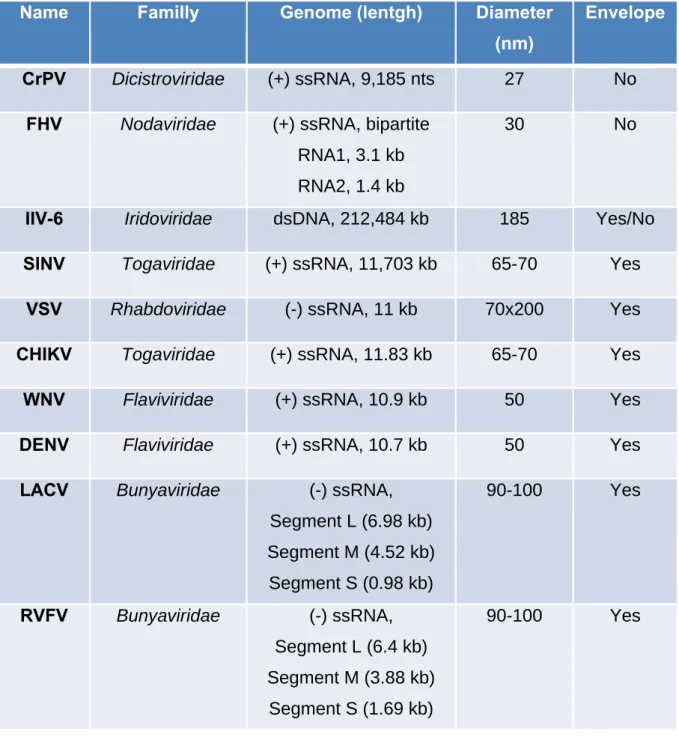
In 2009, the team of Dr. Shou-Wei Ding proposed an original approach to discover new viruses in invertebrates (Wu et al., 2010). Based on the analysis of small RNA libraries, the authors reassembled viral genomes from v-siRNAs. They discovered four previously unknown viruses in Drosophila Schneider 2 (S2) cells: the (+) ssRNA viruses American Nodavirus (ANV) and Drosophila Tetravirus (DTrV) and the dsRNA viruses Drosophila Birnavirus (DBV) and Drosophila Totivirus (DTV). The pathogenicity and replication cycle of these viruses are not yet characterized. Viral small RNAs matching with ANV, DBV, DCV, DTrV, DXV and Nora Virus were found in Drosophila OSS cells. Overall, these results highlight that Drosophila S2 and OSS cells were probably persistently co-infected by five and six RNA viruses, respectively. Their molecular characterization awaits more investigations. Natural Drosophila viruses presented above are summarized in Table 1.
Name Familly Genome (lentgh) Diameter
(nm)
Transmission Envelope
DCV Dicistroviridae (+) ssRNA, 9,264 nts 25-30 Horizontal No
DPV Unclassified (+) ssRNA 25-30 Horizontal and Vertical
No
DAV Unclassified (+) ssRNA 25-30 Horizontal and Vertical No DXV Birnaviridae dsRNA, 3,360 bp (Seg.A), 3,243 bp (Seg.B) 70 Horizontal No
DFV Reoviridae dsRNA,
10 segments
60-70 Horizontal No
SIGMAV Rhabdoviridae (-) ssRNA, 10-15 kb 45-100 Vertical Yes
Nora Virus
Unclassified (+) ssRNA, 12,333 nts
27-30 Horizontal No
Table 1. Overview of viruses naturally infecting Drosophila melanogaster.
1.2. Other viruses infecting Drosophila
Cricket Paralysis virus (CrPV) was isolated in 1970 from two species of Australian
field crickets, Teleogryllus oceanicus and T. commodus, that rapidly succumbed after paralysis (Reinganum et al., 1970). CrPV is able to infect a broad range of insect species, including Drosophila melanogaster flies (Wang et al., 2006) and cell lines (Moore et al., 1980; Scotti, 1975). CrPV injection into flies is highly pathogenic (Wang et al., 2006). The CrPV genome is composed of two ORFs encoding non-structural and structural proteins via IRES mediated translation. Both CrPV IRES were widely used as models to understand cap-independent mechanisms of translation (Deniz et al., 2009; Landry et al., 2009). The sequencing of the genome allowed its classification in the Dicistroviridae family, which also includes DCV (Wilson et al., 2000). CrPV genome is a (+) ssRNA with VPg protein attached at the 5’ extremity (King and Moore, 1988) and a poly(A) tail at the 3’ extremity. The virus is transmitted horizontally (Moore and Tinsley, 1982). CrPV infection leads to a shutoff of host translation in Drosophila cells (Garrey et al., 2010). Intriguingly, the infection is increased at higher temperature (37°C instead of 25°C) in Drosophila S2 cells (Cevallos and Sarnow, 2010). At higher temperature, CrPV RNA genome and viral proteins production are increased but unexpectedly, viral infectious particles are not. It was proposed that cellular responses at high temperature, including selective expression of heat shock proteins (Hsp) at the expense of other host proteins (Klemenz et al., 1985), may provide a beneficial environment for viruses. At high temperatures, the host protein synthesis is reduced in mammals, allowing some IRES-containing RNA viruses to hijack the translation machinery (Kim and Jang, 2002). Furthermore, several mammalian viruses are able to use Hsp for their replication (Burch and Weller, 2005; Glotzer et al., 2000; López et al., 2006), capsid formation (Chromy et al., 2003), or uncoating (Chromy et al., 2006). In Drosophila, it was reported that Hsp90
facilitates the Flock House virus replication (Kampmueller and Miller, 2005). The mechanism facilitating CrPV replication and protein synthesis at high temperature remains to be elucidated.
Flock House Virus (FHV) was originally isolated in 1983 from the grass grub
Costelytra zaelandica, near the Flock House Agricultural Research Station in New Zealand
(Scotti et al., 1983). FHV is able to replicate in plants, insects, yeast and mammalian cells, including different mosquito and Drosophila cell lines (Dasgupta et al., 2003, 2007). FHV is not a natural pathogen of Drosophila. Intrathoracic injection of FHV in flies is highly pathogenic and the virus spreads to multiple tissues, infecting the fat body, muscles and trachea (Galiana-Arnoux et al., 2006). FHV is a small non-enveloped, bipartite (+) ssRNA virus that belongs to Nodaviridae family (Venter and Schneemann, 2008). The genome of FHV consists of two RNAs: RNA1 (3.1 kb) encodes the viral RdRp (FHV protein A) and a subgenomic RNA3 (0.4 kb) containing two overlapping ORFs encoding proteins B1 (unknown function) and B2, a viral suppressor of RNAi (Li et al., 2002). RNA2 (1.4 kb) encodes the precursor protein of the viral capsid. Both RNA1 and RNA2 are capped at their 5’ extremity, but are not polyadenylated. FHV replication occurs in viral factories that were visualized by electron microscopy tomography and reconstructed by three dimensional analysis (Kopek et al., 2007). The depletion of two enzymes involved in phosphatidylcholine biosynthesis (cytidylyltransferase (cct) 1 and cct2) affects FHV replication, indicating that glycerophospholipid metabolism positively regulates FHV replication (Castorena et al., 2010). FHV induces the formation of spherule-like vesicles between the inner and outer mitochondrial membranes that support new RNA synthesis by protein A. The two genomic RNAs are transported to the cytoplasm for translation and encapsidation into provirions is performed by the single protein α. Mature virions are produced after the autocatalytic cleavage of protein α into proteins β and γ, which confers physicochemical stability to the viral particle (Venter and Schneemann, 2008). As mentioned previously, the Hsp90 appears to facilitate FHV replication (Kampmueller and Miller, 2005), by promoting efficient synthesis of the viral RdRp in Drosophila S2 cells (Castorena et al., 2007). The molecular chaperon Hsp90 plays a role in the replication of a broad spectrum of viruses and appears to have virus-specific functions at unique steps in the viral cycle (Geller et al., 2012). This is one example of the complexity and diversity of the mechanisms employed by viruses to appropriate cellular pathways for their own purposes. FHV infectious cycle ends by the induction of
apoptosis through a caspase-dependent pathway in infected DL-1 cells (Settles and Friesen, 2008). Virus-induced apoptosis may favor the release of infectious particles and their subsequent dissemination to neighboring cells (Best, 2008). The American Nodavirus
(ANV) is a variant of FHV that was found to persistently infect Drosophila S2 cells (Wu et
al., 2010). The genome of ANV was assembled from v-siRNAs found in S2 cells and shared 89% and 82% of identity with RNA1 and RNA2 molecules of FHV respectively.
Invertebrate Iridescent virus 6 (IIV-6) is an icosahedral double stranded
desoxyribonucleic acid (dsDNA) virus that infects invertebrates, mainly insects and terrestrial isopods. Its name comes from the opalescent hues observed in heavily infected hosts. The surface of paracrystalline arrays of virus particles reflects light that interferes with incident light resulting in diffraction, thus causing the iridescent hues in highly infected hosts. The iridescence was proposed to be a visual indicator of particles size, but this relationship is still debated (Williams, 2008). IIV-6, also named Chilo Iridescent virus (CIV), belongs to
Iridoviridae family (Williams et al., 2005). Its dsDNA genome is 212,484 base pair (bp) long
and encodes for 211 putative ORFs (initially 468 ORF were predicted (Jakob et al., 2001) but a reannotation of the genome was performed by (Eaton et al., 2007). IIV-6 is able to replicate in flies after intrathoracic injection, and iridescence is visualized in the abdomen of infected flies (Bronkhorst et al., 2012; Kemp et al., 2013; Teixeira et al., 2008). The infection is not lethal in wild-type flies, even if the injected dose is high. No DNA viruses that naturally infect
Drosophila melanogaster have been discovered so far, although a dsDNA virus has recently
been identified in wild-caught Drosophila innubila (Unckless, 2011). IIV-6, among others, is used in our laboratory as a model to study the antiviral immunity against DNA viruses in
Drosophila.
Sindbis virus (SINV) is an arthropod-borne virus (arbovirus) first isolated in 1952
from Culex pipiens and Culex univittatus mosquitoes collected in the Sindbis district, near Cairo in Egypt (Taylor et al., 1955). SINV belongs to the Togaviridae family, in the alphavirus genus. Alphaviruses are transmitted by arthropods, typically the mosquitoes, and replicate in both arthropod and vertebrate hosts worldwide (Jose et al., 2009). SINV is the most widely distributed among alphaviruses causing arthritis in humans, which include Chikungunya, o'nyong-nyong, Mayaro and Ross River viruses (Tesh, 1982). However, it is the least dangerous for public health. The wide tropism of alphaviruses and genome