Nephrol Dial Transplant (2011) 26: 1211–1220 doi: 10.1093/ndt/gfq560
Advance Access publication 15 September 2010
Expression of the chemokine receptor CCR6 in human renal
inflammation
Desa Welsh-Bacic
1,2, Maja Lindenmeyer
1,3, Clemens D. Cohen
1,3, Dan Draganovici
4,
Jana Mandelbaum
4, Ilka Edenhofer
2, Urs Ziegler
5, Heinz Regele
6, Rudolf P. Wüthrich
1and Stephan Segerer
1,21
Division of Nephrology, University Hospital, Zurich, Switzerland,2Department of Anatomy, University of Zurich, Switzerland,
3
Institute of Physiology with Zurich Center of Integrative Human Physiology, University of Zurich, Zurich, Switzerland,
4
Medizinische Poliklinik-Innenstadt, University of Munich, Munich, Germany,5Center of Microscopy, University of Zurich, Zurich, Switzerland and6Clinical Institute of Pathology, University of Vienna, Vienna, Austria
Correspondence and offprint requests to: Stephan Segerer; E-mail: [email protected]
Abstract
Background. Nodular inflammatory cell infiltrates with defined microarchitecture, i.e. tertiary lymphoid organs, develop in the tubulointerstitium during chronic renal in-flammation. CCR6 and the corresponding ligand CCL20 are involved in the formation of gut-associated lymphatic tissue. We hypothesized that CCR6 might be involved in the formation of nodular infiltrates in the kidney.
Methods. CCR6- and CD20-positive B cells were localized in renal biopsies with IgA nephropathy (n = 13), membra-nous nephropathy (n = 12), crescentic glomerulonephritis (cGN, n = 11) and chronic interstitial nephritis (n = 13), and in pre-implantation biopsies as controls (n = 8). The mRNA expression of CCR6 and the ligand CCL20 was quantified by real-time RT–PCR in 51 renal biopsies of the same disease entities.
Results. In the pre-transplant biopsies, CCR6 was ex-pressed by endothelial cells of peritubular and glomerular capillaries. In patients with glomerulonephritis, infiltrating cells were positive particularly in areas of nodular inflam-matory cell accumulations. A major part of the CCR6-positive cells were CD20-CCR6-positive B cells, but a part of the CD3-positive T cells were also found to be positive. The constitutive expression of CCR6 on the endothelium of glomerular capillaries was lost in biopsies with progressive injury. Tubular epithelial cells expressed CCR6 in inflamed kidneys, most commonly on the basolateral side.
Conclusions. CCR6 and the corresponding ligand CCL20 might therefore be involved in the recruitment of T and B cells to organized nodular infiltrates in chronic renal in-flammation. The functional role of endothelial CCR6 needs to be evaluated in further studies.
Keywords: B cells; CCL20; CCR6; chemokine receptor; glomerulonephritis
Introduction
Lymphocytes, macrophages and dendritic cells accumulate in the tubulointerstitium of the human kidney during pro-gressive diseases [1–3]. The numbers of interstitial infiltrat-ing T cells and macrophages correlate with renal function at the time of biopsy [2,4]. In addition, to diffuse infiltrates, leucocytes form nodular structures in about one-third of the biopsies with chronic diseases [3,5]. These structures are called tertiary lymphoid organs (TLOs), and the functional role of TLOs in the kidney is under intense investigation [6–8].
Chemokines are members of a large family of chemotac-tic cytokines, which orchestrate the recruitment of inflam-matory cell subsets under homeostatic and pathological condition [9]. Expression and presentation of chemokines in particular microenvironments are involved in the forma-tion of lymphoid tissue as well as in chronic renal inflam-mation [10–12]. The chemokine receptor CCR6 signals after binding the chemokine CCL20, which is the only known ligand for this receptor so far [13,14]. CCR6 is ac-quired during B-cell maturation and is expressed by all bone marrow, and peripheral blood-derived naive and memory B cells [15]. CCR6 expression has been shown in memory T cells, regulatory T cells, and IL-17-producing CD4-positive T cells (a distinct subset of cells now re-ferred to as Th17 cells) [16–19]. CCR6 is also expressed on a subset of dendritic cells [17,20,21].
CCR6-positive B cells play an important role in the for-mation of gut-associated lymphatic tissue, i.e. Peyer’s patches and isolated lymphoid follicles [22]. The forma-tion of TLOs during chronic inflammaforma-tion might in part be a recapitulation of the embryonic formation of second-ary lymphoid organs [5]. Therefore, we hypothesized that CCR6 might be involved in the formation of nodular infil-trates in chronic renal inflammation. Therefore, we studied
© The Author 2010. Published by Oxford University Press on behalf of ERA-EDTA. All rights reserved. For Permissions, please e-mail: [email protected]
the expression of CCR6 and the ligand CCL20 in the most common forms of human glomerulonephritis, as a pre-requisite for further functional studies.
Materials and methods Study population
Renal biopsies were formalin-fixed, and paraffin-embedded following routine protocols. The diagnosis was based on light microscopy, immuno-histochemistry and electron microscopy. Archival sections were used for the current study from patients with crescentic glomerulonephritis (cGN, n = 11), IgA nephropathy (n = 13), membranous nephropathy (n = 12) and chronic interstitial nephritis (n = 13). Allograft biopsies taken before implantation served as controls (n = 8).
Immunohistochemistry
Immunohistochemistry was performed as previously described [3,23]. In brief, sections were dewaxed in xylene, rehydrated in a graded series of ethanol, and incubated in 3% hydrogen peroxide. The Avidin/Biotin block-ing Kit (Vector, Burlblock-ingame, CA, USA) was used to block endogenous bio-tin. An autoclave oven (or microwave treatment) was used for heat-based antigen retrieval in Antigen retrieval solution (Vector). Incubation with the primary antibody was performed for 1 h. Incubation with biotinylated sec-ondary antibodies (Vector) for 30 min was followed by the ABC reagent (Vector). 3,3′Diaminobenzidine (DAB, Sigma, Taufkirchen, Germany) with metal enhancement was used as a detection system. Two polyclonal antibodies against CCR6 raised in two different rabbits were tested and re-sulted in the same pattern (anti-human CCR6 by MBL International Cor-poration, Woburn, MA, USA and Chemicon, Temecula, CA, USA). The primary polyclonal antibody against CCR6 (Chemicon) was used in a di-lution of 1:1000 in 10% non-fat dry milk on renal biopsies. Other anti-bodies used were a monoclonal antibody against CD20 (clone L26, DakoCytomation, Dako Deutschland, Hamburg, Germany), an anti-CD3 antibody (clone: CD3-12, rat anti-human, Serotec, Oxford, UK), and an anti-CD34 antibody (Accurate Chemical and Scientific Corporation, Westbury, NY, USA).
CD34 was stained on a total of nine biopsies on consecutive sec-tions with CCR6 (four biopsies with chronic interstitial nephritis and five controls).
The numbers of CD20- and CCR6-positive cells were scored semi-quantitatively: 0, no infiltrating cells; 1, scattered positive infiltrating cells between tubules; 2, accumulations of several infiltrating cells between tubules; and 3, nodular appearance of the infiltrates by an observer blinded to the disease entities.
Immunofluorescence
Double-labelling immunofluorescence was performed as described pre-viously [3]. As secondary reagents, biotinylated antibodies (Vector), Streptavidin/FITC complex (Vector), Cy3-labelled anti-rat antibody (Jack-son Immunoresearch Laboratories Inc., West Grove, PA, USA), and Texas red- and FITC-labelled anti-mouse IgG (Vector) were used.
Real-time RT–PCR
To quantify the mRNA expression of CCL20 and CCR6, we used real-time RT–PCR as described [24]. The biopsies were from patients with cGN (n = 8), IgA nephropathy (n = 14), membranous nephropathy (n = 20) and allo-graft biopsies taken before implantation served as controls (n = 9). The renal biopsies were obtained from a multicentre renal biopsy bank (the European Renal cDNA Bank, ERCB). Informed consent was obtained before renal biopsies were performed. The microdissected tubulointersti-tial compartments were used.
Real-time RT–PCR was performed on a TaqMan ABI 7700 Sequence Detection System (Applied Biosystems, Darmstadt, Germany) using heat-activated TaqDNA polymerase (Amplitaq Gold, Applied Biosystems). Quantification of the given templates was performed according to the standard curve method. Commercially available pdeveloped TaqMan re-agents were used for the target genes CCR6 (NM_031409) and CCL20 (NM_004591) (all from Applied Biosystems), and two endogenous control genes (18S rRNA, GAPDH; Applied Biosystems). The normalization to the two reference genes (housekeeper genes) gave comparable results. The data shown in the text and figures are normalized to GAPDH. All measurements were performed in duplicates. Controls consisting of bidistilled H20 were negative in all runs. The samples used for the
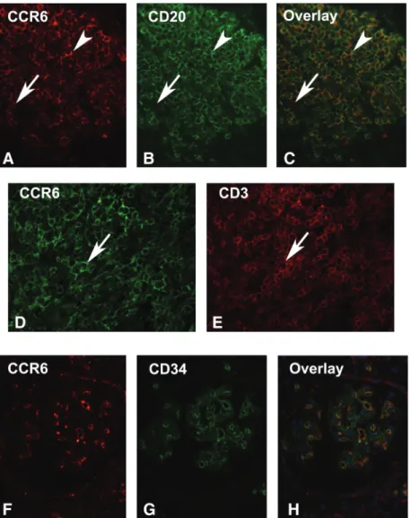
Fig. 1. Establishment of the anti-CCR6 antibody and expression in normal human kidney. Immunohistochemistry was performed on an allograft nephrectomy (A–C), and a pre-transplant biopsy (D and E) with a polyclonal antibody against CCR6 (B, D and E, Chemicon), a non-immune rabbit serum (A), and after pre-absorption of the antiserum with the peptide (C, original ×250 D, E, original ×200 in A–C). The negative controls performed with non-immune rabbit serum (A) and pre-absorption with the peptide used for the induction of the antiserum did not demonstrate a positive colour product (C). Prominent signal of a nodular infiltrate in the allograft nephrectomy (B). In the well-preserved pre-transplant biopsies, a positive signal was present on endothelial cells of glomerular capillaries (arrowheads, D) and peritubular capillaries (E, arrows).
mRNA analysis were from a different cohort than the biopsies used for immunohistochemistry.
Quantification and statistical analysis
The semi-quantitative scores for the interstitial CCR6-positive cells and the percentage of CCR6 staining per glomerular area were evaluated by observers blinded to the diagnosis. For the comparison of medians, the non-parametric Kruskal–Wallis test and Dunn’s multiple comparisons test were used. Spearman rank correlations were performed for the correla-tions with clinical and morphological data (InStat®software, version 3.05, Intuitive Software for Science, San Diego, CA, USA). A P <0.05 was considered to be significant. Error bars illustrate standard error of the mean (SEM).
Results
Description of the antibody and CCR6 expression in pre-transplantation biopsies
For the establishment of the polyclonal CCR6 anti-serum, we used tissue sections from allograft nephrectomies
(Figure 1A–C). A reliable staining pattern was achieved using a heat-based antigen retrieval. Expression of CCR6 was present on inflammatory cells and on endothelial cells in these positive controls (Figure 1B). A second antiserum against CCR6 resulted in the same pattern (not illustrated). A non-immune rabbit serum did not result in a black colour product (Figure 1A). Additionally, the pre-incubation with the peptide used for the generation of the antibody com-pletely abolished the signal (Figure 1C).
Double labelling of an allograft nephrectomy confirmed that CD20-positive B cells formed a major part of the CCR6-positive infiltrating cells (Figure 2A–C). CD3-positive T cells represent additional CCR6-CD3-positive cells (Figure 2D and E). Positivity of endothelial cells was con-firmed in double-labelling studies with CD34, an endothe-lial marker (Figure 2F–H).
Eight allograft biopsies taken before implantation were used as controls (Figure 1D and E). These pre-transplantation biopsies demonstrated well-preserved renal tissue, with vari-able degrees of global glomerulosclerosis. Mild interstitial
Fig. 2. Double immunofluorescence for CCR6. Double Immunofluorescence was performed on tissue sections from an allograft nephrectomy. A–C. Double labelling for CCR6 (A), CD20 (B) and the overlay in C illustrates the majority of this nodular aggregate being CCR6-positive B cells (arrows). D and E illustrate an area with a high number of CD3/CCR6 double-positive cells (arrowhead). F–H illustrate CCR6-positive endothelial cells in a glomerulus (F: CCR6, G: CD34, H: overlay, original ×400).
infiltration was present surrounding sclerosed glomeruli, a feature commonly found in ageing kidneys. These bi-opsies therefore cannot be regarded as normal, as these were exposed to cold ischaemia and the circumstances of brain-dead of the deceased donors, but the morphology demonstrated well-preserved tissue architecture.
In these biopsies, we found CCR6 to be expressed by endothelial cells of glomerular and peritubular capil-laries without prominent variation between the biopsies (Figure 1D and E). Some circulating cells in the lumen of open glomerular capillaries or peritubular capillaries were found to be CCR6 positive, but these cells were rare. Expression of CCR6 by tubular epithelial cells was only occasionally found.
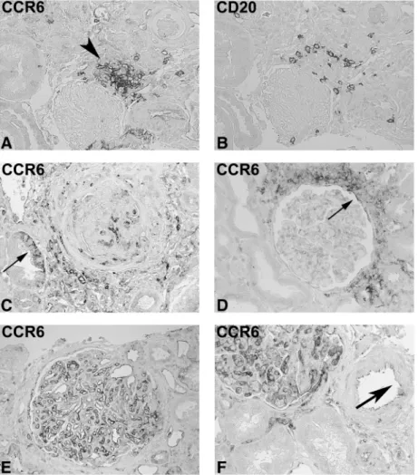
CCR6 expression in IgA nephropathy
Thirteen biopsies from patients with IgA nephropathy were studied (Table 1). Expression of CCR6 was found on endo-thelial cells of glomerular and peritubular capillaries. The staining was less intense as compared with controls. Fur-thermore, segmental to global loss of staining was present in glomeruli (Figure 3A). Parietal epithelial cells were found to be positive in a few glomeruli (Figure 3D). Tubular epithelial cells showed weak basolateral staining with focal distribution, particularly in tubules adjacent to sites of inter-stitial inflammation (Figure 3C). Infiltrating CCR6-positive Table 1. Basic clinical parameters of the patients included in the
immunohistochemistry cohort Disease n Age (years) Creatinine (mg/dL) Proteinuria (g/24 h) IgA N 13 37 (14–59) 1.5 (0.8–8.3) 1.5 (0–7) Membranous N 12 45 (13–65) 1.1 (0.6–6.4) 7 (2.4–22) cGN 11 63 (35–84) 3.8 (1.4–8.4) 0.4 (0.3–8) c Int 13 44 (3–81) 4.8 (1.8–13) 0.6 (0.08–25)
Controls 8 27 (0–67) n.a. n.a.
Data are presented as median (range) except n. n.a., not available; N, nephropathy; cGN, crescentic glomerulonephritis; c Int, chronic interstitial nephritis.
Fig. 3. CCR6 expression in renal biopsies from patients with IgA nephropathy and membranous nephropathy. Immunohistochemistry was performed on renal biopsies from patients with IgA nephropathy (A–D) and with membranous nephropathy (E–F) with a polyclonal antibody against CCR6 (A, C–F) and a monoclonal antibody against CD20 (B; original ×100 in C–F, ×250 in A and B). Consecutive section in A and B demonstrates that only the periphery of the CCR6-positive small nodular infiltrate consists of CD20-positive B cells. In C and D, the glomerular endothelial staining is lost. Tubular CCR6 staining is illustrated in C (arrow). D demonstrates prominent CCR6-positive peri-glomerular infiltration and CC6 expression by parietal epithelial cells (arrow). The staining pattern was similar in membranous nephropathy (E and F). Endothelial cells of arteries were rarely found to be CCR6 positive.
cells were found scattered throughout the cortex and at times accumulated around the glomeruli. Nodular infiltrates were present in 5 out of 13 biopsies. In small accumulations, CD20-positive cells were outnumbered by CCR6-positive cells not expressing CD20 (Figure 3A and B).
CCR6 in membranous nephropathy
Included were 12 biopsies from patients with membranous nephropathy (Table 1). This was the group of patients with the best preserved renal function. On intrinsic renal cells, CCR6 was found to be expressed as in IgA nephropathy (Figure 3E and F). Infiltrating CCR6-positive cells were only found with diffuse distribution, and no formation of nodular structures was present both in the CCR6 and CD20 staining (Figure 3E and F).
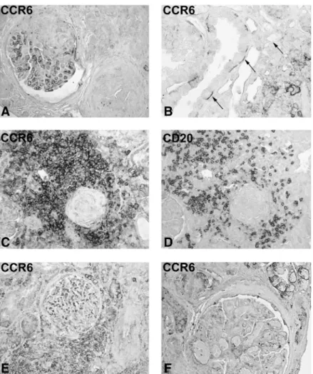
CCR6 expression in cGN
Eleven biopsies from patients with crescentic glomerulo-nephritis were included. All of these were pauci-immune by immunohistochemistry. Therefore, no crescentic IgA
nephritis or anti-glomerular basement membrane diseases were included in this group.
CCR6 was found to be expressed on glomerular endo-thelial cells in preserved glomeruli, as described for nor-mal controls, but segmental loss of positive staining was common (Figure 4A). Tubular epithelial cells were CCR6-positive in 8 out of 10 biopsies (Figure 4B). Oc-casionally, intercalating cells of collecting ducts were found to be positive (Figure 4B). An intensive accumulation of CCR6-positive infiltrating cells was present in the tubulointerstitium, but not in the glomeruli (Figure 4C and E). Nodular infiltrates were almost uniformly CCR6 positive (Figure 4C and D). These structures were commonly found around the completely sclerotic glomeruli (Figure 4C and D). On the other hand, scattered infiltrating cells were only occasionally CCR6 positive. The distribution of CCR6-positive cells matched only about half of the CD20-positive cells (Figure 4C and D). In cGN, a high number of macrophages are usually present in the glomeru-lar tuft and in crescents [25]. The absence of CCR6 staining on infiltrating cells in glomerular tufts and crescents indi-cates that CCR6 is mainly expressed by lymphocytes (loca-Fig. 4. CCR6 expression in biopsies with crescentic GN and chronic interstitial nephritis. Immunohistochemistry was performed on renal biopsies from patients with cGN (A–D) and patients with chronic interstitial nephritis (E and F) with a polyclonal antibody against CCR6 (A–C, E and F) and with a monoclonal antibody against CD20 (D, original ×250 in A–E, and ×400 in F). A segmental loss of the endothelial staining in the upper part of the glomerulus is illustrated in A. Basal tubular expression of CCR6 is shown in B. A prominent accumulation of CCR6-positive cells was found around globally sclerosed glomeruli, with CD20-positive cells being a major part of the infiltrate (C and D). In chronic interstitial nephritis, both interstitial accumulation of CCR6-positive cells (E) and loss of endothelial CCR6 were present (F).
lized in the tubulointerstitium), but not macrophages re-cruited to glomeruli.
CCR6 in chronic interstitial nephritis
Included were 13 biopsies with chronic interstitial neph-ritis (Table 1). The pattern of CCR6 expression was com-parable to the glomerular diseases presented above. Glomerular endothelial cells stained positive for CCR6, but the staining intensity was weaker than in controls. Occasionally, parietal epithelial cells demonstrated a positive signal. In 10 out of 13 biopsies, tubular epithelial
cells were found to be focally CCR6-positive on basolateral membranes of atrophic tubules of cortex and medulla (Figure 4F). Inflammatory cells positive for CCR6 were mainly scattered diffusely throughout the renal cortex (Figure 4E). Additionally, in 9 out of 13 biopsies, nodular structures were present, where the majority of these cells were CCR6 positive (not illustrated).
In four biopsies, CD34 was stained in parallel with CCR6. Complete loss of CD34 and CCR6 was present in globally sclerosed glomeruli. Furthermore, some glom-eruli demonstrated a less prominent CCR6 staining, and also, CD34 was preserved.
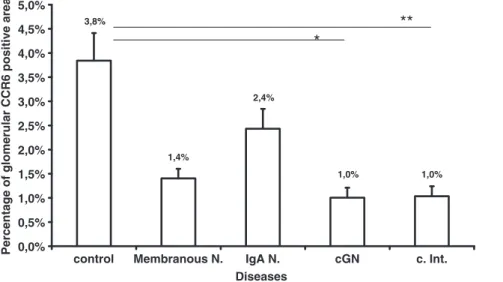
Fig. 5. Comparison glomerular CCR6 expression between the glomerular diseases. Immunohistochemistry was performed with a polyclonal antibody against CCR6 on renal biopsies before implantation (pre Tx control, A), with IgA nephropathy (B, IgA N.), with membranous nephropathy (membr. N., C) and with cGN (D, all original ×250). A weaker endothelial staining was found in all three glomerulopathies with segmental to global loss of the staining pattern, even in preserved glomerular capillaries.
1,0% 1,0% 2,4% 1,4% 3,8% 0,0% 0,5% 1,0% 1,5% 2,0% 2,5% 3,0% 3,5% 4,0% 4,5% 5,0%
control Membranous N. IgA N. cGN c. Int.
Diseases
Percentage of glomerular CCR6 positive area
**
*
Fig. 6. Morphometric quantification of glomerular CCR6 expression. Mean percentage of CCR6-positive colour product per glomerular area is illustrated (*P < 0.05, **P < 0.01).
Common pattern of CCR6 staining in different forms of renal inflammation
The staining pattern for CCR6 was not associated with par-ticular renal disease entities (Figure 5). The constitutive ex-pression of CCR6 on endothelial cells on peritubular and glomerular capillaries decreases in biopsies with chronic glomerular injury as well as in chronic interstitial nephritis. Particularly, in cGN, a prominent segmental to global loss of CCR6 expression was present (Figure 6). As could be ex-pected, globally sclerosed glomeruli demonstrated complete loss of CCR6 staining. The loss of glomerular staining in patients with chronic interstitial nephritis indicates that this is not specific for the glomerular injury process, but rather due to nephrosclerosis which is also present in chronic inter-stitial nephritis (with globally sclerosed glomeruli). The glomerular area of CCR6 expression did not correlate with serum creatinine, proteinuria, or the overall percentage of sclerosed glomeruli (not significant).
A semi-quantitative score was used to characterize the tubulointerstitial infiltration by CD20- and CCR6-positive cells (Table 2). There was an extremely significant correl-ation between the CD20 and CCR6 scores (Spearman r = 0.69, 95% confidence interval 0.51–0.81, P < 0.0001). Furthermore, CCR6 scores were associated with serum creatinine at the time of biopsy (Spearman r = 0.47, 95% confidence interval 0.17–0.69, P = 0.0027), but not with proteinuria (not significant). The median creatinine was significantly higher in the interstitial scores 1–3 vs 0, with a progressive increase from scores 1–3 (not illu-strated). The interstitial CD20 scores were not associated with serum creatinine and proteinuria (not significant), consistent with previously published data [3].
A negative correlation was found between the percent-age of CCR6 expression in glomeruli and the interstitial CCR6 scores (Spearman r =−0.42, 95% confidence inter-val from−0.64 to −0.13, P = 0.0049).
CCR6 and CCL20 mRNA expression in patients with glomerulonephritis
The mRNA expression of CCL20 and the corresponding receptor CCR6 was quantified by real-time RT–PCR in the microdissected tubulointerstitium in renal biopsies with IgA nephropathy, membranous nephropathy and cGN (Table 3). All biopsies demonstrated a prominent
ex-pression of CCL20 mRNA (Figure 7A). Exex-pression of CCR6 was most prominently induced in biopsies with IgA nephropathy and cGN (Figure 7B). A corresponding expression was found for CCL20 and CCR6 mRNA, but only the induction of the receptor reached the level of sig-nificance (Figure 7A and B). The expression of CCL20 mRNA correlated significantly with the expression of the corresponding receptor CCR6 mRNA (P < 0.05), but not with serum creatinine or proteinuria at the time of biopsy.
Discussion
The list of human diseases in which CCR6 plays a signifi-cant role is rapidly growing and contains common ones like colorectal carcinoma [26], Crohn’s disease, and rheumatoid arthritis [27]. Particularly, the description of CCR6 expression on Th17 cells sparked the recent interest in this chemokine receptor [28]. The ligand receptor pair CCL20/CCR6 is involved in the normal development of gut-associated lymphatic tissue, as well as in the men-tioned common human diseases [29].
In the mouse gut, the deficiency of CCR6 leads to reduced sizes of Peyer’s patches with loss of B cells and T cells, and particularly a reduction of regulatory CD4-positive T cells [30]. M cells, a specialized cell type within Peyer’s patches, are also reduced in number [31]. CCR6-deficient mice are protected from oral Yersinia ente-rocolitica infection, which exploits M cells for infection [31]. Isolated lymphoid follicles are organized lymphoid structures in the gut in which B cells are the major CCR6-positive population. The formation of these structures was significantly disturbed due to a reduction of B-cell influx in CCR6-deficient mice [22]. These studies highlight the role of CCR6 in lymphogenesis of the gut. CCR6 has also re-cently been documented on mouse regulatory T cells and in IL-17-producing CD4-positive T cells. CCR6 deficiency aggravated renal injury in mouse nephrotoxic nephritis and increased mortality illustrating the recruitment of regulatory T cells through CCR6 [32]. Future studies need to address the expression of CCR6 by inflammatory subsets in the hu-man kidney.
CCL20 can also be induced under inflammatory condi-tions [29]. Therefore, we hypothesized that the formation of tertiary lymphoid organs might in part be a recapitula-tion of embryogenesis. We localized CCR6 in human kid-Table 2. Morphological results of the study population
Disease n Interstitial CD20 scores Interstitial CCR6 scores
IgA N 13 1 (0–3) 1 (0–3)
Membranous N 12 1 (0–2)* vs cGN 1 (0–3)
cGN 11 2 (1–3)*** vs control 2 (1–3)* vs control c Int 13 2 (1–3)** vs control 2 (1–3)** vs control
Controls 8 0 (0–1) 0 (0–1)
Data are presented as median (range) except n. n.a., not available; N, nephropathy; cGN, crescentic glomerulonephritis; c Int, chronic interstitial nephritis. * P < 0.05. ** P < 0.01. ***P < 0.001.
Table 3. Basic clinical parameters of the patients included in the mRNA expression cohort Disease n Age (years) Creatinine (mg/dL) Proteinuria (g/24 h) IgA N 14 52 (17–72) 2.3 (1.5–6.2) 3.0 (0.5–6.3) Membranous N 20 55 (17–85) 1.0 (0.7–2) 4.7 (0.6–9.8) cGN 8 53 (27–69) 1.3 (0.9–6.4) 0.7 (0.1–1.8)
Controls 9 56 (27–70) n.a. n.a.
Data are presented as median (range) except n. n.a., not available; N, nephropathy; cGN, crescentic glomerulonephritis; c Int, chronic interstitial nephritis.
ney diseases in which the formation of tertiary lymphoid or-gans has previously been described [3,33]. This is the first detailed analysis on the expression of CCR6 in human renal biopsies from patients with glomerular and tubulointerstitial diseases. Consistent with our hypothesis, we found that a prominent number of B cells and T cells were CCR6-positive in the tubulointerstitium. The distribution of CCR6-positive cells was interesting as these were mainly found at sites of larger, nodular lymphocyte accumulations. Staining for CD20-positive cells demonstrated that a major part (about half) of the CD20-positive cells were also CCR6-positive. The majority of the remaining cells are most likely T cells (lymphocytes according to the morphology).
In contrast to the loss of CCR6 on glomerular endothelial cells, we found an increase of interstitial CCL20 and CCR6 mRNA expression particularly in IgA nephropathy. There was an inverse correlation between the glomerular expres-sion of CCR6 and the tubulointerstitial scores. Therefore,
the interstitial expression is consistent with the immunohis-tochemistry finding.
As previously described, T and B cells are present within the tubulointerstitium in high numbers during chronic renal inflammation, but not within the glomerular tuft [3,34]. This mirrors the distribution of CCR6 infiltrating cells in renal tissue. Furthermore, glomerular macrophages do not seem to express CCR6 because CCR6-positive cells were not present in this tissue compartment. In contrast to the high number of CCR6-positive cells in TLOs, the diffuse intersti-tial infiltrates were rarely CCR6 positive. This could be ex-plained in two ways, either a particular recruitment of CCR6-positive cells to tertiary lymphoid organs or an in-duction of CCR6 in nodular infiltrates. This is the first che-mokine receptor described in the human kidney which is expressed both on B cells and T cells. Other chemokine re-ceptors have been shown to be expressed on T cells (e.g. CCR5 [34]) or on B cells (e.g. CXCR5 [3]). CCR6 might
0 0,001 0,002 0,003 0,004 0,005 0,006 0,007
Pre TX IgA N. membran. N. cGN
Mean CCL20 mRNA expression /GAPDH
0,0E+00 5,0E-04 1,0E-03 1,5E-03 2,0E-03 2,5E-03 3,0E-03
Pre TX IgA N. membran. N. cGN
Mean CCR6 mRNA expression/ GAPDH
A
B
**
*
Fig. 7. Real-time expression of CCL20 (A) and CCR6 (B) in the tubulointerstitium of renal biopsies from patients with IgA nephropathy (IgA N.), membranous nephropathy (membran. N.) and cGN. Allograft biopsies taken before implantation were used as controls (Pre Tx, *P < 0.05, **P < 0.01).
therefore be involved in orchestrating a rendezvous between T cells and B cells, particularly at sites of nodular infiltrates. The only study on CCL20 and CCR6 in human kidneys has been published by Woltman et al. [35]. In this study, the authors described the expression of CCL20 by tubular epi-thelial cells, and CCR6 by a subset of inflammatory cells in rejecting allografts [35]. Particularly, CCR6 was localized to dendritic cells in rejecting renal allografts [35]. Our study is the first addressing the expression in endogenous kidney diseases. We could confirm the expression of CCR6 by den-dritic cells in allograft nephrectomies (not illustrated).
The constitutive expression of CCR6 on endothelial cells of glomerular and peritubular capillaries was an un-expected finding. This expression was reduced during in-flammation. Evidence for CCR6 expression on endothelial cells has been provided for human umbilical vein endothe-lial cells, saphenous vein endotheendothe-lial cells, and dermal and lung microvascular endothelial cells [36,37]. Expression of CCR6 has been found to be induced on human umbilical vein endothelial cells after infection with human herpes virus 8 [38]. Induction of CCR6 on human umbilical vein endothelial cells was also described after combined stimu-lation with hepatocyte growth factor and vascular endothe-lial growth factor (VEGF) [36]. Loss of CCR6 was present in focal and global glomerulosclerosis. Due to limitations in materials, we cannot at the moment exclude that there is a regulation of CCR6 independent of endothelial cell loss. This and the functional role of a constitutive expression of CCR6 on renal endothelial cells remain to be evaluated. Another ligand receptor pair, i.e. CXCL12/CXCR4, has been shown to be involved in angiogenesis of the kidney during development but also in neoangiogenesis in malig-nant tumours [39,40]. Mechanisms involved in endothelial biology might be the recruitment of endothelial stem cells or a direct induction of migration/proliferation of endothe-lial cells. Interactions between CXCR4 and CCL20 have been shown to play a role in malignant diseases; therefore, an interaction of these systems might be envisioned in the renal vasculature [41].
In summary, we found CCR6 to be expressed by infiltra-ting lymphocytes, both B cells and T cells, predominantly in nodular accumulation and, to a lesser extent, in diffuse inter-stitial infiltrates. Furthermore, there might be a role for CCR6 on intrinsic renal cells particularly endothelial cells. The functional evaluation will have to await further in vitro studies and studies on CCR6 mutant mice. These studies are currently hampered as useful mouse models of tertiary lymphoid organ formation in the kidney associated with chronic inflammation wait to be established.
Acknowledgements. S.S. is supported by a grant by the Novartis Stiftung (No. 09B26), a grant by the University of Zurich (Forschungskredit) and a grant by the Swiss National Foundation (32003B_129710/1), and C.D.C. is supported by the Else Kröner-Fresenius Foundation. We thank all par-ticipating centres of the European Renal cDNA Bank-Kroener-Fresenius biopsy bank (ERCB-KFB) and their patients for their cooperation. Active members at the time of the study: Clemens David Cohen, Holger Schmid, Michael Fischereder, Lutz Weber, Matthias Kretzler, Detlef Schlöndorff, Munich/Zurich/AnnArbor/New York; Jean Daniel Sraer, Pierre Ronco, Paris; Maria Pia Rastaldi, Giuseppe D'Amico, Milano; Peter Doran, Hugh Brady, Dublin; Detlev Mönks, Christoph Wanner, Würzburg; Andy Rees, Aberdeen; Frank Strutz, Gerhard Anton Müller, Göttingen; Peter Mertens, Jürgen Floege, Aachen; Norbert Braun, Teut Risler, Tübingen; Loreto
Gesualdo, Francesco Paolo Schena, Bari; Jens Gerth, Gunter Wolf, Jena; Rainer Oberbauer, Dontscho Kerjaschki, Vienna; Bernhard Banas, Bernhard Krämer, Regensburg; Moin Saleem, Bristol; Rudolf Wüthrich, Zurich; Walter Samtleben, Munich; Harm Peters, Hans-Hellmut Neumayer, Berlin; Mohamed Daha, Leiden; Katrin Ivens, Bernd Grabensee, Düsseldorf; Francisco Mampaso (†), Madrid; Jun Oh, Franz Schaefer, Martin Zeier, Hermann-Joseph Gröne, Heidelberg; Peter Gross, Dresden; Giancarlo Tonolo; Sassari; Vladimir Tesar, Prague; Harald Rupprecht, Bayreuth; Hans-Peter Marti, Bern.
Conflict of interest statement. None declared.
References
1. Segerer S, Heller F, Lindenmeyer MT et al. Compartment specific expression of dendritic cell markers in human glomerulonephritis. Kidney Int 2008; 74: 37–46
2. Segerer S, Banas B, Wornle M et al. CXCR3 is involved in tubuloin-terstitial injury in human glomerulonephritis. Am J Pathol 2004; 164: 635–649
3. Heller F, Lindenmeyer MT, Cohen CD et al. The contribution of B cells to renal interstitial inflammation. Am J Pathol 2007; 170: 457–468 4. Sean Eardley K, Cockwell P. Macrophages and progressive
tubuloin-terstitial disease. Kidney Int 2005; 68: 437–455
5. Segerer S, Schlondorff D. B cells and tertiary lymphoid organs in renal inflammation. Kidney Int 2008; 73: 533–537
6. Steinmetz OM, Velden J, Kneissler U et al. Analysis and classifica-tion of B-cell infiltrates in lupus and ANCA-associated nephritis. Kidney Int 2008; 74: 448–457
7. Sarwal M, Chua MS, Kambham N et al. Molecular heterogeneity in acute renal allograft rejection identified by DNA microarray profiling. N Engl J Med 2003; 349: 125–138
8. Zarkhin V, Kambham N, Li L et al. Characterization of intra-graft B cells during renal allograft rejection. Kidney Int 2008; 74: 664–673 9. Sallusto F, Baggiolini M. Chemokines and leukocyte traffic. Nat
Immunol 2008; 9: 949–952
10. Segerer S, Nelson PJ, Schlondorff D. Chemokines, chemokine recep-tors, and renal disease: from basic science to pathophysiologic and therapeutic studies. J Am Soc Nephrol 2000; 11: 152–176 11. Segerer S. The role of chemokines and chemokine receptors in
progressive renal diseases. Am J Kidney Dis 2003; 41: S15–S18 12. Panzer U, Steinmetz OM, Stahl RA et al. Kidney diseases and
chemokines. Curr Drug Targets 2006; 7: 65–80
13. Murphy PM, Baggiolini M, Charo IF et al. International union of pharmacology. XXII. Nomenclature for chemokine receptors. Phar-macol Rev 2000; 52: 145–176
14. Baba M, Imai T, Nishimura M et al. Identification of CCR6, the spe-cific receptor for a novel lymphocyte-directed CC chemokine LARC. J Biol Chem 1997; 272: 14893–14898
15. Krzysiek R, Lefevre EA, Bernard J et al. Regulation of CCR6 chemokine receptor expression and responsiveness to macrophage in-flammatory protein-3alpha/CCL20 in human B cells. Blood 2000; 96: 2338–2345
16. Sallusto F, Kremmer E, Palermo B et al. Switch in chemokine recep-tor expression upon TCR stimulation reveals novel homing potential for recently activated T cells. Eur J Immunol 1999; 29: 2037–2045 17. Liao F, Rabin RL, Smith CS et al. CC-chemokine receptor 6 is
expressed on diverse memory subsets of T cells and determines respon-siveness to macrophage inflammatory protein 3 alpha. J Immunol 1999; 162: 186–194
18. Voo KS, Wang YH, Santori FR et al. Identification of IL-17-producing FOXP3+ regulatory T cells in humans. Proc Natl Acad Sci USA 2009; 106: 4793–4798
19. Acosta-Rodriguez EV, Rivino L, Geginat J et al. Surface phenotype and antigenic specificity of human interleukin 17-producing T helper memory cells. Nat Immunol 2007; 8: 639–646
20. Sozzani S, Allavena P, Vecchi A et al. The role of chemokines in the regulation of dendritic cell trafficking. J Leukoc Biol 1999; 66: 1–9 21. Kucharzik T, Hudson JT3rd, Waikel RL et al. CCR6 expression distinguishes mouse myeloid and lymphoid dendritic cell subsets:
demonstration using a CCR6 EGFP knock-in mouse. Eur J Immunol 2002; 32: 104–112
22. McDonald KG, McDonough JS, Wang C et al. CC chemokine recep-tor 6 expression by B lymphocytes is essential for the development of isolated lymphoid follicles. Am J Pathol 2007; 170: 1229–1240 23. Neusser MA, Kraus AK, Regele H et al. The chemokine receptor
CXCR7 is expressed on lymphatic endothelial cells during renal allo-graft rejection. Kidney Int 2010; 77: 801–808
24. Cohen CD, Frach K, Schlondorff D et al. Quantitative gene expres-sion analysis in renal biopsies: a novel protocol for a high-throughput multicenter application. Kidney Int 2002; 61: 133–140
25. Segerer S, Cui Y, Hudkins KL et al. Expression of the chemokine monocyte chemoattractant protein-1 and its receptor chemokine re-ceptor 2 in human crescentic glomerulonephritis. J Am Soc Nephrol 2000; 11: 2231–2242
26. Ghadjar P, Rubie C, Aebersold DM et al. The chemokine CCL20 and its receptor CCR6 in human malignancy with focus on colorectal cancer. Int J Cancer 2009; 125: 741–745
27. Tanida S, Yoshitomi H, Nishitani K et al. CCL20 produced in the cyto-kine network of rheumatoid arthritis recruits CCR6 + mononuclear cells and enhances the production of IL-6. Cytokine 2009; 47: 112–118 28. Brand S. Crohn's disease: Th1, Th17 or both? The change of a para-digm: new immunological and genetic insights implicate Th17 cells in the pathogenesis of Crohn's disease. Gut 2009; 58: 1152–1167 29. Williams IR. CCR6 and CCL20: partners in intestinal immunity and
lymphorganogenesis. Ann NY Acad Sci 2006; 1072: 52–61 30. Lugering A, Floer M, Westphal S et al. Absence of CCR6 inhibits
CD4 + regulatory T-cell development and M-cell formation inside Peyer's patches. Am J Pathol 2005; 166: 1647–1654
31. Westphal S, Lugering A, von Wedel J et al. Resistance of chemokine receptor 6-deficient mice to Yersinia enterocolitica infection: evidence of defective M-cell formation in vivo. Am J Pathol 2008; 172: 671–680
32. Turner JE, Paust HJ, Steinmetz OM et al. CCR6 recruits regulatory T cells and Th17 cells to the kidney in glomerulonephritis. J Am Soc Nephrol 21: 974–985
33. Cohen CD, Calvaresi N, Armelloni S et al. CD20-positive infiltrates in human membranous glomerulonephritis. J Nephrol 2005; 18: 328–333 34. Segerer S, Mack M, Regele H et al. Expression of the C-C chemokine
receptor 5 in human kidney diseases. Kidney Int 1999; 56: 52–64 35. Woltman AM, de Fijter JW, van der Kooij SW et al. MIP-3alpha/
CCL20 in renal transplantation and its possible involvement as dendritic cell chemoattractant in allograft rejection. Am J Transplant 2005; 5: 2114–2125
36. Gerritsen ME, Tomlinson JE, Zlot C et al. Using gene expression profiling to identify the molecular basis of the synergistic actions of hepatocyte growth factor and vascular endothelial growth factor in human endothelial cells. Br J Pharmacol 2003; 140: 595–610 37. Hillyer P, Mordelet E, Flynn G et al. Chemokines, chemokine
recep-tors and adhesion molecules on different human endothelia: discrim-inating the tissue-specific functions that affect leucocyte migration. Clin Exp Immunol 2003; 134: 431–441
38. Punj V, Matta H, Schamus S et al. Induction of CCL20 production by Kaposi sarcoma-associated herpesvirus: role of viral FLICE inhibi-tory protein K13-induced NF-kappaB activation. Blood 2009; 113: 5660–5668
39. Li M, Ransohoff RM. The roles of chemokine CXCL12 in embryonic and brain tumor angiogenesis. Semin Cancer Biol 2009; 19: 111–115 40. Takabatake Y, Sugiyama T, Kohara H et al. The CXCL12 (SDF-1)/ CXCR4 axis is essential for the development of renal vasculature. J Am Soc Nephrol 2009; 20: 1714–1723
41. Beider K, Abraham M, Begin M et al. Interaction between CXCR4 and CCL20 pathways regulates tumor growth. PLoS ONE 2009; 4: e5125 Received for publication: 20.12.09; Accepted in revised form: 19.8.10
Nephrol Dial Transplant (2011) 26: 1220–1228 doi: 10.1093/ndt/gfq558
Advance Access publication 14 September 2010
Anti-C1q autoantibodies do not correlate with the occurrence or
severity of experimental lupus nephritis
Cornelia Bigler
1, Helmut Hopfer
2, Doris Danner
1, Monica Schaller
1, Michael J. Mihatsch
2and Marten Trendelenburg
11
Clinical Immunology, Department of Biomedicine, University Hospital Basel, Switzerland and2Institute for Pathology, University Hospital Basel, Switzerland
Correspondence and offprint requests to: Marten Trendelenburg; E-mail: [email protected]
Abstract
Background. In systemic lupus erythematosus patients, a strong association between the occurrence of antibodies against complement C1q (anti-C1q) and lupus nephritis can be observed. However, the predictive value of anti-C1q titres for a renal flare remains to be determined. In-creasing titres of anti-C1q before the occurrence of clinical apparent nephritis might not only serve as a clinical param-eter but also indicate a direct pathogenic mechanism of anti-C1q.
Methods. The aim of this study was to analyse the occur-rence of anti-C1q before the onset of experimental lupus nephritis in MRL/MpJ +/+ mice and to correlate anti-C1q titres with the type and severity of glomerulonephritis (GN) developing at advanced age.
Results. As judged by a number of morphological and im-munological analyses, GN in MRL/MpJ +/+ mice re-sembled human lupus nephritis and occurred in variable degrees of severity. We also observed an abundant and early presence of anti-C1q. However, anti-C1q neither correlated © The Author 2010. Published by Oxford University Press on behalf of ERA-EDTA. All rights reserved.