HAL Id: hal-01510341
https://hal.univ-brest.fr/hal-01510341
Submitted on 19 May 2020
HAL is a multi-disciplinary open access
archive for the deposit and dissemination of sci-entific research documents, whether they are pub-lished or not. The documents may come from teaching and research institutions in France or abroad, or from public or private research centers.
L’archive ouverte pluridisciplinaire HAL, est destinée au dépôt et à la diffusion de documents scientifiques de niveau recherche, publiés ou non, émanant des établissements d’enseignement et de recherche français ou étrangers, des laboratoires publics ou privés.
Sensitivity of eastern oyster (Crassostrea virginica)
spermatozoa and oocytes to dispersed oil: Cellular
responses and impacts on fertilization and embryogenesis
J. Vignier, A. K. Volety, A. Rolton, Nelly Le Goïc, F. -L. E. Chu, R. Robert,
Philippe Soudant
To cite this version:
J. Vignier, A. K. Volety, A. Rolton, Nelly Le Goïc, F. -L. E. Chu, et al.. Sensitivity of eastern oyster (Crassostrea virginica) spermatozoa and oocytes to dispersed oil: Cellular responses and im-pacts on fertilization and embryogenesis. Environmental Pollution, Elsevier, 2017, 225, pp.270-282. �10.1016/j.envpol.2016.11.052�. �hal-01510341�
1
Please note that this is an author-produced PDF of an article accepted for publication following peer review. The definitive publisher-authenticated version is available on the publisher Web site.
Environmental Pollution
June 2017, Volume 225 Pages 270-282 http://dx.doi.org/10.1016/j.envpol.2016.11.052 http://archimer.ifremer.fr/doc/00377/48851/
Achimer
http://archimer.ifremer.fr
Sensitivity of eastern oyster ( Crassostrea virginica)
spermatozoa and oocytes to dispersed oil: Cellular
responses and impacts on fertilization and embryogenesis
✯Vignier J. 1, 2, Volety A. K. 1, 5, *, Rolton A. 1, 2, Le Goic N. 2, Chu F. -L. E. 3, Robert Rene 4, Soudant P. 2
1
Florida Gulf Coast Univ, Coll Arts & Sci, Dept Marine & Ecol Sci, Ft Myers, FL 33965 ,USA.
2
IUEM UBO, Lab Sci Environm Marin, UMR LEMAR 6539, Technopole Brest Iroise, F-29280 Plouzane, France.
3
Virginia Inst Marine Sci, Coll William & Mary, Dept Aquat Hlth Sci, Gloucester Point, VA 23062, USA.
4
IFREMER, Unite Littoral, Ctr Bretagne, CS 10070, F-29280 Plouzane, France.
5
Univ North Carolina Wilmington, 601S Coll Rd, Wilmington, NC 28403, USA.
* Corresponding author : A. K. Volety, email address : voletya@uncw.edu
✯ This paper has been recommended for acceptance by Dr. Harmon Sarah Michele.
Abstract :
The 2010 Deepwater Horizon (DWH) oil spill released millions of barrels of oil and dispersant into the Gulf of Mexico. The timing of the spill coincided with the spawning season of Crassostrea virginica. Consequently, gametes released in the water were likely exposed to oil and dispersant. This study aimed to (i) evaluate the cellular effects of acute exposure of spermatozoa and oocytes to surface slick oil, dispersed mechanically (HEWAF) and chemically (CEWAF), using flow-cytometric (FCM) analyses, and (ii) determine whether the observed cellular effects relate to impairments of fertilization and embryogenesis of gametes exposed to the same concentrations of CEWAF and HEWAF. Following a 30-min exposure, the number of spermatozoa and their viability were reduced due to a physical action of oil droplets (HEWAF) and a toxic action of CEWAF respectively. Additionally, reactive oxygen species (ROS) production in exposed oocytes tended to increase with increasing oil concentrations suggesting that exposure to dispersed oil resulted in an oxidative stress. The decrease in fertilization success (1-h), larval survival (24-h) and increase in abnormalities (6-h and 24-h) may be partly related to altered cellular characteristics. FCM assays are a good predictor of sublethal effects especially on fertilization success. These data suggest that oil/dispersant are cytotoxic to gametes, which may affect negatively the reproduction success and early development of oysters.
2
Please note that this is an author-produced PDF of an article accepted for publication following peer review. The definitive publisher-authenticated version is available on the publisher Web site.
Graphical abstract
Highlights
► Deepwater Horizon oil spill coincided with the reproductive season of oysters. ► Crassostrea
virginica spermatozoa and oocytes were exposed acutely to dispersed oil. ► Effects on early life stages
were evaluated using FCM analysis and static bioassays. ► Toxic effects of oil/dispersant may be related to altered cellular characteristics. ► FCM assays are a good predictor of sublethal effects on fertilization success.
1
1. Introduction
The recent Deepwater Horizon (DWH) oil spill in the Gulf of Mexico, the U.S’ largest accidental release of crude oil into the ocean in history, resulted in the discharge of millions of barrels of oil into the offshore waters between April 20th and July 15th 2010.1-3 Although subsurface application of several million liters of the dispersant Corexit 9500A® near the wellhead (1500 m depth) enabled the retention of a considerable portion of oil in the water column and the creation of a “plume”, oil also moved to the upper surface waters to form a slick.4-7 The cumulative surface coverage of that oil slick was estimated at ≈ 112,000 km2.8 The dispersant Corexit 9500A® is a complex mixture, primarily containing dioctyl sodium sulfosuccinate or DOSS (surfactant), petroleum distillates (solvent) and propylene glycol (stabilizer).9 Acute toxicity of Corexit 9500A® to early life stages of oysters has been recently shown.10-14 When applied under the appropriate conditions, dispersants can break up oil slicks into small droplets and facilitate the mixing and the biodegradation of the oil into the water column.15-17 However, lethal toxicity of chemically dispersed oil is primarily associated with the bioavailability of oil droplets and the dissolved aromatic fractions of the oil.18-21 Eastern oysters,
Crassostrea virginica, are abundant in the northern region of the Gulf of Mexico. They are
economically important as they are highly valued as food, but their ecological significance for the region is even more important as oyster reefs provide valuable habitat for many estuarine species.22-24 During spawning, oysters release their gametes into the surrounding waters where fertilization takes place. In the case of the DWH event, the spill coincided with oyster spawning season, which occurs in the Gulf from late spring through late fall.25 Consequently, eggs and sperm were likely exposed to the toxic effects of petroleum hydrocarbons released during the DWH oil spill. Numerous studies have shown that acute exposure of sperm and eggs to organic pollutants could negatively impact fertilization success and embryogenesis of oysters.12,26-32 For example, acute exposure of C. virginica gametes to chemically enhanced water accommodated fraction (CEWAF), consisting of 100 mg L-1 of a surrogate of Macondo oil dispersed with 10 mg L-1 of Corexit 9500A (equivalent to 71 µg tPAH L-1), negatively altered fertilization success after 4 h incubation.32 Similarly, our previous work suggested that oyster gametes were sensitive to oil exposure and dispersant: 30 min exposure to CEWAF concentration of 110 mg L-1 of oil (equivalent to 29.9 µg tPAH50 L-1) and high energy water accommodated fraction (HEWAF)
2
concentration of 1371 mg oil L-1 (equivalent to 2250 µg tPAH50 L-1) reduced the fertilization success of gametes by 50%.20 After 24 h of continuous exposure, embryogenesis was also significantly affected (50% abnormal) at a CEWAF concentration of 56 mg oil L-1, corresponding to 14.9 µg tPAH50 L-1, and a HEWAF concentration of 157 mg oil L-1, corresponding to 267 µg tPAH50 L-1.12 We also found that, at equivalent nominal doses, Corexit alone and CEWAF exposures inhibited fertilization success and early development in a similar manner.12 In our previous study, eggs and sperm were exposed simultaneously to oil and dispersant for ecological relevance. However, it is unclear from these results whether the reduced fertilization success and subsequent altered embryogenesis were the consequences of functionally impaired spermatozoa or oocyte, or the result of an interaction of both.
Spermiotoxicity is typically assessed by exposing sperm to the toxic agent for a short time period, then adding exposed sperm to eggs and following up to fertilization success and hatching.27,33-36 Successful fertilization for any species depends on the production of spermatozoa with appropriate motility and fertilizing capability.37 Success of fertilization is also related to viability, DNA integrity, acrosomal integrity, mitochondrial activity or production of reactive oxygen species (ROS) which can be rapidly measured on spermatozoa using flow-cytometry (FCM).38-42 A previous study had demonstrated that increasing exposure concentrations of DWH oil prepared as CEWAF (6.25 – 100 mg oil L-1) and exposure to dispersant alone (0.625 – 10 mg L-1 nominal Corexit) reduced the viability of sperm cells, altered acrosomal integrity and inhibited ROS production and mitochondrial metabolism.14 In parallel, the above mentioned reduced fertilization shown in our previous study12 might have partially resulted from alterations of cellular functions of spermatozoa. Cellular alterations of spermatozoa could reduce their motility and fertilization capabilities as suggested by several authors.29,31,43,44 Furthermore, as a result of dispersion, particulate oil (droplets) may negatively interact with oyster gametes, resulting in reduced fertilization ability. Therefore, additional research is required to better understand the mechanisms of toxicity of DWH oil spill contaminants on the fertilization process, particularly on the related cellular functions of oocytes and spermatozoa.
Generally, the morphology of oocytes, using simple visual observation, and subsequent fertilization success can be estimated using light microscopy to assess egg quality following exposure to metals or to phycotoxins.44,45 In addition, oocyte viability can be determined using fluorescent dyes and epifluorescence microscopy.46,47 A recent study has demonstrated that the use of FCM was a reliable method to assess oocyte quality produced by the Pacific oyster,
3
Crassostrea gigas by determining their viability and ROS production.48 However, very few
studies have examined precisely how oil/PAHs decrease fertilization success by assessing cellular characteristics of exposed gametes using FCM techniques, particularly the fertilization ability of oocytes that have been exposed to oil.
The aims of the present study were (i) to evaluate the sublethal effects of DWH oil, dispersed mechanically or chemically, on the cellular parameters of spermatozoa and oocytes stripped from C. virginica, and to see how the observed cellular effects relate to the success of fertilization and embryogenesis, and (ii) to compare the relative sensitivity of spermatozoa and oocytes to oil and/or dispersant.
2. Materials and methods
2.1 HEWAF and CEWAF preparation
Crude oil was obtained under chain of custody during the Deepwater Horizon response efforts. The DWH surface slick oil (“Slick A”) was collected near the source on July 29th , 2010, from the hold of barge number CTC02404, which received surface slick oil from various skimmer vessels near the Macondo well (sample CTC02404-02). The dispersant Corexit 9500A® (Nalco Environmental Solutions LLC, Sugar Land, TX, USA) was provided by the DWH Trustees. For all exposure solutions, we added contaminants to UV-sterilized and 0.1 µm-filtered seawater (FSW), maintained at a salinity of 20–25 PSU. HEWAF and CEWAF exposure solutions were prepared following a standardized procedure.12 The high-energy preparation (HEWAF) method was adopted to artificially recreate the actions of currents, winds, and waves on oil slicks. CEWAF solutions contained a 1:10 dispersant to oil ratio using the dispersant Corexit 9500A®. CEWAF and HEWAF preparations generate large amounts of micron-size droplets and their chemical composition corresponds closely with that of the whole oil.49 Two concentrations for CEWAF (50 mg L-1 and 100 mg L-1 oil loading rates) and HEWAF (250 mg L-1 and 500 mg L-1 oil loading rates) were tested. These doses were previously demonstrated to cause sub-lethal effects on fertilization success and embryogenesis following 24 h of exposure of gametes to oil/dispersant.12,14 Previous works also showed that, at equivalent dispersant nominal doses, Corexit only and CEWAF exposures inhibited fertilization success and early development in a similar manner .12,14 As a result, we focused the present work on testing HEWAF and CEWAF.
4 2.2 Oyster sperm and oocytes
Ripe C. virginica broodstock were collected from Estero Bay, Florida, in September 2013, and acclimated at the experimental hatchery for two weeks, at 22˚C ± 1, a salinity of 20-25 PSU, and fed ad libitum with Tisochrysis lutea and Chaetoceros muelleri cultured in the hatchery. Fresh gametes were collected by stripping oyster gonads with a scalpel, and were then re-suspended in FSW.50 Oocytes and spermatozoa were examined under a microscope for motility (sperm), shape and absence of atresia (oocytes) and abnormal gametes were discarded. For the exposure experiment using FCM, cellular parameters were performed on gametes from individual genitors. Sperm from 3 males, and oocytes (mean diameter ≈ 50 µm) from 3 females were kept separately in FSW in 50 mL sterile beakers, after successive sieving through 150 µm and 55 µm mesh and washing to remove gonadal tissue and debris. For experiments that tested the success of fertilization and embryogenesis after exposure to dispersed oil, gametes were pooled from the same genitors used for cellular parameters. Sperm was pooled from 3 males and oocytes pooled from 3 females. Pooled gametes were kept in FSW, eggs in a sterile 1 L beaker and sperm in 100 mL beaker. Three subsamples of 100 µL of FSW/egg mixture were then taken after continuous and gentle mixing. Subsamples of eggs were stained with 1% Lugol, and counted using a Sedgwick-Rafter® cell and a dissecting microscope.
2.3 Cellular characteristics of exposed gametes
Cellular characteristics of exposed gametes were analyzed employing FCM. All FCM analyses were performed on an EasyCyte 6HT cytometer (Guava Merck Millipore®) equipped with a 100 µm capillary opening, a 488 nm argon laser, and 3 fluorescence detectors: green (525 nm ± 15), yellow (583 nm ± 13), and red (680 nm ± 15). Samples were acquired during 30 s at a flow rate of 0.59 µL s-1. Collected data from the FCM were analyzed with the software InCyte (Millipore®).
2.3.1. Sperm exposure
Sperm suspensions stripped from 3 individual males as described in section 2.2 were exposed separately to two concentrations of HEWAF (final nominal concentrations of 250 ppm and 500 ppm), and two concentrations of CEWAF (final nominal concentrations of 50 ppm and 100 ppm) for 30 min at 25˚C, prior to FCM analysis. Briefly, 160 µL of prepared oil solution was mixed with 40 µL of sperm suspension to obtain a final concentration of ≈ 1 x 10⁶ cells mL-1. Suspensions of exposed spermatozoa (200 µL) were then processed for FCM assays
5
including morphology, viability, mitochondrial membrane potential and ROS production using specific fluorescent probes.41,42 Sperm solutions incubated in FSW were used as controls.
Spermatozoa morphology and viability
Spermatozoa morphological characterization was based upon relative flow-cytometric
measurements of Forward SCatter (FSC: relative cell size) and Side SCatter (SSC: relative cell complexity). Based on FSC and SSC, sperm cell populations were first separated (or gated) from debris and aggregates into R1 single cell population (Fig. 1A and 1C) on which viability
parameters were determined.
Viability of spermatozoa was evaluated using a dual staining procedure with SYBR-14 and propidium iodide (PI) (Live/Dead® Sperm Viability kit, Molecular Probes). After 20 min of exposure to two different concentrations of HEWAF or CEWAF, sperm was stained with both SYBR-14 (final concentration 1 µM) and PI (final concentration 10 µg mL-1) for 10 min in the dark at 25⁰C. The proportion of live cells was estimated with SYBR-14, which only penetrates sperm cells with intact membranes, binds to double-stranded DNA and then emits in the green fluorescence range (516 nm) (Fig. 1B and 1D). Cell mortality was measured with PI, which penetrates only spermatozoa with damaged membranes, and then emits in the red fluorescence range (617 nm) (Fig. 1B and 1D). Dying sperm cells were indicated by staining with both SYBR-14 and PI (Fig. 1B and 1D). Single cell spermatozoa population (R1) was thus separated into three sub-populations (live, dying and dead cells). Results were expressed as mean percentages of live, dying and dead spermatozoa. Based on red and green fluorescence (Fig. 1D) also allowed further segregating (or gated) population of “oil- free” sperm cells (i.e. not aggregated with oil) from oil droplets and oil-aggregated sperm cell populations to more precisely assess their FSC and SCC parameters.
Spermatozoa mitochondrial membrane potential
Mitochondrial membrane potential (MMP) of spermatozoa was measured using the potential-dependent JC-1 (BD™ MitoScreen, BD Bioscience). JC-1 can enter selectively into mitochondria and exists as two forms, monomeric or aggregate, depending upon membrane potential. The monomer form which predominates in cells with low MMP emits in the green wavelength (525-530 nm), whereas the aggregate form which accumulates in mitochondria with
6
high membrane potential emits in the orange/yellow wavelength (590 nm). The JC-1 aggregate/monomer ratio is assumed to be proportional to MMP.41,51,52
Aliquots of 200 µL of spermatozoa (adjusted to a final concentration of ≈ 1 x 10⁷ cell mL-1) were exposed for 30 min to oil treatments and incubated with JC-1 (final concentration 5 µM) for 10 min in the dark at 25⁰C, and then diluted at 1:10 to stop the reaction prior to FCM analysis.41
Spermatozoa ROS production
Determination of ROS production was performed using 2’7’-dichlorofluorescein diacetate (DCFH-DA: Molecular probes, Invitrogen), a membrane permeable, non-fluorescent dye. Inside cells, the DCFH-DA is first hydrolyzed into DCFH by esterase enzymes. Intracellular hydrogen peroxide (H2O2) and superoxide ion (O2-•) then oxidize DCFH to the fluorescent DCF molecule.
DCF green fluorescence range (525 nm), detected on the FCM, is proportional to ROS production of spermatozoa.
Prior to FCM analysis, the spermatozoa suspension (200 µL) was incubated with oil solutions and DCFH-DA (final concentration 10 µM) simultaneously in the dark at 25⁰C, to reach 30 min total incubation time.
2.3.2. Oocyte exposure
Oocyte suspensions stripped from 3 individual females as described in section 2.2 were exposed separately at a ratio of 4:1 (oil: oocyte suspension) to two concentrations of HEWAF (final concentrations of 250 and 500 ppm) and two concentrations of CEWAF (final concentrations of 50 and 100 ppm) for 30 min at 25˚C, prior to FCM analysis. Oocytes incubated in FSW were used as controls. Oocyte suspensions were left to settle and collected using a Pasteur-pipette (200 µL) and adjusted at 8000 oocytes mL-1, and then analyzed by FCM for morphology, viability, and ROS production using specific fluorescent dyes48 and briefly described hereafter.
Oocytes morphology and viability
Values from FSC and SSC detectors were used as descriptors of oocyte morphological characteristics. Viability of exposed oocytes was measured using propidium iodide (PI), which evaluates cell mortality by penetrating only oocytes with compromised membrane, and emits in the red fluorescence range (550-600 nm). Results were expressed as percentages of live cells.
7
Aliquots of 200 µL of exposed oocytes (final concentration of 8000 mL-1) were stained with PI (final concentration 10 µg mL-1) for 10 min in the dark at 25⁰C.
Oocyte ROS production
Measurements of ROS were conducted as previously described for sperm. Aliquots of 200 µL of oocytes (at 8000 mL-1) were incubated with oil solutions and DCFH-DA (final concentration 10 µM) simultaneously for 30 min in the dark at 25⁰C before FCM analyses.
2.4 Fertilization and embryogenesis assay
Before fertilization, pooled sperm from 3 males and pooled eggs from 3 females obtained in section 2.2 were exposed for 30 min separately to the same 2 nominal concentrations of HEWAF (250 and 500 ppm) and CEWAF (50 and 100 ppm) described in 2.3 (n=4 replicates for each concentration). Sperm suspensions (10 mL at concentration of ≈ 1 x 10⁶ cells mL-1,
n=4 replicates) were incubated in 40 mL HEWAF or CEWAF. Simultaneously, about 4500 oocytes were incubated in 200 mL HEWAF or CEWAF (n=4 replicates). After the 30 min incubation, the 200 mL egg solutions from each exposure replicate were fertilized with 10 mL of sperm from corresponding sperm exposure replicates. In addition, oil-exposed oocytes were cross-fertilized with sperm incubated in FSW. Control groups consisted of sperm incubated in FSW fertilized with oocytes incubated in FSW.
Exposure beakers were maintained in darkness at 25˚C and at a salinity of 23 PSU for 24 h. One hour, 6 h and 24 h post-fertilization (PF), 10 mL subsamples were taken and preserved in 10% buffered formalin for later measurements (fertilization success, abnormality, mortality). To determine fertilization success (%), presence of the first cell cleavage of embryos (a minimum of 50 embryos were examined for each replicate) was verified under light microscopy. Abnormality was evaluated according to the following criteria: (6 h) embryos that did not reach the blastula stage, with abnormally shaped cells, delayed and/or arrested development (polar body to 2-3 cells);53,54 (24 h) D-larvae with indented shell margin, incomplete shell, protruded mantle, convex hinge, and arrested development at the embryo stage (Fig. 4).55 Larval mortality was also assessed at the end of the 24-h exposure by observation of opened valves and/or translucent shells (no clear internal organization) as well as unfertilized eggs.
8 2.5 Statistical analyses
All statistical analyses were performed using SPSS 22.0 statistical software. Percentage data were arcsine-square root transformed prior to statistical analyses to improve normality. The assumption of homogeneity of variance was verified using Levene’s test, and normality was verified using Shapiro-Wilk test. To compare the effects of treatments (HEWAF and CEWAF) on the different cellular measurements and endpoints, one-way analyses of variance (ANOVA) were performed. When requirements of homogeneity were met, Tukey HSD multiple comparison post-hoc tests were used to identify differences between individual treatments. Dunnett’s T3 post-hoc tests were used to identify differences between treatments when homogeneity requirements were not met. Differences were considered significant when p-values were ≤ 0.05.
3. Results
3.1 Water quality
Throughout the exposure, temperature and salinity were 25˚C ± 0.1 and 23 PSU respectively. Dissolved oxygen (D.O.) and pH averaged 6.9 mg L-1 ± 0.1 and 8.0 respectively. Filtered seawater (FSW) used for the control treatments showed levels of PAHs at background concentrations.
3.2 Effects of oil/dispersant on sperm cellular parameters
Cytograms from FCM analyses of Crassostrea virginica sperms cells in the control and 250 ppm HEWAF treatments are shown in Figure 1. Figures 1A to D show sperm cell population R1 (blue) separated from debris and aggregates based on relative size (Forward Side Scatter, FSC) and internal complexity (Side Scatter, SSC) in the control (Fig. 1A), further separation of R1 population into live, intermediate (or dying) and dead sperm cells based on red and green fluorescence in the control (Fig. 1B), separation between R1 population (blue) and the oil-droplet population (black) contained in the 250 ppm HEWAF treatment (Fig. 1C), and separation of this “oil-free” sperm cell population in the 250 ppm HEWAF treatment into live, dying and dead cells (Fig. 1D). There was a reduction in the number of sperm cells in HEWAF treatment compared to the control (Fig. 1B vs Fig. 1D) and an increase in the number of oil droplets (Fig. 1C).
9
Figures 1E to H describe cytograms from FCM analyses of the population of sperm cells R1 (blue) in the control (Fig. 1E & 1F) and the HEWAF 250 ppm treatments (Fig.1G & 1H) used for measurements of mitochondrial membrane potential (MMP). Based on yellow and green fluorescence, the sperm cell population R2 (derived from gated R1 population) was relatively well separated from the oil droplets population in the HEWAF 250 ppm treatment (Fig. 1H). According to this cytogram, MMP could be determined on an “oil-free” sperm cell population exposed to both HEWAF doses.
Figures 1I to L illustrate cytograms from FCM analyses of sperm cells R1 population (blue) in the control (Fig. 1I & 1J) and the HEWAF 250 ppm treatments (Fig. 1K & 1L) used for measurements of reactive oxygen species (ROS) production. Based on SSC and green fluorescence, a “clean, oil-free” sperm cell population (Fig. 1K) could not be separated from the oil droplet population due to the auto-fluorescence of oil droplets interfering with DCFH-DA fluorescence, resulting in a weak signal and low number of sperm cells to analyze (Fig.1L). As a result of these overlapping signals, ROS production could not be measured on “oil-free” sperm cell population exposed to both HEWAF doses.
10
Figure 1: (A) sperm cell population R1 (blue) separated from debris and aggregates; (B) separation of R1
population from the control into live, intermediate and dead sperm cells; (C) separation between R1 population (blue) and oil droplet population (black) in 250 ppm HEWAF treatment; (D) separation of R1 population from 250 ppm HEWAF treatment into live, intermediate and dead sperm cells; (E, F) oil-free sperm cells R1 population (blue) in the control treatment and (G, H) in the HEWAF 250 ppm treatments used for measurements of MMP; (I, J) population of sperm cells R1 (blue) in the control and (K, L) in the HEWAF 250 ppm treatment used for measurements of ROS production. HEWAF: HEWAF, high energy water accommodated fraction; R1: sperm cell region; R2: region gated on R1; ROS: reactive oxygen species; MMP: Mitochondrial membrane potential.
3.2.1 Spermatozoa concentration and morphology
A significant reduction in the number of sperm cells was observed at the 250 ppm (Fig. 1B vs 1D) and 500 ppm doses of HEWAF (F2, 6 = 24.209, p=0.0013), down to 24 % ± 3 and 7 % ± 3
of the control respectively (Fig. 2A). Conversely, number of “oil-free” sperm cells was not affected by exposure to CEWAF solutions (Fig. 2A).
11
In addition, the relative size of sperm cells (FSC) decreased significantly when exposed to both concentrations of HEWAF (F2, 6 = 16.19, p<0.001). Sperm incubated for 30 min with 50 and
100 ppm CEWAF did not result in any modification of cell size (FSC) compared to control (data not shown).
3.2.2 Spermatozoa viability
When exposed to 100 ppm of CEWAF, the percentage of viable spermatozoa was significantly reduced down to 59.5% ± 12.6 (F4, 10 = 14.56, p=0.001, Fig. 2B), with a concomitant increase of
dying cells reaching 30% ± 12.6 (F4, 10 = 8.41, p=0.003, Fig. 2C). No significant effect of 50
ppm CEWAF was observed for either percentage of live and dying spermatozoa. Similarly, percentages of live and dying sperm cells of HEWAF-exposed sperm were not statistically different from the control (Fig. 2B and C).
3.2.3 Mitochondrial membrane potential
Incubation of sperm with oil did not have a significant effect on mitochondrial membrane potential (MMP) ratio in the range of concentrations tested for HEWAF (F2, 6 = 3.779, p=0.086,
Fig. 2D) and CEWAF (F2, 6 = 2.675, p=0.147, Fig. 2D). Nonetheless, a trend appeared, with an
increase of active mitochondria in spermatozoa exposed to oil concentrations (F4, 10 = 2.32,
p=0.128, Fig. 2D). Overall, the ratio of mitochondrial membrane potential was higher in
oil-exposed sperm than the control group, suggesting an increased mitochondrial activity related to exposure.
3.2.4 ROS production
Incubation of sperm with CEWAF had a significant effect on ROS production (F2, 6 = 7.669,
p=0.0222; Fig. 2E). A dose-dependent decrease of ROS was noted. When spermatozoa were
exposed to the highest dose of CEWAF (100 ppm), the reduction of ROS was significant compared to the control (11.2 ± 0.4 and 22.3 ± 3.5 respectively) (F2, 6 = 7.669, p=0.016; Fig.
2E). For the HEWAF-exposed sperm, ROS production could not be determined (Fig. 2E) because of oil droplet fluorescence interfering with DCFH-DA fluorescence (e.g. in Fig. 1L).
12
A
Control HEWAF250 HEWAF500 CEWAF50 CEWAF100
N um b e r o f c e lls .1 0 L -1 0 1000 2000 3000 4000 5000 6000 a a a b b
B
Control HEWAF250 HEWAF500 CEWAF50 CEWAF100
L iv e s p e rm c e lls ( % ) 0 20 40 60 80 100 a a a a b
C
Control HEWAF250 HEWAF500 CEWAF50 CEWAF100
D yi n g s p e rm c e lls ( % ) 0 10 20 30 40 50 a a a a b
D
Treatment (ppm)Control HEWAF250 HEWAF500 CEWAF50 CEWAF100
M M P R a ti o ( A .U ) 0 2 4 6 8 10 12 14
E
Treatment (ppm)Control HEWAF250 HEWAF500 CEWAF50 CEWAF100
R O S G re e n ( A .U ) 0 5 10 15 20 25 30 a ab b N/D N/D
Figure 2: (A) Number of Crassostrea virginica “oil-free” sperm cells (i.e. not aggregated with oil droplets) in 10
µL- sperm sample, (B) Percentage of live spermatozoa, (C) Percentage of dying spermatozoa, (D) Mitochondrial membrane potential (MMP) ratio (Yellow/Green) of active sperm cells, and (E) Reactive oxygen species (ROS) production by sperm cells after 30 min of exposure to 2 nominal concentrations of HEWAF (250 and 500 ppm) and CEWAF (50 and 100 ppm). Data are presented as means ± SD (n=3) and expressed as arbitrary units (A.U) or %. Different letters denote statistical difference (α=0.05) (ANOVA). N/D: not determined due to auto-fluorescence of oil droplets (see Fig. 1I-L).
HEWAF: High energy water accommodated fraction; CEWAF: Chemically enhanced water accommodated fraction.
13
3.3 Effects of oil/dispersant on oocytes cellular parameters
Relative size (FSC) and internal complexity (SSC) of oocytes were not affected by the 30 min exposure to any of the oil treatments. Percentages of viable oocytes remained above 90% regardless of oil exposure conditions (Fig. 3A). Although exposure of oocytes to oil prior to fertilization did not significantly affect ROS production compared to control due to high individual variability (F4, 10 = 0.438, p=0.779, Fig. 3B), a clear trend was observed: ROS
production tended to be higher in oil-exposed eggs than control, but no dose-response was observed. Between the control and the highest dose of CEWAF tested, ROS production in oocytes increased more than two fold (3947 to 9070 A.U) after 30 min incubation.
A
Control HEWAF250 HEWAF500 CEWAF50 CEWAF100
Liv e oocy tes (% ) 0 20 40 60 80 100 Treatment (ppm)
B
Treatment (ppm)Control HEWAF250 HEWAF500 CEWAF50 CEWAF100
R OS Gre en (A.U) 0 2000 4000 6000 8000 10000 12000 14000 16000
Figure 3: (A) Percentage of Crassostrea virginica live oocytes, and (B) reactive oxygen species (ROS) production
by oocytes after 30 min of exposure to 2 nominal concentrations of HEWAF (250 and 500 ppm) and CEWAF (50 and 100 ppm). Data are presented as means ± SE (n=3), expressed in percent (%) and arbitrary units (A.U).
HEWAF: High energy water accommodated fraction; CEWAF: Chemically enhanced water accommodated fraction.
3.4 Cross-fertilization: effects on fertilization success, embryogenesis and survival
3.4.1 Effects on fertilization success
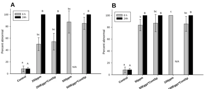
Fertilization success in the control group was 85.7% ± 3.9. When sperm and oocytes were simultaneously incubated with oil, fertilization success was significantly depressed by the highest dose of HEWAF (F6, 21 = 76.6, p<0.001; Fig. 4A), and by both doses of CEWAF (F6, 21
= 315.3, p<0.001; Fig. 4B). Exposure of sperm and oocytes to 500 ppm HEWAF adversely
impacted fertilization success (22.2% ± 9.2, F6, 21 = 76.6, p<0.001; Fig. 4A); whereas 250 ppm
HEWAF did not reduce fertilization success significantly (75.8% ± 3.6, F6, 21 = 76.6, p=0.185;
Fig. 4A). For CEWAF exposures to 50 ppm and 100 ppm, fertilization was significantly impacted with rates falling to 8.7% ± 3.4 and 0.1% ± 0.2 respectively (F6, 21 = 315.3, p<0.001;
14
The fertilization success of oocytes exposed to 500 ppm of HEWAF and fertilized with control sperm (i.e. incubated in FSW only) fell to 46% ± 4.6 (F6, 21 = 76.6, p<0.001; Fig. 4A) but was
not significantly lower in the 250 ppm HEWAF treatment (F6, 21 = 76.6, p=0.99). Oocytes
incubated with 50 and 100 ppm CEWAF and fertilized with non-exposed sperm had significantly reduced fertilization success of 17.2% ± 2.3 and 18.4% ± 4.9 respectively (F6, 21 =
315.3, p<0.001, Fig. 4B).
More specifically, compared to the control group, the fertilization success of oocytes and sperm exposed to 50 ppm of CEWAF was reduced 10 times (≈ 10% Control), whereas the fertilization success was reduced 5 times (≈ 20% Control) when only oocytes were exposed (Fig. 4B). Similarly when both sperm and oocytes were exposed to 100 ppm of CEWAF, fertilization success was drastically reduced, down to ≈ 0.1% of the control; whereas, when only oocytes were exposed to 100 ppm CEWAF, the fertilization success was reduced about 5 times (≈ 20%) compared to control (Fig. 4B). Regarding HEWAF exposure, no significant difference was found for exposing both oocytes and sperm or exposing only oocytes to 250 ppm HEWAF (F6, 21
= 76.6, p>0.05; Fig. 4A). However, significant differences were found for exposing both
oocytes and sperm or exposing only oocytes to 500 ppm HEWAF (F6, 21 = 76.6, p<0.001; Fig.
4A)
A
Cont rol 250ppm 250Eggs*C ontSp 500ppm 500Eggs*C ontSp Fer tiliz ation suc c es s (%) 0 20 40 60 80 100 a a a b cB
Control 50ppm 50Eg gs*C ontSp 100ppm 100Eggs*C ontSp Fer tiliz ation su c c es s (%) 0 20 40 60 80 100 a bc b c bFigure 4: Fertilization success of Crassostrea virginica gametes (oocytes or sperm pooled from 3 individuals)
exposed continuously to (A) HEWAF and (B) CEWAF, expressed as nominal concentrations (ppm or mg oil L
-1). 250 ppm and 500 ppm treatments correspond to exposure of both gametes to HEWAF; “250Eggs*ContSp”
and “500Eggs*ContSp” correspond to cross-fertilization of oocytes exposed to 250 ppm and 500 ppm of HEWAF respectively, with control sperm (i.e. incubated in FSW only). “50 ppm” and “100 ppm” treatments correspond to exposure of both gametes to CEWAF; “50Eggs*ContSp” and “100Eggs*ContSp” correspond to cross-fertilization of oocytes exposed to 50 ppm and 100 ppm of CEWAF respectively, with non-exposed, control sperm. Data are presented as mean percentages of control ± SD (n = 4 replicate). Different letters denote a significant difference at α=0.05 between conditions (ANOVA).
15
3.4.2 Effects on embryogenesis
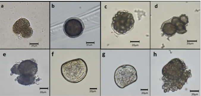
Embryos observed in the control treatment 6 h after fertilization exhibited a normal development (Fig. 5c). However, significantly higher instances of abnormally developed embryos with swollen vitelline membrane (Fig. 5b) or deformed cells (Fig. 5d), and underdeveloped or arrested embryos (Fig. 5e) were observed in the 250 ppm and 500 ppm HEWAF exposures (F6,
21 = 11.1, p=0.026 and p=0.008 respectively; Fig. 6A), and in exposures to 50 ppm and 100
ppm of CEWAF (F6, 21 = 20.2, p=0.014 and p<0.001 respectively; Fig. 6B). Embryos resulting
from both HEWAF- and CEWAF-exposed gametes (eggs and sperm) showed a dose-dependent increase in the number of abnormalities (F6, 21 = 11.1, p<0.001 and F6, 21 = 20.2, p<0.001
respectively; Fig. 6). Whereas, for oocytes fertilized with control, non-exposed sperm, only HEWAF exposure showed a dose-dependent increase in abnormalities (F6, 21 = 11.1, p=0.042;
Fig. 6A). Exposing only oocytes to CEWAF resulted in high abnormalities, but no dose response was observed (F6, 21 = 20.2, p=0.068; Fig. 6B). Moreover, no significant differences in
the percentage of abnormal embryos were observed between cross-fertilization of oil-exposed oocytes with control sperm and oil-exposed oocytes crossed with oil-exposed sperm (F6, 21 =
11.1; p>0.05; F6, 21 = 20.2, p>0.05) (Fig. 6A and B).
Figure 5: Normal Crassostrea virginica 1 h-old embryo (a); Abnormally fertilized embryo with swelling of
vitelline envelope after 1h (b); Normal 6-h embryo (c); 6-h embryos with abnormal cell shape (d) and an arrested development, at 3-cell stage (e); Normal 24 h-old larva (f), and abnormal larvae (g, h) after 24 h of exposure to
Deepwater Horizon oil.
After 24 h, 100% of embryos exposed to HEWAF or CEWAF were either abnormal (at the lowest doses tested) or dead (at the highest doses tested) regardless of whether oocytes and
16
sperm were both exposed, or only oocytes were exposed (Fig. 6A and B). Abnormalities after 24 h, consisting of D-larvae with indented shell margin (Fig. 5g), protruded mantle (Fig. 5h), convex hinge, or underdeveloped larvae at the embryo stage, were observed in the treatment exposed to 250 ppm HEWAF (F5, 18 = 6216.5, p<0.001; Fig. 6A) and in the treatment exposed
to 50 ppm CEWAF (F5, 18 = 2314.7, p<0.001; Fig. 6B). However, no statistical difference was
recorded after 24h of exposure to 250 ppm of HEWAF and 50 ppm of CEWAF and their respective cross-fertilization treatments (F5, 18 = 6216.5; p>0.05; F5, 18 = 2314.7, p>0.05) (Fig.
6A and B). Absence of live larvae (N/A) when oocytes and sperms were exposed for 24 h to 500 ppm HEWAF and 100 ppm CEWAF did not allow evaluation of abnormalities or comparison with oil-exposed oocytes conditions.
A Control 250p pm 250Eg gs*Con tSp 500p pm 500Eg gs*Con tSp Per c en t ab no rmal 0 20 40 60 80 100 6 h 24h a bc bc c bc A B B B N/A B B B
B
Control 50ppm 50Egg s*Co ntSp 100p pm 100Eg gs*Con tSp P er cen t abnor mal 0 20 40 60 80 100 6 h 24h a ab b bc c bc A B B B N/AFigure 6: Percentages of Crassostrea virginica abnormal 6 h-old embryos (grey) and 24 h-old D-larvae (black)
resulting from cross-fertilized exposed gametes (sperm or oocytes) to (A) HEWAF and (B) CEWAF, expressed as nominal concentrations (ppm or mg oil L-1). 250 ppm and 500 ppm treatments correspond to exposure of both gametes to HEWAF; “250Eggs*ContSp” and “500Eggs*ContSp” correspond to cross-fertilization of eggs exposed to 250 ppm and 500 ppm of HEWAF respectively, with control sperm (i.e. incubated in FSW only). “50 ppm” and “100 ppm” treatments correspond to exposure of both gametes to CEWAF; “50Eggs*ContSp” and “100Eggs*ContSp” correspond to cross-fertilization of eggs exposed to 50 ppm and 100 ppm of CEWAF respectively, with control, non-exposed sperm. Data are presented as mean percentages ± SD (n=4). Different letters denote a statistical difference at α=0.05 (ANOVA). N/A: no live larvae were observed, i.e. 100% mortality.
3.4.3 Effects on larval mortality
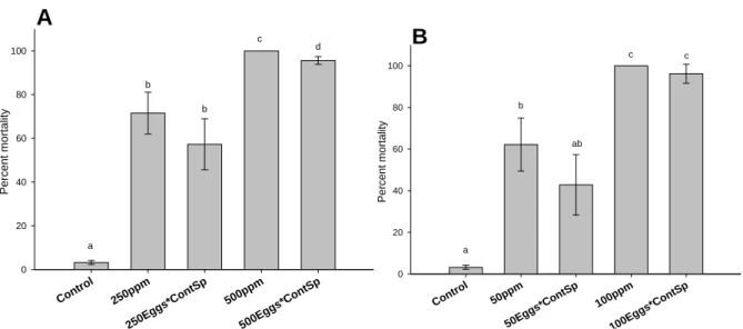
At the end of the exposure, survival was negatively impacted with significant dose-dependent increases of larval mortality in treatment groups exposed to both HEWAF (F6, 21 = 246.9,
p<0.001) and CEWAF (F6, 21 = 108.3, p<0.001) (Fig. 7). Final mortality of both HEWAF and
CEWAF exposures showed that no significant differences existed between larvae derived from cross-fertilization of oil-exposed oocytes with control sperm and fertilization of oil-exposed
17
oocytes with oil-exposed sperm (p>0.05; Fig. 7A and B), except at the highest tested dose of HEWAF (500 ppm) (F6, 21 = 246.9, p<0.05; Fig. 7A).
A
Control 250ppm 250E ggs* ContSp 500ppm 500E ggs* ContSp P ercent mo rtal it y 0 20 40 60 80 100 a b b c d B Contr ol 50ppm 50Egg s*Co ntSp 100p pm 100E ggs*Co ntSp P ercent m ort ali ty 0 20 40 60 80 100 a b c c abFigure 7: Final mortality of Crassostrea virginica D-larvae resulting from cross-fertilized exposed gametes (sperm
or oocytes) to (A) HEWAF and (B) CEWAF for 24 h, expressed as nominal concentrations (ppm or mg oil L-1). 250 ppm and 500 ppm treatments correspond to exposure of both gametes to HEWAF; “250Eggs*ContSp” and “500Eggs*ContSp” correspond to cross-fertilization of oocytes exposed to 250 ppm and 500 ppm of HEWAF respectively, with control sperm (i.e. incubated in FSW only) . “50 ppm” and “100 ppm” treatments correspond to exposure of both gametes to CEWAF; “50Eggs*ContSp” and “100Eggs*ContSp” correspond to cross-fertilization of eggs exposed to 50 ppm and 100 ppm of CEWAF respectively, with control sperm. Data are presented as mean percentages ± SD (n=4). Different letters denote a statistical difference at α=0.05 (ANOVA).
4. Discussion
The first objective of this study was to evaluate the effects of Deepwater Horizon (DWH) acute oil exposure, dispersed chemically (CEWAF) or mechanically (HEWAF), on cellular parameters of Crassostrea virginica spermatozoa and oocytes and to relate the effects to fertilization success and embryogenesis. Flow-cytometry (FCM) is a foremost technique to assess sperm quality and has already been incorporated in cryopreservation protocols for sperm assessment in oysters,47 and has shown a great potential in ecotoxicological studies using oyster sperm.31,41,42,56 Furthermore, recent works demonstrated that application of FCM was a reliable method to evaluate oocyte quality of environmental stressor-exposed oysters and non-exposed oysters.48,57 In our study, morphology, viability, mitochondrial membrane potential (MMP) and the production of reactive oxygen species (ROS) were selected as indicators of sperm and oocyte function and quality.
18
4.1 Sperm cellular characteristics
A reduction in the number of sperm cells was observed following HEWAF exposure (Fig. 1B vs 1D and Fig. 2A), although these spermatozoa were still viable (≥ 87%, Fig. 2B). Sperm cells in these HEWAF treatments may have aggregated with oil droplets, resulting in a reduction in the number of viable “oil- free” sperm cells available for fertilization. The reduced fertilization success observed at 500 ppm (22.2%, Fig. 4A) may be due to a decrease in the number of viable “oil-free” sperm cells available for fertilization. Based on the flow rate and volume of solution tested, the number of “oil-free” sperm cells was estimated and a spermatozoa to oocyte ratio was determined as < 40:1 in this treatment. It has been reported for C. gigas that a spermatozoa: oocyte ratio much lower than the optimum ranges (from 100:1 to 5000:1) limits the successful fertilization of eggs with sperms.58 Additionally, sperm exposed for 30-min to HEWAF induced significant modifications in relative size of spermatozoa. These morphological alterations of sperm cells could affect their motility and hence reduce their fertilization capabilities. The reduction of relative size may be caused by a toxic action of oil/PAHs on sperm cells, resulting in a “shriveling” of exposed spermatozoa. Although sperm deformities can cause a reduction of sperm motility and thus fertilization capacity59, a reduction of “oil-free” spermatozoa is most likely responsible for the reduction in fertilization success observed at 500 ppm of HEWAF.
In our study, HEWAF exposure had no significant effect on the MMP of spermatozoa. Adenosine triphosphate (ATP), produced by the mitochondria, is typically consumed in sperm flagella to provide energy for motility and is crucial for maintaining chemical gradients over membranes.60 The trend of increasing MMP, which was apparent after incubation with HEWAF, may nonetheless indicate an increase in energy demand due to the toxic effects of oil/PAHs. It is also possible that if oil droplets were associated with the sperm, spermatozoa may have expended more energy towards swimming. In regard to ROS, the abundance of droplet-associated oil within the HEWAF, which had a similar fluorescence as the DCFH-DA dye, may have interfered and impeded the determination of ROS production in sperm. Indeed, cytograms of HEWAF-exposed sperm cells showed overlap of these parameters (Fig. 1J-L).
The present study also showed that exposure to 100/10 ppm of CEWAF (oil/dispersant) significantly reduced the viability and ROS production in sperm cells which may have inhibited the fertilization success. These cellular impairments could be associated with the toxic effect of the dispersant contained in the CEWAF as demonstrated in our previous studies.12,14 A reduction in reactive oxygen species (ROS) production was also recorded following exposure to CEWAF and is in agreement with Volety et al. (2016) who found a dose-related decrease of ROS after
19
exposure of C. virginica sperm to CEWAF of DWH oil and to dispersant alone.31 During mitochondrial respiration, some of the consumed oxygen is reduced to water and, also to superoxide ion (O2
•-), which can then be converted to hydrogen peroxide (H2O2) or the highly
reactive OH•. The production of these ROS can cause toxic effects.61 On the other hand, a decrease in ROS may indicate a disruption of the cellular respiration pathway. The decline in intracellular ROS production observed after incubation of sperm with CEWAF, corresponded with an increase in sperm mortality. This result could suggest that dispersant contained in CEWAF might have induced a disruption of cellular respiration, by modification of the respiration pathway, and caused sperm death. Several studies showed evidence of acute toxicity of surfactant (one of the main compounds in Corexit 9500A) to fish, mollusks, or crustaceans by disruption of respiratory cells, which may lead to hypoxia and asphyxia.62-65 Indeed, as described previously, MMP tended to increase upon exposure to CEWAF possibly to compensate for disruption of cellular respiration and oxidative phosphorylation decoupling. Although not statistically significant, MMP increased upon exposure to CEWAF. An increase of MMP was negatively correlated to ATP content and percentage of motile spermatozoa during the motility phase of C. gigas sperm.66
4.2 Oocyte cellular characteristics
Overall, a 30-min exposure of oocytes to either HEWAF or CEWAF did not induce any significant morphological changes or significant effect on oocyte viability. A trend of increasing complexity was however noted after exposure to HEWAF, but not to CEWAF. This leads us to believe there may have been aggregation of oil droplets (abundant in HEWAF but not in CEWAF) on the surface of the oocytes, causing this morphological modification. The structure of the oocyte and the presence of a protective membrane, the vitelline envelope, may explain the lack of effects of oil/PAHs on the viability of exposed eggs.67 Also, the duration of the exposure (i.e. 30 min) was not long enough to induce significant effects on oocytes viability. However, we frequently observed abnormal embryos exhibiting swellings of the vitelline envelope in the CEWAF treatments (Fig. 5b). This modification in the oocyte membrane has previously been suggested to be linked to the surfactant contained in the Corexit 9500A and its potential effect on membranes.64,65 PAHs may also be responsible for the loss of membrane structure: a non-specific mode of PAH toxicity which can cause physical disturbance of bio-membrane structure has been proposed.68
Although there was no significant dose-response relationship of reactive oxygen species (ROS) production in oocytes among the exposed groups, the production of these potentially toxic ROS
20
more than doubled after 30 min of incubation with CEWAF, suggesting that oil and/or dispersant caused an oxidative stress. Oxidative stress (such as excessive ROS production) has been shown to cause toxic effects on human oocytes and has been linked to reduced fertilization success.69 Numerous authors have shown that stimulation of ROS production in bivalve tissues, including oocytes, can be induced by many environmental stresses, such as pollutants, biotoxins, pathogens, heat shock, UV exposure, or hypoxia.48,57,70-74 Several studies described oxidative stress and membrane impairments in bivalve tissues following exposure to petroleum hydrocarbons.75,76 Microscopic observations of membrane damage and cytolysis in cells of larva exposed for 24 h to oil (Fig. 5h) could be the result of an oxidative damage caused by PAHs on oocytes. It can be hypothesized that excessive production of ROS in oocytes observed herein may ultimately have severe implications for the subsequent fertilization and embryo development as it can lead to DNA damage.77 Using the Comet assay, PAHs were shown to induce high rates of abnormal embryos/larvae of C. gigas due to DNA strand breakage.17 This method has been commonly used to assess DNA damage in cells, including bivalve sperm or oyster embryos.30,31,37, 78-81 It would be interesting to test this method with oocyte following oil and/or dispersant exposure as a complementary approach to FCM analysis.
4.3 Relative contribution of exposed gametes to successful fertilization and early development
The second objective of the present study was to determine the respective contributions of exposed spermatozoa and oocytes to successful fertilization, embryogenesis and larval survival. Results from the cross-fertilization study showed that responses were highly dependent on the exposure duration and the doses tested. Fertilization success (measured at 1 h PF) was significantly reduced when both sperm and oocytes were exposed to the highest concentration of HEWAF and both concentrations of CEWAF. Although somewhat ameliorated by crossing exposed oocytes with control (non-exposed) sperm, fertilization success was still significantly reduced. This result suggests that alterations in sperm parameters, such as viability, dying cells, MMP and ROS paralleled the observed reduction in fertilization, particularly in those exposed to the highest dose of HEWAF and CEWAF.
While fertilization success was improved somewhat when oil-exposed oocytes were cross-fertilized with non-exposed sperm, this was not evident in embryogenesis or larval survival. Regardless of whether exposed or non-exposed sperm were used, abnormalities in 6 h and 24 h-embryos and larval survival were similar, suggesting that the effect on h-embryos was primarily a consequence of oocyte exposure. This result also suggests that oocytes may be sensitive to DWH oil/dispersant toxicity, and exposure of oocytes may be a better predictor of embryo/larval
21
development and survival than sperm exposure. It is important to note, however, that abnormalities and larval survival data were determined based on low numbers of fertilized embryos, especially in the CEWAF exposure. It has been well documented that not only surfactant, one of the main components of Corexit 9500A,9 but also PAHs can adversely affect membrane structures and induce increased permeability of the membrane, a loss of barrier function or an osmotic imbalance.63,65,82,83 As previously mentioned, microscopic observations of embryos and larvae after 6 or 24 h of exposure to CEWAF indicated severe damages to the vitelline envelope, possibly imputable to either Corexit 9500A or PAHs. Furthermore, the increasing ROS measured in exposed oocytes, though not significant, is a typical sign of reduced fertility and could explain developmental defects and high mortalities found in exposed embryos and larvae.69
We could speculate that due to the protective vitelline envelope in oocytes and the higher surface/volume ratio of spermatozoa compared to oocytes, sperm cells may have been more vulnerable in the short term (i.e. 30 min) to oil/dispersant toxic effects than oocytes.67,84,85 However, as embryos and larvae developed and utilized reserves laid down in the egg, it becomes apparent that in the long term (i.e. 6 h onward), oocytes were more affected by oil/dispersant. Consequently, detrimental effects of DWH oil/dispersant on early development of oyster larvae may be largely attributed to impairment of oocyte functions.
5. Conclusion
The present study demonstrated that chemically or mechanically dispersed DWH oils were cytotoxic to oyster gametes and could impact sensitive processes of the early life stage of C.
virginica. Our results showed that oil-derived PAHs as well as physical properties of oil (i.e.
droplets aggregation) and chemical characteristics of the dispersant Corexit 9500A (i.e. surfactant) could impair spermatozoa and oocyte cellular functions, resulting in inhibited fertilization and depressed performance of embryo and larvae. Results from the cross-fertilization study showed that responses were highly dependent on the exposure duration. For instance, sperm exposure to oil/dispersant impaired normal spermatozoa functions, affecting fertilization success; whereas, exposure of oocytes seemed to be a better predictor of embryo/larval development and survival.
To the best of our knowledge, this is the first time FCM was used to analyze cellular responses of oyster oocytes following exposure to petroleum hydrocarbons and dispersant. Although
22
cellular parameters could explain some of the observed impairments on fertilization and subsequent development, further optimization of the methodology and additional assays should be developed to find one that will best parallel fertilization responses to toxicants. The use of FCM assay appeared to be, nonetheless, a good predictor of sublethal effects on sperm, especially on fertilization success. It is clear that the use of FCM will not replace standard aquatic testing procedures using representative endpoints and life stages of an ecologically relevant species such as oyster. Nevertheless, FCM assays are relatively easy and quick to implement, less time-consuming than microscopic observations, and a good predictor of biological effects. For these reasons, FCM should be used as a complementary approach to bioassays in the ecotoxicological assessment of oil/dispersant.
Overall, oyster gametes experienced more deleterious effects from acute exposure to CEWAF than HEWAF. This suggests that the use of Corexit to disperse oil at the time of the spawning season of oysters could be more detrimental to oyster population (by affecting reproductive processes) than the oil on its own. However, our study specifically addressed the case of oyster, and these results may not apply to other aquatic species. As a result, in order to provide effective responses to oil spills, additional research using other filter-feeding species is required to broaden the impact of our study and to establish the trade-off on the use of chemical dispersion (i.e. CEWAF) or natural dispersion (i.e. HEWAF).
Acknowledgements
This work was supported in part by funds provided as part of the natural resources damage assessment (NRDA) for the Deepwater Horizon oil spill. Data presented here is a subset of a larger toxicological database that is being generated as part of the Deepwater Horizon NRDA. We are thankful to Kelsey McEachern and Emily Nickols at the Vester Marine Field Station/FGCU for their technical assistance. We also thank Jeffrey Morris, Michael Carney, Claire Lay and Michelle Krasnec at Abt Associates for their input on data analyses and early reviews of this manuscript. This collaborative work was also partially supported by Université de Bretagne Occidentale and the “Laboratoire d’Excellence” LabexMER (ANR-10-LABX-19), an Outgoing Ph.D. Fellowship, co-funded by a grant from the French government under the program “Investissements d’Avenir”.
23
References
(1) National commission on the BP deep ocean horizon oil spill and offshore drilling. Deep water: the Gulf oil disaster and the future of offshore drilling. 2011. Report to the president. www.oilspillcommission.gov. Accessed June 2014
(2) U.S. District Court. In re: oil spill by the oil rig “Deepwater Horizon” in the Gulf of Mexico, on April 20, 2010, No. MDL 2179, Section 7 (Revised September 9, 2014) (“Findings of fact and conclusions of law: phase one trial”), Figure 1. 2014. United States District Court for the Eastern District of Louisiana.
(3) U.S. District Court. In re: oil spill by the oil rig “Deepwater Horizon” in the Gulf of Mexico, on April 20, 2010, No. MDL 2179, 2015 WL 225421 (La. E.D. Jan. 15, 2015) (“Findings of fact and conclusions of law: phase two trial”). 2015. United States District Court for the eastern district of Louisiana.
(4) Camilli, R., Reddy, C. M., Yoerger, D. R., Van Mooy, B. A., Jakuba, M. V., Kinsey, J. C., McIntyre, C. P., Sylva, S. P. & Maloney, J. V. Tracking hydrocarbon plume transport and biodegradation at Deepwater Horizon. Science, 2010, 330(6001), 201-204.
(5) OSAT-1. Summary report for sub-sea and sub-surface oil and dispersant detection: sampling and monitoring. Prepared for Paul F. Zunkunft, Federal On-Scene Coordinator, Deepwater Horizon MC252. 2010 December 17.
(6) Kujawinski, E. B., Kido Soule, M. C., Valentine, D. L., Boysen, A. K., Longnecker, K., & Redmond, M. C. Fate of dispersants associated with the Deepwater Horizon oil spill. Environmental Science &
Technology, 2011, 45(4), 1298-1306.
(7) U.S. Coast Guard. On Scene Coordinator Report: Deepwater Horizon oil spill. Submitted to the National Response Team. September. U.S. Department of Homeland Security, U.S. Coast Guard, Washington, DC. 2011. Available: http://www.uscg.mil/foia/docs/dwh/fosc_dwh_report.pdf. Accessed July 7, 2015.
(8) Environmental Response Management Application 2015. Deepwater gulf response. National Oceanic and Atmospheric Administration. Available: http://response.restoration.noaa.gov/maps-and-spatial-data/environmental-response-management-application-erma/erma-gulf-response.html , (accessed 7/30/2015) (9) Nalco Energy Services. Material Safety Data Sheet for Corexit 9500A®. 2012. Available at http://www.nalcoesllc.com/nes/documents/MSDS/NES-LLC-COREXIT-EC9500A-March_2012.pdf (last consulted on August 10th 2014)
(10) Langdon, C. J., Stefansson, E. S., Pargee, S. M., Blunt, S. M., Gage, S. J., & Stubblefield, W. A. Chronic effects of non‐weathered and weathered crude oil and dispersant associated with the deepwater horizon incident on development of larvae of the eastern oyster, Crassostrea virginica. Environmental Toxicology and Chemistry,
2016, 35(8): 2029-2040.
(11) Stefansson, E. S., Langdon, C. J., Pargee, S. M., Blunt, S. M., Gage, S. J., & Stubblefield, W. A. Acute effects of non‐weathered and weathered crude oil and dispersant associated with the deepwater horizon incident on the development of marine bivalve and echinoderm larvae. Environmental Toxicology and Chemistry, 2016, 35(8): 2016-2028.
(12) Vignier, J., Donaghy, L., Soudant, P., Chu, F. L. E., Morris, J. M., Carney, M. W., Lay, C., Krasnec, M., Robert, R., & Volety, A. K. Impacts of Deepwater Horizon oil and associated dispersant on early development of the Eastern oyster Crassostrea virginica. Marine pollution bulletin, 2015, 100(1), 426-437. http://dx.doi.org/10.1016/j.marpolbul.2015.08.011
(13) Vignier, J., Soudant, P., Chu, F. L. E., Morris, J. M., Carney, M. W., Lay, C. R., Krasnec, M. O., Robert, R. & Volety, A. K. Lethal and sub-lethal effects of Deepwater Horizon slick oil and dispersant on oyster (Crassostrea virginica) larvae. Marine Environmental Research, 2016, 120: 20-31. http://dx.doi.org/10.1016/j.marenvres.2016.07.006
(14) Volety, A.K., Boulais, M., Donaghy, L., Vignier, J., & Soudant, P. Application of flow-cytometry to assess
Deepwater Horizon oil toxicity on the Eastern oyster Crassostrea virginica spermatozoa. Journal of Shellfish Research, 2016, 35(1), 1-9.
24
(15) Canevari, G.P.. Development of the next generation chemical dispersants. Proceedings of Joint Conference
on Prevention and Control of Oil Spills. 1973
(16) NRC National Research Council. Oil spill dispersants: efficacy and effects. The National Academy Press, Washington, DC. 2005. http://dx.doi.org/10.17226/11283
(17) Venosa, A.D., Zhu, X. Biodegradation of crude oil contaminating marine shorelines and freshwater wetlands. Spill Sci. Technol. Bull., 2003, 8 (2), 163–178.
(18) Almeda, R., Wambaugh, Z., Wang, Z., Hyatt, C., Liu, Z., Buskey, E.J. Interactions between zooplankton and crude oil: toxic effects and bioaccumulation of polycyclic aromatic hydrocarbons. PLoS One, 2013, 8 (6), e67212.
(19) Anderson, J.W., Neff, J.M., Cox, B.A., Tatem, H.E., Hightower, G.M. Characteristics of dispersions and water-soluble extracts of crude and refined oils and their toxicity to estuarine crustaceans and fish. Mar. Biol.,
1974, 27 (1), 75–88.
(20) NRC, National Research Council. Using Oil Spill Dispersants on the Sea. National Academy Press, Washington, DC (Chapter 3), 1989.
(21) Ramachandran, S. D., Hodson, P. V., Khan, C. W., & Lee, K. Oil dispersant increases PAH uptake by fish exposed to crude oil. Ecotoxicology and environmental safety, 2004, 59(3), 300-308.
(22) Coen, L. D., Luckenbach, M. W., & Breitburg, D. L. (1999, April). The role of oyster reefs as essential fish habitat: a review of current knowledge and some new perspectives. In American Fisheries Society
Symposium 1999 (Vol. 22, pp. 438-454).
(23) Wells, H.W. The fauna of oyster beds, with special reference to the salinity factor. Ecological Monographs.
1961, 31(3): 239-266.
(24) Volety, A. K., Haynes, L., Goodman, P., & Gorman, P. Ecological condition and value of oyster reefs of the Southwest Florida shelf ecosystem. Ecological Indicators, 2014, 44, 108-119. http://dx.doi.org/10.1016/j.ecolind.2014.03.012 .
(25) Ingle, R.M. (1951). “Spawning and setting of oysters in relation to seasonal environmental changes.”
Bull. Mar. Sci. Gulf Carib. 1951, 1: 111-135
(26) Renzoni, A. Toxicity of three oils to bivalve gametes and larvae. Marine Pollution Bulletin 1975, 6(8): 125. (27) Geffard, O., Budzinski, H., Augagneur, S., Seaman, M. N., & His, E. “Assessment of sediment contamination by spermiotoxicity and embryotoxicity bioassays with sea urchins (Paracentrotus lividus) and oysters (Crassostrea gigas).” Environmental toxicology and chemistry, 2001, 20(7), 1605-1611.
(28) Lyons, B. P., Pascoe, C. K., & McFadzen, I. R. B. Phototoxicity of pyrene and benzo [a] pyrene to embryo-larval stages of the pacific oyster Crassostrea gigas. Marine environmental research, 2002, 54(3): 627-631.
(29) Jeong, W.-G. and Cho, S. M. The effects of polycyclic aromatic hydrocarbon exposure on the fertilization and larval development of the Pacific oyster, Crassostrea gigas. Journal of Shellfish Research, 2005, 24(1): 209. (30) Wessel, N., Rousseau, S., Caisey, X., Quiniou, F., & Akcha, F. Investigating the relationship between embryotoxic and genotoxic effects of benzo[a]pyrene, 17α-ethinylestradiol and endosulfan on Crassostrea gigas embryos. Aquatic Toxicology, 2007, 85(2), 133-142.
(31) Akcha, F., Spagnol, C., & Rouxel, J. Genotoxicity of diuron and glyphosate in oyster spermatozoa and embryos. Aquatic Toxicology, 2012, 106, 104-113.
(32) Laramore, S., Krebs, W., & Garr, A. Effects of Macondo Canyon 252 oil (naturally and chemically dispersed) on larval Crassostrea virginica (Gmelin, 1791). Journal of Shellfish Research, 2014, 33(3), 709-718.
25
(33) Pagano, G., Esposito, A., & Giordano, G. G. Fertilization and larval development in sea urchins following exposure of gametes and embryos to cadmium. Archives of environmental contamination and toxicology, 1982,
11(1), 47-55.
(34) Dinnel, P. A., Link, J. M., Stober, Q. J., Letourneau, M. W., & Roberts, W. E. Comparative sensitivity of sea urchin sperm bioassays to metals and pesticides. Archives of environmental contamination and
toxicology, 1989, 18(5), 748-755.
(35) Ringwood, A. H. Comparative sensitivity of gametes and early developmental stages of a sea urchin species (Echinometra mathaei) and a bivalve species (Isognomon californicum) during metal exposures. Archives of environmental contamination and toxicology, 1992, 22(3), 288-295.
(36) Bellas, J., Beiras, R., & Vázquez, E. Sublethal effects of trace metals (Cd, Cr, Cu, Hg) on embryogenesis and larval settlement of the ascidian Ciona intestinalis. Archives of environmental contamination and
toxicology, 2004, 46(1), 61-66.
(37) Lewis, C., and Ford, A. T. Infertility in male aquatic invertebrates: a review. Aquatic Toxicology, 2012,
120, 79-89.
(38) Cordelli, E., Eleuteri, P., Leter, G., Rescia, M., & Spanò, M. Flow cytometry applications in the evaluation of sperm quality: semen analysis, sperm function and DNA integrity. Contraception, 2005, 72(4), 273-279. (39) Gillan, L., Evans, G., & Maxwell, W. M. C. Flow cytometric evaluation of sperm parameters in relation to fertility potential. Theriogenology, 2005, 63(2), 445-457.
(40) Martínez‐Pastor, F., Mata‐Campuzano, M., Álvarez‐Rodríguez, M., Alvarez, M., Anel, L., & De Paz, P. Probes and techniques for sperm evaluation by flow cytometry. Reproduction in Domestic Animals, 2010,
45(s2), 67-78.
(41) Le Goïc, N., Hégaret, H., Fabioux, C., Miner, P., Suquet, M., Lambert, C., & Soudant, P. Impact of the toxic dinoflagellate Alexandrium catenella on Pacific oyster reproductive output: application of flow cytometry assays on spermatozoa. Aquatic Living Resources, 2013a, 26 (3), 221-228.
(42) LeGoïc, N., Hégaret, H., Lambert, L., Fabioux, C., Soudant, P. Cellular changes of Crassostrea gigas spermatozoa upon exposure to the toxic dinoflagellate Alexandrium catenella. Aquaculture 2013, Feb 21-25th 2013, Nashville, TN, U.S.A. 2013b Meeting abstract nr: 832, poster nr 209.
(43) Nice, H. E. Sperm motility in the Pacific oyster (Crassostrea gigas) is affected by nonylphenol. Marine
pollution bulletin, 2005, 50(12), 1668-1674.
(44) Fitzpatrick, J. L., Nadella, S., Bucking, C., Balshine, S., & Wood, C. M. The relative sensitivity of sperm, eggs and embryos to copper in the blue mussel (Mytilus trossulus). Comparative Biochemistry and Physiology
Part C: Toxicology & Pharmacology, 2008, 147(4), 441-449.
(45) Basti, L., Nagai, K., Tanaka, Y., & Segawa, S. Sensitivity of gametes, fertilization, and embryo development of the Japanese pearl oyster, Pinctada fucata martensii, to the harmful dinoflagellate, Heterocapsa
circularisquama. Marine biology, 2013, 160(1), 211-219.
(46) Valdez-Ramirez, M. E., Le Pennec, M., Dorange, G., Devauchelle, N., & Nonnotte, G. Assessment of female gamete quality in the Pacific oyster Crassostrea gigas. Invertebrate reproduction & development, 1999,
36(1-3), 73-78.
(47) Paniagua-Chávez, C. G., Jenkins, J., Segovia, M., & Tiersch, T. R. Assessment of gamete quality for the eastern oyster (Crassostrea virginica) by use of fluorescent dyes. Cryobiology, 2006, 53(1), 128-138.
(48) Le Goïc, N., Hégaret, H., Boulais, M., Béguel, J. P., Lambert, C., Fabioux, C., & Soudant, P. (2014). Flow cytometric assessment of morphology, viability, and production of reactive oxygen species of Crassostrea gigas oocytes. Application to Toxic dinoflagellate (Alexandrium minutum) exposure. Cytometry Part A. 2014, 85(12), 1049-1056.