HAL Id: hal-01100238
https://hal.archives-ouvertes.fr/hal-01100238
Submitted on 6 Jan 2015
HAL is a multi-disciplinary open access
archive for the deposit and dissemination of
sci-entific research documents, whether they are
pub-lished or not. The documents may come from
teaching and research institutions in France or
abroad, or from public or private research centers.
L’archive ouverte pluridisciplinaire HAL, est
destinée au dépôt et à la diffusion de documents
scientifiques de niveau recherche, publiés ou non,
émanant des établissements d’enseignement et de
recherche français ou étrangers, des laboratoires
publics ou privés.
application to Co-based systems
P. Jonnard, Z.-S. Wang, J.-T. Zhu, C. Meny, J.-M. André, M.-H. Hu, K. Le
Guen
To cite this version:
P. Jonnard, Z.-S. Wang, J.-T. Zhu, C. Meny, J.-M. André, et al.. Characterization of multilayers and
their interlayers: application to Co-based systems. Chinese Optics Letters, Optical Society of America
(imprimé) / OSA publishing (en ligne), 2013, 11 (S1), pp.S10601. �10.3788/COL201311.S10601�.
�hal-01100238�
Characterization of multilayers and their interlayers:
applicationto Co-based systems
P. Jonnard1∗, Z.-S. Wang2, J.-T. Zhu2, C. Meny3, J.-M. Andr´e1, M.-H. Hu1, and K. Le Guen1
1
Laboratoire de Chimie Physique-Mati`ere Rayonnement, UPMC Univ Paris 06, CNRS UMR 7614, 11 rue Pierre et Marie Curie, F-75231 Paris cedex 05, France
2
MOE Key Laboratory of Advanced Micro-Structured Materials, Institute of Precision Optical Engineering, Department of Physics, Tongji University, Shanghai 200092, China
3
Institut de Physique et Chimie des Mat´eriaux de Strasbourg,
CNRS UMR 7504, 23 rue du Loess, BP 43, F-67034 Strasbourg cedex 2, France
∗Corresponding author: [email protected]
Received December 2, 2012; accepted January 5, 2013; posted online May 29, 2013
We use complementary analysis techniques to determine the structure of nanometric periodic multilayers and particularly their interfaces. We focus on Co-based multilayer which can be used as efficient optical component in the extreme ultraviolet (EUV) range. The samples are characterized using reflectivity measurements in order to determine the thickness and roughness of the various layers, X-ray emission and nuclear magnetic resonance (NMR) spectroscopies to identify the chemical state of the atoms present within the stack and know if they interdiffuse. Results are validated through the use of destructive techniques such as transmission electron microscopy or secondary ion mass spectrometry.
OCIS codes: 230.4170, 240.1485, 300.6560, 340.7480. doi: 10.3788/COL201311.S10601.
Nanometric periodic multilayers are nowadays widely used for optical applications in the extreme ultraviolet (EUV, 10–100 eV) as well as the soft (100 eV – 5 keV) and hard X-ray (5–50 keV) ranges. They can be used as optical components for spatial telescopes, synchrotron beamlines, diffraction apparatus or of new EUV and X-ray sources. They can also be used to perform spec-troscopy in the soft-ray range as the thickness of their layers is of the order of the X-ray wavelengths. However, to efficiently reflect or diffract the radiation, the quality of the multilayer should be high, that is to say: the lay-ers should have an uniform thickness, which is now well mastered with the modern deposition techniques; the interfaces should be as sharp as possible, with minimal interdiffusion or geometrical roughness, which depends on the chosen materials and also on the deposition con-ditions.
To these basic requirements, time and thermal stabil-ities are also often required as the multilayers can be subject to high thermal loads for long times.
It is thus important to characterize the interfaces in order to understand the phenomena leading to a degra-dation of the optical performances and then anticipate improved deposition processes and new combinations of materials. If X-ray reflectivity (XRR) and EUV reflec-tivity can be applied to determine the thickness and roughness of the various layers of a stack, they gener-ally consider the interface as a transition layer, where both interdiffusion and geometrical roughness are en-compassed in the same phenomena. To go further, it is necessary to gain some information about the chemical state of the elements present within the stack. This in-formation can be obtained in a non-destructive way from X-ray emission spectroscopy (XES) or nuclear magnetic resonance (NMR) spectroscopy. If destructive analysis is allowed, transmission electron microscopy (TEM) can
be performed on cross sections. The high spatial reso-lution allows determining the quality of the layers and their interfaces and also the chemical state of the various involved elements if electron energy loss spectroscopy (EELS) is available. These results can also be com-plemented by secondary ion mass spectrometry (SIMS) which can perform depth profiling with a nanometer res-olution.
In this communication we present the application of these techniques to the characterization of Co-based mul-tilayers and their interfaces. These are mulmul-tilayers where one of the layers is made of Co. Depending on the mate-rial with which the Co layers are associated, multilayers can be designed to work from the EUV to the soft X-ray range. Because of the magnetic character of the Co atoms, magneto-optical applications can also be envis-aged. Finally, we also consider the thermal stability of these stacks.
The simulations are obtained from computations that model the optical properties (reflectance, transmittance, absorbance, etc.) of multilayer films through solving the Maxwell equations for periodic media. This is an im-portant step as the structure of the multilayer, i.e., the thickness of the various layers, the number of periods, the nature of the substrate, the nature and thickness of an eventual capping layer etc., can be defined. Indeed, these parameters can be optimized in order to obtain the highest reflectance, the narrowest bandwidth etc., at a given application wavelength.
The studied periodic multilayers are prepared in a cali-brated high vacuum direct current magnetron sputtering system with high purity targets of Mg, Co, and Zr in Ar gas. The base pressure is 5 × 10⁻⁵ Pa and the working
pressure is 1 × 10⁻¹ Pa of Ar gas. The power applied
on the Co, Mg, and Zr targets are 20, 15, and 20 W, re-spectively. Multilayers are deposited onto ultra-smooth
polished Si substrates with 0.3 root mean square (RMS) surface roughness. Each sample is made of at least 30 periods. A 3.50-nm-thick B4C capping layer is deposited
at the surface of the samples to prevent oxidation. The reflectivity measurements are performed in the hard X-ray range at grazing incidence with a laboratory apparatus. The Cu Kα radiation at 0.154-nm wave-length is used. From the simulation of the experimental curve, the thickness, roughness, and density of the var-ious layers within a stack can be determined. At the application wavelength of the multilayer, i.e., in the soft X-ray or EUV range, measurements are performed with synchrotron radiation to take advantage of the high flux and tunability of the source. In the soft X-ray and EUV ranges, the optical indices are more sensitive to the chem-ical state of the elements than in the hard X-ray range. This allows distinguishing between roughness and inter-diffusion which are generally encompassed in a single transition layer when fitting hard XRR curves.
If an element is interacting with another element, as when interdiffusion or chemical reaction occurs at the interfaces of some multilayers, then the energy distribu-tion of the valence electron is modified. This happens because these electrons are the less tightly bound and they participate to the chemical bond. XES allows de-termining the density of occupied valence states of an element through the observation of an emission band, i.e., an electron transition between the valence band and given core level. For example, in the Mg/SiC system the examination of the Mg 3p states showed the formation of the Mg2Si interfacial compound upon annealing[1],
while if no interaction would have taken place between the Mg and SiC layers, the spectrum of Mg metal should have been observed. In the case of Mo/Si, more than one interfacial compound have been identified and the thickness of the interlayer has been estimated.
The chemical state of the Co atoms within the multi-layers can be probed using zero-field NMR spectroscopy. In this case the static field is the one induced by the Co atoms themselves. The experiment is performed at low temperature (4.2 K) using a scanning broadband spec-trometer. The NMR spectra represent the distribution of Co atoms as a function of their resonance frequency[2].
The NMR frequency strongly depends on the nature, number and arrangement of the atoms in the neighbor-hood of the Co atoms.
TEM technique is destructive because it is necessary to prepare thin slabs, transparent to the electron beam, in order to observe a cross section of the multilayers by transmission. The energy of the incident electrons is generally 100 or 200 keV and the thickness of the cross section a few tens of nm. From the images, the quality of the stack can be evaluated and in the high-resolution mode, the presence of crystallites, the existence of interfa-cial zones as well as their asymmetry can be emphasized. An electron spectrometer can be associated to the microscope. From the characteristic energy loss of the electrons, it is possible to identify the elements present in the various layers and to know the chemical state of some elements through their unoccupied density of states.
In time-of-flight SIMS a surface is bombarded with in-cident ions. Following the interaction of the sample with the incident ions, secondary ions are produced and then
detected to identify the elements present at the surface of the sample. The analysis is performed by selecting the mass-to-charge ratio of the detected ions through their time of flight. If the technique is associated to a sputtering ion gun, then an elemental depth profile can be obtained.
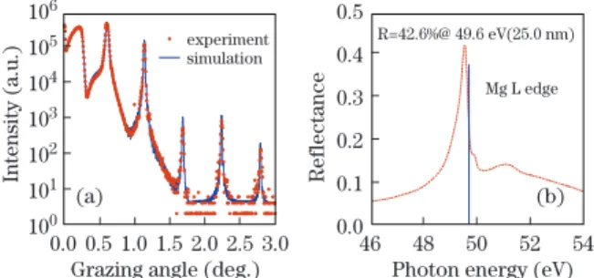
Simulations have shown that Co/Mg multilayers can lead to a high reflectance (50%) for an application wave-length close to the Mg L absorption edge, around 25 nm or 50 eV. Then, for a perfect multilayer, without rough-ness or interdiffusion, having a period of 17 nm, a re-flectance of 55% is expected for an s-polarized radiation under a 45◦ glancing angle. In this case the
thick-ness of the Co and Mg layers are 2.55 and 14.45 nm respectively[3].
We show in Fig. 1 the XRR characterization and EUV reflectivity measurements[3]. The good quality of
the stack is evidenced through the observationof a large number of Bragg peaks on the XRR curve and the 0.426 experimental reflectance, close to the one for the perfect multilayer, on the EUV curve. The fit of the reflectiv-ity curve indicates Co and Mg thicknesses very close to the aimed one in the design and only 0.5 nm interfacial roughness.
The Mg XES and Co NMR spectra of the multilayer are shown in Fig. 2[3]. They are compared to the bulk
Mg and a simulation of a Co thin film respectively. Clearly, the spectra of the multilayer are very close to the corresponding pure materials, showing that there is no significant interaction between Co and Mg layers. Thus, spectroscopies reveal no interfacial compound and confirm the existence of sharp interfaces.
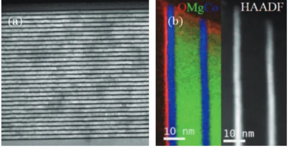
A scanning TEM image of the multilayer is presented in Fig. 3[4]. The 30 periods are clearly seen and the
Fig. 1. (Color online) (a) XRR at 0.154 nm and (b) EUV for s-polarized radiation at 45◦reflectivity curves of the Co/Mg
multilayer.
Fig. 2. (Color online) (a) XES and (b) NMR spectra of the Co/Mg multilayer. They are compared to the bulk Mg and simulation of a Co film respectively.
Fig. 3. (a) TEM image in bright field mode; (b) composite EELS Mg, Co and O maps compared to dark field image of the two first periods of the Co/Mg multilayer.
layers are smooth. The diffuse nature of the interfaces is mainly due to loss of resolution following multiple scat-tering in the thicker parts of the sample. The EELS map presented in Fig. 3 and obtained from the combination of the Co, Mg, and O elemental maps, demonstrates that the layers are chemically well ordered. The presence of oxygen is due to the surface oxidation of the cross section following its preparation. The only departure between the perfect structure expected from the simulation is the existence of a 0.5 nm interfacial roughness.
The good quality of the multilayer and its interfaces is in agreement with SIMS depth profiles. The Co− ions
show a highly contrasted and symmetrical profile on Fig. 4, characteristic of sharp interfaces[5].
It has been experimentally and theoretically estab-lished that introducing a thin film of a third material in the periodic of a multilayer can improve the reflectance. This can be due to two effects: firstly, the optimiza-tion of the optical path within the stack; secondly, the diffusion barrier or smoothing effect of the introduced material. Preliminary experiments have shown that B4C
is not a good candidate because of a strong intermixing with the Co layers, while it has been shown efficient as a diffusion barrier for the Mo/Si or Sc/Si systems for example.
Thus we considered the introduction of a 1.5-nm-thick layer of 4d and 5d transition metal in the Co/Mg stack[6].
Depending on the interface where the thin film is in-serted, the reflectance can vary by more than 10%, as can be seen from the simulations of the corresponding perfect tri- and quadrilayers in Fig. 5. The highest re-flectance has been obtained for the Co/Mg/Zr system. Then, such a sample has been produced, as well as the Co/Zr/Mg system.
Fig. 4. SIMS depth profile of 12 periods of the Co/Mg mul-tilayer. The curve shows the profile of the Co−ions.
Fig. 5. (Color online) Simulated reflectances for various per-fect Co/Mg stacks where a 1.5-nm-thick film of 4d or 5d tran-sition metal (noted X) has been inserted either at the Co-on-Mg or Co-on-Mg-on-Co interfaces or at both interfaces.
We have confirmed the improvement of the reflectance upon insertion of a thin Zr layer in the Co/Mg structure by measuring the EUV reflectivity[7]. For the Co/Mg/Zr
structure, the value is 50.0% at 25.1 nm for an s-polarized radiation incident at 45◦. With respect to the simple
Co/Mg system, the experimental reflectance increases by about 7% while in the simulations it increases only by about 3%. This means that in addition to its optical effect, the Zr layer also produces a material effect, which improves the quality of the stack.
The comparison of the two trilayers, Co/Mg/Zr and Co/Zr/Mg, is interesting[5]. The SIMS profile of the
Co− ions, Fig. 6, clearly shows that the interfaces
of Co/Zr/Mg structure present some interdiffusion, whereas the ones of Co/Mg/Zr are much more symmet-rical. This is confirmed by NMR spectroscopy, Fig. 6, showing the disappearance of the pure Co signal for Co/Zr/Mg, whereas for Co/Mg/Zr the presence of Co hcp is attested. In this last case, the observed shoulders toward lower frequencies are only due to the interfacial Co atoms in contact with the Zr and Mg layers. For both trilayers, XES spectra show that Mg atoms stay in the same state as pure metal, that it is to say do not react with the Co or Zr layers. Thus, the SIMS and NMR fea-tures observed with Co/Zr/Mg are due to an intermixing between the Co and Zr layers. Reflectivity measure-ments and their fit confirm this point: it is possible to adjust the reflectivity curve of Co/Zr/Mg with a model of stack represented by a CoxZry/Mg bilayer, while for
Co/Mg/Zr the three layer model is mandatory[5].
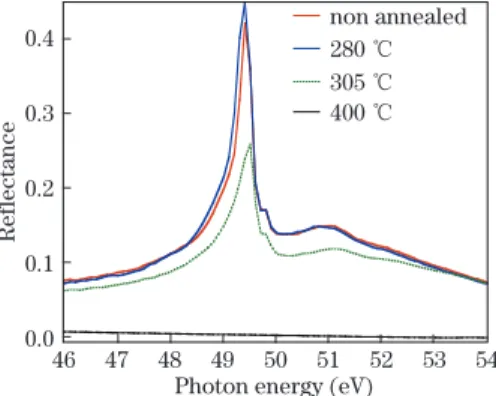
The Co/Mg multilayer has been annealed under high vacuum during one hour up to 400 ◦C. Figure 7 shows
the EUV reflectivity curve obtained for the differently annealed samples. The first annealing a little below 300 ◦C only produces a small increase of reflectance
with respect to the non-annealed sample. However, for annealing slightly above 300◦C, a strong reflectivity
de-crease is observed. Finally at 400 ◦C there is no more
reflectivity.
We have been able to understand this behavior by combining XES and NMR spectroscopies and electron microscopy images[4,8]. XES showed that the Mg layers
did not react with the neighboring ones whatever the an-nealing temperature. NMR measurements also showed that pure cobalt was always present within the multi-layer. However, the signal of the interfacial Co atoms in contact with the Mg layers almost disappeared. This was interpreted as a coalescence of the Co layer upon
Fig. 6. (Color online) (a) SIMS depth profile of four periods of the Co/Mg bilayer and the two Co/Mg/Zr trilayers. The curves show the profile of the Co− ions; (b) NMR spectra
of the three studied stacks. I, II, and III are the frequency characteristics of Co hcp, Co bound with Zr, and Co bound with Mg, respectively.
Fig. 7. (Color online) EUV reflectivity curve of the Co/Mg multilayer for s-polarized radiation at 45◦incidence as a
func-tion of the annealing temperature.
annealing. Electron microscopy clearly showed that above 300◦C some delamination of the multilayer from
its substrate occurred leading to the observed reflectance loss. The delamination was due to the stress introduced within the stack following the transformation of the Co layers. At 400 ◦C the images showed that even some
delamination within the multilayer existed, leading to its destruction.
The trilayer systems have also been tested upon an-nealing up to 500◦C[8]. In this case, the Zr layer clearly
act against the destruction of the multilayer and it is possible to still have a significant reflectance upon an-nealing at 400 ◦C.
Part of this work was performed within the frame-work of the ANR-NSFC COBMUL project (ANR-10-INTB-902-01 and NSFC11061130549). TEM measure-ments were performed at LPS, Paris-Sud University, Orsay, France, within the framework of the METSA net-work. Reflectivity measurements in the EUV range were performed on the BEAR beamline at the Elettra syn-chrotron, Trieste, Italy. SIMS experiments took place at LPS, Chimie Paris Tech, France.
References
1. H. Maury, P. Jonnard, K. Le Guen, J.-M. Andr´e, Z. Wang, J. Zhu, J. Dong, Z. Zhang, F. Bridou, F. Del-motte, C. Hecquet, N. Mahne, A. Giglia, and S. Nan-narone, Eur. Phys. J. B 64, 193 (2008).
2. P. Panissod and C. Meny, Appl. Mag. Reson. 19, 447 (2000).
3. K. Le Guen, M.-H. Hu, J.-M. Andr´e, P. Jonnard, S. K. Zhou, H. Ch. Li, J. T. Zhu, Z. S. Wang, and C. Meny, J. Phys. Chem. C 114, 6484 (2010).
4. M.-H. Hu, K. Le Guen, J.-M. Andr´e, S. K. Zhou, H. Ch. Li, J. T. Zhu, Z. S. Wang, C. Meny, N. Mahne, A. Giglia, S. Nannarone, I. Est`eve, M. Walls, and P. Jonnard, Appl. Phys. A 106, 737 (2012).
5. K. Le Guen, M.-H. Hu, J.-M. Andr´e, S. K. Zhou, H. Ch. Li, J. T. Zhu, Z. S. Wang, C. Meny, A. Galtayries, and P. Jonnard, Appl. Phys. Lett. 98, 251909 (2011). 6. M.-H. Hu, K. Le Guen, J.-M. Andr´e, P. Jonnard, S. K.
Zhou, H. Ch. Li, J. T. Zhu, and Z. S. Wang, AIP Conf. Proc. 1221, 56 (2010).
7. K. Le Guen, M.-H. Hu, J.-M. Andr´e, P. Jonnard, S. K. Zhou, H. C. Li, J. T. Zhu, Z. S. Wang, N. Mahne, A. Giglia, and S. Nannarone, Appl. Phys. A 102, 69 (2011). 8. J. T. Zhu, S. K. Zhou, H. Ch. Li, Z. S. Wang, P. Jonnard, K. Le Guen, M.-H. Hu, J.-M. Andr´e, H. J. Zhou, and T. L. Huo, Opt. Express 19, 21849 (2011).