A contactless electrical stimulator: application
to fabricate functional skeletal muscle tissue
The MIT Faculty has made this article openly available.
Please share
how this access benefits you. Your story matters.
Citation
Kishida, Masako, and Richard D. Braatz. “Ellipsoidal Bounds on
State Trajectories for Discrete-Time Systems with Linear Fractional
Uncertainties.” Optimization and Engineering 16.4 (2015): 695–711.
As Published
http://dx.doi.org/10.1007/s10544-012-9692-1
Publisher
Springer US
Version
Author's final manuscript
Citable link
http://hdl.handle.net/1721.1/104901
Terms of Use
Article is made available in accordance with the publisher's
policy and may be subject to US copyright law. Please refer to the
publisher's site for terms of use.
A contactless electrical stimulator: application to fabricate
functional skeletal muscle tissue
Samad Ahadian&Javier Ramón-Azcón& Serge Ostrovidov&Gulden Camci-Unal& Hirokazu Kaji&Kosuke Ino&Hitoshi Shiku& Ali Khademhosseini&Tomokazu Matsue
Published online: 11 September 2012
# Springer Science+Business Media, LLC 2012
Abstract Engineered skeletal muscle tissues are ideal candidates for applications in drug screening systems, bio-actuators, and as implantable constructs in tissue engineering. Electrical field stimulation considerably improves the differentiation of muscle cells to muscle myofibers. Currently used electrical stimulators often use direct contact of electrodes with tissue constructs or their culture medium, which may cause hydrolysis of the culture medium, joule heating of the medium, contami-nation of the culture medium due to products of electro-des corrosion, and surface fouling of electroelectro-des. Here, we used an interdigitated array of electrodes combined with
an isolator coverslip as a contactless platform to electri-cally stimulate engineered muscle tissue, which elimi-nates the aforementioned problems. The effective stimulation of muscle myofibers using this device was demonstrated in terms of contractile activity and higher maturation as compared to muscle tissues without apply-ing the electrical field. Due to the wide array of potential applications of electrical stimulation to two- and three-dimensional (2D and 3D) cell and tissue constructs, this device could be of great interest for a variety of biolog-ical applications as a tool to create noninvasive, safe, and highly reproducible electric fields.
Samad Ahadian and Javier Ramón-Azcón contributed equally to this work.
Electronic supplementary material The online version of this article (doi:10.1007/s10544-012-9692-1) contains supplementary material, which is available to authorized users.
S. Ahadian
:
J. Ramón-Azcón:
S. Ostrovidov:
A. Khademhosseini:
T. MatsueWPI-Advanced Institute for Materials Research, Tohoku University,
Sendai 980-8577, Japan
G. Camci-Unal
:
A. Khademhosseini (*)Department of Medicine, Center for Biomedical Engineering, Brigham and Women’s Hospital, Harvard Medical School, 65 Landsdowne Street,
Cambridge, MA 02139, USA e-mail: [email protected] G. Camci-Unal
:
A. KhademhosseiniHarvard–MIT Division of Health Sciences and Technology, Massachusetts Institute of Technology,
77 Massachusetts Avenue, Cambridge, MA 02139, USA
H. Kaji
Department of Bioengineering and Robotics, Graduate School of Engineering, Tohoku University, Sendai 980-8579, Japan
K. Ino
:
H. Shiku:
T. Matsue (*)Graduate School of Environmental Studies, Tohoku University, Sendai 980-8579, Japan
e-mail: [email protected] A. Khademhosseini
Wyss Institute for Biologically Inspired Engineering, Harvard University,
Boston, MA 02115, USA A. Khademhosseini
Department of Maxillofacial Biomedical Engineering and Institute of Oral Biology, School of Dentistry, Kyung Hee University, Seoul 130-701, Republic of Korea
Keywords Skeletal muscle tissue engineering . Contactless electrical stimulation . C2C12 myoblasts . Gelatin
methacrylate (GelMA) hydrogel
1 Introduction
Skeletal muscle tissues have the ability to self-repair; how-ever, they lack the potential to restore damage caused by congenital defects, trauma, denervation, and tumor ablation. In addition, intramuscular injection of myogenic cells, such as satellite cells or myoblasts, does not effectively make tissue repair due to low cell retention and survival, immu-norejection, and functional loss (Rossi et al.2010). Muscle tissue engineering has recently been suggested as a promis-ing method to regenerate or recover damaged muscle tissues (Hinds et al.2011; Koning et al.2009). Engineered muscle tissues also have in vitro applications, such as for drug screening (Ghaemmaghami et al. 2012; Vandenburgh
2009) or as engineered bio-actuators (Hosseini et al. 2012; Fujita et al.2011).
It is well known that muscle cells are able to differentiate under stimulation with an electrical field, which leads to an increase in myosin production and the formation of myo-fibers and contractile proteins (Park et al.2008). Advances in microscale technologies used in biology and regenerative medicine (Khademhosseini et al.2006) can provide reliable methods and devices to enable the use of electrical stimula-tion (ES) for tissue engineering applicastimula-tions in a precise and controllable manner. In our previous work (Ahadian et al.
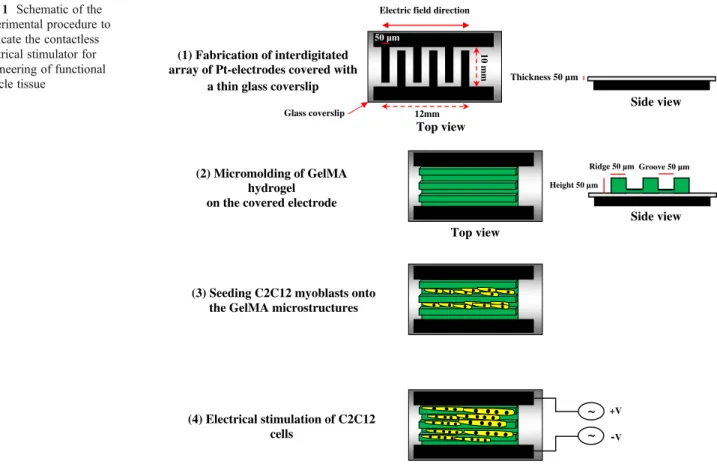
2012), an interdigitated array of electrodes was used for the ES of muscle tissue, which had the following advantages: (a) the electrodes were permanently positioned on the sub-strate and could therefore provide a highly reproducible and well-quantified electric field; (b) this technology made it feasible to fabricate high resolution and complex electrode designs relevant to physiological feature sizes and architec-tures; (c) the ES was synchronized over the whole tissue; (d) lower energy was needed to create a specified electric field compared to the conventional setups for the ES (i.e., pair of conductive electrodes placed in close proximity to the mus-cle tissue). However, any direct contact between electrodes and biological samples as occurred in our suggested device may cause (1) hydrolysis of the culture medium when applied electric potential exceeds the water window and therefore it causes bubble formation; (2) joule heating of the medium; (3) contamination of the culture medium due to products of electrodes corrosion; (4) surface fouling of electrodes as a result of electrochemical reactions on the electrodes surface. To tackle these problems, we propose to use a thin coverslip between the muscle tissue and interdig-itated array of electrodes to make a contactless electrical stimulator as depicted in Fig.1.
In this study, we used gelatin methacrylate (GelMA) hydro-gel, similar to our previous work (Aubin et al. 2010) to fabricate 3D arrays of engineered muscle tissue. Hydrogels have been employed extensively in biological applications because they have a high water content and mimic many features of the natural extracellular matrix (ECM) (Peppas et al.2006; Slaughter et al.2009). GelMA hydrogel is an inex-pensive, photopolymerizable semi-natural hydrogel com-prised of gelatin modified with acrylates (Aubin et al.2010). In this work, we demonstrate the potential advantages of a contactless interdigitated array of Pt (IDA-Pt) electrodes in comparison to IDA-Pt electrodes stimulating muscle tissues made on microgrooved GelMA hydrogel. The engineered tissue was subjected to the ES as a means to accelerate the tissue maturation. In this step, a contactless IDA-Pt electro-des was introduced as a platform for the ES of muscle tissue, and the resulting tissue was characterized and compared with that without applying the electrical field. Due to the wide array of potential applications of ES to two- and three-dimensional (2D and 3D) cell and tissue constructs (Hronik-Tupaj and Kaplan 2012), the proposed device could be of great interest for a variety of biological applications as a tool to create noninvasive, accurate, safe, and highly reproducible direct and alternative electric fields.
2 Materials and methods 2.1 Materials
Positive g-line photoresist (i.e., S1818) and developer (i.e., MF CD-26) were purchased from Shipley Far East Ltd., Japan. Hexamethyldisilazane was purchased from Tokyo Ohka Kogyo Co., Ltd., Japan. Methacrylic anhydride, 3-(trimethoxysilyl)propyl methacrylate (TMSPMA), gelatin Type A made of porcine skin, and penicillin/streptomycin (P/S) were purchased from Sigma-Aldrich Chemical Co., USA. 2-Hydroxy-1-(4-(hydroxyethoxy) phenyl)-2-methyl-1-propanone (i.e., Irgacure 2959) was purchased from Ciba Chemicals, Japan. Polydimethylsiloxane (PDMS) was pur-chased from Dow Corning Toray Co. Ltd., Japan. Trichloro (1H, 1H, 2H, 2H-tridecafluro-n-octyl) silane was purchased from Tokyo Chemical Industry Co., Japan. Fetal bovine serum (FBS) was obtained from Bioserum, Japan. Dulbec-co’s modified Eagle’s medium (DMEM), trypsin/EDTA, MEM essential amino acid, MEM nonessential amino acid, and insulin were obtained by Invitrogen, USA.
2.2 GelMA hydrogel synthesis and preparation of its prepolymer
GelMA hydrogel was synthesized as described in our pre-vious work (Aubin et al.2010). In summary, a high degree
of methacrylation (~80 %) was obtained by adding 8 mL methacrylic anhydride to 10 g of gelatin in PBS for 3 h at 50 °C. The mixture was dialyzed with a 12–14 kDa cutoff dialysis membrane against distilled water for one week at 40 °C and then lyophilized for 1 week. The GelMA hydro-gel was kept at−20 °C until use.
GelMA hydrogel (20 % [w/v]) was combined with the phosphate buffered saline (PBS) and 1 % (w/v) photoinitiator (i.e., Irgacure 2959), kept at 60 °C until fully dissolved to obtain the GelMA prepolymer, and then used for the experiments. 2.3 Cell culture
The C2C12 myoblast cell line was purchased from the American Type Culture Collection (ATCC), USA. The cells were cultured in the DMEM supplemented with 10 % FBS and 1 % P/S, and used for the experiments at passage 7. The C2C12 myoblasts were trypsinized using 0.25 % trypsin/ 0.1 % EDTA when 70–80 % confluence was reached. The cells were maintained in a cell culture incubator (Sanyo, Japan) with a 5 % CO2at 37 °C.
2.4 Microfabrication of GelMA templates for muscle tissue engineering
To generate micromolds for fabricating microengineered GelMA templates (groove-ridge micropatterns with
dimensions: groove 50 μm, ridge 50 μm, and height 50μm), the PDMS stamp (see Supplementary information
for fabrication procedure of PDMS stamp) was initially silanized by using trichloro(1H, 1H, 2H, 2 H-tridecafluro-n-octyl) silane to prevent the adhesion of the GelMA polymer. A coverslip (No. 000, thickness 50μm; Matsunami Co., Japan) mounted on the IDA-Pt electrodes (Ramón-Azcón et al.2012) (see Supplementary informationfor detailed information on design and fabrication of electrodes) was acrylated with the TMSPMA. Note that the coverslip was fixed on the IDA-Pt electrodes using a plastic tape such that there was no direct contact between the electrodes and their surrounding medium. The GelMA hydrogel was then molded on the acrylated coverslip on the IDA-Pt electrodes with the aid of the PDMS stamp such that the GelMA micropattern was perpendicular to the direction of the electrode bands as illustrated in Fig.1. To fabricate the GelMA microstructures, 20 μL of GelMA prepolymer was poured onto the device. Then, the PDMS stamp was gently placed on the surface to completely fill the microgrooves of the stamp with the GelMA hydrogel, and the pattern was then exposed to 7 mW/cm2UV light (Hayashi UL-410UV-1, Hayashi Electronic Shenzen Co., Ltd., Japan) for 150 s. Afterwards, the PDMS stamp was gently removed, leaving the GelMA micropattern on the IDA-Pt electrodes.
For cell culture on the GelMA micropattern, the C2C12 myoblasts were trypsinized, counted, and resuspended in
(1) Fabrication of interdigitated array of Pt-electrodes covered with
a thin glass coverslip
(2) Micromolding of GelMA hydrogel on the covered electrode
Top view
(3) Seeding C2C12 myoblasts onto the GelMA microstructures
~ ~ +V -V (4) Electrical stimulation of C2C12 cells
Electric field direction 50 µm 12mm 10 mm Glass coverslip Thickness 50 µm Side view Top view Side view Height 50 µm Ridge 50 µm Groove 50 µm
Fig. 1 Schematic of the experimental procedure to fabricate the contactless electrical stimulator for engineering of functional muscle tissue
DMEM at a density of 1.5×10⁶cells/mL. Then, 100μL of this suspension was pipetted onto the GelMA micropattern and incubated at 37 °C for 30 min to allow for cell seeding inside the micropattern grooves. Cells loaded within the GelMA micropattern were then cultured after adding suffi-cient culture medium. The procedure described here is shown schematically in Fig.1. After 2 days of culture, the culture medium was replaced with the differentiation medi-um, which was DMEM with 2 % horse sermedi-um, 1 nM insulin, and 1 % P/S. During the culture period, the differentiation medium was replenished every 48 h.
2.5 Immunostaining of engineered muscle tissue
C2C12 muscle cells were fixed with 3–4 % paraformalde-hyde for 12 min, followed by a wash with PBS. The cells were treated with 0.3 % Triton X-100 for 5 min at ambient temperature to make them permeable. Then the cells were exposed to 5 % bovine serum albumin dissolved in PBS for 15 min. A primary mouse monoclonal IgG antibody (ab-7784, Abcam®, Japan) to detect fast skeletal myosin was added to the underlying sample at a dilution of 1:1000 in PBS and incubated at 4 °C for 24 h. The sample was then washed 3 times with PBS and treated with secondary goat anti-mouse AlexaFluor® 488 antibody (Invitrogen, USA) at a dilution of 1:1000 in PBS and incubated at 37 °C for 1 h. To visualize α-actinin, the samples were incubated with monoclonal anti-α-actinin antibody (Sigma-Aldrich, USA) at a dilution of 1:1000 in PBS for 1 h at ambient temperature and then treated with Alexa-Fluor 594-conjugated donkey anti-mouse IgG (Invitro-gen, USA) for 1 h. The samples were stained with 4,6-diami-dino-2-phenylindole (DAPI) (Vector Laboratories Inc., USA) (5 mg/mL in PBS), in order to reveal cell nuclei. The pictures of stained samples were recorded with a fluorescence micro-scope (Carl Zeiss Observer Z.1, Germany).
2.6 Electrical stimulation of the engineered muscle tissue On day 8 of culture, the engineered muscle tissue was electri-cally stimulated through the IDA-Pt electrodes as depicted in Fig. 1. As the control system, the muscle tissue was kept without applying the electrical field. For the ES of muscle tissue, the differentiation medium was replaced with the stim-ulation medium that was composed of DMEM with 2 % horse serum, 1 nM insulin, 2 % MEM essential amino acid, 1 % MEM nonessential amino acid, and 1 % P/S (Kaji et al.2010). Electrical pulses were applied to the muscle tissue using a waveform generator (WF 1946B Multifunction Synthesizer, NF Co., Japan) under an ES regime (voltage 10 V, frequency 1 Hz, and duration 10 ms) for 2 days. An oscilloscope (wave surfer 424; LeCroy Co., Japan) was used to confirm the generated electric current. During ES of the muscle tissue, the stimulation medium was replenished every day.
2.7 RNA extraction and cDNA synthesis
Total RNA was extracted from 3 mg of the muscle tissue. The weighed tissue was placed in liquid nitrogen and thor-oughly ground with a mortar and pestle. RNA was extracted usingβ-mercaptoethanol and purified according to the man-ufacturer’s protocol (RNeasy©microkit, Qiagen, Venlo, Netherlands). Reverse transcription was performed according to the manufacturer’s instructions (Quantitech Reverse Tran-scription, Qiagen, Venlo, Netherlands) for up to 3μg of total RNA. The temperature profile of the cDNA synthesis protocol was as follows: 12 μl of sample (3 μg of total RNA) was diluted with 14μl of RNase-free water and 4 μl of gDNA wipeout buffer and incubated for 2 min at 42 °C and then cooled down to 4 °C. Quantiscript Reverse Transcriptase and Reverse Transcriptase primer mix were subsequently added, and the mixture was incubated for 15 min at 42 °C followed by incubation for 3 min at 95 °C. The samples were kept at 4 °C until use for the quantitative PCR (qPCR). 2.8 Real time PCR
Primer sets for GAPDH, MyoD, myogenin, MRF4, Myf-5, Mef2c, MLP, sarcomeric actin,α-actinin, pn, MHC-IId/x, MHC-IIa, and MHC-IIb were obtained from Operon Biotechnologies (Tokyo, Japan) and validated for qPCR. The primer sequences are listed in the Supplementary infor-mation, Table S1. Real time PCR was performed on a Roche Lightcycler 1.5 (Roche, Mannheim, Germany) using 2μl of cDNA, 2 μl of the primer set (50 μm), and 14 μl of Lightcycler FastStart DNA Master SYBR Green 1 (Roche, Mannheim, Germany). Following an initial denaturation step at 95 °C for 10 min, real time PCR was performed over 45 cycles of 95 °C for 10 s, 62 °C for 10 s, and 72 °C for 20 s, followed by a melt curve analysis. The expression of the target gene was assessed using the comparative method (Berdat et al. 2008), and the results were normalized to the mouse GAPDH gene as the internal reference.
2.9 Statistical analysis
Statistically significant differences were revealed by the independent Student’s t test for 2 groups of data using the MINITAB 16.0 statistical software package (Minitab Inc., USA). All data are represented as average ± standard devi-ation, and p-values less than 0.05 were deemed to be statis-tically significant.
3 Results and discussion
The IDA-Pt electrodes as the electrical stimulator (control device) and our designed contactless electrical stimulator
were compared in terms of pH differences, bubble forma-tion, and appearance due to applied electric field. Figure2
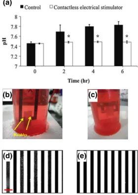
shows such comparison between two devices. We applied an AC electrical field (voltage 10 V, frequency 1 Hz, and duration 10 ms) through devices in the culture medium (i.e., DMEM supplemented with 10 % FBS and 1 % P/S) and evaluated pH changes (as measured by pH meter F-52, Horiba, Japan), bubble formation, and appearance of devi-ces. As can be seen, direct contact between the IDA-Pt electrodes and medium led to noticeable pH changes in culture medium during the stimulation time (Fig. 2-(a)). There was a sharp increase in pH within the control medium during 2 h of stimulation (from pH 7.45 for initial pH buffer to pH 7.69 for the same buffer after 2 h of stimulation); however, it almost remained unchanged until the end of stimulation probably due to the formation of passivation layer on the Pt electrodes. In contrast, pH changes within the medium for the contactless electrical stimulator device during the stimulation time were negligible. In addition, we observed bubble formation within the control device during the stimulation time (Fig.2-(b)) whereas this phenomenon was not observed within the contactless electrical stimulator
(Fig. 2-(c)). This gas evolution was also observed in our previous work where Pt microelectrodes were employed to stimulate cardiac myocytes (Nishizawa et al.2008). Finally, it seems that the ES through control device for 24 h caused Pt corrosion and damage to the device (Fig.2-(d)); however, Pt electrodes remained fully intact within the contactless electrical stimulator (Fig. 2-(e)). The latter finding is in accordance with previous studies in which Pt corrosion as a result of long-time ES was studied using mass spectrom-etry (Hibbert et al.2000) and reciprocal derivative chrono-potentiometry (Musa et al.2011) and it was shown that Pt underwent corrosion during the ES causing to release trace quantities of byproducts that could be toxic to tissues. To solve this problem, Nagamine et al. (Nagamine et al.2011) used Pt microelectrodes coated with poly(3,4-ethylenediox-ythiophene) as a conductive material for the ES of muscle tissue. This chemical modification of Pt electrodes led to the chemical stability of electrodes within the ES period. Note that they did not directly culture the muscle cells on the Pt microelectrodes and therefore as shown by authors, the electric field was dramatically decreased inversely propor-tional to the distance from the electrodes. In contrast, our designed electrical stimulator enabled close placement of electrodes with the muscle tissue through a thin coverslip leading to an efficient and homogeneous electric field within the muscle tissue.
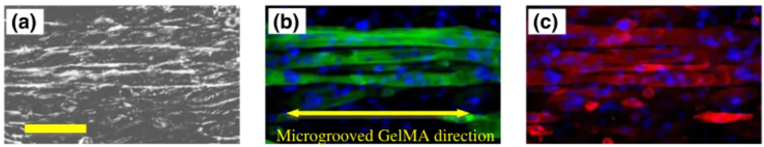
Most C2C12 myoblast cells that were loaded onto the GelMA micropattern oriented and elongated along the ridge-groove direction. This phenomenon is referred to as contact guidance, in which most cells tend to orient and elongate along grooves or fiber axes (Riboldi et al.2005). A high degree of C2C12 myoblast alignment is needed to obtain highly aligned myotubes (as a fundamental require-ment for functional muscle tissues) because the myoblasts fuse together to form myotubes in an end-to-end configura-tion (Clark et al. 2002). Myotube alignment is crucial to maximize the contractility of muscle tissue (Bian et al.
2011). Figure 3 demonstrates the myotube alignment ob-served after 10 days of culture (i.e., after the ES). It also shows high expression levels of myosin heavy chain and α-actinin within multinucleated myotubes in the muscle tissue due to an efficient ES through contactless electrical stimu-lator. Expression levels of target genes (i.e., MyoD, Myf-5, myogenin, MRF4, Mef2c, MLP, sarcomeric actin,α-actinin, perinatal myosin heavy chain (MHC-pn), MHC-IId/x, MHC-IIa, and MHC-IIb) were also evaluated after applying the ES using the designed contactless electrical stimulator on day 10 of culture. The cultures that were electrically stimulated were compared to the corresponding samples without ES (control samples) (Fig. 4). As shown, except myogenin and MRF4, the expression levels of all underly-ing genes significantly increased upon the ES application. Indeed, MRF4 and myogenin interact with muscle LIM
Bubbles
(b) (a)
(d) (e)
(c)
Fig. 2 Comparison between interdigitated array of Pt electrodes as the electrical stimulator (control) and corresponding secured device with glass coverslip (contactless electrical stimulator). (a) pH changes ver-sus the stimulation time for both control and contactless electrical stimulator devices. Evaluation of bubble formation during the electrical stimulation within control (b) and contactless electrical stimulator devices (c). Appearance of control (d) and contactless electrical stim-ulator devices (e) after electrical stimulation (voltage 10 V, frequency 1 Hz, and duration 10 ms) for 24 h. Scale bar shows 100μm (*p<0.05)
protein during muscle maturation and are highly expressed in mature adult skeletal muscle (Arber et al.1994). Contrac-tion of myotubes due to ES is associated with sarcomere development. As an electric field is applied, the sarcomere assembly is increased due to the manipulation of the intra-cellular Ca2+transient through the depolarization of the cell membrane potential (Nagamine et al.2010). Note that ES promoted the contraction of myotubes as indicated in the Supplementary information, Movie S1. In contrast, no such phenomenon was observed for the myotubes without apply-ing the ES. The expression levels of all underlyapply-ing target genes for sarcomere protein development (i.e., sarcomeric actin, α-actinin, perinatal myosin heavy chain (MHC-pn), MHC-IId/x, MHC-IIa, and MHC-IIb) were increased upon applying the electrical field that is in agreement with the contractile activity of our fabricated muscle tissue. An engi-neered muscle tissue may provide a good model to investigate a wide variety of biological phenomena in vitro. However, there is still a demand to develop a cellular model of muscle tissues having contractile ability (Kaji et al.2010). Various effects of muscle tissue contraction on muscle cells such as immune responses, metabolic changes, even angiogenesis can only be evaluated on a muscle tissue model having contractile ability (Nedachi et al.2008). Therefore, such cellular system
is of great interest. Taken together, this work introduced a contactless electrical stimulator that was effective for the ES of muscle tissue.
Applying efficient electric field through thin isolated barriers (e.g., PDMS layer or glass coverslip) has already been developed and employed for a wide range of cell manipulation applications using the dielectrophoresis tech-nique (Park et al. 2009; Shafiee et al. 2009; Shafiee et al.
2010); however, here, we reported the first application of this system for the ES of an engineered tissue. Eventually, a major hurdle to widespread clinical applications of neered muscle tissues is cost and time to fabricate an engi-neered product (Adam2012). Using the suggested electrical stimulator, it is feasible to employ cheap, disposable, and low stable electrodes because we do not need to worry about the corrosion problem of electrode materials in high corro-sive physiological mediums. Therefore, the contactless elec-trical stimulator may decrease the final cost of an engineered tissue. Due to the wide array of potential applications of ES to two- and three-dimensional (2D and 3D) cell and tissue constructs (Hronik-Tupaj and Kaplan 2012), the proposed device could be of great interest for a variety of biological applications as a tool to create noninvasive, accurate, safe, and highly reproducible direct and alternative electric fields.
Microgrooved GelMA direction
(a) (b) (c)
Fig. 3 Phase contrast (a) and corresponding immunofluorescence images of myosin heavy chains (green)-cell nuclei (blue) (b) andα-actinin (red)-cell nuclei (blue) (c) for myotubes cultured on the microgrooved GelMA hydrogel on day 10 of culture. Scale bar shows 100μm
MyoD MHC-IIa MHC-IIb
0.000 0.005 0.010 0.015 Electrically stimulated Control 2 -Δ ct MHC-pn Sarcomeric actin 0.0 0.2 0.4 0.6 0.8 1.0 2 -Δ ct
MLP Myogenin Myf5 Mef2c MHC-IId/x α-actinin MRF4
0.00 0.01 0.02 0.03 0.04 2 -Δ ct
*
*
*
*
*
*
*
*
*
*
Fig. 4 Changes in the expression levels of MHC-pn, sarcomeric actin, MyoD, MHC-IIa, MHC-IIb, MLP, myogenin, myf5, mef2c, MHC-IId/x, α-actinin, and MRF4 as a result of electrical stimulation (voltage 10 V, frequency 1 Hz, and duration 10 ms) and control (without electrical stimulation). Expression levels were normal-ized with respect to the internal reference gene GAPDH (*p<0.05)
4 Conclusions
Here, we proposed a contactless electrode to electrically stimulate engineered muscle tissue, which minimizes hydro-lysis of the culture medium, joule heating of the medium, contamination of the culture medium due to products of electrodes corrosion, and surface fouling of electrodes. The efficient stimulation of skeletal muscle tissues using this platform was also shown in terms of contractile activity and higher maturation compared to muscle tissues without applying the electrical field.
Acknowledgments S.A. conceived the idea. S.A. and J.R. designed the research. S.A., J.R., H.K., H.S., A.K., and T.M. analyzed the results. S.A. wrote the paper. G.C-U. synthesized the GelMA hydrogel. S.A. and J.R. performed all other experiments. H.K., H.S., A.K., and T.M. super-vised the research. All authors read the manuscript, commented on it, and approved its content. This work was supported by the World Premier International Research Center Initiative (WPI), MEXT, Japan.
References
C. Adam, Endogenous musculoskeletal tissue engineering - a focused perspective. Cell Tissue Res. 347, 489–499 (2012)
S. Ahadian, J. Ramón-Azcón, S. Ostrovidov, G. Camci-Unal, V. Hosseini, H. Kaji, K. Ino, H. Shiku, A. Khademhosseini, T. Matsue, Interdigitated array of Pt electrodes as a new platform for the electrical stimulation of engineered muscle tissue. Lab Chip. 12, 3494–3503 (2012)
S. Arber, G. Halder, P. Caroni, Muscle LIM protein, a novel essential regulator of myogenesis, promotes myogenic differentiation. Cell 79, 221–231 (1994)
H. Aubin, J.W. Nichol, C.B. Hutson, H. Bae, A.L. Sieminski, D.M. Cropek, P. Akhyari, A. Khademhosseini, Directed 3D cell align-ment and elongation in microengineered hydrogels. Biomaterials 31, 6941–6951 (2010)
D. Berdat, A.C. Martin Rodriguez, F. Herrera, M.A.M. Gijs, Label-free detection of DNA with interdigitated micro-electrodes in a fluidic cell. Lab Chip 8, 302–308 (2008)
W. Bian, M. Juhas, T.W. Pfeiler, N. Bursac, Local tissue geometry determines contractile force generation of engineered muscle net-works. Tissue Eng. Part A. 18, 957–967 (2011)
P. Clark, G.A. Dunn, A. Knibbs, M. Peckham, Alignment of myoblasts on ultrafine gratings inhibits fusion in vitro. Int. J. Biochem. Cell Biol. 34, 816–825 (2002)
H. Fujita, T. Van Dau, K. Shimizu, R. Hatsuda, S. Sugiyama, E. Nagamori, Designing of a Si-MEMS device with an integrated skeletal muscle cell-based bio-actuator. Biomed. Microdevices 13, 123–129 (2011)
A.M. Ghaemmaghami, M.J. Hancock, H. Harrington, H. Kaji, A. Khademhosseini, Biomimetic tissues on a chip for drug discovery. Drug Discov. Today 17, 173–181 (2012)
D.B. Hibbert, K. Weitzner, B. Tabor, P. Carter, Mass changes and dissolution of platinum during electrical stimulation in artificial perilymph solution. Biomaterials 21, 2177–2182 (2000) S. Hinds, W. Bian, R.G. Dennis, N. Bursac, The role of extracellular
matrix composition in structure and function of bioengineered skeletal muscle. Biomaterials 32, 3575–3583 (2011)
V. Hosseini, S. Ahadian, S. Ostrovidov, G. Camci-Unal, S. Chen, H. Kaji, M. Ramalingam, A. Khademhosseini, Engineered
contractile skeletal muscle tissue on a microgrooved methacry-lated gelatin substrate. Tissue Eng. Part A. (2012)
M. Hronik-Tupaj, D.L. Kaplan, A review of the responses of two- and three-dimensional engineered tissues to electric fields. Tissue Eng. Part B: Rev. 18, 167–180 (2012)
H. Kaji, T. Ishibashi, K. Nagamine, M. Kanzaki, M. Nishizawa, Elec-trically induced contraction of C2C12 myotubes cultured on a porous membrane-based substrate with muscle tissue-like stiff-ness. Biomaterials 31, 6981–6986 (2010)
A. Khademhosseini, R. Langer, J. Borenstein, J.P. Vacanti, Microscale technologies for tissue engineering and biology. Proc. Natl. Acad. Sci. U. S. A. 103, 2480–2487 (2006)
M. Koning, M.C. Harmsen, M.J.A. van Luyn, P.M.N. Werker, Current opportunities and challenges in skeletal muscle tissue engineer-ing. J. Tissue Eng. Regen. Med. 3, 407–415 (2009)
S. Musa, D.R. Rand, C. Bartic, W. Eberle, B. Nuttin, G. Borghs, Coulometric detection of irreversible electrochemical reactions occurring at Pt microelectrodes used for neural stimulation. Anal. Chem. 83, 4012–4022 (2011)
K. Nagamine, T. Kawashima, T. Ishibashi, H. Kaji, M. Kanzaki, M. Nishizawa, Micropatterning contractile C2C12 myotubes em-bedded in a fibrin gel. Biotechnol. Bioengin. 105, 1161–1167 (2010)
K. Nagamine, T. Kawashima, S. Sekine, Y. Ido, M. Kanzaki, M. Nishizawa, Spatiotemporally controlled contraction of micropat-terned skeletal muscle cells on a hydrogel sheet. Lab Chip 11, 513–517 (2011)
T. Nedachi, H. Fujita, M. Kanzaki, Contractile C2C12 myotube model for studying exercise-inducible responses in skeletal muscle. Am. J. Physiol.-Endocrinol. Metab. 295, E1191–E1204 (2008) M. Nishizawa, H. Nozaki, H. Kaji, T. Kitazume, N. Kobayashi, T.
Ishibashi, T. Abe, Electrodeposition of anchored polypyrrole film on microelectrodes and stimulation of cultured cardiac myocytes. Biomaterials 28, 1480–1485 (2008)
H. Park, R. Bhalla, R. Saigal, Effects of electrical stimulation in C2C12 muscle constructs. J. Tissue Eng. Regen. Med. 2, 279–287 (2008)
K. Park, H.-J. Suk, D. Akin, R. Bashir, Dielectrophoresis-based cell manipulation using electrodes on a reusable printed circuit board. Lab Chip 9, 2224–2229 (2009)
N.A. Peppas, J.Z. Hilt, A. Khademhosseini, R. Langer, Hydrogels in biology and medicine: from molecular principles to bionanotech-nology. Adv. Mater. 18, 1345–1360 (2006)
J. Ramón-Azcón, S. Ahadian, R. Obregon, G. Camci-Unal, S. Ostrovidov, V. Hosseini, H. Kaji, K. Ino, H. Shiku, A. Khademhosseini, T. Matsue, Gelatin methacrylate as a promising hydrogel for 3D microscale organization and proliferation of dielectrophor-etically patterned cells. Lab Chip 12, 2959–2969 (2012) S.A. Riboldi, M. Sampaolesi, P. Neuenschwander, G. Cossu, S. Mantero,
Electrospun degradable polyesterurethane membranes: potential scaffolds for skeletal muscle tissue engineering. Biomaterials 26, 4606–4615 (2005)
C.A. Rossi, M. Pozzobon, P. De Coppi, Advances in musculoskeletal tissue engineering: moving towards therapy. Organogenesis 6, 167–172 (2010)
H. Shafiee, J. Caldwell, M. Sano, R. Davalos, Contactless dielectro-phoresis: a new technique for cell manipulation. Biomed. Micro-devices 11, 997–1006 (2009)
H. Shafiee, M.B. Sano, E.A. Henslee, J.L. Caldwell, R.V. Davalos, Selective isolation of live/dead cells using contactless dielectro-phoresis (cDEP). Lab Chip 10, 438–445 (2010)
B.V. Slaughter, S.S. Khurshid, O.Z. Fisher, A. Khademhosseini, N.A. Peppas, Hydrogels in regenerative medicine. Adv. Mater. 21, 3307–3329 (2009)
H. Vandenburgh, High-content drug screening with engineered mus-culoskeletal tissues. Tissue Eng. Part B: Rev. 16, 55–64 (2009)