ORIGINAL ARTICLE
Expression of MAGE-C1/CT7 and selected cancer/testis
antigens in ovarian borderline tumours and primary
and recurrent ovarian carcinomas
Anne-Katrin Zimmermann&Jochen Imig&Agnes Klar&
Christoph Renner&Dimitri Korol&Daniel Fink&
Sylvia Stadlmann&Gad Singer&Alexander Knuth&
Holger Moch&Rosmarie Caduff
Received: 13 July 2012 / Revised: 3 March 2013 / Accepted: 7 March 2013 / Published online: 26 March 2013 # Springer-Verlag Berlin Heidelberg 2013
Abstract C1/CT7, NY-ESO-1, GAGE and MAGE-A4 are members of the cancer/testis (CT) antigen family, which have been proposed as potential targets for cancer immunother-apy. To determine the prevalence and biologic relevance of the novel CT antigen MAGE-C1/CT7 and other antigens, 36 ovar-ian borderline tumours (BTs), 230 primary ovarovar-ian carcinomas (OCs) and 80 recurrent OCs were immunohistochemically analysed using the monoclonal antibodies CT7-33 (MAGE-C1/CT7), E978 (NY-ESO-1), clone 26 (GAGE) and 57B (MAGE-A4). Positivity of at least one CT antigen was present in 39.5 % (81/205) of primary OC and in 50 % (26/52) of all recurrences. Expression of the novel CT antigen MAGE-C1/CT7 was most commonly seen with positivity in 24.5 % of primary and 35.1 % of recurrent OC. MAGE-A4, GAGE and NY-ESO-1 expressions were seen in 22.7, 13.9 and 7.1 % of primary and 22.6, 17.5 and 8.9 % of recurrent OC, respec-tively. Analysis of histological subtypes (serous, endometrioid,
clear cell, mucinous and transitional) exhibited variable expression with negativity in all mucinous OC. High-grade serous OC revealed CT antigen expression in 5.6 to 28 % with MAGE-C1/CT7 being the most frequent, but without corre-lation with stage or overall survival. MAGE-C1/CT7 expres-sion and coexpresexpres-sion of CT antigens were significantly correlated with grade of endometrioid OC. None of the BT showed CT antigen expression. No significant correlation was seen with stage, overall survival or response to chemo-therapy. In summary, CT antigens are expressed in a certain subset of OC with no expression in BT or OC of mucinous histology. These findings may have implications for the design of polyvalent vaccination strategies for ovarian carcinomas.
Keywords NY-ESO-1 . MAGE-C1 . GAGE . CT7 . Ovarian tumour . Cancer/testis antigens
A.-K. Zimmermann (*)
:
H. Moch:
R. CaduffInstitute of Surgical Pathology, University Hospital Zurich, Schmelzbergstrasse 12,
8091 Zurich, Switzerland
e-mail: [email protected] J. Imig
Institute of Pharmaceutical Science, ETH Zurich, Wolfgang-Pauli-Str. 10,
8093 Zurich, Switzerland A. Klar
Tissue Biology Research Unit, Department of Surgery, University Children’s Hospital, August-Forel-Str. 7,
8032 Zurich, Switzerland C. Renner
Onkozentrum Hirslanden Zürich, Klinik Hirslanden, Witellikerstrasse 40,
Zurich 8032, Switzerland
D. Korol
Institute of Social and Preventive Medicine, University of Zurich, Vogelsangstrasse 10,
8091 Zurich, Switzerland
D. Fink
Department of Gynecology, University Hospital Zurich, Frauenklinikstrasse 10,
8091 Zurich, Switzerland
S. Stadlmann
:
G. SingerInstitute of Pathology, Kantonsspital Baden AG, 5404 Baden, AG, Switzerland
A. Knuth
Clinic of Oncology, University Hospital Zurich, Rämistrasse 100, 8091 Zurich, Switzerland
Introduction
At the time of initial diagnosis, more than half of the women with ovarian carcinomas (OCs) present with an advanced stage (FIGO stage III and IV). Treatment options include debulking surgery with maximal reduction of the tumour mass. In addition, platinum-based drugs and taxanes are used for systemic cyto-toxic therapy. However, prognosis is still poor with 5-year survival rates of only 18–47 % for advanced stages [1]. Since the beginning of the 1990s, a standstill of treatment results is seen that can hopefully be overcome by new treatment strategies. Immunotherapy using cancer vaccines may represent a novel approach to ameliorate outcome in women with ovarian carci-nomas [2,3]. Cancer/testis (CT) antigens are attractive targets for immunotherapy as they display a restricted expression pat-tern with occurrence in germ cells and a variety of malignant tumours but not in normal tissues. So far, more than 100 CT genes have been identified, which belong to at least 44 distinct gene families. Most CT antigens correspond to chromosome X-linked genes, but their function is largely unknown [4,5]. Some studies reported association of CT antigen expression with pro-gression of cancer growth, dedifferentiation and/or poorer sur-vival rates [6–10]. Furthermore, CT antigens or peptides derived from CT antigens are candidates for cancer vaccination due to their high immunogenicity. CT antigens have already been evaluated as target antigens in clinical trials in patients with advanced carcinomas and melanomas [4, 11–15]. Effective application of immunotherapy depends on the prevalence and intratumoural expression heterogeneity of CT antigens [16].
Ovarian cancer belongs to the group of cancers with frequent expression of CT antigens. Members of the MAGE family [17–19], GAGE [18, 20], BAGE [18], XAGE [21], OY-TES-1 [22], SP17 [23], SCP-1 [24, 25], SSX [26], NY-ESO-1 [27–29], AKAP and LAGE [28] have been demonstrated at the RNA/DNA level. Accordingly, immunotherapy could be an option for the treatment of ovarian cancer after failure of first- and second-line thera-pies [3,30,31]. However, only a limited number of ovarian cancers respond to such therapies yet [30]. Some studies using antibodies against GAGE [32], NY-ESO-1 [28, 29,
33], MAGE-A1 and -A4 [29,32,34,35], SCP-1 [24], SP17 [23] and OY-TES-1 [22] have analysed the CT antigen expression in OC at the protein level with divergent results. MAGE-C1 was previously identified as a novel CT anti-gen. The MAGE-C1/CT-7 gene has significant homology with the MAGE-C2/CT-10 gene, and both genes map in close proximity to chromosome Xq27 [36]. The CT7-33 monoclo-nal antibody recognises MAGE-C1/CT7 in formalin-fixed, paraffin-embedded tissues. Only nine OCs were recently stud-ied using the MAGE-C1/CT7 antibody [37].
Expression of MAGE-C1/CT7, GAGE, NY-ESO-1 and MAGE-A4 was determined by immunohistochemistry in a large number of ovarian neoplasms. CT antigen expression
was correlated with clinico-pathological parameters and patient outcome. Subgroup analysis of serous and endometrioid OC, the most common histological subtype, was conducted. Our data show CT antigen expression in 40–50 % of primary and recurrent OC with surprisingly low expression of NY-ESO-1.
Material and methods Patients
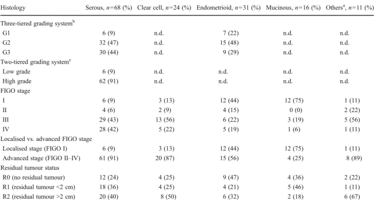
One hundred fifty consecutive primary OC specimens and 36 borderline tumours (BTs) were retrieved from the archives of the Institute for Surgical Pathology, University Hospital Zurich, Switzerland, covering the period from 1995 to 2005. Tissue samples were fixed in 4 % neutral buffered formaldehyde, embedded in paraffin and then used to construct one tissue micro array (TMA) with cores of 150 OCs, 36 BTs and 6 normal tissue samples of the fallopian tube, as described previously [38]. Routine haematoxylin and eosin sections were used for histopathological evaluation. All tumours were reviewed by one gynaecologic pathologist (RC). The tumour stage was assessed according to the International Federation of Gynecology and Obstetrics (FIGO) staging system. The tumour grade and histo-logical subtype were defined according to the 2003 WHO classification. Additionally to the Silverberg grading proposed by WHO 2003 [39], serous carcinomas were graded according to the two-tiered MD Anderson system [40]. Clinical and path-ological characteristics were taken from clinical database and pathology records. The median age at carcinoma diagnosis was 61 years (range 20–87 years), and overall survival was 2.25 years (range 0–128 months). A detailed overview of the clinico-pathological characteristics of carcinomas of this cohort from Zurich is given in Table1. Ovarian BT included 20 serous, 14 mucinous, 1 endometrioid BT, and 1 BT with Brenner histology. Median age at diagnosis of BT was 60.5 years (range 30– 85 years), and median survival was 6.2 years (range 4– 125 months).
A second patient cohort with paired tissue samples from 80 patients with advanced (FIGO II and III) high-grade (MD Anderson grading) primary serous ovarian carcinomas and their corresponding recurrences after chemotherapy was available and was used to construct a second TMA. This TMA consisted of formalin-fixed, paraffin-embedded tu-mour tissue of OC from the Institutes of Pathology from the University Hospital Bale, Cantonal Hospital St. Gallen, Cantonal Hospital Baden and Cantonal Hospital Liestal, diagnosed and treated between 1985 and 2003. Median age at diagnosis of OC was 59 years (range 20–77 years), and median recurrence-free survival (RFS) was 9 months (range 1–85 months). Recurrence was defined as an eleva-tion of CA-125 levels with tumour confirmaeleva-tion by radio-logical examination and/or during secondary surgical
procedures. Chemoresistance was defined as OC recurrence within 6 months after finishing chemotherapy. Clinico-pathological details of this cohort have been recently reported [41,42] and are shown in Table2.
The complete tumour cohort included 230 primary OCs, 80 recurrent high-grade serous OCs and 36 ovarian BTs. The project was approved by the local ethics review boards
of Bale and Zurich (Kantonale Ethikkomission Zurich, StV 27–2009).
Histology and immunohistochemistry
Three-micrometre-thick sections of TMA blocks and formalin-fixed, paraffin-embedded tissues were mounted on glass slides (Super-Frost Plus, Menzel, Braunschweig, Germany), deparaffinised, rehydrated and stained with haematoxylin–eosin using standard histological techniques. For immunohistochemi-cal staining of TMA and large sections, the Ventana Benchmark automated staining system (Ventana Medical Systems, Tucson, AZ, USA) and Ventana reagents were used. After deparaffinisation in xylene, the slides were rehydrated in decreas-ing concentrations of ethanol. Endogenous peroxidase was blocked using the Ventana endogenous peroxidase blocking kit after a rinse with distilled water. For antigen retrieval, the slides were heated with cell conditioning solution (CC1, Ventana) according to the manufacturer’s instructions. For the detection of the MAGE-A4 protein, the 57B monoclonal antibody (1:50, kindly provided by Dr. G.C. Spagnoli, University of Basel, Switzerland) was used, which recognises most of the MAGE-A family members, but predominantly the MMAGE-AGE-MAGE-A4 protein. Primary antibodies against MAGE-C1/CT-7 (clone CT7-33, Table 1 Clinico-pathological characteristics of primary ovarian carcinomas of the Zurich cohort
Histology Serous, n=68 (%) Clear cell, n=24 (%) Endometrioid, n=31 (%) Mucinous, n=16 (%) Othersa, n=11 (%) Three-tiered grading systemb
G1 6 (9) n.d. 7 (22) n.d. n.d.
G2 32 (47) n.d. 15 (48) n.d. n.d.
G3 30 (44) n.d. 9 (29) n.d. n.d.
Two-tiered grading systemc
Low grade 6 (9) n.d. n.d. n.d. n.d. High grade 62 (91) n.d. n.d. n.d. n.d. FIGO stage I 6 (9) 3 (13) 12 (44) 12 (75) 1 (11) II 4 (6) 2 (9) 4 (15) 0 (0) 2 (22) III 29 (43) 13 (56) 6 (22) 3 (19) 5 (56) IV 28 (42) 5 (22) 5 (19) 1 (6) 1 (11)
Localised vs. advanced FIGO stage
Localised stage (FIGO I) 6 (9) 3 (13) 12 (44) 12 (75) 1 (11)
Advanced stage (FIGO II–IV) 61 (91) 20 (87) 15 (56) 4 (25) 8 (89)
Residual tumour status
R0 (no residual tumour) 12 (24) 4 (25) 9 (47) 4 (36) 2 (22)
R1 (residual tumour <2 cm) 18 (36) 4 (25) 4 (21) 5 (46) 1 (11)
R2 (residual tumour >2 cm) 20 (40) 8 (50) 6 (32) 2 (18) 6 (67)
n.d. not done
a
Nine transitional and one anaplastic carcinoma and one malignant Mullerian mixed tumour
bThree-tiered grading system according to WHO 2003 only for serous carcinomas (Silverberg grading) and endometrioid carcinomas c
Two-tiered MD Anderson grading only for serous carcinomas
Table 2 Clinico-pathological characteristics of ovarian carcinomas of the Bale cohort
High-grade advanced serous ovarian carcinomasa n=80 (%) Residual tumour status
R0 (no residual tumour) 32 (42)
R1 (residual tumour <2 cm) 23 (30) R2 (residual tumour >2 cm) 22 (28) Response to chemotherapyb Sensitive 57 (72) Resistant 22 (28) a
Grading was performed according to the MD Anderson grading system. All carcinomas were of advanced FIGO stage (FIGO≥II)
b
Chemosensitivity was defined as RFS>6 months after chemotherapy; chemoresistance was defined as RFS<6 months after chemotherapy
1:80, Dako, Barr, Switzerland), GAGE (clone 26, reacts with GAGE-3, -4, -5, -6 and -7B proteins; 1:2,000; BD Transduction Laboratories, San Jose, CA, USA) and NY-ESO-1 (clone E978, 1:50, Zymed Laboratories, South San Francisco, CA, USA) were applied adjusted to the Ventana Benchmark system after performing titrations. iVIEW-DAB was used as chromogen.
Immunoreactivity was cytoplasmic for MAGE-A4, MAGE-C1/CT-7 and GAGE and both nuclear and cytoplas-mic for NY-ESO-1. CT antigen expression was scored
according to the percentage of positive cells as negative (0 %), focal (1–25 %), moderate (26–50 %), and diffuse (>50 % positive cells). Testicular tissue served as a positive control. Homogenous staining was defined as positivity in more than 50 % of tumour cells.
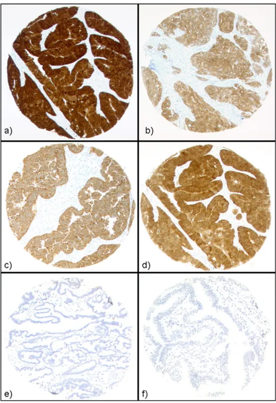
Normal fallopian tubal epithelium and ovarian stroma tissue were consistently negative for MAGE-A4, MAGE-C1/CT7 (see Fig.1) and GAGE. Diffuse weak staining of NY-ESO-1 was initially observed in the epithelium of the fallopian tube. Fig. 1 CT antigen expression
in ovarian neoplasms. Strong and homogeneous staining of NY-ESO-1 (a), GAGE (b), MAGE-A4 (c) and MAGE-C1/ CT7 (d) was observed in ovarian carcinomas. Borderline tumours (e) and epithelium of the fallopian tube (f) did not exhibit expression of MAGE-C1/CT7 (shown) or other CT antigens
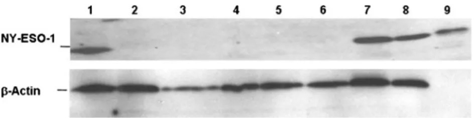
By RT-PCR analyses, low amounts of NY-ESO-1 mRNA could be identified in tissue of the fallopian tube, but NY-ESO-1 protein was absent in Western blot analyses (see Fig.2). In Western blot, strong bands of NY-ESO-1 protein were seen in immunohistochemically positive OC and the three controls, whereas immunohistochemically negative OC and BT did not demonstrate NY-ESO-1 protein in Western blot analysis. Consequently, NY-ESO-1 antibody was diluted to the point where tubal epithelium was negative (1:50). Statistical analysis
Statistical analyses were performed using SPSS version 20 software. Associations between categorical groups (i.e. CT antigen expression and clinico-pathological parameters) were tested using the Pearsonχ2test or Fisher’s exact test. Survival analysis was performed on 150 OCs of different histology. For univariate analysis, Kaplan–Meier analysis survival curves were constructed using the product limit method. The logrank test was applied to assess the statistical significance of the association between variables and patient survival. Two-tailed p≤0.05 was considered to be significant.
Results
CT antigen expression in primary ovarian neoplasms CT antigen expression was analysed in TMA tumour tissue cores of 230 primary OCs and 36 ovarian BTs. Evaluation of the markers was not always possible in all tumour samples due to loss of tissue cores related to the TMA technology. In primary OC, MAGE-C1/CT7 was seen most frequently (24. 5 %, 52/212), followed by MAGE-A4 (22.7 %, 49/216), GAGE (13.9 %, 30/216) and NY-ESO-1 (7.1 %, 15/211). Examples of CT antigen staining are shown in Fig. 1. Expression differed among histological subtypes of OC, though without significant correlation. Whereas all mucinous carcinomas were CT antigen negative (0/15), other histologi-cal subtypes (serous, clear cell, endometrioid and transitional) exhibited variable CT antigen expression between 4 to 30 %
(an overview from the Zurich cohort is given in Table3). In serous carcinomas of both cohorts, MAGE-C1/CT7 was most frequently expressed (36/131, 27.5 %), followed by MAGE-A4 (35/135, 25.9 %), GAGE (18/135, 13.3 %) and NY-ESO-1 (8/130, 6.2 %); see Table4.
Tissue cores of OC exhibited heterogenous staining (<50 % of tumour cells stained) of MAGE-A4, NY-ESO-1, MAGE-C1/CT7 and GAGE in 55.NY-ESO-1, 33.3, 80.8 and 53. 3 %, respectively. To better evaluate the prevalence of CT antigen expression, large sections of 20 OCs with negativity for NY-ESO-1 in the TMA cores were additionally immunohistochemically analysed. One tumour with diffuse NY-ESO-1 positivity, three tumours with focal NY-ESO-1 positivity, four tumours with focal GAGE, three tumours with focal MAGE-C1/CT7 and two tumours with diffuse MAGE-A4 positivity were additionally identified. BTs were negative for all CT antigens analysed (example of negative MAGE-C1/CT7 staining is shown in Fig.1e).
CT antigen expression and clinico-pathological parameters FIGO stage was significantly associated with survival (p=0. 006), but there was no significant correlation between CT antigen expression and FIGO stage or survival. Comparison of clinico-pathological variables with CT antigen expression in the Zurich cohort is summarised in Table 3. A separate subgroup analysis of serous carcinomas from the combined Zurich and Bale cohort (see Table4) was conducted but did not reveal significant association with grade (MD Anderson grading) or FIGO stage (localised vs. advanced). Also, there was no significant association with response to chemother-apy. Though higher frequencies of MAGE-C1/CT7 and GAGE expression were seen in recurrent serous carcinomas (20/57 (35.1 %) and 10/57 (17.5 %)) compared to tissue from primary manifestation (16/64 (25 %) and 5/67 (7.5 %)), this was not significant. In recurrent OC, MAGE-C1/CT7 (35.1 %, 20/57) was most frequent, followed by MAGE-A4 (22.6 %, 14/62), GAGE (17.5 %, 10/57) and NY-ESO-1 (8.9 %, 5/56). MAGE-C1/CT7 ex-pression was significantly associated with grade in the sub-group of endometrioid carcinomas (p=0.04).
Fig. 2 Western blot analysis with anti-NY-ESO-1 antibody E978 on tumour and non-tumour tissues. Ovarian carcinoma with positivity for NY-ESO-1 in immunohistochemistry (column 1), NY-ESO-1 immunohistochemically negative ovarian carcinoma (column 2),
fallopian tube (column 4) and borderline tumours (columns 5 and 6). Negative control: myocardial tissue (column 3). Positive controls: mela-noma cell line SK-MEL-37 (column 7), multiple myeloma cell line U266 (column 8) and bacterial recombinant NY-ESO-1 protein (column 9)
Coexpression of CT antigens
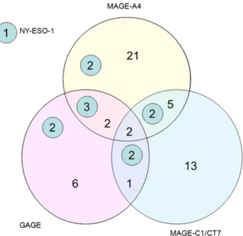
At least one of the four examined CT antigens was expressed in 39.5 % (81/205) of all primary and 50 % (26/52) of recurrent ovarian carcinomas. Two or more CT antigens were expressed in 10.7 % (22/205) of primary OC and 11.5 % (6/52) of recurrent OC. The pattern of coexpression from cases where all four tested CT antigens could be evaluated is visualised in a Venn diagram (see Fig. 3). Interestingly, only one tumour sample showed isolated expression of NY-ESO-1, whereas most NY-ESO-1 positive cases (11/12, 92 %) revealed coexpression of MAGE-A4, MAGE-C1/CT7 and/or GAGE. Coexpression of CT antigens was signif-icantly associated with histological subtype (p=0.02); see Table 5. Presence of CT antigen coexpression was not associated with overall survival.
The subgroup analysis for all serous carcinomas revealed no significant correlation of CT antigen coexpression and
grade (MD Anderson), FIGO stage, response to chemotherapy or recurrence. In the subgroup of endometrioid carcinomas, coexpression was significantly associated with grade (p=0.03).
Discussion
We demonstrate protein expression data of C1, MAGE-A4, NY-ESO-1 and GAGE in ovarian BT as well as in primary and recurrent OC. Whereas none of the borderline tumours showed expression of CTA, positive immunohistochemistry was seen in about 40 to 50 % of primary and recurrent OC. This finding is of clinical significance since previous studies have shown that CT antigens are able to induce specific immune responses. Antibodies directed against MAGE and other CT antigens were detected in the serum of (ovarian) cancer patients [2,43–45], suggesting that these antigens are targets for peptide vaccination in patients with advanced ovarian cancer.
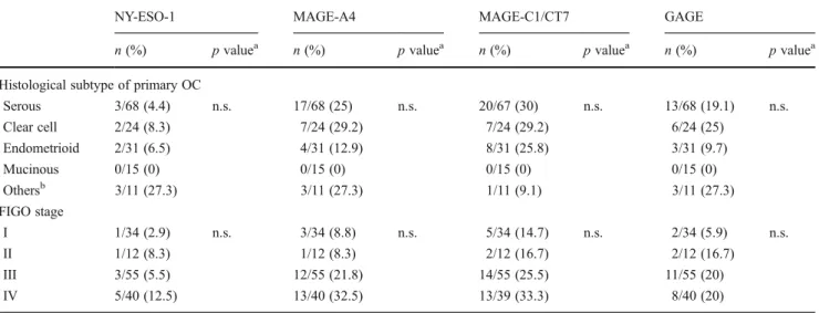
Table 3 CT antigen expression in primary ovarian carcinomas (Zurich cohort) according to clinico-pathological characteristics
NY-ESO-1 MAGE-A4 MAGE-C1/CT7 GAGE
n (%) p valuea n (%) p valuea n (%) p valuea n (%) p valuea
Histological subtype of primary OC
Serous 3/68 (4.4) n.s. 17/68 (25) n.s. 20/67 (30) n.s. 13/68 (19.1) n.s. Clear cell 2/24 (8.3) 7/24 (29.2) 7/24 (29.2) 6/24 (25) Endometrioid 2/31 (6.5) 4/31 (12.9) 8/31 (25.8) 3/31 (9.7) Mucinous 0/15 (0) 0/15 (0) 0/15 (0) 0/15 (0) Othersb 3/11 (27.3) 3/11 (27.3) 1/11 (9.1) 3/11 (27.3) FIGO stage I 1/34 (2.9) n.s. 3/34 (8.8) n.s. 5/34 (14.7) n.s. 2/34 (5.9) n.s. II 1/12 (8.3) 1/12 (8.3) 2/12 (16.7) 2/12 (16.7) III 3/55 (5.5) 12/55 (21.8) 14/55 (25.5) 11/55 (20) IV 5/40 (12.5) 13/40 (32.5) 13/39 (33.3) 8/40 (20) n.s. not significant a
p value, Fisher’s exact test (two sided)
bOther histological subtypes comprise nine transitional carcinomas, one anaplastic carcinoma and one malignant Mullerian mixed tumour. Only
transitional carcinomas showed positivity for CT antigens
Table 4 Subgroup analysis of CT antigen expression in prima-ry serous ovarian carcinomas
Zurich and Bale cohort com-bined, n=148
NY-ESO-1 MAGE-A4 MAGE-C1/CT7 GAGE
n (%) n (%) n (%) n (%)
Positive cases 8/130 (6.2) 35/135 (25.9) 36/131 (27.5) 18/135 (13.3) MD Anderson grading
Low grade 1/6 (16.7) 1/6 (16.7) 1/6 (16.7) 1/6 (16.7)
High grade 7/124 (5.6) 34/129 (26.4) 35/125 (28) 17/129 (13.2) Localised vs. advanced FIGO stage
Localised stage (FIGO I) 0/6 (0) 0/6 (0) 2/6 (33.3) 0/6 (0) Advanced stage (FIGO II–IV) 8/123 (6.5) 35/128 (27.3) 34/124 (27.3) 18/128 (14)
The new CT antigen MAGE-C1/CT7 was the most fre-quent CT antigen in primary OCs (24.5 %, 52/212) and recurrent carcinomas (35.1 %, 20/57). Whereas no association was seen with FIGO stage, overall survival or response to chemotherapy, in the subgroup of endometrioid carcinomas, MAGE-C1/CT7 expression correlated significantly with grade. In ovarian neoplasms, MAGE-C1/CT7 has primarily been analysed at the level of gene transcription, but protein expression has not yet been studied to a larger extent. One small MAGE-C1/CT7 protein expression study reported immuno-positivity in six of nine (66 %) OCs [37]. Humoral and cellular immunoresponses were demonstrated in multiple myeloma patients [44,46], suggesting that MAGE-C1/CT7 could be a potential target for vaccination strategies.
GAGE protein was recently not identified in OC, but only ten OC cases were studied [32]. GAGE mRNA expres-sion was reported in 10 and 26.8 % of ovarian neoplasms [17,47]. This GAGE mRNA expression is consistent with our data on protein level, with expression rate of 13.9 % (30/216). Our MAGE-A4 data with expression in 22.7 % (49/212) of primary OC are consistent with two previous studies, reporting MAGE-A4 immunoreactivity in 13.9 % (17/122) and 11 % (13/117) [34,35]. In contrast, Yakirevich et al. identified MAGE-A4 positivity in 57 % (30/53) of serous OC [29], using large sections and less diluted anti-bodies (1:20).
NY-ESO-1 as one of the best characterised CT antigens was recently ranked by an NCI panel as among the top ten antigens for the development of human cancer vaccines [48]. Therefore, the prevalence of NY-ESO-1-positive OC is of high clinical relevance. We identified NY-ESO-1 pro-tein expression in 7.1 % of primary OC (15/211) and 8.9 % of recurrent OC (5/56). This was unexpectedly low com-pared to previously published studies with expression rates of 18.8 % (10/53) [29] and 43 % (62/142) [28]. In contrast, Gjerstorff et al. were not able to detect NY-ESO-1 protein at all in ten OC cases [32]. Several parameters can influence immunohistochemical results.
Three different antibodies (D8.38, E978 and ES121) with different specificity were used in previous studies. D8.38 is reactive with NY-ESO-1 as well as LAGE1, a CT antigen with 94 % homology to NY-ESO-1. Initial publication of E978 and ES121 antibodies described similar specific im-munohistochemical staining patterns [49], though ES121 was generated against a shorter NY-ESO-1 recombinant protein than E978 and is thus possibly less specific. One would expect that the highest results were detected with antibody D8.38. This was not the case; 18.9 % (10/53) of ovarian carcinomas were positive with antibody D8.38 [29], less than half than with antibody ES121 [28], where 43 % Fig. 3 Venn diagram with coexpression of CT antigens. The numbers
indicate the absolute amount of positive cases. Numbers in the small circles indicate the NY-ESO-1-positive tissue samples
Table 5 Coexpression of can-cer/testis antigens according to clinico-pathological parameters (Zurich cohort)
n.s. not significant
a
Pearsonχ2test (two sided)
bOther histological subtypes
comprise nine transitional carci-nomas, one anaplastic carcinoma and one malignant Mullerian mixed tumour. Only transitional carcinomas showed expression of CT antigens
One CT antigen Two CT antigens More than two CT antigens p value
n (%) n (%) n (%)
Histological subtype of primary OC
Serous 1/67 (24) 9/67 (13.4) 5/67 (13.4) 0.02a Clear cell 3/24 (12.5) 3/24 (12.5) 4/24 (16.7) Endometrioid 5/31 (16.1) 0/31 (0) 4/31 (12.9) Mucinous 0/15 (0) 0/15 (0) 0/15 (0) Othersb 0/11 (0) 0/11 (11.1) 3/11 (27.3) FIGO stage 1 3/34 (8.8) 1/34 (2.9) 2/34 (5.9) n.s. 2 1/12 (8.3) 1./12 (8.3) 1/12 (8.3) 3 13/55 (23.6) 5/55 (9.1) 5/55 (9.1) 4 7/39 (4.5) 5/39 (12.8) 6/39 (15.4)
(62/142) of OC were positive. Another study that used the same antibody as we did (E978) did not detect protein expression in OC at all (0/10 [32]).
CT antigens are known for their restricted expression pat-tern in germs cells and malignant tumours and no expression in normal tissue. Initially, diffuse weak staining of NY-ESO-1 was observed in fallopian tubal epithelium, all BT and 44 % of OC. In our analysis, we performed extensive positive and negative control studies. In Western blot analyses, NY-ESO-1 protein could not be detected in tubal epithelium, BT and immunohistochemically negative OC, whereas it was clearly present in immunohistochemically positive tumours and pos-itive controls. Consequently, we applied for immunohisto-chemistry a NY-ESO-1 antibody dilution resulting in negativity of tubal epithelium (1:50).
Intratumoural heterogeneity of CT antigen expression has been reported and is a particular problem in tissue microarray approaches. Lack of expression in a substantial number of cancer cells in CT antigen-positive tumours has decisive implications for the development of CT antigen-targeted ovarian cancer ther-apies because only a subset of cancer cells are potentially affect-ed by a tumour vaccination approach. Therefore, immunohistochemical evaluation of CT antigen distribution is important in the response evaluation of CT antigen-targeted therapy. Heterogenous staining (<50 % of tumour cells) of the CT antigens MAGE-A4, NY-ESO-1, MAGE-C1/CT7 and GAGE was the predominant pattern in most OC tissue cores on the TMA. Our large section analysis with the monoclonal NY-ESO-1-specific antibody E978 [49–51] revealed that we missed in the TMA analysis 4 of 20 (20 %) tumours with NY-ESO-1 positivity due to intratumoural expression heterogeneity. Further, four, three and two tumours with GAGE (20 %), MAGE-C1/CT7 (15 %) and MAGE-A4 (10 %) expression were identified in large sections, which were initially missed on the TMA analysis. Yakirevich et al. have also reported intratumoural expression heterogeneity for MAGE-A4 and NY-ESO-1. In their study, NY-ESO-1 was focally (<25 % positive cells) seen in 9 %, moderately (<50 % positive cells) in 4 % and diffusely (>50 % positive cells) in 6 % of serous ovarian cancers [29]. Therefore, the TMA approach underestimates the prevalence of NY-ESO-1 protein and other CT antigens.
CT antigens were not expressed in BT, confirming previous MAGE-A4 data by Yakirevich et al. [29]. Coexpression of different CT antigens (two or more CT antigens) was more often seen in recurrent serous OC (50 %) compared to primary serous OC (39.5 %) though this was not significant. Coexpression was significantly associated with tumour differentiation grade of endometrioid carcinomas. Information on CT antigen coexpression is crucial for the design of polyvalent vaccine strategies because OC patients can be selected for combined or sequential vaccination with two or more tumour antigens. Such strategies have the potential of reducing or even preventing the in vivo selection of tumour cell variants with antigen loss.
Subgroup analysis of serous OC, representing the most common histological subtype of OC, was conducted for several clinico-pathological parameters but did not reveal a significant correlation. Interestingly, mucinous OC was neg-ative for all CT antigens. This result was congruent with previous reports [28,34] and is consistent with the hypoth-esis that mucinous OC has a different histogenhypoth-esis and biologic behaviour [52]. The few transitional OC that were included in our study cohort showed a surprisingly high (co) expression rate of the examined CT antigens.
In summary, our data provide evidence that a subset of primary and recurrent OC do express the examined CT antigens MAGE-A4, NY-ESO-1, MAGE-C1/CT7 and GAGE, and these tumours are potentially attackable with immunotherapies. In our cohort, the new CT antigen MAGE-C1/CT7 is the most frequently expressed antigen and thus seems to be interesting for further investigation. Acknowledgments The authors thank Martina Storz and Silvia Behnke for their excellent technical assistance. This work was supported by the Cancer Research Institute and the Zurich Cancer League (Zurich).
Conflict of interest The authors declare that they have no conflict of interest.
Ethics statement The project was approved by the local ethics review boards of Bale and Zurich (Kantonale Ethikkomission Zurich, StV 27–2009). The aim of this retrospective study was immunohisto-chemical analysis of tumour tissue of patients with ovarian neoplasias (carinomas and borderline tumours). We tried to find a tissue marker that gives evidence for biologic behaviour (prognostic marker, good/bad prognosis) or can possibly be used as therapeutic targets for immunotherapies (therapeutic markers).
All tissue samples were taken from the archives after diagnostic processes were completed. Samples (186) were retrieved from the archives of the Institute for Surgical Pathology, University Hospital Zurich, Switzerland, covering the period from 1995 to 2005. Tissue samples of further 80 patients were available from the Institutes of Pathology, University Hospital Bale, Cantonal Hospital St. Gallen, Cantonal Hospital Baden and Cantonal Hospital Liestal from the period between 1985 and 2003.
According to the ethical rules of our local ethics committee, patient data were pseudoanonymised with patient identification numbers. To our knowledge, the results of this study have no influence on patient’s treatment (yet). However, should our results be of importance for the therapy of individual patients, they can again be identified.
In agreement with the local ethics committee, informed consent of individual patients was not obtained. Most patients of the collective examined are already dead. Thus, we relinquished to get patient con-sent or concon-sent of family members in order not to upset them.
References
1. Heintz AP, Odicino F, Maisonneuve P et al (2003) Carcinoma of the ovary. Int J Gynaecol Obstet 83(Suppl 1):135–166
2. Odunsi K, Sabbatini P (2008) Harnessing the immune system for ovarian cancer therapy. Am J Reprod Immunol 59:62–74
3. Kandalaft LE, Powell DJ Jr, Singh N et al (2011) Immunotherapy for ovarian cancer: what's next? J Clin Oncol 29:925–933 4. Simpson AJ, Caballero OL, Jungbluth A et al (2005) Cancer/testis
antigens, gametogenesis and cancer. Nat Rev Cancer 5:615–625 5. Caballero OL, Chen YT (2009) Cancer/testis (CT) antigens:
po-tential targets for immunotherapy. Cancer Sci 100:2014–2021 6. Brasseur F, Rimoldi D, Lienard D et al (1995) Expression of MAGE
genes in primary and metastatic cutaneous melanoma. Int J Cancer 63:375–380
7. Kocher T, Zheng M, Bolli M et al (2002) Prognostic relevance of MAGE-A4 tumor antigen expression in transitional cell carcinoma of the urinary bladder: a tissue microarray study. Int J Cancer 100:702–705 8. Kurashige T, Noguchi Y, Saika T et al (2001) Ny-ESO-1 expres-sion and immunogenicity associated with transitional cell carcino-ma: correlation with tumor grade. Cancer Res 61:4671–4674 9. Patard JJ, Brasseur F, Gil-Diez S et al (1995) Expression of MAGE
genes in transitional-cell carcinomas of the urinary bladder. Int J Cancer 64:60–64
10. van Baren N, Brasseur F, Godelaine D et al (1999) Genes encoding tumor-specific antigens are expressed in human myeloma cells. Blood 94:1156–1164
11. Davis ID, Chen W, Jackson H et al (2004) Recombinant NY-ESO-1 protein with ISCOMATRIX adjuvant induces broad integrated antibody and CD4(+) and CD8(+) T cell responses in humans. Proc Natl Acad Sci U S A 101:10697–10702
12. Jager D, Jager E, Knuth A (2001) Immune responses to tumour antigens: implications for antigen specific immunotherapy of can-cer. J Clin Pathol 54:669–674
13. Jager D, Jager E, Knuth A (2001) Vaccination for malignant melanoma: recent developments. Oncology 60:1–7
14. Jager E, Karbach J, Gnjatic S et al (2006) Recombinant vaccinia/ fowlpox NY-ESO-1 vaccines induce both humoral and cellular NY-ESO-1-specific immune responses in cancer patients. Proc Natl Acad Sci U S A 103:14453–14458
15. Sadanaga N, Nagashima H, Mashino K et al (2001) Dendritic cell vaccination with MAGE peptide is a novel therapeutic approach for gastrointestinal carcinomas. Clin Cancer Res 7:2277–2284 16. Scanlan MJ, Simpson AJ, Old LJ (2004) The cancer/testis genes:
review, standardization, and commentary. Cancer Immun 4:1 17. Gillespie AM, Rodgers S, Wilson AP et al (1998) MAGE, BAGE
and GAGE: tumour antigen expression in benign and malignant ovarian tissue. Br J Cancer 78:816–821
18. Russo V, Dalerba P, Ricci A et al (1996) MAGE BAGE and GAGE genes expression in fresh epithelial ovarian carcinomas. Int J Cancer 67:457–460
19. Yamada A, Kataoka A, Shichijo S et al (1995) Expression of MAGE-1, MAGE-2, MAGE-3/-6 and MAGE-4a/-4b genes in ovarian tumors. Int J Cancer 64:388–393
20. Duan Z, Duan Y, Lamendola DE et al (2003) Overexpression of MAGE/GAGE genes in paclitaxel/doxorubicin-resistant human cancer cell lines. Clin Cancer Res 9:2778–2785
21. Egland KA, Kumar V, Duray P et al (2002) Characterization of overlapping XAGE-1 transcripts encoding a cancer testis antigen expressed in lung, breast, and other types of cancers. Mol Cancer Ther 1:441–450
22. Tammela J, Uenaka A, Ono T et al (2006) OY-TES-1 expression and serum immunoreactivity in epithelial ovarian cancer. Int J Oncol 29:903–910
23. Straughn JM Jr, Shaw DR, Guerrero A et al (2004) Expression of sperm protein 17 (Sp17) in ovarian cancer. Int J Cancer 108:805–811 24. Tammela J, Jungbluth AA, Qian F et al (2004) SCP-1 cancer/testis antigen is a prognostic indicator and a candidate target for immu-notherapy in epithelial ovarian cancer. Cancer Immun 4:10 25. Tureci O, Sahin U, Zwick C et al (1998) Identification of a
meiosis-specific protein as a member of the class of cancer/testis antigens. Proc Natl Acad Sci U S A 95:5211–5216
26. Valmori D, Qian F, Ayyoub M et al (2006) Expression of synovial sarcoma X (SSX) antigens in epithelial ovarian cancer and identi-fication of SSX-4 epitopes recognized by CD4+ T cells. Clin Cancer Res 12:398–404
27. Chen YT, Gure AO, Tsang S et al (1998) Identification of multiple cancer/testis antigens by allogeneic antibody screening of a mela-noma cell line library. Proc Natl Acad Sci U S A 95:6919–6923 28. Odunsi K, Jungbluth AA, Stockert E et al (2003) NY-ESO-1 and
LAGE-1 cancer-testis antigens are potential targets for immuno-therapy in epithelial ovarian cancer. Cancer Res 63:6076–6083 29. Yakirevich E, Sabo E, Lavie O et al (2003) Expression of the
MAGE-A4 and NY-ESO-1 cancer-testis antigens in serous ovarian neoplasms. Clin Cancer Res 9:6453–6460
30. Leffers N, Daemen T, Helfrich W et al (2010) Antigen-specific active immunotherapy for ovarian cancer. Cochrane Database Syst Rev. doi:10.1002/14651858.CD007287.pub2
31. Liu B, Nash J, Runowicz C et al (2010) Ovarian cancer immuno-therapy: opportunities, progresses and challenges. J Hematol Oncol 3:7. doi:10.1186/1756-8722-3-7
32. Gjerstorff MF, Johansen LE, Nielsen O et al (2006) Restriction of GAGE protein expression to subpopulations of cancer cells is independent of genotype and may limit the use of GAGE proteins as targets for cancer immunotherapy. Br J Cancer 94:1864–1873 33. Cioca DP, Deak E, Cioca F et al (2006) Monoclonal antibodies
targeted against melanoma and ovarian tumors enhance dendritic cell-mediated cross-presentation of tumor-associated antigens and efficiently cross-prime CD8+ T cells. J Immunother 29:41–52 34. Bolli M, Kocher T, Adamina M et al (2002) Tissue microarray
evaluation of melanoma antigen E (MAGE) tumor-associated an-tigen expression: potential indications for specific immunotherapy and prognostic relevance in squamous cell lung carcinoma. Ann Surg 236:785–793, discussion 793
35. Sato E, Olson SH, Ahn J et al (2005) Intraepithelial CD8+ tumor-infiltrating lymphocytes and a high CD8+/regulatory T cell ratio are associated with favorable prognosis in ovarian cancer. Proc Natl Acad Sci U S A 102:18538–18543
36. Gure AO, Stockert E, Arden KC et al (2000) CT10: a new cancer-testis (CT) antigen homologous to CT7 and the MAGE family, iden-tified by representational-difference analysis. Int J Cancer 85:726–732 37. Jungbluth AA, Chen YT, Busam KJ et al (2002) CT7 (MAGE-C1) antigen expression in normal and neoplastic tissues. Int J Cancer 99:839–845
38. Noske A, Zimmermann AK, Caduff R et al (2011) Alpha-methylacyl-CoA racemase (AMACR) expression in epithelial ovarian cancer. Virchows Arch 459:91–97
39. Silverberg SG (2000) Histopathologic grading of ovarian carcino-ma: a review and proposal. Int J Gynecol Pathol 19:7–15 40. Malpica A (2008) Grading of ovarian cancer: a histotype-specific
approach. Int J Gynecol Pathol 27:175–181
41. Stadlmann S, Gueth U, Reiser U et al (2006) Epithelial growth factor receptor status in primary and recurrent ovarian cancer. Mod Pathol 19:607–610
42. Stadlmann S, Gueth U, Wight E et al (2007) Expression of perox-isome proliferator activated receptor gamma and cyclo-oxygenase 2 in primary and recurrent ovarian carcinoma. J Clin Pathol 60:307–310
43. Fossa A, Berner A, Fossa SD et al (2004) NY-ESO-1 protein expression and humoral immune responses in prostate cancer. Prostate 59:440–447
44. Lendvai N, Gnjatic S, Ritter E et al (2010) Cellular immune responses against CT7 (MAGE-C1) and humoral responses against other cancer-testis antigens in multiple myeloma patients. Cancer Immun 10:4
45. Stockert E, Jager E, Chen YT et al (1998) A survey of the humoral immune response of cancer patients to a panel of human tumor antigens. J Exp Med 187:1349–1354
46. Curioni-Fontecedro A, Knights AJ, Tinguely M et al (2008) MAGE-C1/CT7 is the dominant cancer-testis antigen targeted by humoral immune responses in patients with multiple myeloma. Leukemia 22:1646–1648
47. Zhang S, Zhou X, Yu H et al (2010) Expression of tumor-specific antigen MAGE, GAGE and BAGE in ovarian cancer tissues and cell lines. BMC Cancer 10:163
48. Cheever MA, Allison JP, Ferris AS et al (2009) The prioriti-zation of cancer antigens: a national cancer institute pilot pro-ject for the acceleration of translational research. Clin Cancer Res 15:5323–5337
49. Jungbluth AA, Chen YT, Stockert E et al (2001) Immunohistochemical analysis of NY-ESO-1 antigen expression in normal and malignant human tissues. Int J Cancer 92:856–860
50. Fujita S, Wada H, Jungbluth AA et al (2004) NY-ESO-1 expression and immunogenicity in esophageal cancer. Clin Cancer Res 10:6551–6558 51. Vaughan HA, Svobodova S, Macgregor D et al (2004)
Immunohistochemical and molecular analysis of human melano-mas for expression of the human cancer-testis antigens NY-ESO-1 and LAGE-1. Clin Cancer Res 10:8396–8404
52. Harrison ML, Jameson C, Gore ME (2008) Mucinous ovarian cancer. Int J Gynecol Cancer 18:209–214