Research Article
Mycobacterial lipoarabinomannans modulate
cytokine production in human T helper cells by interfering
with raft/microdomain signalling
A. K. Shabaanaa, b, dK. Kulangaraa, e, I. Semaca, f, Y. Parelc, S. Ilangumarana, g, K. Dharmalingamb,
C. Chizzolinic and D. C. Hoesslia,*
aDepartment of Pathology and Immunology, Centre médical universitaire, University of Geneva Medical School,
1 rue Michel-Servet, 1211 Geneva 4 (Switzerland), e-mail: [email protected]
bSchool of Biotechnology, Madurai Kamaraj University, Madurai 625021 (India)
cDepartment of Immunology and Allergy, University Hospital, 1211 Geneva 14 (Switzerland) dCurrent address: Trudeau Institute Inc., Saranac Lake, New York (USA)
eCurrent address: Swiss Institute of Technology, Cellular Neurobiology Laboratory, Life Sciences Faculty,
Lausanne (Switzerland)
fCurrent address: SpinX Technologies, Meyrin, Geneva (Switzerland)
gCurrent address: Department of Pediatrics/Immunology Division CHUS/CRC, Sherbrooke, Québéc J1H 5N4 (Canada)
Received 9 September 2004; received after revision 14 October 2004; accepted 11 November 2004
Abstract. Lipoarabinomannans (LAMs) are major lipo-glycans of the mycobacterial envelope and constitute im-munodominant epitopes of mycobacteria. In this paper, we show that mannose-capped (ManLAM) and non-man-nose-capped (PILAM) mycobacterial lipoglycans insert into T helper cell rafts without apparent binding to known receptors. T helper cells modified by the insertion of PILAM responded to CD3 cross-linking by decreasing type 1 (IL-2 and IFN-g) and increasing type 2 (IL-4 and
IL-5) cytokine production. Modification by the mannose-capped ManLAMs had similar, but more limited effects DOI 10.1007/s00018-004-4404-5
© Birkhäuser Verlag, Basel, 2005
CMLS
Cellular and Molecular Life Scienceson T helper cell cytokine production. When incorporated into isolated rafts, PILAMs modulated membrane-associ-ated kinases in a dose-dependent manner, inducing in-creased phosphorylation of Src kinases and Cbp/PAG in Th1 rafts, while decreasing phosphorylation of the same proteins in Th2 rafts. Mycobacterial lipoglycans thus modify the signalling machineries of rafts/microdomains in T helper cells, a modification of the membrane organi-zation that eventually leads to an overall enhancement of type 2 and inhibition of type 1 cytokine production.
Key words. Th1/Th2; raft; signalling; cytokine; lipoarabinomannan.
*Corresponding author.
A. K. Shabaana and K. Kulangara made equal contributions to this work.
munodominant epitopes of mycobacteria [3, 4] that ac-cumulate in the infected host and induce strong antibody responses [5]. Mannose capping of the LAM arabinan polymers (ManLAM) was originally considered to occur mostly in mycobacterial strains of high virulence [1], but this view was later revised when LAMs from
Mycobac-terium tuberculosis were all found to be mannose-capped,
in both virulent and less virulent strains [6, 7]. Mannose-The lipoarabinomannan (LAM) lipoglycans span the
my-cobacterial envelope [1], and are thought to insert into the mycobacterial plasma membrane by way of their glyco-sylphosphatidylinositol (GPI) anchor [2]. LAMs are
im-capped LAMs were less efficient than the non-mannose-capped LAMs (PILAMs) in modulating the cytokine pro-duction of macrophages [6–10], but recent studies have documented potent biological activities of ManLAMs [11, 12], which may be active in different contexts than PILAMs. The non-mannose-capped PILAM molecules are actually capped with a phospho-myo-inositol and should no longer be named AraLAMs, the term AraLAM being only appropriate for the rare and fully non-capped LAMs elaborated by M. chelonae [13].
Several surface receptors have been identified with the capacity to bind LAMs. In particular, mannose receptors on phagocytes bind ManLAMs, but not AraLAMs lack-ing terminal di-mannosyl units [14, 15]. Likewise, the dendritic cell surface receptor DC-SIGN (dendritic cell-specific intercellular adhesion molecule-3-grabbing non-integrin) specifically recognizes capped LAMs [16]. The GPI-anchored CD14 pattern recognition receptor allows phagocytes to sense bacterial products in their environ-ment, and binds several lipoglycans including LAMs [17] and peptidoglycans [18]. Likewise, distinct Toll-like re-ceptors function as pattern-recognition rere-ceptors and me-diate macrophage responses to LPS-CD14 or PILAM-CD14 complexes [19]. Lastly, the CD1 surface molecules accommodate ManLAMs in their large hydrophobic binding groove [20, 21] and induce immune responses [3, 4]. The ManLAM molecule can be sampled by different group I CD1 receptors in endosomes of mycobacteria-laden, antigen-presenting cells [22], and presented to a variety of CD1-restricted T cells [23].
T lymphocytes are devoid of such LAM-binding recep-tors, but are nonetheless functionally modified following interaction with LAMs [8, 24, 25]. In particular, Jurkat cells preincubated with LAMs downregulate mRNA lev-els for interleukin IL-2, IL-3 and granulocyte/macro-phage-colony-stimulating factor (GM-CSF) when mito-gen-activated [26], but neither the direct effects of LAMs on T lymphocytes with polarized cytokine production patterns, nor the interference of LAMs with the raft sig-nalling platform, have been investigated.
Through differential cytokine release, T lymphocytes in-duce cell-mediated or humoral responses to pathogens. Among CD4 T cells, type 1 helper (Th1) lymphocytes synthesize the IL-2 and IFN-g cytokines generally
as-sociated with resistance to intracellular infection, and favour cell-mediated immune responses such as delayed-type hypersensitivity and activation of cytotoxic T cells and macrophages, while the Th2 lymphocytes predomi-nantly release IL-4 and IL-5 to promote B cell activation and humoral responses [27, 28]. The spectrum of leprosy skin lesions is typically polarized and ranges from tuber-culoid type 1 to lepromatous type 2 responses. Patients with tuberculoid lesions present with a localized form of the disease and a strong cell-mediated response. The lepromatous leprosy patients present with multibacillary
lesions and high titers of specific antibodies but poor cell-mediated immune responses against M. leprae anti-gens. Furthermore, the detection of high levels of IL-2 and IFN-g mRNA in the total RNA extracted from
lesions of tuberculoid leprosy patients contrasts with the high levels of IL-4 and IL-5 measured in lepromatous leprosy lesions [29, 30].
To further define the interaction of M. leprae lipoglycans with helper T cells, we investigated the effects of LAMs on the raft signalling platform and the polarized patterns of cytokine production of helper T cells. We show that LAMs insert into sphingolipid-rich rafts/microdomains of the plasma membrane of polarized T helper cells and modulate raft-associated protein kinases in a dose-dependent man-ner. To document the functional consequences of such events, we have analysed the modifications in the cytokine profiles of activated T helper cells after incubation with AraLAMs or ManLAMs. Our results show that LAMs inserted in T helper lymphocyte rafts alter patterns of cytokine production, most likely by modifying the transbi-layer organization of the raft signalling platform.
Materials and methods
Mycobacterial lipoglycans
LAMs from a rapidly growing unclassified
Mycobac-terium species (PILAMs) and a virulent strain (Erdman)
of M. tuberculosis (ManLAMs) were isolated as previ-ously described [1], and found free of contaminating mycobacterial products. LAMs were obtained in freeze-dried form, reconstituted in sterile water and stored at –20 °C. Deacylated PILAM was prepared by mild alka-line hydrolysis [31]. One hundred micrograms of PILAMs was incubated in 100 ml of 0.1 N NaOH for 2 h at 37 °C,
neutralized with acetic acid and desalted through Bio-Gel P-10 (Bio-Rad, Hercules, Calif.) in PBS. The eluted, dea-cylated PILAM was lyophilized and reconstituted in PBS. An aliquot of PILAM was processed similarly but incu-bated with water instead of NaOH. Lipopolysaccharide (LPS) and polymyxin B were obtained from Sigma (Fluka, Buchs, Switzerland).
Antibodies
The anti-CD3 OKT3 monoclonal antibody (mAb)-pro-ducing hybridoma was from ATCC (Bethesda, Md.). For dot-blotting, the anti-CD3e and anti-CD4 mAbs were from
Dako (DakoCytomation, Zug, Switzerland), CD55 was detected with the IA-10 mAb (BD Pharmingen, Basel, Switzerland), CD59 with MEM-43 mAb and CD45 with the MEM-28 mAb. The GM1 ganglioside was detected with the peroxidase-labelled B subunit of cholera toxin (CTB; Sigma). LAMs (AraLAM and ManLAM) were de-tected with the anti-LAM mAb CS35. All MEM mAbs were kindly donated by Dr. V. Horejsi, Academy of
Sci-ences of the Czech Republic, Prague. For Western blotting, the anti-Lck and anti-Fyn polyclonal anti-peptide antibod-ies were from Santa Cruz (LabForce, Nunningen, Switzer-land) and Cbp/PAG was detected with the MEM-255 mAb. Generation of Th1 and Th2 cell clones
Th1 and Th2 cell clones were generated from peripheral blood of normal individuals upon antigen activation and cloning by limiting dilution in RPMI-1640 medium sup-plemented with IL-2 (20 U/ml), penicillin (50 U/ml), strep-tomycin (50 mg/ml), 5% human AB serum, 10% FCS,
irra-diated (3500 rad) allogeneic PBMCs, and PHA (1 mg/ml)
as described elsewhere [32]. Growing cells were further expanded and characterized for their capacity to produce IFN-g and IL-4 upon CD3 cross-linking. High IFN-g/low
IL-4 producers were defined as Th1 whereas low IFN-g
high IL-4 producers were Th2 [31]. Cultured T cells were harvested 15 days after stimulation, washed extensively and suspended in RPMI-1640 medium for further studies. Sucrose gradient centrifugation of Th1 and Th2 cells Th1 and Th2 (50 ¥ 106) cells were incubated in 100 ml of
serum-free medium containing 20 mg PILAMs or
Man-LAMs (200 mg/ml) for 30 min at 37 °C. This corresponds
to 0.4 mg LAMs for 1 ¥ 106cells, the same ratio of LAMs
to cells used to evaluate the effects of LAM on cytokine mRNA and protein production. After one wash in PBS, the cells were lysed in 1% TX-100 before equilibrium sucrose gradient centrifugation as previously described and an equal volume of each fraction (20 ml) was sampled
and adsorbed on nitrocellulose using the BioRad dot-blot apparatus [33]. Raft fractions (low-density fractions 3–5) were pooled, the detergent-resistant membranes pelleted
by ultracentrifugation and their contents analysed by Western blotting [34].
Evaluation of the effects of mycobacterial lipoglycans on the production of T helper cell cytokines
For cytokine mRNA levels, 4 ¥ 106Th1 or Th2 cells were
incubated in 270 ml of serum-free RPMI medium
contain-ing PILAMs or ManLAMs (6 mg/ml) for 30 min at 37 °C.
Control cells were incubated under the same conditions without mycobacterial lipoglycans. Unbound LAM was removed by washing with medium containing 10% FCS. The cells were suspended in 1 ml complete medium and stimulated or not in the presence of 10 mg/ml of OKT3 and
PMA (5 ng/ml) for 6 h at 37 °C. Cytokine protein levels were determined in cell-free supernatants.
RNA extraction, cDNA preparation and amplification Th1 or Th2 cells were pelleted and lysed in 1 ml Trizol (Life Technologies), the lysate mixed with 0.2 ml chloro-form, centrifuged, and the RNA precipitated from the aqueous phase by addition of 1.5 ml isopropylalcohol (Sigma). The RNA pellet was resuspended in 25 ml
DEPC-treated water and its concentration measured spectrophoto-metrically. Complementary DNA was synthesized from RNA (0.5 mg) using 200 U of Moloney murine leukaemia
virus-reverse transcriptase (Life Technologies, Gaithers-burg, Md.) in a total reaction volume of 20 ml, for 1 h at
37 °C, and used as template for amplification by PCR. The sequences of oligonucleotide primers specific for b-actin,
IL-2, IL-4, IL-5, IL-10 and IFN-g were designed as shown
in table 1. All primers were used at a final concentration of 1 mM. PCR was performed in a 40 ml reaction mix
con-taining 5 ml cDNA, 1.25 units of Taq polymerase (Life
Table 1. Cytokine primers used in this study.
Primers Sequence Length (bp) Size of product (bp)
b-actin Primer 1 GTGGGGCGCCCCAGGCACCA 20 540 Primer 2 CTCCTTAATGTCACGCACGATTTC 24 IL-2 Primer 1 CAGGATGCTCACATTTAAGTTTTACA 26 91 Primer 2 CTCGAGAGGTTTGAGTTCTTCTTCTA 26 IL-4 Primer 1 ATGGGTCTCACCTCCCAACTG 21 462 Primer 2 TCAGCTCGAACACTTTGAATATTTCTCTCTCAT 33 IL-5 Primer 1 CAAACGCAGAACGTTTCAGA 20 137 Primer 2 GCAGTGCCAAGGTCTCTTTC 20 IL-10 Primer 1 TGGTGAAACCCCGTCTCTAC 20 163 Primer 2 CTGGAGTACAGGGGCATGAT 20 IFN-g Primer 1 TGCAGAGCCAAATTGTCTCCTTTTAC 26 299 Primer 2 TTACTGGGATGCTCTTCGACCTCGAAACAGGAT 33
Technologies), 1.0 mM MgCl2and 0.1 mM dNTP
(Pro-mega Catalysis AG, Wallisellen, Switzerland). Semiquan-titative PCR analysis was carried out for 18, 22, 26 and 30 cycles and the PCR products electrophoresed on a 2% agarose gel, visualized with ethidium bromide and the re-sulting documents stored and processed with ImageQuant. Amplification of b-actin mRNA was used as an internal
control. b-Actin levels were normalized for all samples
such that the intensities of the PCR products at 18, 22, 26 and 30 cycles in different samples were identical and lin-ear when analysed by ImageQuant. The semiquantitative analyses shown in figure 2 utilized cDNAs obtained from Th1 and Th2 cells that were pre-treated with PILAMs or ManLAMs, and stimulated with OKT3 and PMA. Cytokine protein measurement in culture supernatants
Production in 6-h culture supernatants of IFN-g, IL-4
(Hoffmann-La Roche, Basel, Switzerland), IL-2 and IL-5 (R & D Systems, Minneapolis, Minn.) by Th1 or Th2 cells was assessed by ELISA. The sensitivity threshold was 25 pg/ml for all assays.
In vitro kinase assays
Raft fractions (fractions 3–5, cf. fig. 1) were obtained from sucrose gradients loaded with 50 ¥ 106Th1 or Th2
cells, pooled and aliquoted, ultracentrifuged and resus-pended in 50 ml 10 mM Hepes pH 7.4, 156 mM NaCl,
containing 0, 25 or 125 mg/ml of PILAMs. After 30 min
at 37 °C, raft membranes were again ultracentrifuged, re-suspended in 30 ml kinase buffer and assayed for
endoge-nous kinases as described elsewhere [35]. The levels of phosphorylation were measured by PhosphorImager and analysed by ImageQuant.
Results
PILAM and ManLAMs incorporate into rafts of Th1 and Th2 cells
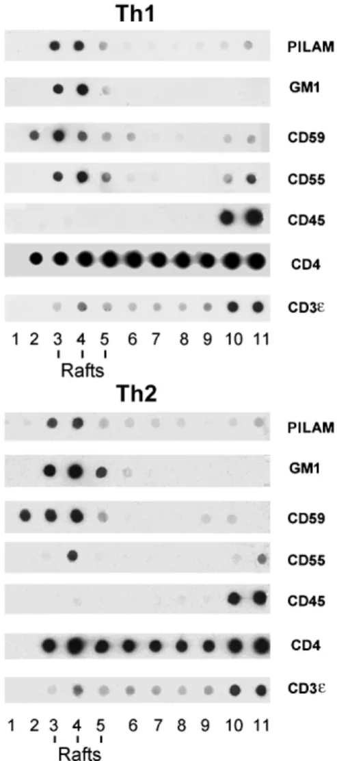
Th1 and Th2 cells incubated with PILAMs or ManLAMs were washed free of unbound LAMs and subjected to sub-cellular fractionation in the presence of TX-100, to isolate the detergent-resistant (or raft) fractions. Dot-blot analysis of each gradient fraction of either Th1 or Th2 cells (fig. 1) showed that GM1 detected with CTB and the GPI-linked proteins CD55 and CD59 were selectively enriched in the low-density raft fractions 3–5. In contrast, the major T cell surface protein, the transmembrane CD45, was re-covered in the high-density fractions 9–11 containing the TX-100-soluble proteins. The CD3e protein was found
mostly in the soluble fractions, with 10–15% reproducibly found in the raft fractions. The CD4 coreceptor distrib-uted predominantly in the raft fractions (60%) of both Th1 and Th2 cells. The incorporated PILAM lipoglycans
were only detectable in the raft fractions 3–5 with the CS35 anti-LAM mAb and the same gradient distribution was found for ManLAM. Likewise, deacylation of PILAMs or ManLAMs (see below) resulted in more than 70% inhibi-tion of raft associainhibi-tion. Lastly, the overall protein and ganglioside composition of rafts was not altered quantita-tively or qualitaquantita-tively by LAM incorporation.
PILAM-treated cells upregulate Th2
and downregulate Th1 cytokines upon stimulation by T cell receptor agonists, while ManLAM-treated cells only upregulate Th2 cytokines.
To assess the role of LAMs in cytokine production, Th1 or Th2 cells were exposed for 30 min to PILAMs or
Man-Figure 1. PILAM inserts preferentially in raft fractions of Th1 and Th2 cells. Dot-blot analysis of sucrose density gradient fractions. Each gradient was loaded with 50 ¥ 106 Th1 or Th2 cells, previously
incubated with PILAMs. Fractions 3–5 correspond to the interface between 5 and 36% sucrose (TX-100-insoluble raft membranes) and fractions 9–11 to the bulk of the TX-100-soluble proteins. Twenty microlitres was dotted for each fraction and incubated with the antibodies listed in Materials and methods, or CTB for the detection of GM1.
LAMs (0.4 mg/ml per 1 ¥ 106cells), leading to
incorpora-tion of lipoglycans in rafts as shown in figure 1. Under these conditions, the basal levels of cytokine mRNA were not affected. However, following activation with anti-CD3 mAb and PMA, significant changes in cytokine mRNA levels were detected by semiquantitative RT-PCR analy-sis in LAM-treated compared to untreated T helper cells. Exposure to PILAMs significantly decreased IL-2 and IFN-g in Th1 cells. In contrast, IL-4 mRNA levels were
increased by PILAMs, and to a lesser extent by
Man-LAMs, in Th2 cells (fig. 2A, B). PILAMs only increased IL-5 mRNA levels in Th2 cells (fig. 2B).
The results observed at the protein level are illustrated in figure 2C for one representative Th1 and one Th2 clone, and are comparable to the results observed at the mRNA level (fig. 2B). With all Th1 and Th2 clones tested, PILAM preincubation slightly but consistently inhibited IFN-g and
IL-2 to 80.0% ± 0.06 of the control value (p = 0.022, n = 6). Conversely, PILAMs enhanced IL-4 (149.2% ± 10.0) and IL-5 protein production (172.3% ± 3.3, p = 0.042, n = 6. ManLAM pre-incubation had no effect on IFN-g (109.6%
± 9.8 of the control), while it enhanced IL-4 (119.8% ± 2.8) and IL-5 (192.3% ± 13.3) protein production.
LAM effects are independent of LPS but depend upon the acyl moieties of the LAM molecule
Both preparations of PILAMs and ManLAMs contained 9 ng/ml of endotoxin, measured by the Limulus assay. The level of LPS contamination was not increased following deacylation. LPS is unlikely to have been responsible for the modulation of cytokine messages, since PILAMs and ManLAMs had different effects, despite similar contents of LPS. Moreover, upregulation of IL-4 and downregu-lation of IL-2 and IFN-g by PILAMs were not affected
by pre-treatment of cells with the LPS inhibitor poly-myxin B.
The biological effect of PILAMs depends upon the hy-drophobic part of the molecule that permits membrane insertion. Deacylated LAMs were no longer inserted into cellular membranes and thus ineffective. For instance, using Th1 cells, the inhibitory effect of PILAMs on Th1 cytokine message levels was abrogated by deacylation, while mock-treated PILAMs (i.e. subjected to the same separation steps as alkali-treated PILAMs) retained their inhibitory properties on IL-2 and IFN-g messages (fig. 3).
Raft distribution of Lck and FynT kinases in Th1 and Th2 cells
In both Th1 and Th2 cells, the 60-kDa form of the Lck ki-nase was found exclusively in the raft fractions (fig. 4, top panel). The main Lck form detected in non-raft fractions
Figure 2. Effects of PILAMs and ManLAMs on cytokine mRNA and protein levels in Th1 and Th2 cells. (A, B) RT-PCR analysis of the IL-2, IFN-gand IL-4, IL-5 responses of Th1 and Th2 cells to anti-CD3 and PMA, following pre-incubation with PILAMs or ManLAMs. Control cells were stimulated with anti-CD3 and PMA without prior incubation with mycobacterial lipoglycans. (A) Semi-quantitative PCR for 18, 22, 26 and 30 cycles with the appropriate cytokine primers and b-actin control. (B) The amounts of PCR products (ethidium bromide labelled) recovered after 18, 22, 26 and 30 cycles were measured by scanning and expressed as percent of control (no LAM treatment). (C) ELISA measurement of the indicated cytokines in supernatants of one representative Th1 and one Th2 clone.
Figure 3. Deacylated PILAM fails to downregulate IL-2 and IFN-g
messages. RT-PCR of the IL-2 and IFN-gresponses of Th1 cells to anti-CD3 and PMA, with pre-incubation with deacylated PILAMs, and mock-treated PILAMs. The semiquantitative analysis of PCR products was carried out for 18, 22, 26 and 30 cycles.
was the nominal 56-kDa form. Less than 10% of the total Lck detected in non-raft fractions was 60 kDa. In both Th1 and Th2 cells, the amount of Lck found in rafts cor-responded to approximately 40% of the total cellular Lck (data not shown). The FynT kinase was detected as a 60-kDa protein in both raft and non-raft fractions of both cell types with a raft/non-raft ratio of about 4 to 1 (fig. 4, bot-tom panel).
AraLAM incorporation in rafts modulates the in vitro activity of associated kinases
Incorporation of increasing amounts of PILAM (25 and 125 mg/ml) in Th1 raft membranes resulted in a
dose-de-pendent increase in Src kinase (open bars) and Cbp/PAG (filled bars) phosphorylation, reaching 2.5- to 3-fold at 125 mg/ml (fig. 5A). The main phosphorylated proteins
were the Src kinases (Lck and FynT) at 60 kDa and Cbp/PAG at 83 and 95 kDa. These phosphoproteins were identified by Western blotting and two-dimensional elec-Figure 4. Raft and non-raft distribution of Lck and FynT protein
tyrosine kinases in Th1 and Th2 cells. Western blot analysis. Lck (top) and FynT (bottom). Raft: pooled fractions 3–5 (20 ml); Non-raft: pooled fractions 9–11 (20 ml). The sucrose gradient was devel-oped and fractionated as in figure 1.
Figure 5. In vitro kinase assays on rafts isolated from Th1 cells in the presence of increasing concentrations of PILAMs (0, 25 and 125 mg/ml). (A) PhosphoImager measurement of Src kinase (p60Lck and FynT, open bars) and Cbp/PAG (83- and 95-kDa bands, filled bars) phosphorylation. (B) In vitro kinase assays following incuba-tion of isolated rafts with 0, 25 and 125 mg/ml PILAMs. (C) West-ern blots with anti-Fyn and anti-Lck antibodies in the raft aliquots subjected to in vitro kinase assays. These results are representative of three independent experiments.
Figure 6. In vitro kinase assays on rafts isolated from Th2 cells in the presence of increasing concentrations of PILAM (0, 25 and 125 mg/ml). (A) PhosphoImager measurement of Src kinase (p60Lck and FynT, open bars) and Cbp/PAG (83- and 95-kDa bands, filled bars) phosphorylation. (B) In vitro kinase assays following incuba-tion of isolated rafts with 0, 25 and 125 mg/ml PILAMs. (C) West-ern blots with anti-Fyn and anti-Lck antibodies in the raft aliquots subjected to in vitro kinase assays. These results are representative of three independent experiments.
trophoresis (not shown). The amounts of FynT and Lck kinases were not modified following incubation with PIL-AMs (fig. 5B). Incorporation of PILPIL-AMs in Th2 raft membranes under the same conditions (fig. 6A, B) caused a 25% increase in total phosphorylations with 25
mg/ml PILAMs, but all phosphorylations were
down-modulated at 125 mg/ml of AraLAMs. This
downmodula-tion was not the result of any loss of Src kinases (fig. 6C). ManLAMs caused no significant changes in the phos-phorylation of Src kinases and Cbp/PAG in either Th1 or Th2 rafts (data not shown).
Discussion
This study shows that mycobacterial lipoglycans insert in raft microdomains and differentially modulate cytokine production by activated type 1 and type 2 helper T cells. Among those lipoglycans, PILAMs especially decrease mRNA levels and protein amounts of Th1 cytokines, while increasing the transcription and translation of the Th2 cy-tokine genes. The mannose-capped lipoglycan (ManLAM) was only efficient at increasing IL-4 and IL-5 production by Th2 cells. This is in agreement with previous reports showing that T lymphocytes respond to mycobacterial lipoglycans [24, 26], but our data show in addition that T helper cells, with GPI-linked mycobacterial LAMs inserted in their signalling platforms, respond to CD3 cross-linking by differently regulating their cytokine pro-duction.
A number of recent studies have shown a regulatory and organizing role for rafts/microdomains in signalling through the T cell receptor (TCR) [reviewed in ref. [36], as well as in cytokine signalling pathways [37, 38]. In this study, we show that insertion of mycobacterial lipoglycans in isolated rafts/microdomains alters the catalytic activity of raft-associated kinases. In particular, the autophospho-rylation of Src kinases and the phosphoautophospho-rylation of the Cbp/PAG adaptor protein [39, 40] are modulated. The dis-tinct dose-dependent modulations of Src kinase and Cbp/ PAG phosphorylations measured in Th1 and Th2 rafts fol-lowing LAM incorporation suggest a transbilayer effect of LAM on the raft signalling platform with functional modification of the raft-associated tyrosine and serine/ threonine protein kinases. The different effects of LAMs on membrane-bound kinases may reflect differences in the membrane environment of FynT and Lck ratios in Th1 versus Th2 rafts and influence the functional relationships between kinases [41]. However, additional studies are needed to define which signaling pathways are affected by LAM insertion into the membrane and how the transcrip-tion factors involved in controlling Th1/Th2 cytokine pro-duction react to those proximal events.
In the Th1 and Th2 rafts analysed in this study, the form of Lck detected is reproducibly the hyperphosphorylated p60
molecule that is preferentially phosphorylated in vitro. This raft-specific form is similar to the hyperphosphory-lated p60 Lck isoform expressed in activated T cells [42], but given its measurable catalytic activity, it appears distinct from the ‘closed’ (and catalytically less active) Lck isoform described in Jurkat cell rafts by Kabouridis [43].
The transbilayer effect of LAMs on T helper cell raft ki-nases is thus similar to the Lck modulation obtained with exogenous gangliosides in Jurkat T cells [44], Lyn modu-lation in neuronal cells [45] and protein kinase C modula-tion by leishmanial lipophosphoglycans in reconstituted membrane vesicles [46]. A similar transmembrane mod-ulation by LAMs was recently reported on the Hck ki-nase in polymorphonuclear leucocytes [47]. In the pre-sent view of how rafts influence signalling via the TCR [48], rafts are not part of the T cell immune synapse, but rather form a concentric network around the synapse [49] and a heterogeneous population of rafts contribute distinct components to the TCR signalling machinery [50]. We propose that rafts containing GPI-anchored LAM perform differently as signalling platforms when stimulated with TCR agonists and autocrine cytokine signals. For instance, raft-associated LAMs may interact with other carbohy-drate moieties on sphingolipids and glycoproteins in the context of the ‘glycosynapse’ on the extracellular surface of the raft [51], or inserted LAMs may modulate trans-membrane receptor functions, as do gangliosides with transmembrane receptor tyrosine kinases [52].
Recent models of T cell activation envision that in the im-mune synapse, the TCR acts as a ‘decoder’ that analyzes the quality and quantity of ligand and initiates signalling, and rafts are ‘amplifiers’, providing the necessary adap-tors and signalling proteins [53]. Our data suggest that rafts may not only amplify the response, but also qualita-tively influence the T cell effector function. Indeed, we have shown that mycobacterial lipoglycans insert prefer-entially into sphingolipid-rich rafts of T helper cells [36] and modify the kinases associated with the raft signalling platform. Such alterations of the raft platforms probably affect both the TCR and cytokine pathways and thus alter T cell responsiveness to different stimuli. The different in vitro phosphorylation responses to LAMs measured in isolated Th1 and Th2 rafts most likely reflect the differ-ences in the nature of signalling proteins associated with rafts [54], and further suggests that inserted LAMs may alter cellular responses by modifying the lateral and transmembrane organization of raft-associated signalling proteins without causing major changes in raft lipid and protein composition.
Acknowledgements. We thank Drs P. J. Brennan and J. T. Belisle for supplying LAMs as part of NIH contract NO1-AI-75320, ‘Tuber-culosis Research Materials and Vaccine Testing’. This work was supported by the Indo-Swiss Collaboration in Biotechnology, De-partment of Biotechnology, ETH Zürich, Switzerland, and Swiss
National Science Foundation Grant 3100AO-102158 and OncoSwiss Grant OCS 01408-08-2003 to D. C. H. and SNSF Grant 3100A0-100478 to C. C. K. D thanks the Department of Biotechnology, New Delhi, India, for grants DO:BT/TI-O7/35/SWS/Dharma/95 and CGESM:BT/03/002/87. A. K. S. was a recipient of a Research Fel-lowship from the Council of Scientific and Industrial Research, New Delhi, India.
1 Chatterjee D., Hunter S. W., McNeil M. and Brennan, P. J. (1992) Lipoarabinomannans: multiglycosylated form of the my-cobacterial mannosylphosphatidylinositols. J. Biol. Chem. 267: 6228–6233
2 Hunter S. W. and Brennan P. J. (1990) Evidence for the presence of a phosphatidylinositol anchor on the lipoarabinomannan and lipomannan of Mycobacterium tuberculosis. J. Biol. Chem. 265: 9272–9279
3 Hetland G., Wiker H. G., Hoagsen K., Hamasur B., Svenson S. B. and Harbon M. (1998) Involvement of anti-lipoarabinoman-nan antibodies in classical complement activation in tuberculo-sis. Clin. Diagn. Lab. Immunol. 5: 211–218
4 Sousa A. O., Henry S., Maroja F. M., Lee F. K., Brum L., Singh M. et al. (1998) IgG subclass distribution of antibody re-sponses to protein and polysaccharide mycobacterial antigens in leprosy and tuberculosis patients. Clin. Exp. Immunol. 111: 48–55
5 Glatman-Freedman A., Mednick A. J., Lendval N. and Casade-vall A. (2000) Clearance and organ distribution of Mycobac-terium tuberculosis lipoarabinomannan (LAM) in the presence and absence of LAM-binding immunoglobulin M. Infect. im-mun. 68: 335–341
6 Khoo K.-H., Dell A., Morris H. R., Brennan P. J. and Chatterjee D. (1995) Inositol phosphate capping of the non-reducing termini of lipoarabinomannan from rapidly growing strains of Mycobacterium: mapping of the non-reducing termini of LAMs. J. Biol. Chem. 270: 12380–12389
7 Vercellone A., Nigou J. and Puzo, G. (1998) Relationships be-tween the structure and the roles of lipoarabinomannans and related glycoconjugates in tuberculosis pathogenesis. Front. Biosci. 3: 149–163
8 Barnes P. F., Chatterjee D., Abrams J. S., Lu S., Wang E., Yama-mura M. et al. (1992) Cytokine production induced by My-cobacterium tuberculosis lipoarabinomannan: relationship to chemical structure. J. Immunol. 149: 541–547
9 Sibley L. D., Hunter S. W., Brennan P. J. and Krahenbuhl, J. L. (1988) Mycobacterial lipoarabinomannan inhibits gamma in-terferon-mediated activation of macrophages. Infect. Immun.
56: 1232–1236
10 Sibley L. D., Adams L. B. and Krahenbuhl J. L. (1990) Inhibi-tion of interferon-gamma-mediated activaInhibi-tion in mouse macro-phages treated with lipoarabinomannan. Clin. Exp. Immunol.
80: 141–148
11 Collins H. L., Schaible U. E. and Kaufmann, S. H. E. (1998) Early IL-4 induction in bone marrow lymphoid precursor cells by mycobacterial lipoarabinomannan. J. Immunol. 161: 5546– 5554
12 Dahl K. E., Shiratsuchi H., Hamilton B. D., Ellner J. J., and Toossi Z. (1996) Selective induction of transforming growth factor b in human monocytes by lipoarabinomannan of My-cobacterium tuberculosis. Infect. Immun. 64: 399–405 13 Nigou J., Gilleron M. and Puzo G. (2003)
Lipoarabinoman-nans: from structure to biosynthesis. Biochimie 85: 153–166 14 Schlesinger L. S., Hull S. R. and Kaufman T. M. (1994)
Bind-ing of the terminal mannosyl units of lipoarabinomannan from a virulent strain of Mycobacterium tuberculosis to human macro-phages. J. Immunol. 152: 4070–4079
15 Schlesinger L. S., Kaufman T. M., Iyer S., Hull S. R. and Marchi-ando L. K. (1996) Differences in mannose receptor-mediated uptake of lipoarabinomannan from virulent and attenuated
strains of Mycobacterium tuberculosis by human macrophages. J. Immunol. 157: 4568–4575
16 Maeda N., Nigou J., Herrmann J.-L., Jackson M., Amara A., Lagrange P. et al. (2003) The cell surface receptor DC-SIGN discriminates between Mycobacterium species through selec-tive recognition of the mannose caps on lipoarabinomannan. J. Biol. Chem. 278: 5513–5516
17 Pugin J., Heumann D., Tomasz A., Kravchenko V. V., Akmatsu Y., Nishijima M. et al. (1994) CD14 is a pattern recognition receptor. Immunity 1: 509
18 Gupta D., Kirkland T. N., Viriyakosol S. and Dziarski R. (1996) CD14 is a cell-activating receptor for bacterial peptidoglycan. J. Biol. Chem. 271: 23310–23316
19 Mens T. K., Lien E., Yoshimura A., Wang S., Golenbock D. T., and Fenton M. J. (1999) The CD14 ligands lipoarabinomannan and lipopolysaccharide differ in their requirements for Toll-like receptors. J. Immunol. 163: 6748–6755
20 Ernst W. A., Maher J., Cho S., Niazi K. R., Chatterjee D., Mooda D. B. et al. (1998) Molecular interactions of CD1b with lipoglycan antigens. Immunity 8: 331–340
21 Zeng Z.-H., Castano A. R., Segelke B. W., Stura E. A., Peterson P. A. and Wilson I. A.W. (1997) Crystal structure of mouse CD1: an MHC-like fold with a large hydrophobic binding groove. Sci-ence 277: 339–345
22 Schaible U. E., Hagens K., Fischer K., Collins H. L. and Kauf-mann S. H. E. (2000) Intersection of group I CD1 molecules and mycobacteria in different intracellular compartments of den-dritic cells. J. Immunol. 164: 4843–4852
23 Sieling P. A., Ochoa M.-T., Jullien D., Leslie D. S., Sabet S., Rosat J.-P. et al. (2000) Evidence for human CD4+ T cells in the CD1-restricted repertoire: derivation of mycobacteria-reactive T cells from leprosy lesions. J. Immunol. 164: 4790–4796 24 Berman J. S., Blumenthal R. L., Kornfeld H., Cook J. A.,
Cruik-shank W. W., Vermeulen M. W. et al. (1996) Chemotactic activity of mycobacterial lipoarabinomannans for human blood T lym-phocytes in vitro. J. Immunol. 156: 3828–3835
25 Moreno C., Mehlert A. and Lamb J. (1988) The inhibitory ef-fects of mycobacterial lipoarabinomannan and polysaccharides upon polyclonal and monoclonal human T cell proliferation. Clin. Exp. Immunol. 74: 206–210
26 Chujor C. S., Kuhn B., Schwerer B., Bernheimer H., Levis W. R. and Bevec D. (1992) Specific inhibition of mRNA accumulation for lymphokines in human T cell line Jurkat by mycobacterial lipoarabinomannan antigen. Clin. Exp. Immunol. 87: 398–403 27 Romagnani S. (1997) The Th1/Th2 paradigm. Immunol. Today
18: 263–266
28 Seder R. A. and Paul W. E. (1994) Acquisition of lymphokine-producing phenotype by CD4+ T cells. Annu. Rev. Immunol.
12: 635–673
29 Salgame P., Abrams J. S., Clayberger C., Goldstein H., Convit J., Modlin R. L. et al. (1991) Differing lymphokine profiles of functional subsets of human CD4 and CD8 T cell clones. Sci-ence 254: 279–282
30 Yamamura M., Uyemura K., Deans R. J., Weinberg K., Rea T. H., Bloom et al. (1991) Defining protective responses to pathogens: cytokine profiles in leprosy lesions. Science 254: 277–279 31 Ilangumaran S., Arni S., Poincelet M., Thele J. M., Brennan P.
J. Nasiruddin et al. (1995) Integration of mycobacterial lipoara-binomannans into glycosylphosphatidylinositol-rich domains of lymphomonocytic cell plasma membranes. J. Immunol. 155: 1334–1342
32 Chizzolini C., Chicheportiche R., Burger D. and Dayer J.-M. (1997) Human Th1 cells preferentially induce interleukin (IL)-1bwhile Th2 cells induce IL-1 receptor antagonist production upon cell/cell contact with monocytes. Eur. J. Immunol. 27: 171–177
33 Ilangumaran S., Arni S., Chicheportiche Y., Briol A. and Hoessli D. C. (1996) Evaluation by dot-immunoassay of the differential distribution of cell surface and intracellular proteins in
glyco-sylphosphatidylinositol-rich plasma membrane domains. Anal. Biochem. 235: 49–56
34 Semac I., Palomba C., Kulangara K., Echten-Deckert G. van, Borisch B. and Hoessli D. C. (2003) Perturbations of plasma membrane rafts/microdomains following exposure of B lym-phoid cells to anti-CD20 therapeutic antibody. Cancer Res. 63: 534–540
35 Ilangumaran S., Arni S., Echten-Deckert G. van, Borisch B. and Hoessli D. C. (1999) Microdomain-dependent regulation of Lck and Fyn protein tyrosine kinases in T lymphocyte plasma membranes. Mol. Biol. Cell 10: 891–905
36 Hoessli D. C., Ilangumaran S., Soltermann A., Robinson P. J., Borisch B. and Nasir-ud-Din (2000) Signaling through sphin-golipid microdomains of the plasma membrane: the concept of signaling platform. Glycoconj. J. 17: 1–7
37 Marmor M. D. and Julius M. (2001) Role for lipid rafts in regulating interleukin-2 receptor signaling. Blood 98: 1489– 1497
38 Goebel J., Forrest K., Morford L. and Roszman T. L. (2002) Differential localization of IL-2 and -15 receptor chains in membrane rafts of human T cells. J. Leuk. Biol. 72: 199– 206
39 Brdicka T., Pavlistova D., Leo A., Bruyns E., Korinek V., Ange-lisova P. et al. (2000) Phosphoprotein associated with glyco-sphingolipid-enriched microdomains (PAG), a novel ubiqui-tously expressed transmembrane adaptor protein, binds the protein tyrosine kinase Csk and is involved in regulation of T cell activation. J. Exp. Med 191: 1591–1604
40 Kawabuchi M., Satomi Y., Takao T., Shimonishi Y., Nada S., Nagai K. et al. (2000) Transmembrane phosphoprotein Cbp regulates the activities of Src-family tyrosine kinases. Nature
404: 999–1003
41 Filipp D., Zhang J., Leung B. L., Shaw A., Levin S. D., Veillette A. et al. (2003) Regulation of Fyn through translocation of activated Lck into lipid rafts. J. Exp. Med. 197: 1221–1227 42 Schröder A. J., Quehl P., Müller J. and Samstag Y. (2000)
Con-version of p56lck to p60lck in human peripheral blood T lym-phocytes is dependent on co-stimulation through accessory receptors: involvement of phospholipase C, protein kinase C and MAP-kinases in vivo. Eur. J. Immunol. 30: 635–643
43 Kabouridis P. S. (2003) Selective interaction of LAT (linker of activated T cells) with the open-active form of Lck in lipid rafts reveals a new mechanism for the regulation of Lck in T cells. Biochem. J. 371: 907–915
44 Gouy H., Debré P. and Bismuth G. (1995) Triggering of a sus-tained calcium response through a p56lck-dependent pathway by exogenous ganglioside GM1 in human T lymphocytes. J. Immunol. 155: 5160–5166
45 Kasahara K., Watanabe Y., Yamamoto T. and Sanai Y. (1997) Association of Src tyrosine kinase Lyn with ganglioside GD3 in rat brain: possible regulation of Lyn by glycosphingolipid in caveolae-like domains. J. Biol. Chem. 272: 29947–29963 46 Giorgione J., Turco S. J. and Epand R. M. (1996) Transbilayer
inhibition of protein kinase C by the lipophosphoglycan from Leishmania donovani. Proc. Natl. Acad. Sci. USA 93: 11634– 11639
47 Astarie-Dequeker C., Nigou J., Puzo J. and Maridonneau-Parini I. (2000) Lipoarabinomannans activate the protein tyrosine kinase Hck in human neutrophils. Infect. Immun. 68: 4827– 4830
48 Ilangumaran S., He H.-T. and Hoessli D. C. (2000) Micro-domains in lymphocyte signalling: beyond GPI-anchored pro-teins. Immunol. Today 21: 2–7
49 Dustin M. L. (2002) Membrane domains and the immunologi-cal synapse: keeping T cells resting and ready. J. Clin. Invest.
109: 155–160
50 Schade A. E. and Levine A. D. (2002) lipid raft heterogeneity in human peripheral blood T lymphoblasts: a mechanism for regulating the initiation of TCR signal transduction. J. Immunol.
168: 2233–2239
51 Hakomori S.-i. (2002) The glycosynapse. Proc. Natl. Acad. Sci. USA 99: 225–232
52 Miljan E. A. and Bremer E. G. (2002) Regulation of growth factor receptors by gangliosides. Science’s STKE 2002, re15 53 Lanzavecchia A. and Sallusto F. (2001) Antigen decoding by T
lymphocytes: from synapses to fate determination. Nat. Im-munol. 2: 487–492
54 Balamuth F., Leitenberg D. J. U., Mellman I. and Bottomly, K. (2001) Distinct patterns of membrane microdomain partition-ing in Th1 and Th2 cells. Immunity 75: 729–738