REVIEW
Causes of and diagnostic approach to methylmalonic acidurias
B. Fowler&J. V. Leonard&M. R. BaumgartnerReceived: 10 December 2007 / Submitted in revised form: 21 February 2008 / Accepted: 10 March 2008 #SSIEM and Springer 2008
Summary Several mutant genetic classes that cause isolated methylmalonic acidurias (MMAuria) are known based on biochemical, enzymatic and genetic complementation analysis. The mut0and mutjdefects result from deficiency of MMCoA mutase apoenzyme which requires adenosyl-cobalamin (Ado-Cbl) as co-enzyme. The cblA, cblB and the variant 2 form of cblD complementation groups are linked to processes unique to Ado-Cbl synthesis. The cblC, cblD and cblF complementation groups are associated with defective methyl-cobalamin synthesis as well. Mutations in the genes associated with most of these defects have been described. Recently a few patients have been described with mild MMAuria associated with mutations of the MMCoA epimerase gene or with neurological symp-toms due to SUCL mutations. A comprehensive diag-nostic approach involves investigations at the level of
metabolites, genetic complementation analysis and en-zymatic studies, and finally mutation analysis. MMA levels in urine range from 10–20 mmol/mol creatinine in mild disturbances of MMA metabolism to over 20 000 mmol/mol creatinine in severe MMCoA mutase deficiency, but show considerable overlap and are of limited value for differential diagnosis. The underlying defect in isolated MMAuria can be characterized in cultured skin fibroblasts using several assays, e.g. conversion of propionate to succinate, specific activity of MMCoA, cobalamin adenosyltransferase assay, cellular uptake of CN-[57Co] cobalamin and its con-version to cobalamin coenzymes and complementation analysis. The reliable characterization of patients with isolated MMAuria pinpoints the correct gene for mu-tation analysis. Reliable classification of these patients is essential for ongoing and future prospective studies on treatment and outcome.
Abbreviations
MMCoA Methylmalonyl-CoA MMA methylmalonic acid MMAuria methylmalonic aciduria Me-Cbl methylcobalamin Ado-Cbl adenosylcobalamin
Introduction
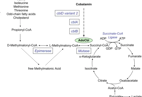
Methylmalonyl-CoA (MMCoA), theD-isomer, is a key metabolite in the catabolism of propionate, which is derived from the breakdown of four amino acids, pyrimidines, odd-chain fatty acids and cholesterol side-chains (Fig. 1). D-MMCoA is converted to succinyl-CoA through its L form by the successive action of
DOI 10.1007/s10545-008-0839-4
Communicating editor: Marinus Duran Competing interests: None declared
References to electronic databases: Methylmalonic aciduria due to methylmalonyl-CoA mutase deficiency, mut0and mutj defects: OMIM 251000. cblA: OMIM 251100. cblB: OMIM 251110. cblD: OMIM 277410. Cobalamin adenosyltransferase: EC 2.5.1.17. MMCoA mutase: EC 5.4.99.2.
B. Fowler (*)
Metabolic Unit, University Children_s Hospital, Roemergasse 8, Basel, CH-4058, Switzerland e-mail: Brian.Fowler@unibas.ch
J. V. Leonard
Institute of Child Health, London, UK M. R. Baumgartner
Division of Metabolism and Molecular Pediatrics, University Children_s Hospital, Zu¨rich, Switzerland
MMCoA racemase (epimerase) and MMCoA mutase. The mutase enzyme requires adenosylcobalamin (Ado-Cbl) as coenzyme so that the integrity of methylmalonic acid (MMA) metabolism is inextricably linked to vitamin B12(cobalamin), its adequate intake and correct uptake, transport and intracellular metabo-lism. Deficiency of MMCoA mutase leads to elevated levels of MMA in body fluids. In humans vitamin B12is a co-factor for one other enzyme, methionine synthase. This enzyme requires methylcobalamin (Me-Cbl) for activity. Deficiency of this enzyme results in accumula-tion of homocysteine and occurs in isolaaccumula-tion or com-bined with MMCoA mutase deficiency in certain genetic defects of intracellular cobalamin metabolism
as well as in nutritional deficiency or disturbed uptake or transport of vitamin B12 (Rosenblatt and Fenton 2001; Suormala et al 2004). In the combined defects (cblC, cblD and cblF) both MMA and homocysteine will accumulate and these defects must be borne in mind when characterizing methylmalonic aciduria.
Genetic disorders causing methylmalonic aciduria (MMAuria)
Several distinct mutant genetic classes, based on biochemical, enzymatic and genetic complementation analysis are known (Table1).
Succinate Fumarate Malate Oxaloacetate Citrate Isocitrate α -Ketoglutarate Succinyl-CoA Acetyl-CoA Pyruvate Lactate L-Methylmalony-CoA D-Methylmalonyl-CoA
free Methylmalonic Acid
Mutase Propionyl-CoA Valine Isoleucine Methionine Threonine Odd-chain fatty acids
Cholesterol AdoCbl Epimerase Succinate-CoA Ligase ATP GDP GTP Cobalamin cblD variant 2 cblA cblB ADP Fig. 1 Metabolic
interrelation-ships of methylmalonic acid. Epimerase, methylmalonyl CoA epimerase (EC:5.1.99.1); mutase, methylmalonyl-CoA mutase (EC 5.4.99.2); AdoCbl, adenosylcobalamin; Ligase, succinate-CoA ligase (EC 6.2.1.4); ADP, adenosine diphosphate; ATP, adenosine triphosphate; GDP, guanosine diphosphate; GTP, guanosine triphosphate
Table 1 Methylmalonic acidurias: complementation groups
Complementation group Impaired synthesis Primary defect Elevated metabolite
mut0/mutj Mutase apoenzyme MMCoA mutase MMA
cblA AdoCbl ? Mitochondrial Cbl import MMA
cblB AdoCbl ATP:cobalamin adenosyl-transferase MMA
cblD-Var2 AdoCbl ? Cytosolic Cbl transport MMA
cblC AdoCbl, MeCbl ? Cytosolic Cbl transport MMA, Hcy
cblD AdoCbl, MeCbl ? Cytosolic Cbl transport MMA, Hcy
cblF AdoCbl, MeCbl Lysosomal Cbl release MMA, Hcy
cblE MeCbl MS-reductase Hcy
cblG MeCbl Methionine synthase (MS) Hcy
cblD-Var1 MeCbl ? Cytosolic Cbl transport Hcy
MMCoA, methylmalonyl-coenzyme A; MMA, methylmalonic acid; Hcy, homocyst(e)ine; cbl, cobalamin; AdoCbl, adenosylcobala-min; MeCbl, methylcobalaadenosylcobala-min; MS, methionine synthase
Table 2 Summary of mutations causing MMAuria Enzyme/protein Structure Short name Gene Locus No. of exons ORF (bp) Pathogenic mutations References No. of mutations a Common mutations b Methylmalonyl-CoA mutase Homo- dimer Mutase MUT 6p21 13 2250 188 c.655A>T (p.N219Y) Ledley et al ( 1988 ) c.1106G>A (p.R369H) (mut 0 patients) Worgan et al ( 2006 ) c.2080C>T (p.R694W) (mut j patients) Lempp et al ( 2007 ) c.322C>T (p.R108C) (Hispanic patients) c.2150G>T (p.G717V) (Black patients) Methylmalonyl-CoA epimerase Homo- dimer MMCoA epimerase MCEE 2p13.3 3 525 3 None Bikker et al ( 2006 ) Dobson et al ( 2006 ) Gradinger et al ( 2007 ) ? Mitochondrial translocation of vitamin B12 Homo- dimer cblA MMAA 4q31.1 7 1254 28 c.433C>T (p.R145X) Dobson et al ( 2002a ) ? Protection of mutase from inactivation c.592_595delACTG Lerner-Ellis et al ( 2004 ) Korotkova and Lidstrom ( 2004 ) Banerjee ( 2006 ) Merinero et al ( 2008 ) ATP-cobalamin adenosyltransferase Homo- trimer cblB MMAB 12q24 9 750 24 c.556C>T (p.R186W) Dobson et al ( 2002b ) Saridakis et al ( 2004 ) c.700C>T (p.Q234X) Lerner-Ellis et al ( 2006a ) Merinero et al ( 2008 ) c.197-1G>T ? Channeling of vitamin B12 to its cytosolic and mitochondrial targets ? cblD MMADHC 2q23.2 8 891 13 None Coelho et al ( 2008 ) Coelho et al ( 2008 ) ? Binding and intracellular trafficking of cobalamin ? cblC MMACHC 1p34.1 4 849 44 c.271dupA (R91KfsX14) Lerner-Ellis et al ( 2006b ) c.331C>T (R111X) Nogueira et al ( 2008 ) c.394C>T (R132X) aAs reported in The Human Gene Mutation Database ( http://www.hgmd.cf.ac.uk ). bMutations with an overall allele frequency of >10% were considered as common mutations. ORF, open reading frame; bp, base pairs.
The mut0and mutjdefects (OMIM 251000) belong to a single genetic complementation class and result from deficiency of MMCoA mutase apoenzyme due to mutations of the MUT gene (Ledley et al 1988). Differentiation of these defects is based on the presence (mutj) or absence (mut0) of residual enzyme activity in cultured fibroblasts and response to vitamin B12 in vitro and in vivo. MMCoA mutase (MCM, EC 5.4.99.2) is a mitochondrial enzyme requiring 5¶-deoxyadenosylcobalamin. The gene (MUT) maps to 6p21 and its full-length cDNA codes for a 750-amino-acid protein of 88.2 kDa. The gene contains 13 exons spanning more than 35 kb and so far approximately 200 mutations have been identified (Table 2). The enzyme contains an N-terminal mitochondrial leader sequence of 32 amino acids and the mature form is a dimer of identical subunits of 78.5 kDa. Each subunit contains two main functional domains, an N-terminal (ba)8 barrel (residues 88–422) with the substrate binding site and a C-terminal (ba)5 Ado-Cbl binding domain (residues 578–750). The two domains are connected by a long linking region (residues 423–577) (Fenton et al2001).
The cblA (OMIM 251100) and cblB (OMIM 251110) complementation groups are linked to processes unique to Ado-Cbl synthesis and cause isolated MMAuria. Genes for the cblA (MMAA) and cblB (MMAB) groups have recently been described (Dobson et al 2002a,b).
The MMAA gene is located at 4q31.1-q31.2 and spans about 17.1 kb containing 7 exons, but exon 1 is untranslated. The full-length cDNA codes for a 418-amino-acid protein of 46.5 kDa. At least 28 mutations have been identified in 37 cblA patients (Table2). The cblA protein contains an N-terminal mitochondrial leader sequence and cleavage site as well as Walker A and B ATP-binding motifs, a Mg2+-binding site and a GTP-binding site, but its exact function is unclear. It was first thought to be responsible for translocation of cobalamin into mitochondria prior to the final steps of AdoCbl synthesis (Dobson et al2002a). More recent evidence, however, points to a role of the protein in the assembly and stabilization of holo-MMCoA-mu-tase analogous to its bacterial homologue meaB. This enzyme forms a complex with MMCoA mutase, stimulates its activity and protects the enzyme from irreversible inactivation (Padovani and Banerjee2006).
The MMAB gene for cobalamin adenosyltransferase (EC 2.5.1.17) maps to 12q24; it consists of 9 exons extending over 18.87 kb and its full-length cDNA codes for a 250-amino-acid protein (27.3 kDa) (Dobson et al 2002b). The cblB enzyme contains a leader sequence and signal cleavage site consistent with localization to
the mitochondrion. At least 24 mutations associated with low Ado-Cbl synthesis and mainly involving amino acids from the active site have been found in patients in the cblB complementation group.
Defective synthesis of both Ado-Cbl and Me-Cbl occurs in the cblC, cblF and combined form of cblD complementation groups as well as in nutritional deficiency or disturbed uptake or transport of vitamin B12(for review see Whitehead2006).
The gene associated with the cblC defect is MMACHC which maps to chromosome 1p34.1; it comprises 5 exons and its full length cDNA contains 846 base pairs corresponding to a protein of 282 amino acids with a molecular weight of 31.7 kDa (Lerner-Ellis et al.2006b). The protein appears to locate to the cytosol and homol-ogy with bacterial cobalamin transport proteins suggests a role in intracellular channelling of Cbl (Table2).
The cblD complementation group (OMIM 277410) was first associated with combined MMAuria and homocystinuria, but it is now known that two addi-tional variant forms of this mutant class exist with either isolated homocystinuria (cblD-variant 1) or iso-lated MMAuria (cblD-variant 2) (Coelho et al 2008; Suormala et al2004).
The previously described single patient (Watkins et al. 2000) who was thought to belong to a new complementation group (cblH Watkins et al2000) also belongs to the cblD-variant 2 mutant class.
The cblF defect is caused by defective release of Cbl from lysosomes (Suormala et al2004) and its gene has not yet been identified.
Recently a few patients have been described with mild MMAuria associated with a mutation of the MMCoA epimerase gene (MCEE), but these patients had no consistent clinical phenotype, as outlined below.
Diagnostic approach
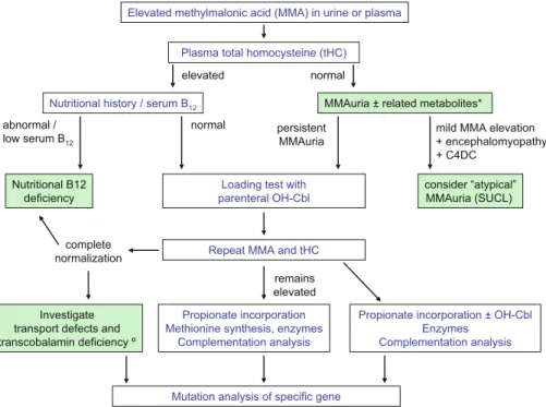
A comprehensive diagnostic approach involves inves-tigations at the level of metabolites, genetic comple-mentation analysis and enzymatic studies, and finally mutation analysis (Fig.2).
Metabolites
The measurement of MMA and related metabolites such as 3-hydroxypropionate and methylcitrate in urine provides a reliable first-line diagnostic approach in patients suspected to have a disorder of MMA metabolism.
In normal subjects MMA concentrations are age-dependent with median values (P95 limits) of 1.1 (4.8) mmol/mol creatinine below 30 days, 5.2 (49) mmol/mol creatinine at 1–6 months, and 0.8 (6.5) mmol/mol creatinine at 6–12 months of age (Boulat et al2003).
Elevated levels range from the order of 10–20 mmol/mol creatinine in mild disturbances of MMA metabolism to over 20 000 mmol/mol creatinine in severe MMCoA mutase deficiency (Fig.3) and can be accompanied by increased levels of a range of related
metabolites such as methylcitrate, 3-hydroxypropio-nate, lactate and propionylglycine.
Since several possible causes of elevated MMA are known (Table 3), each must be carefully considered, taking into account the clinical presentation and nutritional state of the patient (Fig. 2).
Although concentrations of MMA are generally higher in the genetic disorders than in absorption and transport defects or nutritional vitamin B12deficiency, there is considerable overlap. Thus the values them-selves are of limited value in the differential diagnosis because of this overlap (see Fig.3).
In addition, several factors such as protein intake, the catabolic state and associated metabolic decompensation, renal function as well as vitamin B12 status can greatly influence MMA levels, especially in urine. Also there appears to be wide variation in MMA levels independent of the changes in these factors. Furthermore, analytical variation as shown by results within the ERNDIM EQA scheme for organic acids can contribute to apparent varia-tion in individual subjects (http://www.erndim.unibas.ch). This variation makes comparison of concentrations obtained in different laboratories of limited value. Analytical improvements such as the use of GC-MS stable-isotope dilution methods and participation of labo-ratories in external QA will improve analytical variation. Use of MMA levels in plasma (normal <0.27 mmol/L) may well be a more reliable approach (Chandler et al 2007), but more evidence is needed.
CSF measurements of MMA (control values 0.19– 0.67 mmol/L) are generally not necessary for diagnostic Elevated methylmalonic acid (MMA) in urine or plasma
Plasma total homocysteine (tHC)
Investigate transport defects and transcobalamin deficiency º
MMAuria ± related metabolites* Nutritional history / serum B12
Loading test with parenteral OH-Cbl Nutritional B12
deficiency
Mutation analysis of specific gene Propionate incorporation Methionine synthesis, enzymes
Complementation analysis
Propionate incorporation ± OH-Cbl Enzymes Complementation analysis normal elevated normal remains elevated complete
normalization Repeat MMA and tHC
mild MMA elevation + encephalomyopathy + C4DC consider “atypical” MMAuria (SUCL) persistent MMAuria abnormal / low serum B12
Fig. 2 Diagnostic flowchart. *Related metabolites such as methylcitrate, 3-hydroxypropionate, lactate and propionylglycine. -For further details see Whitehead (2006). C4DC, succinyl- or methymalonylcarnitine >10'000 5'000 1'000 100 Mutº cblB MCEE Benign MMA Cbl A mut-SUCLG1 Vit B12 def Cbl C/D/F SUCLA2 mmol/mol creat.
Fig. 3 Urinary MMA levels in various types of disorders. The levels shown are approximations based on ranges of values reported in the literature. Note the arbitrary nonlinear scale. Complementation groups, mut0, mutj, cblA, cblB, cblC, cblD, cblF. MCEE, methylmalonyl-CoA epimerase deficiency; MMA, methylmalonic acid; SUCLG1, succinate-CoA ligase, alpha subunit; SUCLA2, succinate-CoA ligase, ADP-forming beta subunit
purposes although this may be useful in the investiga-tion of atypical forms, especially in those with mainly neurological involvement (Mayatepek et al1996).
Increasingly, measurement of acyl carnitine esters in dried blood spots is being applied to diagnosis of the MMAurias, in particular in extended newborn screen-ing programmes (Schulze et al 2003; Wilcken et al 2003). Note, however, that MMAuria is not included in all such schemes as, for example, in Germany (discontinued in 2005).
It is likely that in future, as more and more laboratories undertake metabolic screening and also newborn screening by acylcarnitine measurements, a more common marker of MMA metabolism will be C4-dicarboxylic acylcarnitine (C4DC) but this can be either methylmalonyl- or succinylcarnitine.
Plasma total homocysteine measurement (Fowler and Jakobs1998) is essential in the differential diag-nosis of elevated MMA levels of all degree to exclude combined forms of disorders of intracellular cobalamin metabolism as well as transport defects and nutritional causes (Fig.2).
Testing of the response to vitamin B12
Defects of the translocation and intracellular synthesis of the active cofactors of vitamin B12often respond to pharmacological doses of the vitamin with clinical and biochemical improvement. CblC and cblD defects are
routinely treated with hydroxocobalamin but not all patients with defects of synthesis of adenosylcobalamin (cblA and cblB) will respond. While most patients with cblA will improve, only about one-third of cblB patients will do so (Matsui et al 1983). This response was first noted in a patient reported by Rosenberg and his colleagues (Rosenberg et al 1968) and since then many responsive patients have been identified. Some patients in the mut complementation group will also respond, particularly mutj(Ho¨rster et al 2007). Mut0 patients have also been reported to have responded to parenteral vitamin B12(Matsui et al1983), but a genu-ine response is rare.
The difficulty in interpreting previous reports is that the testing with vitamin B12has never been standardized and what constitutes a response never clearly defined. In a survey of European centres there was a very marked variation in practice (Zwicker et al2008). Vitamin B12 was given as hydroxocobalamin intramuscularly or intravenously or as cyanocobalamin intramuscularly. The response was assessed by changes in plasma or urine methylmalonate or in blood spot propionylcarnitine. One to three specimens were collected before giving the vitamin and 1–7 specimens afterwards. The response was judged by a fall in metabolite concentration of between 10% and 90% or by a fall in methylmalonate to a value below 1000 mmol/mol creatinine. Clearly, using some of these criteria makes it doubtful that there was a genuine response and such variation in practice makes it impossible to make any comparisons.
A standardized test is proposed. The purpose of this test is to identify those patients who genuinely benefit from vitamin B12. It is important to remember that no child should be given unnecessary or ineffective treatment, particularly by injection.
Parallel to this test the enzymology including in vitro response to adenosylcobalamin and if possible the complementation group should be determined (see below). The vitamin B12test should not be delayed by waiting for the results of these tests.
1. The patient should be clinically stable on the same treatment for at least one month. The protein and energy intake should be specified.
2. If the patient is already receiving cobalamin, this should be stopped for at least one month before the test. If the patient appears to deteriorate, restart vitamin B12 and defer the test. Note: As a general rule patients with MMA excretion greater than 10 000 mmol/mol creatinine and those who are clinically unstable rarely respond to vitamin B12. 3. Baseline urine collections. At least three
speci-mens should be collected on different days. Plasma
Table 3 Causes of elevated MMA in plasma and urine Non-genetic
Nutritional deficiency
Subjects on strict vegetarian diet/vegans
(Breastfed) babies of vegan mothers or mothers with unrecognized disturbed vitamin B12metabolism Reduced intestinal absorption (elderly persons) Genetic disorders
Disorders of MMA-CoA conversion Methlylmalonyl-CoA mutase
Methlylmalonyl-CoA epimerase (racemase) Disorders of absorption and transport of cobalamin
Hereditary intrinsic factor deficiency Defective transport of Cbl by enterocytes (Imerslund–Gra¨sbeck syndrome) Haptocorrin (R Binder) deficiency Transcobalamin (TC) Deficiency Disorders of intracellular utilization of Cbl
Combined deficiencies of AdoCbl and MeCbl AdoCbl deficiency
Atypical MMAurias associated with encephalomyopathy SUCLG1, succinate-CoA ligase, alpha subunit
concentrations may be used only if a sensitive assay (stable-isotope dilution assay) is available. 4. Give hydroxocobalamin 1 mg intramuscularly on
three consecutive days.
5. After the cobalamin injection, collect urine (or plasma) specimens on alternate days for 10 days. 6. The urine or plasma samples should be analysed in
the same run in a laboratory participating in a recognized quality control scheme for methylma-lonic acid using GCMS.
7. A decrease of the mean urine or plasma MMA concentrations of more than 50% should be regarded as indicative of a beneficial response.
Enzymatic and complementation analysis
Once nutritional causes have been excluded, the complexity of causes of inherited isolated MMAuria necessitates characterization of the underlying defect in cultured skin fibroblasts using several assays.
The overall conversion of propionate to succinate is determined by the incorporation of label from [14C]propionate into cell proteins (Willard et al1976) (Fig. 4). Specific activity of MMCoA mutase can be measured using a radioactive substrate in the presence and absence of Ado-Cbl (Baumgartner1983) (Fig.5). Distinction of isolated MMAurias into mut0 or mutj is based on the somewhat arbitrary biochemical
assessment of in vitro response to hydroxocobalamin. Mut0 is defined as very low mutase activity together with a ratio of propionate incorporation with or without supplemented hydroxocobalamin lower than 1.5 which is the upper limit found in control cell lines. Mutj is defined as low to moderate residual mutase activity and a ratio of >1.5 although it must be borne in mind that in our series some cell lines showed borderline values of 1.2–1.7 and assays needed to be repeated several times to enable definite classification (Lempp et al2007).
Cobalamin adenosyltransferase can be measured by monitoring the conversion of OH-[57Co]cobalamin to Ado-Cbl. The reaction must be performed in the dark and under hydrogen to provide reducing conditions (Fenton and Rosenberg1981).
The cellular uptake of CN-[57Co]cobalamin and its conversion to cobalamin coenzymes can be measured in intact fibroblasts in monolayer culture. Human serum is used as a source of transcobalamin to facilitate cellular uptake (Fowler and Jakobs1998).
Complementation analysis is performed by measuring the incorporation of [14C]propionate into protein in patient cells mixed with cells of known complementation groups and treated with 40% polyethyleneglycol (PEG) which results in the formation of multinuclear cells (Fig.6) (Zavadakova et al2002).
In the investigation of combined MMAuria and homocystinuria the function of methionine synthase
0 1000 2000 3000 4000 5000 6000 7000
mut- mut° mut- mut°
MEM Ø MEM +
pmol /mg protein
Lower Control range
Fig. 4 Propionate incorporation assay in 66 mut0and 17 mutj cell lines. Intact fibroblasts in monolayer culture are incubated with [14C]propionate (0.2 mmol/L) in medium containing 15% serum for 16 h at 37-C. Then cells are harvested with trypsin followed by precipitation of protein with 5% trichloroacetic acid. Protein is solubilized in 1 mol/L NaOH . The protein concentra-tion is determined by Lowry and radioactivity is measured by
scintillation counting. Activity is expressed as pmol propionate fixed per mg protein per 16 h. Shaded bars represent the ranges with median values depicted by solid bars. MEM; = fibroblasts grown in minimal essential medium without added hydroxoco-balamin. MEM+ = fibroblasts grown in medium with 10 mg/L added hydroxocobalamin
can be determined by measurement of conversion of [14C]formate to methionine in intact fibroblasts in monolayer culture (Fowler et al1997).
The results of these assays, the most important being the determination of propionate incorporation and MMCoA mutase activity in fibroblasts grown in medium with and without supplemented hydroxoco-balamin, allow the reliable characterization of patients
with isolated MMAuria and identify the correct gene for mutation analysis (Table4).
Mutation analysis
Many different complementation classes exist and these cannot be distinguished by clinical presentation and/or response to high-dose parenteral vitamin B12 administration. Enzymatic and/or complementation analysis is necessary before proceeding to mutation analysis of single genes (Fig. 2). Table 2 summarizes present knowledge on genes and mutations relevant to isolated MMAuria.
Approaching 200 mutations of the MUT gene have been described in MMCoA mutase deficient patients (Lempp et al 2007; Worgan et al 2006). Most are private, although a few occur in between 10% and 20% of mutant alleles. The p.R694W mutation is associated with the mutj form (Lempp et al 2007). Two others have been identified in particular ethnic groups with a relatively high frequency (Worgan et al2006).
In the cblA defect the p.R145X mutation was found to be present in 43% of mutant alleles and was associ-ated with a common haplotype (Lerner-Ellis et al2004). The p.R186W mutation of the MMAB gene accounted for 33% of mutant alleles in cblB patients who were of European origin and presented with a severe clinical phenotype (Lerner-Ellis et al2006a). 0 50 100 150 200 250 300 350
mut- mut° mut- mut° mut- mut° mut- mut° MEM Ø, Ø AdoCbl MEM Ø, + AdoCbl MEM+, ØAdoCbl MEM+, +AdoCbl
Lower Control range
575 585
pmol/min/mg. protein
Fig. 5 Methylmalonyl-CoA mutase assay. Fibroblast extract is incubated with [14C]MMCoA (4 mmol/L) at pH 7.4, 37-C for 60 min, without (holomutase) and +50 mmol/L AdoCbl (total mutase). Free [14C]succinic acid is released by alkaline hydrolysis, unlabelled succinic acid is added as carrier and is separated by anion exchange HPLC. Fractions are collected and counted for
radioac-tivity. Activity is expressed as pmol succinate formed per minute per mg extract protein. Shaded bars represent the ranges, with median values depicted by solid bars. MEM; =fibroblasts grown in minimal essential medium without added hydroxocobalamin. MEM+ = fibroblasts grown in medium with 10 mg/L added hydroxocobalamin
Fig. 6 Complementation analysis of cblD-variant 2 cell line. Fibroblasts of a particular patient were mixed with fibroblasts belonging to known complementation groups, fused with poly-ethyleneglycol 1500 (PEG) treatment (shaded columns), and 3 days later propionate incorporation was measured. Parallel cultures of mixed unfused cells were used as background controls (unshaded columns). Self-fusions were used as a negative control
Mutations of the recently identified gene responsible for the cblD defect (MMADHC) have been described in four subjects with combined MMAuria and homocysti-nuria and in four subjects with the isolated MMAuria form of this defect (Coelho et al2008).
At least 44 mutations have been described in the MMACHC gene responsible for the cblC defect (Lerner-Ellis et al 2006b, Nogueira et al 2008). The c.271dupA mutation occurred in about 40% of alleles and correlated with early-onset disease. In addition, two nonsense mutations, R111X and R132X, were found in 10–20% of mutant alleles. Interestingly, one of these (R132X) was associated with late-onset disease.
Miscellaneous disorders with mildly elevated MMA MCoA epimerase deficiency
Mutations of the gene (MCEE) coding for this enzyme deficiency have been reported in six patients. The first patient with MMA levels of 142–300 mmol/mol creatinine and increased propionylcarnitine (Bikker et al2006) was also affected by sepiapterin reductase deficiency with a clinical phenotype reminiscent of that disorder. Dobson and colleagues (2006) reported a child previously thought to have MMA due to the cblA defect who presented at 13.5 months with severe metabolic acidosis requiring intensive care, having pre-viously failed to thrive. MMA and methylcitrate were elevated in urine and plasma MMA was 11.1 mmol/L (normal <0.27). Urine MMA while on a normal protein intake with oral cobalamin was 180–1456 mmol/mol creatinine. Growth and mental develop-ment are normal. An older sister showed MMA levels of 95–166 mmol/mol creatinine on a self restricted low-protein intake but has remained asymptomatic.
It is likely that this enzyme deficiency leads to only a partial block in MMCoA catabolism owing to the ability of D-MMCoA to interconvert with L-MMCoA through deacetylation and acetylation of free MMA (Fig.1). This is reflected in the relatively modest eleva-tion of urine MMA as well as the only partially reduced levels of propionate incorporation into macromolecules in cultured fibroblasts. Gradinger and colleagues (2007) reported pathogenic mutations in 5 of 229 patients with unexplained MMAuria but there was no consis-tent clinical phenotype. Three of these five patients had two mutant alleles but propionate incorporation was decreased in only two. The other two subjects were only heterozygous for a single MCEE mutation and normal propionate incorporation was found in their cultured fibroblasts.
Thus conclusions on the clinical consequences of MMACoA epimerase deficiency cannot be made. Atypical MMA with neurological symptoms due to SUCL mutations
The succinate-CoA ligase (SUCL) enzyme complex catalyses the conversion of succinyl-CoA to succinate in the tricarboxylic acid cycle (Fig. 1). Two forms of the complex exist, each comprising a common alpha unit and one of two different beta subunits which impose specificity for either GDP or ADP in the reaction. Mutations of the ADP-forming beta subunit SUCLA2 are associated with depletion of mitochon-drial DNA (Elpeleg et al. 2005) and deficiency of complexes I, III and IV of the respiratory chain. Further reported patients (Carrozzo et al. 2007; Ostergaard et al. 2007a) characterized by Leigh-like encephalomyopathy, dystonia and deafness have been shown to excrete mildly elevated levels of MMA. The mild MMAuria ranged from 16 to 212 mmol/mol
cre-Table 4 Methylmalonic acidurias: findings in fibroblast assays (N = normal)
Medium OHCbl Propionate incorporation MMA-CoA mutase CN-[57Co]Cbl uptake
jAdoCbl +AdoCbl Total AdoCbl MeCbl
mut0 j , T, , N N N + , , , mutj j , T, , N TN N + upto N , TN cblA j , T, N N , N + N T, N cblB j , T, N N , N + , or N , N cblD-var2 j , T, N N , N + N , N
atinine and is accompanied by a range of increased metabolites such as methylcitrate, 3-hydroxypropionate, propionylcarnitine, MMA, methylmalonylcarnitine, succinylcarnitine, lactate, citrate and succinate. Thus MMA is likely to constitute a marker for the disease rather than to be responsible for the phenotype.
Fatal lactic acidosis of neonatal onset with mtDNA depletion was recently described in two patients with mutations in the alpha subunit, SUCLG1 (Ostergaard et al. 2007b) with mildly increased urinary MMA (68 mmol/mol creatinine) in one of them.
Further cases, some of which have been reported previously (Yano et al.2003) presented with a milder phenotype including developmental delay, generalized muscular hypotonia and bilateral papillary oedema but without deafness (A. Burlina, C. Dionisi-Vici and R. Wevers, personal communication 2007). One of them showed increased urinary MMA (285 mmol/mol cre-atinine) and related organic acids and elevated pro-pionylcarnitine and methylmalonylcarnitine (Yano et al2003; patient 1).
In conclusion, several possible causes of elevated MMA are known (Table3) and each must be carefully considered in relation to the clinical presentation and nutritional state of the patient. The diagnostic ap-proach and methods described here allow a definitive classification of this heterogeneous group of disorders. It must be emphasized that such investigations need to be performed in laboratories with comprehensive experience. Reliable classification of these patients is essential for ongoing and future prospective studies on treatment and outcome.
References
Banerjee R (2006) B12trafficking in mammals: a case for coenzyme escort service. ACS Chem Biol 1: 149–159.
Baumgartner R (1983) Activity of the cobalamin-dependent methylmalonyl-CoA mutase. In: Hall CA, ed. The Cobalamins, Methods in Hematology, vol. 10, Edinburgh, New York: Churchill Livingston, 181–195.
Bikker H, Bakker HD Abeling NGGM, et al (2006) A homo-zygous missense mutation in the methylmalonyl-CoA epim-erase gene (MCEE) results in mild methylmalonic aciduria. Hum Mutat 27: 640–643.
Boulat O, Gradwohl M, Matos V, Guignard JP, Bachmann C (2003) Organic acids in the second morning urine in healthy Swiss paediatric population. Clin Chem Lab Med 41: 1642– 1658.
Carrozzo R, Dionisi-Vici C, Steuerwald U, et al (2007) SUCLA2 mutations are associated with mild methylmalonic aciduria, leigh-like encephalomyopathy, dystonia and deafness. Brain 130: 862–874.
Chandler RJ, Sloan J, Fu H, et al (2007) Metabolic phenotype of methylmalonic acidemia in mice and humans: the role of skeletal muscle. BMC Med Genet 8: 64 [Epub ahead of print].
Coelho D, Suormala T, Stucki M, et al (2008) Gene identifica-tion for the cblD defect of vitamin B12metabolism. N Engl J Med 358: 1454–1464.
Dobson MC, Wai T, Leclerc D, et al (2002a) Identification of the gene responsible for the cblA complementation group of vitamin B12-responsive methylmalonic acidemia based on analysis of prokaryotic gene arrangements. PNAS 99: 15554–15559.
Dobson MC, Wai T, Leclerc D, et al (2002b) Identification of the gene responsible for the cblB complementation group of vitamin B12-dependent methylmalonic aciduria. Hum Mol Genet 11: 3361–3369.
Dobson MC, Gradinger A, Longo N, et al (2006) Homozygous nonsense mutation in the MCEE gene and siRNA suppres-sion of methylmalonyl-CoA epimerase expressuppres-sion: A novel cause of mild methylmalonic aciduria. Mol Genet Metab 88: 327–333.
Elpeleg O, Miller C, Hershkovitz E, et al (2005) Deficiency of ADP-forming succinyl-CoA synthase activity is associated with encephalomyopathy and mitochondrial depletion. Am J Hum Genet 76: 1081–1086.
Fenton WA, Rosenberg LE (1981) The defect in the cbl B class of human methylmalonic acidemia: deficiency of cob(I)ala-min adenosyltransferase activity in extracts of cultured fibroblasts. Biochem Biophys Res Commun 98: 283–289. Fenton WA, Gravel RA, Rosenblatt DS (2001) Disorders of
propionate and methylmalonate metabolism. In: Scriver CR, Beaudet AL, Sly WS, Valle D, eds; Childs B, Kinzler KW, Vogelstein B, assoc, eds. The Metabolic and Molecular Bases of Inherited Disease, 8th edn. New York: McGraw-Hill, 2165–2193.
Fowler B, Jakobs C (1998) Post- and prenatal diagnostic methods for the homocystinurias. Eur J Pediatr 157(Sup-plement 2): S88–93.
Fowler B, Whitehouse C, Wenzel F, Wraith JE (1997) Methio-nine and serine formation in control and mutant human cultured fibroblasts: evidence for methyl group trapping and characterisation of remethylation defects. Pediatr Res 41: 145–151.
Gradinger AB, Be´lair C, Worgan LC, et al (2007) Atypical methylmalonic acidurias: frequency of mutations in the methylmalonyl CoA epimerase gene (MCEE). Hum Mutat 28: 1045.
Ho¨rster F, Baumgartner MR, Viardot C, et al (2007) Long-term outcome in methylmalonic acidurias is influenced by the underlying defect (mut0, mutj, cblA, cblB). Pediatr Res 62: 225–230.
Korotkova N, Lidstrom ME (2004) MeaB is a component of the methylmalonyl-CoA mutase complex required for protec-tion of the enzyme from inactivaprotec-tion. J Biol Chem 279: 13652–13658.
Ledley FD, Lumetta MR, Zoghbi HY, Van Tuinen P, Ledbetter SA, Ledbetter DH (1988) Mapping of human methylma-lonyl CoA mutase (MUT) locus on chromosome 6. Am J Hum Genet 42: 839–846.
Lempp TJ, Suormala T, Siegenthaler R, et al (2007) Mutation and biochemical analysis of 19 probands with mut0and 13 with mutjmethylmalonic aciduria: Identification of seven novel mutations. Mol Genet Metab 90: 284–290.
Lerner-Ellis JP, Dobson MC, Wai T, et al (2004) Mutations in the MMAA gene in patients with the cblA disorder of vitamin B12metabolism. Hum Mutat 24: 509–516.
Lerner-Ellis JP, Gradinger AB, Watkins D, et al (2006a) Mutation and biochemical analysis of patients belonging to the cblB complementation class of vitamin B12-dependent methylmalonic acidurias. Mol Genet Metab 87: 219–225.
Lerner-Ellis JP, Tirone JC, Pawelek PD, et al (2006b) Identifi-cation of the gene responsible for methylmalonic aciduria and homocystinuria, cblC type. Nat Genet 38: 93–100. Matsui SM, Mahoney MJ, Rosenberg LE (1983) The natural
history of the inherited methylmalonic acidemias. N Engl J Med 308: 857–861.
Mayatepek E, Hoffmann GF, Baumgartner R, et al (1996) Atypical vitamin B12-unresponsive methylmalonic aciduria in a sibship with severe progressive encephalomyelopathy: a new genetic disease? Eur J Pediatr 155: 398–403.
Merinero B, Pe´rez B, Pe´rez-Cerda C, et al (2008) Methylmalonic acidaemia: Examination of genotype and biochemical data in 32 patients belonging to mut, cblA or cblB complemen-tation group. J Inherit Metab Dis 31: 55–66.
Nogueira C, Aiello C, Cerone R, et al (2008) Spectrum of MMACHC mutations in Italian and Portuguese patients with combined methylmalonic aciduria and homocystinuria, cblC type. Mol Genet Metab 93: 475–480.
Ostergaard E, Hansen FJ, Sorensen N, et al (2007a) Mitochon-drial encephalomyopathy with elevated methylmalonic acid is caused by SUCLA2 mutations. Brain 130: 853–861. Ostergaard E, Christensen E, Krsitensen E, et al (2007b)
Deficiency of the a subuinit of succinate-CoA ligase causes fatal infantile lactic acidosis with mtDNA depletion. Am J Hum Genet 81: 383–387.
Padovani D, Banerjee R (2006) Assembly and protection of the radical enzyme, methylmalonyl-CoA mutase, by its chaper-one. Biochemistry 45: 9300–9306.
Rosenberg LE, Lilljeqvist A, Hsia YE (1968) Methylmalonic aciduria: metabolic block localization and vitamin B12 dependency. Science 162: 805–807.
Rosenblatt DS, Fenton WA (2001) Inherited disorders of folate and cobalamin transport and metabolism. In: Scriver CR, Beaudet AL, Sly WS, Valle D, eds; Childs B, Kinzler KW, Vogelstein B, assoc, eds. The Metabolic and Molecular Bases of Inherited Disease, 8th edn. New York: McGraw-Hill, 3897–3933.
Saridakis V, Yakunin A, Xu X, Anandakumar P, et al (2004) The structural basis for methylmalonic aciduria. The crystal structure of archaeal ATP: cobalamin adenosyltransferase. J Biol Chem 279: 23646–23653.
Schulze A, Lindner M, Kohlmuller D, Olgemoller K, Mayatepek E, Hoffmann GF (2003) Expanded newborn screening for inborn errors of metabolism by electrospray ionization– tandem mass spectrometry: results, outcome, and implica-tions. Pediatrics 111: 1399–1406.
Suormala T, Baumgartner MR, Coelho D, et al (2004) The cblD defect causes either isolated or combined deficiency of methylcobalamin and adenosylcobalamin synthesis. J Biol Chem 279: 42742–42749.
Watkins D, Matiaszuk N, Rosenblatt DS (2000). Complementa-tion studies in the cblA class of inborn error of cobalamin metabolism: evidence for interallelic complementation and for a new complementation class (cblH). J Med Genet 37: 510–513.
Whitehead M (2006) Acquired and inherited disorders of cobala-min and folate in children. Br J Hematol 134: 125–136. Wilcken B, Wiley V, Hammond J, Carpenter K (2003) Screening
newborns for inborn errors of metabolism by tandem mass spectrometry. N Engl J Med 348: 2304–2312.
Willard HF, Ambani LM, Hart AC, Mahoney MJ, Rosenberg LE (1976) Rapid prenatal and postnatal detection of inborn errors of propionate, methylmalonate and cobalamin metabo-lism. A sensitive assay using cultured cells. Hum Genet 34: 277–283. Worgan LC, Niles K, Tirone JC, et al (2006) Spectrum of mutation in mut methylmaonic acidemia and identification of a common hispanic mutation and haplotype. Hum Mutat 27: 31–43. Yano S, Li L, Le TP, et al (2003) Infantile mitochondrial DNA
depletion syndrome associated with methylmalonic aciduria and 3-methylcrotonyl-CoA and propionyl-CoA carboxylase deficiencies in two unrelated patients: a new phenotype of mtDNA depletion syndrome. J Inherit Metab Dis 26: 481–488.
Zavadakova P, Fowler B, Zeman J, Suormala T, Pristoupilova K, Kozich V (2002) CblE type of homocystinuria due to methionine synthase reductase deficiency: clinical and molecular studies and prenatal diagnosis in two families. J Inherit Metab Dis 25: 461–476.
Zwicker T, Lindner M, Ibrahim HI, et al (2008) Diagnostic work-up and management of patients with isolated methyl-malonic acidurias in European metabolic centres. J Inher Metab Dis. doi:10.1007/s10545-008-0804-2.
![Fig. 4 Propionate incorporation assay in 66 mut 0 and 17 mut j cell lines. Intact fibroblasts in monolayer culture are incubated with [ 14 C]propionate (0.2 mmol/L) in medium containing 15%](https://thumb-eu.123doks.com/thumbv2/123doknet/14862813.635999/7.892.204.688.679.951/propionate-incorporation-intact-fibroblasts-monolayer-incubated-propionate-containing.webp)
![Fig. 5 Methylmalonyl-CoA mutase assay. Fibroblast extract is incubated with [ 14 C]MMCoA (4 mmol/L) at pH 7.4, 37-C for 60 min, without (holomutase) and +50 mmol/L AdoCbl (total mutase)](https://thumb-eu.123doks.com/thumbv2/123doknet/14862813.635999/8.892.180.715.85.390/methylmalonyl-mutase-fibroblast-extract-incubated-mmcoa-holomutase-adocbl.webp)
