Christoph Zubler Bernard Mengiardi Marius R. Schmid Juerg Hodler Bernhard Jost Christian W. A. Pfirrmann Received: 12 February 2006 Revised: 27 May 2006 Accepted: 1 August 2006
Published online: 12 October 2006 # Springer-Verlag 2006
MR arthrography in calcific tendinitis
of the shoulder: diagnostic performance
and pitfalls
Abstract The purpose was to assess the diagnostic performance of MR arthrography to diagnose calcific ten-dinitis of the shoulder and to assess the reasons for diagnostic errors. Standard MR arthrograms of 22 patients with calcific tendinitis and 61 controls were retrospectively analyzed by two inde-pendent and blinded radiologists. All cases were consecutively collected from a database. Conventional radio-graphs were available in all cases serving as gold standard. The supra-spinatus was involved in 16, the infraspinatus in four and the subscap-ularis in two patients. All diagnostic errors were analyzed by two addi-tional readers. Reader 1 correctly detected 12 of the 22 shoulders with and 42 of the 61 shoulders without calcific tendinitis (sensitivity 0.55, specificity 0.66). The corresponding values for reader 2 were 13 of 22 and 40 of 61 cases (sensitivity 0.59,
spec-ificity 0.69). Inter-rater agreement (kappa-value) was 0.42. Small size of the calcific deposits and isointensity compared to the surrounding tissue were the most important reasons for false negative results. Normal hypointense areas within the supra-spinatus tendon substance and attach-ment were the main reason for false positive results. In conclusion, MR arthrography is insufficient in the diagnosis of calcific tendinitis. Nor-mal hypointense parts of the rotator cuff may mimic calcific deposits and calcifications may not be detected when they are isointense compared to the rotator cuff. Therefore, MR imag-ing should not be interpreted without corresponding radiographs.
Keywords Calcification . Joint . Magnetic resonance imaging . Shoulder . Tendons
Introduction
Approximately 50% of patients with calcific tendinitis have shoulder pain [1,2]. Acute calcific tendinitis of the rotator cuff clinically is characterized by acute onset of severe shoulder pain, local tenderness and limited range of motion. The diagnosis is typically made based on standard radiographs of the shoulder. In the acute phase calcifica-tions tend to be cloudy and may even become apparent in the subacromial/subdeltoid bursa. In most cases, clinical symptoms resolve spontaneously within 7–10 days. How-ever, the disease may become chronic [3]. In patients with chronic calcific tendinitis symptoms are often
uncharacter-istic with painful restriction of range of motion and limitation of activities of daily living. These patients will eventually be referred to an imaging center for further evaluation. Since symptoms are uncharacteristic and differ from the classic symptoms of rotator cuff lesions and instability, MR imaging of the shoulder is often requested. MR imaging answers many of the typical questions such as rotator cuff tears. MR arthrography may diagnose additional, often subtle abnormalities which may by associated with uncharacteristic clinical symptoms includ-ing biceps tendinopathy [4], pulley lesions [5], lesions of the biceps anchor [6], abnormalities of the rotator cuff interval [7] and cartilage abnormalities [8]. In our C. Zubler . B. Mengiardi .
M. R. Schmid . J. Hodler . C. W. A. Pfirrmann (*) Radiology,
University Hospital Balgrist, Balgrist, Forchstrasse 340, CH-8008 Zurich, Switzerland e-mail: [email protected] Tel.: +41-1-3863305 Fax: +41-1-3863319 B. Jost Orthopedic Surgery, University Hospital Balgrist, Balgrist, Forchstrasse 340, CH-8008 Zurich, Switzerland
institution MR arthrography is the standard examination to address these questions. Standard radiographs are some-times not available to the radiologists at the time of reporting or calcifications are not visible on the radiographs because of their localization or due to insufficient quality of the radiographs. In such situations it would be desirable to be able to suggest a calcific tendinitis on standard MR images or a MR arthrogram.
To our knowledge, no reports about the diagnostic performance and the diagnostic pitfalls of MR imaging in the detection of calcific tendinitis of the rotator cuff have been published in the radiology literature.
The purpose of our study was to assess the sensitivity and specificity of MR arthrography for the detection of calcific tendinitis in the rotator cuff tendons and to assess the reasons for diagnostic errors.
Materials and methods
Patients and control subjects
Twenty-two patients with a final clinical diagnosis of calcific tendinitis were included in a consecutive fashion using a database. Following inclusion criteria used: (a) availability of a standardized set of radiographs pf the shoulder perfomed at our institution (anteroposterior radio-graph with the humerus in neutral and position and in internal rotation, a supraspinatus outlet view and an axillary view) demonstrating unequivocally a calcific deposit, (b) availability of MR arthrography of the shoulder performed at our institution, (c) no prior shoulder surgery, (d) no full thickness rotator cuff tear. There were 8 men and 14 women. The patients’ age was between 36 to 85 years (mean, 52.1 years). The age range of the women was 36 to 59 years (mean, 47.6 years) and for the men 44 to 85 years (mean, 59.0 years). There were 15 right and seven left shoulders.
The control group was chosen during the same period of time based on the following criteria: (a) availability of a standardized set of radiographs pf the shoulder perfomed at our institution (anteroposterior radiograph with the humer-us in neutral and position and in internal rotation, a supraspinatus outlet view and an axillary view), (b) availability of MR arthrography of the shoulder performed at our institution within three month from the conventional radiographs, (c) no calcific tendinitis, (d) no full thickness rotator cuff tear, (e) no prior shoulder surgery. There were 61 patients (21 women and 40 men). The patients’ age was between 16 and 76 years (mean, 43.2). The age range for women was 16 to 76 years (mean, 49 years) and for men was 18 to 61 years (mean, 40.4 years). There were 33 right and 28 left shoulders.
Our institutional review board does not require its approval or informed consent for the review of patient records or images. Patient rights are protected by a law that requires patients to be informed at the time of examination
about the possibility that their medical records and radiographs will be reviewed for scientific purposes.
MR protocol
MR imaging was performed with a 1.5T system (Sympho-ny; Siemens Medical Solutions, Erlangen, Germany). All examinations were performed according to a standard protocol. 10 mL of diluted (2 mmol/L) gadopentetate dimeglumine (Magnevist; Schering, Berlin, Germany), were injected under fluoroscopic control after verification of intraarticular needle position with 1 ml iodinated contrast material. The MR examination was performed with a 4-channel shoulder coil. The arm position was standardized, with the thumb pointing upward. The MR protocol included oblique coronal proton density-weighted fat saturated fast spin-echo images (TR 2350 ms/TE 13 ms) and T2-weighted fat saturated fast spin-echo images (TR 3000 ms/TE 91 ms; section thickness, 3 mm; field of view, 16 cm), oblique coronal T1-weighted fat saturated spin-echo images (TR 792 ms/ TE 12 ms; section thickness, 3 mm; field of view, 16 cm); oblique sagittal T1-weighted spin echo images (TR 500 ms/TE 12 ms; section thickness, 4 mm; field of view, 16 cm) and transverse T1-weighted spin echo images (TR 500 ms/TE 12 ms; section thickness, 3 mm; field of view, 16 cm). The data acquisition matrix was 512×512 pixels.
Standard radiographs
Radiographs of the shoulder were obtained in a standard-ized fashion using a digital computed radiography system: A neutral and an internally rotated anteroposterior radio-graph, a cross-table view and a supraspinatus outlet view were obtained.
Analysis of MR images
The MR images were evaluated separately by two rearders (a fellowship trained musculoskeletal radiologist with three years experience in musculoskeletal MR (B.M.) and a staff radiologist (M.R.S.) with six years experience in muscu-loskeletal MR) blinded with regard to clinical and radio-graphic findings in a randomized fashion. The evaluation was performed on a PACS workstation. All sequences were available and used for the analysis. The evaluation was performed after a teaching session including six patients with different appearances of calcific tendinitis. These six cases were not included in the study group because they did not fulfill at least one of the inclusion criteria (most cases did not have all four plain radiographs available). Both readers were unaware of the number of cases with calcific tendinitis and the number of control cases.
The following qualitative findings were evaluated. The criteria were chosen by one of the authors based on own clinical experiences. This author was not involved in the image analysis:
(a) signal abnormalities consistent with calcific tendinitis [0: no focal signal alteration, 1: hypointensity on T1-and T2-weighted images in comparison to the surrounding tendon, and 2: mixture of hypo- and hyperintensity on T1- and T2-weighted images (salt and pepper appearance)],
(b) potential ancillary signs of calcific tendinitis [focal swelling of the tendon, bursal fluid (0: none, 1: up to 2 mm and 2: equal or more than 2 mm)],
(c) general appearance of the tendon (normal, tendino-pathy, and partial tear).
If a calcification was found the readers noted the location of the deposit within the tendon (bursal/central or articular) and measured its medio-lateral, cranio-caudal and antero-posterior dimensions to the nearest millimeter. A tendino-pathy was defined by altered signal on T1w images and normal signal on fluid sensitive sequences, without con-trast material entering the tendon substance. Partial tears were diagnosed with additional high signal within the tendon substance on fluid sensitive sequences or contrast material entering the tendon substance.
Analysis of standard radiographs
All measurements described below were performed by one of the authors (Z.Z.) on a PACS workstation. The following findings were noted: Localization of calcific deposits (supraspinatus, infraspinatus, subscapularis). The calcific deposits were localized using the three dimensions which were available by the different projections of the radio-graphs of the shoulder which were perpendicular to each other (anteroposterior, a cross-table and supraspinatus outlet). The medio-lateral, cranio-caudal and antero-poste-rior dimensions of the largest calcification was measured to the nearest millimeter.
Error analysis
Two of the authors not involved in the evaluation of the MR images and the radiographs (C.W.A.P. with eight years and C.Z. two years experience of musculoskeletal imaging) reviewed the MR images in the presence of the standard radiographs. This evaluation was the basis of the error analysis in which the following types of false negative errors were differentiated:
(a) Small size of calcific deposits
(b) Signal of calcific deposits isointense to surrounding tendon tissue on all sequences
The following sources for false positive results were differentiated:
(a) Focal hypointensity of the supraspinatus near the humeral insertion, seen on all sequences and probably representing a normal variation of the tendon.
(b) Focal area of thickening and hypointensity on all sequences at the anterior aspect of the supraspinatus. (c) Hypointense spots seen on all sequences at the
transition between supra- and infraspinatus tendons mimicking a salt and pepper appearance.
(d) Sclerotic, irregular bone changes at the greater tuberosity.
Statistics
The standard radiographs were used as the standard of reference. Sensitivity, specificity, accuracy, positive and negative predictive value were calculated for both blinded readers. Inter-rater agreement was described with kappa values.
Results
The diagnostic performance of MR imaging is presented in Table1. Reader 1 detected 12 of the 22 cases with calcific tendinitis and made a correctly negative diagnosis in 42 of 61 control patients (sensitivity of 0.55, specificity 0.66, accuracy 0.63). The corresponding values for reader 2 were 13 of 22 cases and 40 of 61 cases (sensitivity 0.59, specificity 0.69, accuracy 0.66). Inter-rater agreement (kappa value) was moderate (0.42).
Calcific tendinitis was localized in the supraspinatus tendon in 16 (73%) shoulders (bursal side: 7, central location: 6, articular side: 3). The infraspinatus was involved four (18%) times (all central). The subscapularis was involved twice (9%, both central). In 14 shoulders (64%) with calcific tendinitis a bursal fluid collection (Fig.1) was present, in four of these the maximum width was more than 2 mm. In the control group 21 cases (34%) presented with
Table 1 Diagnostic accuracy of MR arthrography for the diagnosis of calcific tendinitis Reader 1 Reader 2 True positive 12 13 True negative 40 42 False positive 21 19 False negative 10 9 Sensitivity 54.5% 59.1% Specificity 65.6% 68.9% Accuracy 62.7% 66.3%
Negative predictive value 36.4% 40.6% Positive predictive value 80.0% 82.4%
bursal fluid which measured more than 2 mm in five cases. Tendinopathy was diagnosed in ten cases (45%) with calcific tendinitis. In nine shoulders (41%) a partial tear was diagnosed. In the control group eight cases (13%) had a tendinopathy and 32 (52%) had a partial tear. Focal thickening of a rotator cuff tendon (Fig. 2) was seen in eight (36%) cases with calcific tendinitis an in four cases (7%) in the control group.
On MR images the calcific deposits were hypointense in comparison to the surrounding tendon in 16 of the 22 cases. In three cases the calcific deposits were irregular with a salt and pepper appearance (Fig. 3). Three calcific deposits were isointense with regard to the surrounding tissue on both T1- and T2-weighted sequences (Fig.4).
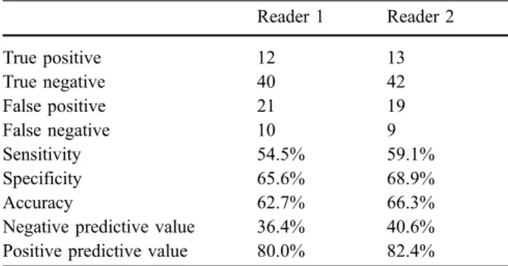
The mean size of the calcific deposits was 9.5×2.9×5.8 ([mm] medio-lateral × [mm] cranio-caudal × [mm] antero-Fig. 1 39-year-old woman with a true positive calcific tendinitis of
the supraspinatus tendon: The anteroposterior radiograph (left image) demonstrates a large calcific deposit (arrowheads). The corresponding coronal oblique T1-weighted fat-saturated (792/12) (middle image) and coronal oblique T2-weighed fat-saturated (3000/91) (right image)
MR images demonstrate the calcification as a hypointense structure within the tendon substance of the supraspinatus near the bursal surface and thickening of the tendon. Note small fluid collection in the subdeltoid bursa (arrow)
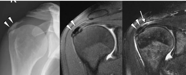
Fig. 2 42-year-old woman with a true positive calcific tendinitis of the supraspinatus tendon. The anteroposterior radiograph (left image) demonstrates a small calcific deposit (arrowheads). The correspond-ing coronal oblique T1-weighted fat-saturated (792/12) (middle image) and coronal oblique T2-weighed fat-saturated (3000/91)
(right image) MR images demonstrate the calcification as a small hypointense structure (white arrowhead) within the tendon substance. Note thickening of the tendon (black arrowheads) and fluid collection in the subdeltoid bursa (arrow)
posterior diameter) on standard radiographs and 8.1×4.6×6.5, on the MR images The size of the calcific deposits not detected on MR images was 5.3×2.9×3.9 mm.
Error analysis
The results of the error analysis are summarized in Table2. There were more false positive than false negative results.
False negative cases
False negative results were diagnosed in nine (reader 1) and ten cases (reader 2). In six (reader 1) and in seven cases (reader 2), the diagnostic error was considered to be caused by small size of the calcific deposits. In three cases (for both readers) the signal of the calcific deposits was isointense to the surrounding tissue and could not be detected even retrospectively (Fig.4).
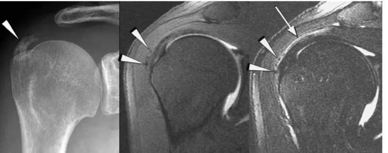
Fig. 3 52-year-old man with a true positive calcific tendinitis of the supraspinatus tendon: The anteroposterior radiograph (left image) demonstrates longitudinal calcific deposit (arrowheads). The corre-sponding coronal oblique T1-weighted fat-saturated (792/12) (middle image) and coronal oblique T2-weighed fat-saturated (3000/91) (right
image) MR images demonstrate the calcification as a small hypo-intense dots structure (salt and pepper appearance, arrowheads) at the bursal surface of the supraspinatus tendon and within the subdeltoid bursa. Note small fluid collection in the subdeltoid bursa (arrow)
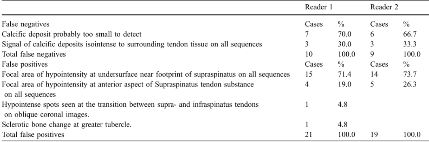
Fig. 4 32-year-old woman with a false negative calcific tendinitis of the supraspinatus tendon: The anteroposterior radiograph (left image) demonstrates a calcific deposit (arrowheads). In the corresponding
coronal oblique T1-weighted fat-saturated (792/12) (middle image) and coronal oblique PD-weighed fat-saturated (2350/13) (right image) MR images the calcification was not visible
False positive cases
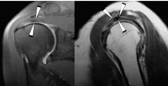
Reader 1 had 19 and reader 2, 21 false positive results. The most common error (reader 1: n=14, reader 2: n=15) related to a focal area of hypointensity at the undersurface of the supraspinatus tendon near the humeral insertion (Fig. 5). This hypointensity is seen on both T1- and T2-weighted sequences. The second most common presumed source for false positive results (reader 1: n=5, reader 2: n=4 cases) was a hypointensity seen within the substance of the supraspinatus tendon anteriorly (Fig.6).
Discussion
In 1907, Painter described calcifications in the shoulder [9]. McCarty and Gatter first recognized that these calcifica-tions are composed of hydroxyapatite (HA) [10,11]. HA
crystals are well recognized as a cause of a painful periarticular inflammatory condition. Some investigators also found HA crystals in the joint fluid which may lead to articular symptoms. This has been experimentally proven by intra-articular injection of HA which resulted in an acute inflammation in animal models [12].
In the acute phase of calcific tendinitis, spontaneous resorption may occur within a period of two to three weeks [2, 13]. This course may be typical for calcium deposits that appear translucent or cloudy and are not clearly circumscribed on standard radiographs [14]. Biopsy specimens from 18 patients suffering from calcific tendi-nitis have demonstrated cell-mediated calcification of living tissue. The process resembles incomplete endochon-dral ossification [15]. There is a significant correlation between severity of pain and histological signs of resorp-tion [16]. In patients with chronic calcific tendinitis, Table 2 Results of the error analysis
Reader 1 Reader 2
False negatives Cases % Cases %
Calcific deposit probably too small to detect 7 70.0 6 66.7
Signal of calcific deposits isointense to surrounding tendon tissue on all sequences 3 30.0 3 33.3
Total false negatives 10 100.0 9 100.0
False positives Cases % Cases %
Focal area of hypointensity at undersurface near footprint of supraspinatus on all sequences 15 71.4 14 73.7 Focal area of hypointensity at anterior aspect of Supraspinatus tendon substance
on all sequences
4 19.0 5 26.3
Hypointense spots seen at the transition between supra- and infraspinatus tendons on oblique coronal images.
1 4.8
Sclerotic bone change at greater tubercle. 1 4.8
Total false positives 21 100.0 19 100.0
Fig. 5 33-year-old woman with a false positive result: A focal area of hypointensity (arrow-heads) at undersurface near in-sertion of supraspinatus is seen on all sequences [coronal oblique intermediate-weighed fat-saturated (2350/13) (left image) and corresponding coro-nal oblique T1-weighted fat-saturated (792/12) (right image)]
calcifications are still present in more than 90 percent after three years [1,14].
Calcific tendinitis of the rotator cuff is common with a reported prevalence of 2.7% [1]. In 20 to 30 percent of patients with calcific tendinitis of the shoulder, both shoulders are involved [1]. The deposits are more common on the right side and are most frequently found in the supraspinatus tendon. Calcific tendinitis may remain asymptomatic [17], be associated with chronic pain or lead to acute exacerbation. It is important to recognize the imaging features of this condition to avoid unnecessary investigations and treatment [18].
The diagnosis of calcific tendinitis is typically made on standard radiographs. Axillary or supraspinatus outlet views are often required to identify involvement of the infraspinatus and subscapularis. Ultrasound (US) has been employed for diagnosis. On US images, calcification is seen as an echogenic focus with or without acoustic shadow. The presence of calcification can interfere with the diagnosis of a rotator cuff tear on US scans [19].
Calcific deposits are most typically associated with signal intensity loss on MR images. At high calcium concentrations (above 30–40%) susceptibility effects and decreases in proton density dominate, leading to signal intensity loss [20]. However, T1 shortening effects resulting in hyperintensity on T1-weighted images also are present. They have been attributed to surface interac-tion of protons with calcified tissue. At lower concentra-tions of calcium, T1 shortening effects dominate, resulting in isointensity or even hyperintensity [20]. Three of our patients had calcific deposits that were isointense to the surrounding tissue.
Gradient-echo sequences such as fast low angle shot (FLASH) or spoiled GRASS (SPGR) may better demon-strate calcific deposits than spin-echo, or STIR sequences due to an artifact consisting of a halo of hyperintense signal surrounding black dots [21]. Gradient-echo sequences may improve sensitivity of MR imaging in calcific tendinitis but are not part of our standard protocol.
Calcific tendinitis of the rotator cuff usually resolves with conservative therapy. In patients with acute or recurrent symptoms in the resorptive phase, needling and lavage can provide relief [22]. Due to scarring associated with this procedure orthopedic surgeons prefer arthro-scopic removal of the calcific deposits [23]. Recent studies have demonstrated promising results with extracorporeal shock wave therapy [24].
There are some study limitations to be considered. In our study we only used MR arthrography which is the standard examination to address the painful shoulder in our institution. Since calcific tendinitis is usually an intra tendinous process, intra-articular contrast material will have little effect on the appearance. Therefore, the same results may also be valid for standard MR examination. We have only used MR examinations which are done no longer than three month from the conventional radiographs. However, calcific deposits may change over time. This may be the reason for some diagnostic errors.
In conclusion, MR imaging is insufficient for the diagnosis of calcific tendinitis. Hypointense areas nor-mally present within the rotator cuff may mimic calcific deposits. Calcifications may be invisible because of the isointense signal compared to the rotator cuff. Therefore, MR imaging should not be interpreted without corre-sponding radiographs.
Fig. 6 41-year-old woman with a false positive result: A focal area of hypointensity (arrow-heads) is seen at anterior aspect of supraspinatus tendon sub-stance on all sequences [coronal oblique intermediate-weighed fat-saturated (2350/13) (left image) and corresponding sag-ittal oblique T1-weighted (500/12) image (right image)
References
1. Bosworth BM (1941) Calcium deposits in shoulder and subacromial bursitis: survey of 12,122 shoulders. JAMA 116:2477–2482
2. McKendry RJ, Uhthoff HK, Sarkar K, Hyslop PS (1982) Calcifying tendinitis of the shoulder: prognostic value of clinical, histologic, and radiologic features in 57 surgically treated cases. J Rheumatol 9:75–80
3. Ark JW, Flock TJ, Flatow EL, Bigliani LU (1992) Arthroscopic treatment of calcific tendinitis of the shoulder. Arthroscopy 8:183–188
4. Zanetti M, Weishaupt D, Gerber C, Hodler J (1998) Tendinopathy and rupture of the tendon of the long head of the biceps brachii muscle: Evalua-tion with MR Arthrography. AJR 170:1557–1561
5. Weishaupt D, Zanetti M, Tanner A, Gerber C, Hodler J (1999) Lesions of the reflection pulley of the long biceps tendon. MR arthrographic findings. Invest Radiol 34:463–469
6. Bencardino JT, Beltran J, Rosenberg ZS, Rokito A, Schmahmann S, Mota J, Mellado JM, Zuckerman J, Cuomo F, Rose D (2000) Superior labrum anteri-or-posterior lesions: diagnosis with MR arthrography of the shoulder. Radiology 214:267–271
7. Chung CB, Dwek JR, Cho GJ, Lektrakul N, Trudell D, Resnick D (2000) Rotator cuff interval: evaluation with MR imaging and MR arthrography of the shoulder in 32 cadavers. J Comput Assist Tomogr 24:738–743 8. Sanders TG, Tirman PF, Linares R,
Feller JF, Richardson R (1999) The glenolabral articular disruption lesion: MR arthrography with arthroscopic correlation [see comments]. AJR Am J Roentgenol 172:171–175
9. Painter CF (1907) Subdeltoid bursitis. Boston Med Surg J 156:345–349 10. McCarty DJ, Gatter RA (1964)
Identi-fication of calcium hydrogen phosphate dihydrate crystals in human fibrocarti-lage. Nature 201:391–392
11. McCarty DJ, Gatter RA (1966) Recur-rent acute inflammation associated with focal apatite crystal deposition. Arthri-tis Rheum 9:804–819
12. Dieppe PA, Crocker P, Huskisson EC, Willoughby DA (1976) Apatite de-position disease. A new arthropathy. Lancet 1:266–269
13. Harmon PH (1958) Methods and re-sults in the treatment of 2,580 painful shoulders, with special reference to calcific tendinitis and the frozen shoulder. Am J Surg 95:527–544 14. Gartner J, Heyer A (1995) Calcific
tendinitis of the shoulder. Orthopade 24:284–302
15. Uhthoff HK (1975) Calcifying tendini-tis, an active cell-mediated calcifica-tion. Virchows Arch A Pathol Anat Histol 366:51–58
16. Uhthoff HK, Sarkar K, Maynard JA (1976) Calcifying tendinitis: a new concept of its pathogenesis. Clin Orthop 164–168
17. Wright V, Haq AM (1976) Periarthritis of the shoulder. I. Aetiological con-siderations with particular reference to personality factors. Ann Rheum Dis 35:213–219
18. Hurt G, Baker CL Jr (2003) Calcific tendinitis of the shoulder. Orthop Clin North Am 34:567–575
19. Farin PU, Rasanen H, Jaroma H, Harju A (1996) Rotator cuff calcifications: treatment with ultrasound-guided per-cutaneous needle aspiration and lavage. Skeletal Radiol 25:551–554
20. Henkelman RM, Watts JF, Kucharczyk W (1991) High signal intensity in MR images of calcified brain tissue. Radiology 179:199–206
21. Beltran J, Marty-Delfaut E, Bencardino J, Rosenberg ZS, Steiner G, Aparisi F, Padron M (1998) Chondrocalcinosis of the hyaline cartilage of the knee: MRI manifestations. Skeletal Radiol 27: 369–374
22. Farin PU, Jaroma H, Soimakallio S (1995) Rotator cuff calcifications: treatment with US-guided technique. Radiology 195:841–843
23. Jerosch J, Strauss JM, Schmiel S (1998) Arthroscopic treatment of cal-cific tendinitis of the shoulder. J Shoulder Elbow Surg 7:30–37 24. Gerdesmeyer L, Wagenpfeil S, Haake
M, Maier M, Loew M, Wortler K, Lampe R, Seil R, Handle G, Gassel S, Rompe JD (2003) Extracorporeal shock wave therapy for the treatment of chronic calcifying tendonitis of the rotator cuff: a randomized controlled trial. Jama 290:2573–2580