C L I N I C A L R E S E A R C H
Clinical Stability of Slipped Capital Femoral Epiphysis
does not Correlate with Intraoperative Stability
Kai Ziebarth MD, Stephan Domayer MD, Theddy Slongo MD, Young-Jo Kim MD, PhD, Reinhold Ganz MD
Received: 24 August 2011 / Accepted: 22 March 2012 / Published online: 10 April 2012 Ó The Association of Bone and Joint Surgeons1 2012
Abstract
Background The most important objective of clinical classifications of slipped capital femoral epiphysis (SCFE) is to identify hips associated with a high risk of avascular necrosis (AVN) — so-called unstable or acute slips; however, closed surgery makes confirmation of physeal stability difficult. Performing the capital realignment pro-cedure in SCFE treatment we observed that clinical estimation of physeal stability did not always correlate with
intraoperative findings at open surgery. This motivated us to perform a systematic comparison of the clinical classi-fication systems with the intraoperative observations. Questions/purposes We asked: (1) Is the classification of an acute versus chronic slip based on the duration of symptoms sensitive and specific in detecting intraoperative disrupted physes in patients with SCFE? (2) Is the stable/ unstable classification system based on clinical symptoms sensitive and specific in detecting intraoperative disrupted physes in patients with SCFE?
Methods We retrospectively reviewed 82 patients with SCFE treated by open surgery between 1996 and 2009. We classified the clinical stability of all hips using the classi-fications based on onset of symptoms and on function. We classified intraoperative stability as intact or disrupted. We determined the sensitivity and specificity of two classifi-cation systems to determine intraoperative stability. Results Complete physeal disruption at open surgery was seen in 28 of the 82 hips (34%). With classification as acute, acute-on-chronic, and chronic, the sensitivity for disrupted physes was 82% and the specificity was 44%. With the classification of Loder et al., the values were 39% and 76%, respectively.
Conclusion Current clinical classification systems are lim-ited in accurately diagnosing the physeal stability in SCFE. Level of Evidence Level III, retrospective diagnostic study. See Guidelines for Authors for a complete descrip-tion of levels of evidence.
Introduction
Slipped capital femoral epiphysis (SCFE) affects the ado-lescent population with an incidence of 0.2 to 10 per 100,000 [4,32]. It is more common in males and usually Each author certifies that he or she, or a member of their immediate
family, has no commercial associations (eg, consultancies, stock ownership, equity interest, patent/licensing arrangements, etc) that might pose a conflict of interest in connection with the submitted article.
All ICMJE Conflict of Interest Forms for authors and Clinical Orthopaedics and Related Research editors and board members are on file with the publication and can be viewed on request. Each author certifies that his or her institution approved the human protocol for this investigation, that all investigations were conducted in conformity with ethical principles of research, and that informed consent for participation in the study was obtained.
K. Ziebarth (&), R. Ganz
Department of Orthopaedic Surgery, Inselspital, University of Bern, Freiburgstrasse, 3010 Bern, Switzerland
e-mail: [email protected]; [email protected] K. Ziebarth, T. Slongo
Department of Pediatric Surgery, Inselspital, University of Bern, Bern, Switzerland S. Domayer
Department of Orthopaedic Surgery, AKH Vienna, 1090 Vienna, Austria
Y.-J. Kim
Department of Orthopaedic Surgery, Children’s Hospital Boston, Boston, MA, USA
Clin Orthop Relat Res (2012) 470:2274–2279
DOI 10.1007/s11999-012-2339-y
and Related Research
®
manifests as pain in the hip or knee and reduced range of hip flexion and internal rotation [4]. The etiology remains unclear; however, epidemiologic data for geographic, racial, and seasonal variations suggest that environmental and genetic factors may influence the development of SCFE [4, 29]. Rapid growth, obesity, and hormonal dis-orders have been recognized as risk factors [6,8,19,48]. Greater displacement in patients with SCFE predicts the development of osteoarthritis [9,10,15,50]. Therefore, to prevent further slip progression, surgical fixation of the epiphysis is the recommended primary treatment [2,4,16,
34, 38, 51]. Most SCFE deformities occur with gradual displacement of the femoral head from the metaphysis and with risk of avascular necrosis (AVN) up to 4.6% [28]. However, abrupt and complete disconnection of the epiph-ysis from the metaphepiph-ysis — a so-called unstable slip — is not uncommon and has been associated with incidences of AVN ranging from 4.7% to 58% [5,13,24,36,37,44,47]. However, the exact cause for AVN developing in patients with SCFE remains unclear. Assuming that mechanical instability of the epimetaphyseal connection of the proxi-mal femur is one of the main causes for development of AVN, a classification scheme that accurately identifies hips with disconnection of the epiphyses from the metaphyses would be of great clinical value. Two clinical classifica-tions [3, 17, 35] have attempted to predict physeal instability by appreciation of the duration of symptoms [3,
17] or of the severity of symptoms regarding the ability to walk [35]; however, confirmation of their accuracy is dif-ficult and indirect [25,26]. In the classification based on the duration of symptoms [3,17], a SCFE was considered acute if the duration of symptoms was less than 3 weeks, as acute-on-chronic if symptoms were present intermittently for more than 3 weeks with a recent exacerbation, and as chronic if symptoms had been constantly present longer than 3 weeks. The classification by Loder et al. [35] focuses on the severity of symptoms as an indicator for mechanical stability; they considered SCFE as unstable if weightbearing on the affected limb was impossible with or without crutches. According to their system the recom-mended treatment for a stable or chronic slip is in situ fixation with pins or screws without regard for the slip
angle. In contrast, the treatment strategies regarding timing of treatment and method of reduction, whether it be open or closed, vary in unstable or acute cases [3,4,18,22,34,38,
39, 47, 49, 52]. Nevertheless, the incidence of AVN in unstable slips stabilized by pinning is high and reportedly ranges from 4.7% to 58% [1, 3, 11, 33, 39, 42, 47] as opposed to stable slips that reportedly have AVN develop in as much as to 4.6% of hips, depending on the amount of slip [2,12,28,35].
During a modified Dunn procedure through a surgical dislocation approach [31, 52], we observed that at times, clinically chronic [3, 17] or stable [35] slips had a dis-connection between the epiphysis and metaphysis. Conversely, hips classified as clinically acute [3, 17] or unstable [35] slips had a mechanically stable physis at the time of surgery. Based on such observations, we presumed current clinical classification schemes for physeal stability had limited diagnostic accuracy.
We therefore asked: (1) Is the classification of an acute versus chronic slip based on the duration of symptoms sensitive and specific in detecting intraoperative disrupted physes in patients with SCFE? (2) Is the stable/unstable classification system based on clinical symptoms sensitive and specific in detecting intraoperative disrupted physes in patients with SCFE?
Patients and Methods
Between 1996 and 2009, we treated 89 patients at two centers (University Bern and Children’s Hospital-Boston) with the modified Dunn procedure through a sur-gical hip dislocation approach [20,21, 31,52]. Sufficient clinical data based on the patients’ records were available for 82 hips to retrospectively classify the SCFE according to the acute/chronic [3,17] and stable/unstable [35] clas-sifications and to classify intraoperative physeal stability as intact or disrupted with visible and demonstrable mobility between metaphysis and epiphysis. Thirty-nine patients were female and 43 were male, with a mean age at surgery of 12 ± 1.7 years (range, 7–18 years), and the average duration of symptoms before surgery was 12 ± 20.7 weeks
Table 1. Characteristics of the patients with intact and disrupted physeal integrity*
Demographics Intraoperative physeal integrity p value
Intact (N = 54 patients) Disrupted (N = 28 patients)
Male:female 30:24 13:15 .291
Age 12.7 ± 1.88 11.7 ± 1.37 .045
Slip angle (degrees) 45 ± 16.7 52 ± 14.0 .02
Duration of symptoms (weeks) 13 ± 23.1 12 ± 15.0 .404
(range, 0–156 weeks) (Table1). The slip angle was as-sessed using the frog lateral or cross-table lateral radiographs and classified as mild, moderate, or severe [7,
45] (Table1). Minimum followup was 2 months (mean, 37 months; range, 2–96 months). Institutional review board approval was obtained for this study in both institutions.
The preoperative status of the hips was classified using the system based on the onset of symptoms [3,17]. Using the acute/chronic classification, 11 slips were classified as acute, 40 as acute-on-chronic, and 31 as chronic. With the stable/unstable classification [35], hips that allowed walk-ing without or with crutches were defined as stable and those that did not allow walking as unstable. Twenty-four patients were unable to walk and therefore were classified as unstable and 58 were stable (Table2).
Only hips classified as unstable, acute, or acute-on-chronic were operated on as emergencies. Subcapital realignment of the epiphysis was performed for the moderate and severe slips using surgical hip dislocation and an extended retinacular soft tissue flap (modified Dunn procedure) [14, 21, 30, 31], whereas mild stable slips only had surgical hip subluxation for osteoplasty of the prominent anterior metaphysis com-bined with pinning in situ. The technique of surgical hip dislocation has been described in detail [20]. Briefly, the dislocation approach includes a trochanteric flip osteotomy, and a Z-shaped capsular incision is used to access the joint. When we were concerned by lack of mechanical stability of the physis or when capsulotomy revealed a hematoma and/or visible disconnection, we avoided the risk of stretching or rupturing the retinacular vessels to the epiphysis by pinning the femoral head in situ before subluxation. To avoid tension or rupture of the retinaculum when reducing the slipped epiphysis, an extended retinacular flap was created [21]. The first step was careful subperiosteal resection of the part of the stable greater trochanter proximal to the physis including all external rotators. Proximally this dissection was extended onto the neck as a longitudinal incision of the periosteum anterior to the retinaculum and distally the dissection was extended starting with a longitudinal periosteal incision reaching the most proximal fibers of the gluteus maximus
tendon. The periosteum then was meticulously peeled off the lateral and posterior neck from the superior border of the lesser trochanter to the attachment of the retinaculum near the border of the epiphysis. The flap so created contains the deep branch of the medial femoral circumflex artery, the anastomoses with the inferior gluteal artery, and its retinac-ular end branches; it clearly is longer than with the retinacretinac-ular tunneling produced with the classic Dunn procedure [14] and therefore allows better compensation of adverse stretching during manipulation. After developing the posterolateral flap portion, we created an anteromedial flap containing a con-stant branch of the medial femoral circumflex artery, running in the synovial surface of Weitbrecht’s ligament and giving blood supply to the inferomedial portion of the epiphysis [43], again with strictly subperiosteal dissection. Both flaps were connected posteriorly and allowed circumferential access to the osseous neck. An important part of the procedure was resection of the callus formation on the posterior neck before manual reorientation of the epiphysis. Resection of callus from the posterior neck before reorientation of the epiphysis is documented in the reports of 68 of the 75 hips with sub-capital reorientation, including all 28 hips with complete physeal disruption.
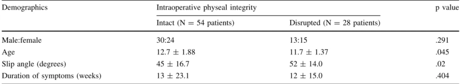
We intraoperatively classified physeal integrity as intact or disrupted. The mechanical stability of the physis was considered intact if the periosteum was intact and if several deep chisel cuts were necessary to separate the epiphysis as part of the reorientation procedure (Fig. 1). The physis was considered disrupted when the epiphysis was completely mobile without the need to free the physis (Fig.2). The presence of an intracapsular hematoma was not considered in this classification and was not always present. Integrity of the retinaculum and of its attachment on the epiphysis was evaluated by visual inspection at the time of surgical dislocation and presentation of the femoral head-neck junction. The intraoperative physeal integrity was intact in 54 hips and disrupted in 28 hips (Table2).
Table 2. Clinical classifications of the 82 hips with SCFE and intraoperative physeal integrity
Classification Chronic Acute-on-chronic Acute
Stable Unstable
Intact Disrupted
Chronic/acute 31 (38%) 40 (49%) 11 (13%)
Loder et al. 58 (71%) 24 (29%)
Physeal integrity 54 (66%) 28 (34%) SCFE = slipped capital femoral epiphysis.
Fig. 1 An intraoperative photograph shows the periosteum of the femoral head is stretched but intact (arrow).
We determined the sensitivity and specificity of the two clinical classification systems (acute/acute-on-chronic/ chronic and stable/unstable) to predict the presence of intraoperatively confirmed physeal instability (disrupted physis). All statistical analyses were performed with Microsoft1 Excel (Microsoft1, Redmond, WA, USA) and SPSS 16.0 (SPSS Institute, Chicago, IL, USA).
Results
For the classification based on duration of symptoms the sensitivity for a disrupted physis was 82% (23 of 28 hips) and the specificity was 44% (24 of 54 hips). Five of 28 hips (18%) were falsely negative (chronic symptoms but intra-operatively disrupted physeal integrity) and 30 of 54 (56%) were falsely positive (acute or acute-on-chronic symptoms but intraoperatively intact physeal integrity) (Table3).
The sensitivity of the stable/unstable classification for a disrupted physis was 39% (11 of 28 hips); however, the specificity was 76% (41 of 54 hips). Seventeen of 28 hips (61%) were falsely negative (clinically classified as stable hip but an intraoperatively disrupted physis) and 13 of 54 (24%) were falsely positive (clinically classified as unsta-ble hip but intraoperatively intact physeal integrity) (Table 4).
Discussion
Acute or unstable SCFEs reportedly are associated with rates of necrosis ranging from 4.7% to 58% [12,27,28,34,
35, 39–41, 46, 47, 49]. Assuming that mechanical insta-bility of the proximal femoral physis is one of the main causes for having AVN develop [35], it would be desirable to detect those cases accurately. The current preferred treatment of acute and unstable slips is percutaneous pin-ning strictly in situ or with gentle closed reduction, which precludes direct inspection of the physeal stability [4, 38,
48, 51]. Performing the capital femoral realignment pro-cedure based on the surgical dislocation approach [21,31,
52], we observed that clinically chronic or stable slips sometimes showed complete epimetaphyseal disruption at surgery and acute or unstable slips sometimes showed no epimetaphyseal disruption. These observations motivated us to perform a systematic comparison of the most com-monly used clinical classification systems with the intraoperative observations. We asked: (1) Is the classifi-cation of an acute versus chronic hip based on the duration of symptoms sensitive and specific in detecting intraoper-ative disrupted physes in patients with SCFE? (2) Is the stable/unstable classification system based on clinical symptoms sensitive and specific in detecting intraoperative disrupted physes in patients with SCFE?
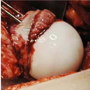
This study has limitations. First, in this retrospective study the definitions acute/chronic and stable/unstable were taken from clinical files that did not always allow rigorous categorization. Second, although using motion between epiphysis and metaphysis as a criterion of instability, some Fig. 2 The intraoperative photograph shows the femoral head is in a
dislocated position. The physis is disrupted, and the periosteum is torn (arrow). A mobile metaphyseal fragment with callous formation (triangle) is present. The femoral head was pinned prophylactically with two 3.0-mm threaded K wires before dislocation (square).
Table 3. Crosstable of physeal integrity and the acute/chronic clas-sification in 82 hips Acute or acute-on-chronic Intraoperative physeal integrity disrupted Total Yes No Yes 23 (82%*) 30 (56%) 53 No 5 (18%) 24 (44%**) 29 Total 28 54 82 * Sensitivity, **specificity.
Table 4. Crosstable of physeal integrity and the Loder classification in 82 hips Loder classification as unstable Intraoperative physeal integrity disrupted Yes No Total Yes 11 (39%*) 13 (24%) 24 No 17 (61%) 41 (76%**) 58 Total 28 54 82 * Sensitivity; **specificity.
hips looked stable at capsulotomy but proved to be unstable after dislocation with better visual and manual access to the epiphysis. It may be possible that dislocation of the hip can contribute to final destabilization of the epiphysis; however all questionable stable hips were prophylactically pinned before dislocation. Third, we had a relatively short fol-lowup in some patients but our primary objective was to understand the correlation between clinical assessment and intraoperative findings of physeal stability. We can draw no conclusions regarding whether these relate to subsequent development of necrosis.
The traditional acute/chronic classification system had high sensitivity (82%) but low specificity (44%) in pre-dicting intraoperative physeal stability. To our knowledge only two studies [3,25] discuss the acute/chronic classifi-cation and the sufficiency in detecting disrupted physes in SCFE. Both studies lack a comparison of clinical estima-tion of slip stability with intraoperative findings of the effective stability of the physis. Aronsson and Loder [3] criticized the acute/chronic classification by claiming that it did not consider the stability of the slipped epiphysis. Kallio et al. [25] stated that this classification is not based on objective findings and therefore is not accurate enough for scientific evaluation. They recommended the unstable/ stable classification proposed by Loder et al. [35], provided there is a satisfactory method for identifying and measuring the degree of instability.
We found the stable/unstable classification of Loder et al. [35] had relatively high specificity (76%) but low sensitivity (39%) for predicting intraoperative physeal stability. The low sensitivity highlights the fact that clinical symptoms alone are insufficient to determine physeal sta-bility. Kallio et al. [25,26] found the ability to bear weight on the affected leg is not necessarily a sensitive clinical indicator of mechanical stability of the physis; they reported a rate of 58% false-negative results when testing unstable hips using the classification of Loder et al. against reduction of the head observed on the postoperative radiographs. However, they found a high correlation of preoperative joint effusion on ultrasound with physeal instability. When joint effusion and the ability to walk were used, they were able to attain 100% sensitivity; however, they attained only 46% test specificity. As mentioned above, these studies did not verify the mechanical stability of the proximal femoral physis by direct observation during surgery.
According to our observations current clinical classifi-cation systems are not able to accurately identify the mechanical stability of the proximal femoral physis in patients with SCFE. Assuming mechanically unstable slips are prone to a greater incidence of AVN, a clinical clas-sification system identifying these hips is desirable. Other approaches to improve assessment of SCFE stability
include imaging techniques such as ultrasound [23,26] or MRI [46] by evaluating effusion, synovitis, or bone mar-row edema as indirect measures of epiphyseal stability; however, more experience with such techniques is neces-sary. Our observations regarding physeal stability as observed at the time of surgical dislocation may add to the understanding of some pathophysiologic aspects in SCFE leading to improved assessment techniques. Additional studies are necessary to develop reliable clinical classifi-cation systems with better sensitivity and specificity to establish appropriate treatment strategies.
Acknowledgments We thank Joseph M. Schwab MD for assistance in the preparation of this manuscript.
References
1. Aadalen RJ, Weiner DS, Hoyt W, Herndon CH. Acute slipped capital femoral epiphysis. J Bone Joint Surg Am. 1974;56: 1473–1487.
2. Aronsson DD, Karol LA. Stable slipped capital femoral epiphy-sis: evaluation and management. J Am Acad Orthop Surg. 1996; 4:173–181.
3. Aronsson DD, Loder RT. Treatment of the unstable (acute) slipped capital femoral epiphysis. Clin Orthop Relat Res. 1996; 322:99–110.
4. Aronsson DD, Loder RT, Breur GJ, Weinstein SL. Slipped cap-ital femoral epiphysis: current concepts. J Am Acad Orthop Surg. 2006;14:666–679.
5. Ballard J, Cosgrove AP. Anterior physeal separation: a sign indicating a high risk for avascular necrosis after slipped capital femoral epiphysis. J Bone Joint Surg Br. 2002;84:1176–1179. 6. Billing L, Eklof O. Slip of the capital femoral epiphysis: revival
of a method of assessment. Pediatr Radiol. 1984;14:413–418. 7. Boyer DW, Mickelson MR, Ponseti IV. Slipped capital femoral
epiphysis: long-term follow-up study of one hundred and twenty-one patients. J Btwenty-one Joint Surg Am. 1981;63:85–95.
8. Burrows HJ. Slipped upper femoral epiphysis: characteristic of a hundred cases. J Bone Joint Surg Br. 1957;39:641–658. 9. Carney BT, Weinstein SL. Natural history of untreated chronic
slipped capital femoral epiphysis. Clin Orthop Relat Res. 1996; 322:43–47.
10. Carney BT, Weinstein SL, Noble J. Long-term follow-up of slipped capital femoral epiphysis. J Bone Joint Surg Am. 1991;73: 667–674.
11. Casey BH, Hamilton HW, Bobechko WP. Reduction of acutely slipped upper femoral epiphysis. J Bone Joint Surg Br. 1972;54: 607–614.
12. Castaneda P, Macias C, Rocha A, Harfush A, Cassis N. Func-tional outcome of stable grade III slipped capital femoral epiphysis treated with in situ pinning. J Pediatr Orthop. 2009;29: 454–458.
13. Dendane MA, Amrani A, El Alami Z, El Medhi T, Gourinda H. [Risk factors of osteonecrosis of the femoral head following slipped capital femoral epiphysis] [in French]. Rev Med Brux. 2010;31:88–92.
14. Dunn DM. The treatment of adolescent slipping of the upper femoral epiphysis. J Bone Joint Surg Br. 1964;46:621–629. 15. Engelhardt P. [Natural course of epiphysiolysis of the femur
16. Exner GU, Schai PA, Notzli HP. [Treatment of acute slips and clinical results in slipped capital femoral epiphysis] [in German]. Orthopade. 2002;31:857–865.
17. Fahey JJ, O’Brien ET. Acute slipped capital femoral epiphysis: review of the literature and report of ten cases. J Bone Joint Surg Am. 1965;47:1105–1127.
18. Fallath S, Letts M. Slipped capital femoral epiphysis: an analysis of treatment outcome according to physeal stability. Can J Surg. 2004;47:284–289.
19. Fidler MW, Brook CG. Slipped capital femoral epiphysis fol-lowing treatment with human growth hormone. J Bone Joint Surg Am. 1974;56:1719–1722.
20. Ganz R, Gill TJ, Gautier E, Ganz K, Krugel N, Berlemann U. Surgical dislocation of the adult hip a technique with full access to the femoral head and acetabulum without the risk of avascular necrosis. J Bone Joint Surg Br. 2001;83:1119–1124.
21. Ganz R, Huff TW, Leunig M. Extended retinacular soft-tissue flap for intra-articular hip surgery: surgical technique, indications, and results of application. Instr Course Lect. 2009;58:241–255. 22. Gholve PA, Cameron DB, Millis MB. Slipped capital femoral
epiphysis update. Curr Opin Pediatr. 2009;21:39–45.
23. Harland U, Krappel FA. [Value of ultrasound, CT, and MRI in the diagnosis of slipped capital femoral epiphysis (SCFE)] [in German]. Orthopade. 2002;31:851–856.
24. Herman MJ, Dormans JP, Davidson RS, Drummond DS, Gregg JR. Screw fixation of Grade III slipped capital femoral epiphysis. Clin Orthop Relat Res. 1996;322:77–85.
25. Kallio PE, Mah ET, Foster BK, Paterson DC, LeQuesne GW. Slipped capital femoral epiphysis: incidence and clinical assess-ment of physeal instability. J Bone Joint Surg Br. 1995;77:752–755. 26. Kallio PE, Paterson DC, Foster BK, Lequesne GW. Classification in slipped capital femoral epiphysis: sonographic assessment of stability and remodeling. Clin Orthop Relat Res. 1993;294:196– 203.
27. Kalogrianitis S, Tan CK, Kemp GJ, Bass A, Bruce C. Does unstable slipped capital femoral epiphysis require urgent stabil-ization? J Pediatr Orthop B. 2007;16:6–9.
28. Kennedy JG, Hresko MT, Kasser JR, Shrock KB, Zurakowski D, Waters PM, Millis MB. Osteonecrosis of the femoral head associated with slipped capital femoral epiphysis. J Pediatr Orthop. 2001;21:189–193.
29. Lehmann CL, Arons RR, Loder RT, Vitale MG. The epidemi-ology of slipped capital femoral epiphysis: an update. J Pediatr Orthop. 2006;26:286–290.
30. Leunig M, Casillas MM, Hamlet M, Hersche O, Notzli H, Slongo T, Ganz R. Slipped capital femoral epiphysis: early mechanical damage to the acetabular cartilage by a prominent femoral metaphysis. Acta Orthop Scand. 2000;71:370–375.
31. Leunig M, Slongo T, Kleinschmidt M, Ganz R. Subcapital cor-rection osteotomy in slipped capital femoral epiphysis by means of surgical hip dislocation. Oper Orthop Traumatol. 2007;19: 389–410.
32. Loder RT. The demographics of slipped capital femoral epiphy-sis: an international multicenter study. Clin Orthop Relat Res. 1996;322:8–27.
33. Loder RT. Unstable slipped capital femoral epiphysis. J Pediatr Orthop. 2001;21:694–699.
34. Loder RT, Aronsson DD, Weinstein SL, Breur GJ, Ganz R, Leunig M. Slipped capital femoral epiphysis. Instr Course Lect. 2008;57:473–498.
35. Loder RT, Richards BS, Shapiro PS, Reznick LR, Aronson DD. Acute slipped capital femoral epiphysis: the importance of physeal stability. J Bone Joint Surg Am. 1993;75:1134–1140. 36. Loyd RD, Evans JP. Acute slipped capital femoral epiphysis.
South Med J. 1975;68:857–862.
37. Lubicky JP. Chondrolysis and avascular necrosis: complications of slipped capital femoral epiphysis. J Pediatr Orthop B. 1996;5:162–167.
38. Mooney JF 3rd, Sanders JO, Browne RH, Anderson DJ, Jofe M, Feldman D, Raney EM. Management of unstable/acute slipped capital femoral epiphysis: results of a survey of the POSNA membership. J Pediatr Orthop. 2005;25:162–166.
39. Parsch K, Weller S, Parsch D. Open reduction and smooth Kirschner wire fixation for unstable slipped capital femoral epiphysis. J Pediatr Orthop. 2009;29:1–8.
40. Phillips SA, Griffiths WE, Clarke NM. The timing of reduction and stabilisation of the acute, unstable, slipped upper femoral epiphysis. J Bone Joint Surg Br. 2001;83:1046–1049.
41. Rattey T, Piehl F, Wright JG. Acute slipped capital femoral epiphysis: review of outcomes and rates of avascular necrosis. J Bone Joint Surg Am. 1996;78:398–402.
42. Schein AJ. Acute severe slipped capital femoral epiphysis. Clin Orthop Relat Res. 1967;51:151–166.
43. Sevitt S, Thompson RG. The distribution and anastomoses of arteries supplying the head and neck of the femur. J Bone Joint Surg Br. 1965;47:560–573.
44. Slavkovic N, Vukasinovic Z, Slavkovic S. [Factors influencing the development of avascular necrosis in non-operative treatment of the acute slipped capital femoral epiphysis] [in Serbian]. Srp Arh Celok Lek. 2007;135:54–60.
45. Southwick WO. Osteotomy through the lesser trochanter for slipped capital femoral epiphysis. J Bone Joint Surg Am. 1967;49: 807–835.
46. Tins B, Cassar-Pullicino V, McCall I. The role of pre-treatment MRI in established cases of slipped capital femoral epiphysis. Eur J Radiol. 2009;70:570–578.
47. Tokmakova KP, Stanton RP, Mason DE. Factors influencing the development of osteonecrosis in patients treated for slipped capital femoral epiphysis. J Bone Joint Surg Am. 2003;85:798–801. 48. Weiner D. Pathogenesis of slipped capital femoral epiphysis:
current concepts. J Pediatr Orthop B. 1996;5:67–73.
49. Weiner DS. Management of unstable/acute slipped capital fem-oral epiphysis. J Pediatr Orthop. 2007;27:363; author reply 363. 50. Weinstein SL. Natural history and treatment outcomes of child-hood hip disorders. Clin Orthop Relat Res. 1997;344:227–242. 51. Witbreuk M, Besselaar P, Eastwood D. Current practice in the
management of acute/unstable slipped capital femoral epiphyses in the United Kingdom and the Netherlands: results of a survey of the membership of the British Society of Children’s Orthopaedic Surgery and the Werkgroep Kinder Orthopaedie. J Pediatr Orthop B. 2007;16:79–83.
52. Ziebarth K, Zilkens C, Spencer S, Leunig M, Ganz R, Kim YJ. Capital realignment for moderate and severe SCFE using a mod-ified Dunn procedure. Clin Orthop Relat Res. 2009;467:704–716.