Background: The number of patients after gastric bypass being referred to plastic surgery units for sec-ondary plastic surgery procedures is increasing. The characteristic abdominal deformity includes a drap-ing apron of panniculus, occasionally associated with previous transverse surgical scars in the upper abdomen. Often a limited abdominoplasty of the low transverse type with limited undermining only up to the level of the umbilicus is performed in order not to compromise blood supply in the zone between the old transverse and the new transverse scar.
Method: We propose a new, modified and safe sur-gical technique to perform a complete abdominoplas-ty with wide undermining up to the xiphoid process in patients with preexisting transverse subcostal scars after gastric bypass surgery, by selectively dissecting and preserving one to three periumbilical abdominal wall perforator vessels to secure flap blood supply. Vessel tunnelling through the rectus sheath and mus-cle and ligation of the cephalad branch of the perfo-rator provide sufficient flap mobility without perfora-tor tension or traction. Flap undermining is performed around these perforator vessels. To match dissected flap perforators with blood-flow, we performed post-operative color-flow duplex scanning.
Results: We treated two patients according to this new technique. In both cases the postoperative course was uneventful and a good aesthetic result was achieved.
Conclusion: We conclude from our experience that with this perforator-sparing abdominoplasty tech-nique, safe and complete abdominoplasty can be per-formed with no additional risk of complications and that a good cosmetic result can be achieved in patients after open gastric bypass surgery.
Key words: Perforator, abdominoplasty, scar, obesity
Introduction
The number of patients being referred for abdomino-plasty following gastric bypass (GBP) surgery1 is
constantly increasing. In these patients, we occasion-ally encounter a long transverse supraumbilical or subcostal scar compromising blood supply from the superior epigastric arteries. The left oblique or left and right oblique incisions are the standard incisions of some surgeons for bariatric surgery, because of a very low rate of incisional hernia compared to verti-cal midline incisons.2-4 Scarring after oblique
inci-sions on both sides is a real problem and a great lim-itation to performing full abdominoplasty later, due to a higher postoperative risk of complications.5On the
one hand, previous scarring and subsequent subder-mal fibrosis can compromise blood supply, leading to partial or complete tissue necrosis. On the other hand, patients following GBP need full abdominoplasty by extensive lifting also above the umbilicus. Limiting the mobilization of the abdominal flap leads to preservation of vascular zones,6but not to an
estheti-cally acceptable result. Traditionally, to perform full abdominoplasty with lifting effects both above und below the navel, a two-stage procedure was neces-sary: the first step was a limited abdominoplasty of the lower abdomen, and the second step consisted of a reversed abdominoplasty with scar positioning in the submammary folds to complete satisfactory body contouring. Beside the two-stage procedure itself, the main disadvantage is an insensible abdominal skin between the two transverse scars.
Perforator-Sparing Abdominoplasty Technique in
the Presence of Bilateral Subcostal Scars after
Gastric Bypass
Ulrich M. Rieger, MD
1; Markus Aschwanden, MD
2; Dominik Schmid, MD
1;
Daniel F. Kalbermatten, MD
1; Gerhard Pierer, MD
1; Martin Haug, MD
1 1Department of Plastic, Reconstructive and Aesthetic Surgery, and
2Department of Internal
Medicine, Division of Angiology, University Hospital of Basel, Basel, Switzerland
Correspondence to: Ulrich M. Rieger, MD, Department of Plastic, Reconstructive and Aesthetic Surgery, University Hospital of Basel, Spitalstrasse 21, 4031 Basel, Switzerland. Fax: ++41 61 265 7301; e-mail: riegeru@uhbs.ch
We describe a new technique that allows perform-ance of a full abdominoplasty in patients with trans-verse supraumbilical or subcostal scars after open GBP surgery as a single-stage procedure. This tech-nique appears not to have higher postoperative com-plication rates than the traditional aforementioned two-stage procedure.
Materials and Methods
Operative Technique
The abdominoplasty procedure was performed by a sin-gle faculty member (M.H.), under general anesthesia with muscle relaxation. Basically, the operative proce-dure was done according to Pitanguy.7However, in order
to prevent hypoperfusion of the flap, we selectively dis-sected and perserved 1 to 3 abdominal wall perforator vessels. Vessel tunnelling through the rectus sheaths and rectus muscles and ligation of the cranial perforator branches provided sufficient flap mobility without perfo-rator tension or traction. Flap undermining was per-formed around those perforator vessels (Figure 1).
The abdominal flap was then elevated to the xiphoid process centrally and the costal margins lat-erally, and excess skin and subcutaneous tissue were excised. Hereby the lifting effect could be extended all over the abdominal flap up to the xiphoid. The umbilicus was circumcised and reinserted in a trian-gular incision on the abdominal flap. In presence of
true diastasis of the rectus muscle, midline suture plication of the fascia was performed, beginning below the xiphoid process and continuing down to the pubis. Layered closure of the abdominal wound was performed over two suction drains. Cephazoline 1 gram was administered intravenously 1 hour pre-operatively for antibiotic prophylaxis.
In order to measure postoperative blood-flow through the dissected perforator vessels, we per-formed color-flow duplex imaging 10 weeks post-operatively. Duplex imaging was done as previous-ly described,8,9 the patient was examined supine,
and a Siemens Sonoline VERSA PRO apparatus (Siemens, Munich, Germany) was used.
Case Reports
Case 1A 37-year-old female underwent GBP surgery for morbid obesity (height 175 cm, weight 106 kg, body mass index, BMI, 42 kg/m2). A bilateral subcostal
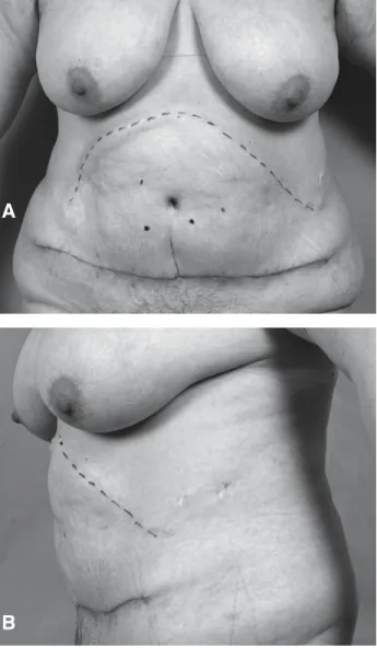
incision was used for this surgery. Seven years later, she presented for abdominoplasty and excess skin resection (weight loss 23 kg, weight 83 kg, BMI 34 kg/m2) (Figure 2).
Perforator-sparing abdominoplasty was performed as described above with two major periumbilical perfora-tors (latero-cephalad to the umbilicus) dissected, with tunneling through the rectus fascia and muscles and lig-ation of the cephalad branch of each perforator. The post-operative course was uneventful with good wound heal-ing (Figure 3). Postoperative duplex imagheal-ing showed blood-flow in both dissected perforators (Figure 4). Case 2
A 32-year-old female underwent GBP surgery for morbid obesity (height 160 cm, weight 130 kg, BMI 50.8 kg/m2). A bilateral subcostal incision was used
for this surgery. Two years later, she presented for abdominoplasty and excess skin resection (weight loss 58 kg, weight 72 kg, BMI 28.1 kg/m2).
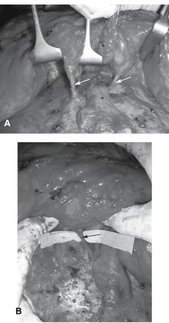
Perforator-sparing abdominoplasty was performed as described above with three major periumbilical per-forators (two lateral to the umbilicus, one cranio-later-al to the umbilicus) dissected, with tunneling through the rectus fascia and muscles and ligation of the cepha-lad branch of each perforator (Figure 5). The postoper-ative course was uneventful with good wound healing.
Figure 1. Outline of perforator-sparing abdominoplasty.
Displayed are the area of undermining, the rectus abdo-minis muscles and one exemplary dissected perforator with ligated ascending branch.
ascending branch of perforator (ligated)
dissected perforator area of undermining rectus abdominis muscle
Results
Both cases had uneventful postoperative courses and were successful in terms of the aesthetic result. Blood supply through the dissected perforator ves-sels was proven by color-flow duplex imaging (blood-flow is indicated by red and yellow color).
Discussion
Extended flap undermining is the surgical standard in abdominoplasty. Nine out of ten plastic surgeons reported that they performed complete undermining up to the costal margin.10Reports describing
limit-ed dissection of the abdominal flap in a triangular shape from the xiphoid to anterior superior iliac spine,11with discontinuous undermining by
dissect-ing the supraumbilical flap by liposuction,12 have
been described to improve flap perfusion. In these reports, adequate flap mobility was achieved with good flap perfusion by arterial perforators.12-14
However, in patients being referred for abdomino-plasty after GBP, a long transverse supraumbilical or subcostal scar is occasionally encountered15that
compromises blood supply from the superior epi-gastric arteries. In a study by De Castro et al5on full
abdominoplasties on previously scarred abdomens, the authors claimed that the supraumbilical scar is a significant problem, and they considered it to be a considerable limitation for abdominoplasty due to a
A
B
Figure 2. Preoperative views (the bilateral subcostal scar
is indicated with a dotted line): A. frontal; B. lateral.
A
B
Figure 3. Postoperative views 6 weeks after
abdomino-plasty: A. front view: two major black dots indicate the two dissected perforator vessels that were detected by color duplex imaging; B. anterolateral view.
higher rate of postoperative complications. One risk factor that is responsible for jeopardizing the blood supply of the abdominal flap is the existence of these previous scars and the subsequent subdermal fibrosis. Propositions have been made to limit the extent of mobilization of the abdominal flap to pre-serve the vascular zones.6A fluorescence
perfusog-raphy study from Germany on effects of abdomino-plasty on abdominal wall perfusion showed signifi-cant impairment of vascular supply of the so-called zone 1 in all patients.16Saulis et al17have reported a
significant reduction of postoperative wound com-plications in midline ventral hernia repairs by preservation of peri-umbilical rectus abdominis per-forator vessels. Selective dissection and preserva-tion of one perforator vessel from the inferior epi-gastric artery has been described in a patient with no risk factors undergoing aesthetic abdominoplasty. The effect was visualized by indocyanine green per-fusography.16Through this technique the blood
sup-ply of the abdominal flap may be changed from a random pattern into an axial pattern blood supply.
This assumption gains significant importance when the random pattern blood supply is compro-mised by oblique subcostal incisions. Oscar et al18
have described a rat model that showed that most of the abdominal rat skin could survive through the supply of a single musculo-cutaneous perforator vessel. This is consistent with the clinical experi-ence that we have had with the deep inferior epigas-tric perforator flap (DIEP). Schoeller et al19 have
reported a case of successful raising of a DIEP flap for breast reconstruction in a patient with subcostal
scars and successful wound closure by dissecting one single perforator to provide perfusion for the distal part of the cranial abdominal flap below the scar for wound closure. This finding of safe abdom-inal flap preparation is consistent with our experi-ence with the two described cases of perforator-sparing abdominoplasty in patients with bilateral subcostal scars after open GBP.
As previously described,20patients requiring
sur-gical skin excision after massive weight loss for functional and/ or aesthetic reasons are challenging, and require individualized approaches. We believe
Figure 4. Blood-flow through dissected perforator vessel
visualized by color-flow duplex imaging 10 weeks postop-eratively.
Figure 5. Intraoperative Views: A. Two dissected
para-umbilical perforator vessels (arrows) and umbilicus (marked *); B. Perforator (arrow) cranio-lateral to umbilicus.
A
that preoperative marking of perforators in the prox-imity of the umbilicus by color duplex imaging can help find the necessary perforator vessels,7 which
may be of great use to a microsurgically less-expe-rienced surgeon. Our approach shows that the dis-tinct use of sonographic equipment enhances not only microsurgical flap surgery, but also may be of great use in abdominoplasty. We recommend a cus-tomized approach using duplex imaging for any “non-standard” abdominoplasty to enhance safety.
Although massive weight loss improves health status in general, the formerly obese patients still often carry significant residual co-morbidities, such as hypertension, diabetes or hypercholesterolemia.19
Through an adequate surgical technique, excellent results can be achieved even in the presence of these co-morbidities using the described perforator-spar-ing technique. In none of our cases was flap survival or wound healing a problem.
In conclusion, our concept of improving blood supply to the abdominal flap by selective dissection of perforator vessels in patients with bilateral sub-costal scars, which enables us to perform a full abominoplasty with complete flap undermining, has not been described before. By color-flow duplex imaging, we show that a major blood supply comes from the dissected perforators.
We suggest that our perforator-sparing abdomino-plasty technique should be considered as a treatment option in patients after open GBP with oblique sub-costal incisions. Furthermore, we recommend pre-operative color flow duplex mapping of the abdom-inal wall for microsurgically less-experienced sur-geons, for easier detection of perforator vessels for “non-standard” abdominoplasties.
We thank Stefan De Maddalena for excellent photo documentation.
References
1. Savage RC. Abdominoplasty following gastrointestinal bypass surgery. Plast Reconstr Surg 1983; 71: 500-9. 2. Alvarez-Cordero R, Aragon-Viruette E. Incisions for
obesity surgery: a brief report. Obes Surg 1991; 1: 409-11.
3. Jones KBJ. The superiority of the left subcostal inci-sion compared to mid-line inciinci-sions in surgery for mor-bid obesity. Obes Surg 1993; 3: 201-5.
4. Jones KBJ. The left subcostal incision revisited. Obes Surg 1998; 8: 225-8.
5. de Castro CC, Aboudib JJ, Salema R et al. How to deal with abdominoplasty in an abdomen with a scar. Aesthetic Plast Surg 1993; 17: 67-71.
6. Huger WEJ. The anatomic rationale for abdominal lipectomy. Am Surg 1979; 45: 612-7.
7. Pitanguy I, Mayer B, Labrakis G. [Abdominoplasty – personal surgical guidelines] Zentralbl Chir 1988; 113: 765-71. German.
8. Chang BW, Luethke R, Berg WA et al. Two-dimen-sional color Doppler imaging for precision preopera-tive mapping and size determination of TRAM flap perforators. Plast Reconstr Surg 1994; 93: 197-200. 9. Rand RP, Cramer MM, Strandness DEJ. Color-flow
duplex scanning in the preoperative assessment of TRAM flap perforators: a report of 32 consecutive patients. Plast Reconstr Surg 1994; 93: 453-9.
10. Grazer FM, Goldwyn RM. Abdominoplasty assessed by survey, with emphasis on complications. Plast Reconstr Surg 1977; 59: 513-7.
11. Baroudi R, Keppke EM, Netto FT. Abdominoplasty. Plast Reconstr Surg 1974; 54: 161-8.
12. Illouz YG. A new safe and aesthetic approach to suc-tion abdominoplasty. Aesthetic Plast Surg 1992; 16: 237-45.
13. Matarasso A. Liposuction as an adjunct to a full abdominoplasty. Plast Reconstr Surg 1995; 95: 829-36. 14. Matarasso A. Liposuction as an adjunct to a full
abdomino-plasty revisited. Plast Reconstr Surg 2000; 106: 1197-202; discussion 1203-5.
15. Jones KB, Afram JD, Benotti PN et al. Open versus laparo-scopic Roux-en-Y gastric bypass: a comparative study of over 25,000 open cases and the major laparoscopic bariatric reported series. Obes Surg 2006; 16: 721-7.
16. Mayr M, Holm C, Hofter E et al. Effects of aesthetic abdominoplasty on abdominal wall perfusion: a quantita-tive evaluation. Plast Reconstr Surg 2004; 114: 1586-94. 17. Saulis AS, Dumanian GA. Periumbilical rectus abdo-minis perforator preservation significantly reduces superficial wound complications in “separation of parts” hernia repairs. Plast Reconstr Surg 2002; 109: 2275-80 ; discussion 2281-2.
18. Oksar HS, Coskunfirat OK, Ozgentas HE. Perforator-based flap in rats: a new experimental model. Plast Reconstr Surg 2001; 108: 125-31.
19. Schoeller T, Huemer GM, Kolehmainen M et al. Management of subcostal scars during DIEP-flap rais-ing. Br J Plast Surg 2004; 57: 511-4.
20. Taylor J, Shermak M. Body contouring following mas-sive weight loss. Obes Surg 2004; 14: 1080-5.