ORIGINAL ARTICLE
Downstream resource utilization following hybrid cardiac
imaging with an integrated cadmium-zinc-telluride/64-slice
CT device
Michael Fiechter&Jelena R. Ghadri&Mathias Wolfrum&Silke M. Kuest&
Aju P. Pazhenkottil&Rene N. Nkoulou&Bernhard A. Herzog&
Cathérine Gebhard&Tobias A. Fuchs&Oliver Gaemperli&Philipp A. Kaufmann
Received: 18 August 2011 / Accepted: 7 November 2011 / Published online: 6 December 2011 # Springer-Verlag 2011
Abstract
Purpose Low yield of invasive coronary angiography and unnecessary coronary interventions have been identified as key cost drivers in cardiology for evaluation of coronary artery disease (CAD). This has fuelled the search for non-invasive techniques providing comprehensive functional and anatomical information on coronary lesions. We have evaluated the impact of implementation of a novel hybrid cadmium-zinc-telluride (CZT)/64-slice CT camera into the daily clinical routine on downstream resource utilization.
Methods Sixty-two patients with known or suspected CAD were referred for same-day single-session hybrid evaluation with CZT myocardial perfusion imaging (MPI) and coro-nary CT angiography (CCTA). Hybrid MPI/CCTA images from the integrated CZT/CT camera served for decision-making towards conservative versus invasive management.
Based on the hybrid images patients were classified into those with and those without matched findings. Matched findings were defined as the combination of MPI defect with a stenosis by CCTA in the coronary artery subtending the respective territory. All patients with normal MPI and CCTA as well as those with isolated MPI or CCTA finding or combined but unmatched findings were categorized as“no match”.
Results All 23 patients with a matched finding underwent invasive coronary angiography and 21 (91%) were revascu-larized. Of the 39 patients with no match, 5 (13%, p<0.001 vs matched) underwent catheterization and 3 (8%, p<0.001 vs matched) were revascularized.
Conclusion Cardiac hybrid imaging in CAD evaluation has a profound impact on patient management and may contrib-ute to optimal downstream resource utilization.
Keywords Downstream resource utilization . CZT . SPECT . Cardiac hybrid imaging
Introduction
In clinical practice diagnosis of coronary artery disease (CAD) has been based on the presence of luminal narrowing of greater than 50% documented by invasive coronary an-giography catheterization (CATH). However, it has been recognized over the last decade that many factors other than lumen size and its narrowing, which cannot be comprehen-sively elucidated by coronary angiography alone, may de-termine whether or not an anatomical coronary lesion induces ischaemia. Therefore, decision-making towards
Michael Fiechter and Jelena R. Ghadri contributed equally to this work.
M. Fiechter
:
J. R. Ghadri:
M. Wolfrum:
S. M. Kuest:
A. P. Pazhenkottil
:
R. N. Nkoulou:
B. A. Herzog:
C. Gebhard:
T. A. Fuchs
:
O. Gaemperli:
P. A. Kaufmann (*)Department of Radiology, Cardiac Imaging, University Hospital Zurich,
Ramistrasse 100, 8091 Zurich, Switzerland e-mail: [email protected]
M. Fiechter
:
P. A. KaufmannZurich Center for Integrative Human Physiology (ZIHP), University of Zurich,
revascularization without proof of ischaemia has been re-cently challenged. In fact, modern guidelines for coronary revascularization [1] stipulate that comprehensive evalua-tion including proof of ischaemia is mandatory for prognos-tically relevant target vessel revascularization in chronic stable CAD.
Nuclear myocardial perfusion imaging (MPI) is a well-established method for the evaluation of myocardial ischae-mia and coronary computed tomography angiography (CCTA) has been established over the past years as a clinical tool for the assessment of coronary anatomy. The combina-tion of MPI with CCTA has been found to be a valuable gatekeeper for invasive coronary angiography [2] and may help to increase the low yield of elective invasive diagnostic coronary angiography (currently less than 40%) [3]. Cardiac hybrid imaging has emerged as one of the latest methodo-logical advancements, integrating information on anatomy (CCTA) with that from MPI into hybrid images [4]. This allows to directly relate individual myocardial perfusion territories to the subtending coronary arteries. Most studies on hybrid MPI/CT imaging are based on fusion of data from a separate gamma camera and a stand-alone CT scanner, as the unfavourable discrepancy of scan length (short CT and long MPI acquisition time) was hampering the widespread use of hybrid single photon emission computed tomography (SPECT)/CT devices in daily clinical routine. With the introduction of new gamma cameras using a cadmium-zinc-telluride (CZT) detector technique [5–8], scan time has been reduced substantially down to a few minutes [9,
10]. This has paved the way for hybrid SPECT/CT scanners integrating a CZT gamma camera with a high-end 64-slice CT device and has increased the interest in cardiac fusion imaging, further supported by recent results confirming the diagnostic strength [4,11] and the prognostic value [12] of cardiac hybrid imaging. However, data on the added clinical value of hybrid cardiac CZT/CT imaging in the decision-making for the appropriate treatment strategy are lacking. The aim of the present study was to evaluate in the daily clinical routine the impact of hybrid cardiac MPI and CCTA imaging obtained with an integrated ultrafast CZT/CT de-vice on downstream resource utilization such as invasive coronary angiography and revascularization.
Materials and methods
Patients and study protocol
We included 62 patients without a history of prior coronary artery bypass graft (CABG) who were consecutively re-ferred for the assessment of known or suspected CAD for a same-day single-session hybrid scan with CZT MPI and CCTA to obtain hybrid cardiac images. All patients were
scanned on a CZT/64-slice CT hybrid camera (Discovery NM/CT 570c, GE Healthcare), and the integrated CZT/CT images were reported to the referring physicians who made a decision towards invasive CATH versus conservative management based on the hybrid imaging finding on the one hand and including the clinical history and the symp-toms on the other hand. Similarly, in the catheterization laboratory the interventional cardiologist based his/her de-cision towards revascularization on the hybrid images and the clinical history, integrating the angiographic findings including fractional flow reserve (FFR) at the operators’ discretion into the clinical decision-making. Downstream resource utilization within 60 days triggered by the hybrid imaging was assessed, including decision for conservative treatment, invasive angiography and revascularization pro-cedure (CABG or percutaneous coronary intervention, PCI). From each study participant, written informed consent was obtained for the use of their imaging and clinical data for research purposes as approved by the Institutional Review Board.
Image acquisition
All patients underwent a 1-day pharmacological stress/rest SPECT MPI protocol according to the guidelines of the European Association of Nuclear Medicine [13] with aden-osine (0.14 mg/kg per min over 6 min) while in patients with contraindications to adenosine (n016) we used dobutamine (incrementally administered, starting at 5μg/kg per min and increasing at 1-min intervals to a maximal dose of 60μg/kg per min until 85% of the age-predicted heart rate had been achieved) according to daily clinical routine in our lab. Approximately 60 min after injection of 99mTc-tetrofosmin (332±36 MBq), stress MPI images were acquired (5 min) using the CZT/CT camera. This was followed by acquisition of rest MPI with the identical acquisition protocol several minutes [14] after administration of a three times higher dose of99mTc-tetrofosmin (955±169 MBq). MPI scans were acquired using a multi-pinhole collimator (effective diame-ter aperture of 5.1 mm) and 19 stationary detectors simulta-neously imaging 19 different views of the heart as previously reported. In brief, each detector contains 32 × 32 pixelated (2.46 × 2.46 mm) CZT elements; the system design allows acquisition without detector or collimator motion, and a 10% symmetrical energy window at 140 keV was used [9]. A low-dose CT scan was performed for attenuation correction of SPECT MPI, which also allows calculation of calcium scoring [15]. Thereafter, all patients underwent contrast-enhanced CCTA with prospective ECG triggering as previously reported [16–18]. Intravenous metoprolol (2.5–25 mg) was administered prior to the ex-amination if necessary in order to obtain optimal image quality for CCTA (target heart rate <65 bpm). All patients
received 2.5 mg isosorbide dinitrate sublingually 2 min prior to the CCTA scan. MPI and CCTA images were fused on a dedicated workstation (Advantage Workstation 4.3, GE Healthcare) projecting the MPI images on the left ventricu-lar epicardial surface obtained from the CCTA using the CardIQ Fusion software package (GE Healthcare) as previ-ously validated [19,20]. The 3-D volume rendered fusion images enable a panoramic view of the coronary artery tree projected onto the left ventricular myocardial perfusion territories. Cardiac hybrid images can be displayed in stan-dard anterior, posterior, lateral and apical views and in freely selectable angles for standardized documentation and reporting. Effective radiation dose for CCTA was calculated from the dose-length product (DLP) using a conversion coefficient for the chest, k00.014 mSv/(mGy × cm); for SPECT MPI a conversion factor of 0.0079 mSv/MBq was used.
Cardiac hybrid image interpretation
Fused MPI and CCTA images were analysed by two expe-rienced nuclear cardiologists by consensus with regard to functionally relevant coronary stenoses. For each patient, myocardial tomograms were grouped into 20 segments. The following 5-point scoring system was used to grade the segmental radionuclide uptake: 00normal, 10equivocal, 20moderate, 30severe reduction of radioisotope uptake and 40absence of detectable tracer in a segment. A pathological scan was defined as one with stress score≥2 in two or more
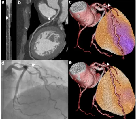
segments. If a stress defect was associated with a stress defect score of 4 with a rest score of 2 or a rest score ≤1, a scan was classified as reversible perfusion defect. As patient management should be ischaemia driven according to best clinical practice, only reversible defects were con-sidered for the following assignment into“matched” vs “no match” finding. A matched SPECT/CT hybrid cardiac im-aging finding was defined as a reversible SPECT MPI defect in a territory served by a coronary artery with a stenosis (defined as narrowing of the coronary luminal diameter ≥50%, Fig.1). All patients with normal SPECT and CCTA as well as those with isolated CCTA or MPI finding or combined but anatomically unmatched finding were categorized as no match.
Invasive coronary angiography data acquisition and analysis
Invasive CATH was performed according to clinical stand-ards by experienced interventional cardiologists. Segmenta-tion of the coronary artery tree was performed as described for CCTA. A coronary stenosis was defined as a luminal narrowing ≥50%. This was based on visual assessment reflecting the clinical routine in our and most other catheterization laboratories worldwide [21,22]. The FFR was determined as the ratio of the simultaneously assessed pressure in the aorta and in the coronary artery distal to a stenosis during maximal hyperaemia (induced by intravenous adenosine administration via a central vein at 140μg/kg per min) [22].
Fig. 1 Cardiac hybrid image and angiographic finding. Coronary artery stenosis (white arrowhead) in proximal left an-terior descending artery is shown in CCTA (a multiplanar and b curved multiplanar reconstruc-tion), cardiac hybrid imaging with normal perfusion at rest (e) and perfusion defect (pink area) at stress (c), matching the territory of the coronary artery with stenosis (white arrowhead), and invasive coronary
angiography (d) confirming the coronary stenosis (white arrowhead)
Statistical analysis
Quantitative variables were expressed as mean±standard deviation and categorical variables as frequencies or percen-tages. SPSS 19.0 (SPSS, Chicago, IL, USA) was used for statistical analysis. The chi-square test was applied to com-pare the revascularization rates between the two different patient groups. A p value from the chi-square test of less than 0.05 was considered statistically significant.
Results
All patients (n062) successfully underwent stress/rest CZT MPI and CCTA using the CZT/CT camera. The patient characteristics are given in Table1.
Overall, the CATH rate was 43% in the study population yielding 96% CAD. CZT revealed 32 patients (52%) with an abnormal MPI with reversibility in 26 patients, while CCTA documented a coronary stenosis in 42 patients (68%) ex-cluding CAD in 20 patients (32%). Three cases with false positive SPECT were corrected into negative by normal CCTA findings. Conversely, a coronary stenosis≥50% on CCTA but without stress-induced regional perfusion defect was found in 17 patients, while 19 patients had a normal finding on both CCTA and MPI. Cardiac hybrid imaging revealed a matched perfusion defect in 23 patients (38%), whereas 39 patients (63%) had no matched findings. The measurement of FFR was performed in 9 patients (in 12 coronary lesions).
During follow-up, all patients with matched cardiac hy-brid findings (n023) were referred to CATH of whom 21 (91%, Fig.2) subsequently underwent PCI or CABG. Of the two patients without revascularization, one had normal cor-onary arteries by CATH and the second patient had a small MPI defect and invasive assessment revealed a stenosis with normal FFR (≥0.8) over two lesions, i.e. in the left anterior descending artery (0.82) and in the left circumflex artery (0.89), both of which therefore did not qualify for coronary revascularization [23]. Only 5 of the 39 patients (13%) with no matched hybrid finding (Fig.2) were sent to CATH and 3 (8%) were revascularized. The reasons for referring a patient to CATH without matched hybrid finding were either severe CAD (left main or three-vessel disease) ≥80% (n02) or massively elevated Agatston coronary calcium scoring (> 1,000; n03). The latter has recently been shown to unmask obstructive CAD in patients with normal SPECT MPI [24]. Revascularization was subsequently performed in three patients despite lack of matched finding as one patient revealed abnormal FFR over a lesion in the left anterior descending artery (0.69) and the other patients showed either severe coronary three-vessel disease or severe steno-sis of the left main artery. There was a significantly lower
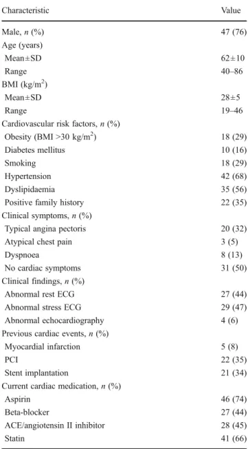
Table 1 Baseline patient characteristics (n062)
Characteristic Value Male, n (%) 47 (76) Age (years) Mean±SD 62±10 Range 40–86 BMI (kg/m2) Mean±SD 28±5 Range 19–46
Cardiovascular risk factors, n (%)
Obesity (BMI >30 kg/m2) 18 (29)
Diabetes mellitus 10 (16)
Smoking 18 (29)
Hypertension 42 (68)
Dyslipidaemia 35 (56)
Positive family history 22 (35)
Clinical symptoms, n (%)
Typical angina pectoris 20 (32)
Atypical chest pain 3 (5)
Dyspnoea 8 (13)
No cardiac symptoms 31 (50)
Clinical findings, n (%)
Abnormal rest ECG 27 (44)
Abnormal stress ECG 29 (47)
Abnormal echocardiography 4 (6)
Previous cardiac events, n (%)
Myocardial infarction 5 (8)
PCI 22 (35)
Stent implantation 21 (34)
Current cardiac medication, n (%)
Aspirin 46 (74)
Beta-blocker 27 (44)
ACE/angiotensin II inhibitor 28 (45)
Statin 41 (66)
BMI body mass index
Fig. 2 Downstream resource utilization after cardiac hybrid imaging. CATH coronary angiography catheterization
downstream CATH resource utilization in patients with no matched findings versus those patients with matched cardiac hybrid findings (p<0.001). Due to subclinical CAD (<50% luminal narrowing) preventive medical intervention was initiated in one patient, while in one patient such medication was withdrawn after excluding any lesion by CCTA.
The effective radiation dose for stress/rest SPECT MPI was 10.2±1.5 mSv, for attenuation correction 0.9±0.1 mSv and for prospective triggered CCTA 1.8±0.6 mSv.
Discussion
Our results demonstrate that cardiac hybrid imaging with a CZT/CT device allows efficient comprehensive evaluation of CAD. This has a profound impact on patient management and may contribute to optimal downstream resource utiliza-tion. In the present study we have shown that in 91% of patients with a matched finding on cardiac hybrid imaging coronary revascularization was performed, whereas only 8% of patients without matched cardiac hybrid finding were revascularized.
With regard to resource utilization the use of hybrid imaging had a strong impact as 100% of patients with a match but only 13% of patients without match were referred for CATH. As a result, in the patient group with a match the CAD per CATH yield was 96% comparing favourably to the yield of less than 38% recently reported for a similar elective large population [3]. The high CAD yield in our study paved the way for the above-mentioned high rate of revasculariza-tion per CATH (91%), which is far superior to figures reported in registries of most European countries, such as for example Germany (36%) [25].
An appropriate selection of patients who will benefit from invasive CATH and eventually from revascularization is crucial not only because of the costs of CATH but also because of its non-negligible rate of procedural in-hospital morbidity and mortality [26, 27]. Although it has been suggested for a long time [28] and repeatedly documented [22] that for best clinical practice functional assessment of coronary lesions is mandatory, invasive CATH has remained not only the gold standard for CAD detection but also for decision-making for or against the need for revasculariza-tion. Our results show that hybrid cardiac imaging is an excellent gatekeeper for further evaluation with CATH. In fact, the high yield of CAD and revascularization rate per CATH documents an appropriateness of invasive resource utilization far beyond that reported from large registries of routine clinical practice [3,25]. By contrast, resource utili-zation was very low in patients with no hybrid match, in whom a decision towards invasive CATH (n05) was mainly driven by extensive coronary calcification (> 1,000 Agatston units), which has recently been shown to unmask CAD in
patients without SPECT MPI abnormalities. Therefore, in the present study a calcium score > 1,000 triggered a CATH overruling the lack of a matched finding. In three of these five patients justification to revascularize was supported either by FFR or the presence of severe multi-vessel disease including main stem involvement [2]. The clinical value of comprehensive CAD assessment by hybrid CZT/CT imag-ing lies in the improvement of evidence-based patient selec-tion for coronary revascularizaselec-tion [12, 29]. Although coronary interventions have been shown to successfully reduce ischaemic symptoms and, therefore, improve patients’ quality of life, periprocedural adverse events re-main important considerations, particularly in low-risk pop-ulations. In stable CAD populations revascularizations have failed to confer a prognostic superiority compared to opti-mal medical therapy [30–33]. However, in patients with moderate to large ischaemia revascularization may be the most effective strategy to reduce the amount of jeopardized myocardium and, consequently, improve prognosis [34]. As hybrid findings have been shown to be superior in predict-ing adverse events over isolated findpredict-ings [12], it appears appropriate that these findings are the predominant target of revascularization. In this context our results show that the implementation of a hybrid CZT/CT camera in daily clinical routine confers an added value for evidence-based decision-making and optimization of adequate downstream resource utilization. Patients with intermediate to high pre-test prob-ability are among those who most likely may benefit from noninvasive evaluation by hybrid imaging. However, in view of the fact that most patients referred for elective invasive coronary angiography reveal normal coronary arteries, even lower pre-test probability patients may be candidates for a hybrid approach as long as unnecessary invasive angiographies can be avoided. A larger study would be needed to provide final proof of cost-effectiveness of the hybrid method, which was beyond the scope of the present pilot study.
We acknowledge the following limitations: A potential limitation of the present study is the fact that all patients underwent two noninvasive tests cumulating radiation ex-posure to the patients. However, several algorithms may be implemented into hybrid imaging such as for example stress-only MPI [35] to reduce radiation. In addition, CZT cameras offer the potential of a low-dose/low-dose 1-day protocol as the count-rate linearity allows subtraction of the counts of the first scan avoiding the need for a threefold increase of the second scan [36]. Finally, although patients were included consecutively, there was no randomization to different treatment options due to the observational nature of this pilot study on the downstream resource utilization. However, our results support the notion that use of a CZT/ CT hybrid device in the daily clinical routine may favour-ably affect resource utilization. Nevertheless, this may also
depend on the awareness of the clinicians about the avail-ability and performance of hybrid imaging as well as on the clinical environment accepting noninvasive imaging as guidance for revascularization.
Acknowledgments The study was supported by grants from the
Swiss National Science Foundation to PAK and to MF. Furthermore, we thank Serpil Bostanci-Kökylidirim, Mirjam De Bloeme, Edlira Loga, Ennio Mueller and Patrick von Schulthess for their excellent technical support.
Conflicts of interest None.
References
1. Task Force on Myocardial Revascularization of the European Society of Cardiology (ESC) and the European Association for Cardio-Thoracic Surgery (EACTS), European Association for Per-cutaneous Cardiovascular Interventions (EAPCI), Wijns W, Kolh P, Danchin N, Di Mario C, Falk V, Folliguet T, et al. Guidelines on
myocardial revascularization. Eur Heart J 2010;31:2501–55.
2. Gaemperli O, Husmann L, Schepis T, Koepfli P, Valenta I, Jenni W, et al. Coronary CT angiography and myocardial perfusion imaging to detect flow-limiting stenoses: a potential gatekeeper for coronary revascularization? Eur Heart J 2009;30:2921–9. 3. Patel MR, Peterson ED, Dai D, Brennan JM, Redberg RF, Anderson
HV, et al. Low diagnostic yield of elective coronary angiography. N
Engl J Med 2010;362:886–95.
4. Namdar M, Hany TF, Koepfli P, Siegrist PT, Burger C, Wyss CA, et al. Integrated PET/CT for the assessment of coronary artery
disease: a feasibility study. J Nucl Med 2005;46:930–5.
5. Pazhenkottil AP, Buechel RR, Herzog BA, Nkoulou RN, Valenta I, Fehlmann U, et al. Ultrafast assessment of left ventricular dyssyn-chrony from nuclear myocardial perfusion imaging on a new
high-speed gamma camera. Eur J Nucl Med Mol Imaging
2010;37:2086–92.
6. Buechel RR, Herzog BA, Husmann L, Burger IA, Pazhenkottil AP, Treyer V, et al. Ultrafast nuclear myocardial perfusion imaging on a new gamma camera with semiconductor detector technique: first
clinical validation. Eur J Nucl Med Mol Imaging 2010;37:773–8.
7. Fiechter M, Ghadri JR, Kuest SM, Pazhenkottil AP, Wolfrum M, Nkoulou RN, et al. Nuclear myocardial perfusion imaging with a novel cadmium-zinc-telluride detector SPECT/CT device: first validation versus invasive coronary angiography. Eur J Nucl Med
Mol Imaging 2011;38:2025–30.
8. Buechel RR, Pazhenkottil AP, Herzog BA, Husmann L, Nkoulou RN, Burger IA, et al. Real-time breath-hold triggering of myocar-dial perfusion imaging with a novel cadmium-zinc-telluride
detec-tor gamma camera. Eur J Nucl Med Mol Imaging 2010;37:1903–8.
9. Herzog BA, Buechel RR, Katz R, Brueckner M, Husmann L, Burger IA, et al. Nuclear myocardial perfusion imaging with a cadmium-zinc-telluride detector technique: optimized protocol for
scan time reduction. J Nucl Med 2010;51:46–51.
10. Herzog BA, Buechel RR, Husmann L, Pazhenkottil AP, Burger IA, Wolfrum M, et al. Validation of CT attenuation correction for high-speed myocardial perfusion imaging using a novel
cadmium-zinc-telluride detector technique. J Nucl Med 2010;51:1539–44.
11. Kajander S, Joutsiniemi E, Saraste M, Pietilä M, Ukkonen H, Saraste A, et al. Cardiac positron emission tomography/computed tomogra-phy imaging accurately detects anatomically and functionally signif-icant coronary artery disease. Circulation 2010;122:603–13. 12. Pazhenkottil AP, Nkoulou RN, Ghadri JR, Herzog BA, Buechel
RR, Küest SM, et al. Prognostic value of cardiac hybrid imaging
integrating single-photon emission computed tomography with coronary computed tomography angiography. Eur Heart J 2011;32:1465–71.
13. Hesse B, Tagil K, Cuocolo A, Anagnostopoulos C, Bardiés M, Bax J, et al. EANM/ESC procedural guidelines for myocardial perfu-sion imaging in nuclear cardiology. Eur J Nucl Med Mol Imaging 2005;32:855–97.
14. Giorgetti A, Rossi M, Stanislao M, Valle G, Bertolaccini P, Maneschi A, et al. Feasibility and diagnostic accuracy of a gated SPECT early-imaging protocol: a multicenter study of the Myoview Imaging Optimization Group. J Nucl Med
2007;48:1670–5.
15. Schepis T, Gaemperli O, Koepfli P, Rüegg C, Burger C, Leschka S, et al. Use of coronary calcium score scans from stand-alone multislice computed tomography for attenuation correction of myocardial
per-fusion SPECT. Eur J Nucl Med Mol Imaging 2007;34:11–9.
16. Husmann L, Valenta I, Gaemperli O, Adda O, Treyer V, Wyss CA, et al. Feasibility of low-dose coronary CT angiography: first
expe-rience with prospective ECG-gating. Eur Heart J 2008;29:191–7.
17. Herzog BA, Husmann L, Burkhard N, Gaemperli O, Valenta I, Tatsugami F, et al. Accuracy of low-dose computed tomography
coronary angiography using prospective
electrocardiogram-triggering: first clinical experience. Eur Heart J 2008;29:3037–42. 18. Buechel RR, Husmann L, Herzog BA, Pazhenkottil AP, Nkoulou R, Ghadri JR, et al. Low-dose computed tomography coronary angiography with prospective electrocardiogram triggering:
feasi-bility in a large population. J Am Coll Cardiol 2011;57:332–6.
19. Gaemperli O, Schepis T, Kalff V, Namdar M, Valenta I, Stefani L, et al. Validation of a new cardiac image fusion software for three-dimensional integration of myocardial perfusion SPECT and stand-alone 64-slice CT angiography. Eur J Nucl Med Mol Imaging
2007;34:1097–106.
20. Gaemperli O, Schepis T, Valenta I, Husmann L, Scheffel H, Duerst V, et al. Cardiac image fusion from stand-alone SPECT and CT:
clinical experience. J Nucl Med 2007;48:696–703.
21. Herzog BA, Husmann L, Buechel RR, Pazhenkottil AP, Burger IA, Valenta I, et al. Rapid cardiac hybrid imaging with minimized radiation dose for accurate non-invasive assessment of ischemic
coronary artery disease. Int J Cardiol 2011;153:10–3. doi:10.1016/
j.ijcard.2010.08.023.
22. Tonino PA, Fearon WF, De Bruyne B, Oldroyd KG, Leesar MA. Ver Lee PN, et al. Angiographic versus functional severity of coronary artery stenoses in the FAME study fractional flow reserve versus angiography in multivessel evaluation. J Am Coll Cardiol
2010;55:2816–21.
23. Tonino PA, De Bruyne B, Pijls NH, Siebert U, Ikeno F, van’ t Veer
M, et al. Fractional flow reserve versus angiography for guiding
percutaneous coronary intervention. N Engl J Med 2009;360:213–
24.
24. Ghadri JR, Pazhenkottil AP, Nkoulou RN, Goetti R, Buechel RR, Husmann L, et al. Very high coronary calcium score unmasks obstructive coronary artery disease in patients with normal SPECT
MPI. Heart 2011;97:998–1003.
25. van Buuren F, Horstkotte D. 24th report of performance statistics of heart catheterization laboratories in Germany. Der Kardiologe
2009;3:512–18.
26. West R, Ellis G, Brooks N, Joint Audit Committee of the British Cardiac Society and Royal College of Physicians of London. Complications of diagnostic cardiac catheterisation: results from a confidential inquiry into cardiac catheter complications. Heart 2006;92:810–4.
27. Shaw LJ, Shaw RE, Merz CN, Brindis RG, Klein LW, Nallamothu B, et al. Impact of ethnicity and gender differences on angiograph-ic coronary artery disease prevalence and in-hospital mortality in the American College of Cardiology-National Cardiovascular Data
28. Kern MJ, Lerman A, Bech JW, De Bruyne B, Eeckhout E, Fearon WF, et al. Physiological assessment of coronary artery disease in the cardiac catheterization laboratory: a scientific statement from the American Heart Association Committee on Diagnostic and Interventional Cardiac Catheterization, Council on Clinical Cardi-ology. Circulation 2006;114:1321–41.
29. Pazhenkottil A, Nkoulou R, Ghadri J, Herzog B, Küest S, Husmann L, et al. Impact of cardiac hybrid SPECT/CT imaging on choice of treatment strategy in coronary artery disease. Eur Heart J
2011;32:2824–9.
30. Boden WE, O’Rourke RA, Teo KK, Hartigan PM, Maron DJ,
Kostuk WJ, et al. Optimal medical therapy with or without PCI
for stable coronary disease. N Engl J Med 2007;356:1503–16.
31. Bucher HC, Hengstler P, Schindler C, Guyatt GH. Percutaneous transluminal coronary angioplasty versus medical treatment for non-acute coronary heart disease: meta-analysis of randomised
controlled trials. BMJ 2000;321:73–7.
32. Yusuf S, Zucker D, Peduzzi P, Fisher LD, Takaro T, Kennedy JW, et al. Effect of coronary artery bypass graft surgery on survival: overview of 10-year results from randomised trials by the Coro-nary Artery Bypass Graft Surgery Trialists Collaboration. Lancet 1994;344:563–70.
33. Pitt B, Waters D, Brown WV, van Boven AJ, Schwartz L, Title LM, et al. Aggressive lipid-lowering therapy compared with an-gioplasty in stable coronary artery disease. Atorvastatin versus Revascularization Treatment Investigators. N Engl J Med 1999;341:70–6.
34. Shaw LJ, Berman DS, Maron DJ, Mancini GB, Hayes SW, Hartigan PM, et al. Optimal medical therapy with or without percutaneous coronary intervention to reduce ischemic burden: results from the Clinical Outcomes Utilizing Revascularization and Aggressive Drug
Evaluation (COURAGE) trial nuclear substudy. Circulation
2008;117:1283–91.
35. Husmann L, Herzog BA, Gaemperli O, Tatsugami F, Burkhard N, Valenta I, et al. Diagnostic accuracy of computed tomog-raphy coronary angiogtomog-raphy and evaluation of stress-only single-photon emission computed tomography/computed
to-mography hybrid imaging: comparison of prospective
electrocardiogram-triggering vs. retrospective gating. Eur
Heart J 2009;30:600–7.
36. Nkoulou RN, Pazhenkottil AP, Kuest SM, Ghadri JR, Wolfrum M, Husmann L, et al. Semiconductor detectors allow low-dose-low-dose 1-day SPECT myocardial perfusion imaging. J Nucl Med 2011;52:1204–9.