HAL Id: hal-02135110
https://hal.archives-ouvertes.fr/hal-02135110
Submitted on 21 May 2019
HAL is a multi-disciplinary open access
archive for the deposit and dissemination of
sci-entific research documents, whether they are
pub-lished or not. The documents may come from
teaching and research institutions in France or
abroad, or from public or private research centers.
L’archive ouverte pluridisciplinaire HAL, est
destinée au dépôt et à la diffusion de documents
scientifiques de niveau recherche, publiés ou non,
émanant des établissements d’enseignement et de
recherche français ou étrangers, des laboratoires
publics ou privés.
US-guided percutaneous release of the first extensor
tendon compartment using a 21-gauge needle in de
Quervain’s disease: a prospective study of 35 cases
Franck Lapègue, Aymeric André, Etienne Pasquier Bernachot, Ezin Jocelyn
Akakpo, Pierre Laumonerie, Hélène Chiavassa-Gandois, Omar Lasfar,
Christophe Borel, Marine Brunet, Olivia Constans, et al.
To cite this version:
Franck Lapègue, Aymeric André, Etienne Pasquier Bernachot, Ezin Jocelyn Akakpo, Pierre
Lau-monerie, et al.. US-guided percutaneous release of the first extensor tendon compartment using a
21-gauge needle in de Quervain’s disease: a prospective study of 35 cases. European Radiology,
Springer Verlag, 2018, 28 (9), pp.3977-3985. �10.1007/s00330-018-5387-1�. �hal-02135110�
O
pen
A
rchive
T
oulouse
A
rchive
O
uverte
(OATAO)
OATAO is an open access repository that collects the work of some Toulouse
researchers and makes it freely available over the web where possible.
This is
an author'sversion published in:
https://oatao.univ-toulouse.fr/23086Official URL :
https://doi.org/10.1007/s00330-018-5387-1
To cite this version :
Any correspondence concerning this service should be sent to the repository administrator: tech-oatao@listes-diff.inp-toulouse.fr
Lapègue, Franck and André, Aymeric and Pasquier Bernachot, Etienne and Akakpo, Ezin
Jocelyn and Laumonerie, Pierre and Chiavassa-Gandois, Hélène and Lasfar, Omar and Borel,
Christophe and Brunet, Marine and Constans, Olivia and Basselerie, Hubert and Sans,
Nicolas and Faruch-Bilfeld, Marie US-guided percutaneous release of the first extensor
tendon compartment using a 21-gauge needle in de Quervain’s disease: a prospective study of
35 cases. (2018) European Radiology, 28 (9). 3977-3985. ISSN 0938-7994
OATAO
US-guided
percutaneous release of the first extensor tendon
compartment
using a 21-gauge needle in de Quervain’s disease:
a
prospective study of 35 cases
Franck Lapègue1,2 & Aymeric André3,4 & Etienne Pasquier Bernachot1 & Ezin Jocelyn Akakpo5 & Pierre Laumonerie3,6 &
Hélène Chiavassa-Gandois1 & Omar Lasfar1 & Christophe Borel1 & Marine Brunet1 & Olivia Constans1 &
Hubert Basselerie1& Nicolas Sans1& Marie Faruch-Bilfeld1
Abstract
Purpose To evaluate the efficacy of ultrasonography-guided percutaneous treatment of de Quervain tenosynovitis with the
combination of a corticosteroid injection and release of the retinaculum of the first extensor compartment tendons
with a 21-gauge needle.
Materials and methods The first part of our study consisted of ten procedures on cadaver wrists followed by dissection to analyse
the effectiveness of the retinaculum release and detect any collateral damage. The second part was a prospective clinical study of
35 procedures. Outcomes were evaluated through a 6-month clinical follow-up and telephone interview at the end of the study. The following parameters were monitored over time: pain level on a visual analogue scale, the QuickDASH and the PRWE.
Patient satisfaction questionnaires were also administered.
Results No complications were found during the cadaver study. However, the release was confirmed as ‘partial’ in all wrists. In
the clinical portion of this study, significant improvement was observed in 91.4 % of cases (32/35) within 1 month and the results
were stable until the end of the study; all of these patients avoided surgery. The release procedure failed in three patients who
eventually required surgical treatment.
Conclusion US-guided partial release and simultaneous corticosteroid injection for treatment of de Quervain’s disease
using a 21-gauge needle is feasible in current practice, with minimal complications.
Key Points
• Ultrasound guided treatment of de Quervain's disease is feasible with a 21G needle. • There was notable regression of clinical signs in 91.4 % of cases.
• The procedure is very safe, no iatrogenic neurovascular or tendinous injuries occurred. • Our procedure requires only one session and 3 days away from work.
Keywords De Quervain disease . Tendon entrapment . Ultrasonography, interventional . Injections, intralesional . Tenotomy
* Franck Lapègue
franck.lapegue@gmail.com
1
Service d’imagerie, CHU de Toulouse Purpan, bâtiment Pierre Paul Riquet, Place du Dr Baylac, TSA, 40031 31059 Toulouse cedex 9, France
2 ELSAN, Polyclinique Le Languedoc, Avenue de la Côte des Roses,
11100 Narbonne, France
3
Laboratoire d’anatomie, faculté de médicine, 31062 Toulouse, France
4
Service de chirurgie orthopédique, clinique Médipole Garonne, 45 rue de Gironis, 31036 Toulouse Cédex1, France
5
ISBA, Faculté de médecine du CNHU de Cotonou, Cotonou, Bénin
6 Institut de l’appareil locomoteur, CHU de Toulouse Purpan, bâtiment
Pierre Paul Riquet, Place du Dr Baylac, TSA, 40031 31059 Toulouse cedex 9, France
Abbreviations
APL Abductor Pollicis Longus
EPB Extensor Pollicis Brevis
PRWE Patient Rated Wrist Evaluation
Quick DASH Quick Disabilities of the Arm,
Shoulder and Hand outcome measure
RNSB Radial nerve’s sensory branch
VAS pain Visual analogue scale for pain
Introduction
De Quervain's disease [1] is a specific form of stenosing
teno-synovitis in the wrist due to a mismatch between the size of the tendons of the first extensor compartment (abductor pollicis longus (APL) and extensor pollicis brevis (EPB)) and the size of the osteofibrous tunnel in which they are lo-cated. This tenosynovitis manifests itself as pain on the radial side of the wrist, which is increased by performing the
Finkelstein test [2]. This is a common disease, with 1.2%
prevalence [3]. It mostly affects women (prevalence of 1.3–
2.8 %) [4,5]) particularly some new mothers who develop
‘baby wrist’ [6]. Men are, however, not immune to this
con-dition (0.6 %[3,5]), particularly manual laborers who perform
repetitive movements [3].
Ultrasonography (US) is an excellent modality for ex-ploring this condition, as it provides an accurate
assess-ment of the local damage and anatomical variations [7,
8]. Hypoechogenic thickening of the retinaculum is present
and measures 2.01 mm on average [8]. It can encompass
both tendons without a septum (type 1 according to Volpe
et al.) [8] or only the EPB with a septum (type 2) [8]. The
presence of this intertendinous septum, which is easily
seen with US [9], contributes to the occurrence of de
Quervain's tenosynovitis and increases friction on the
EPB [10]. Its frequency varies from 29 % to 81 % of cases,
depending on the study [8,9,11–13]. US can also detect a
hyperintense Doppler signal in the retinaculum [8] and
anatomical tendon variations (multi-fasciculated APL [9],
bifid or absent EPB [9,14]).
The initial medical care varies between teams but typi-cally consists of stopping pain-inducing movements,
brac-ing [15], NSAIDs and corticosteroid injections. If
conser-vative treatment fails, surgical treatment (release of radial
styloid pulley) can be proposed to the patient [16]. Various
types of reconstruction procedures have been described
[17–21] to prevent the most typical complication– anterior
tendon dislocation [22].
Up to now, the therapeutic role of US was limited to deter-mining the exact location for corticosteroid injection to
achieve optimal diffusion within the EPB tendon sheath [23]
or around both tendons [24], thereby reducing the probability
of failure [25, 26]. Very few studies apply US-guided
percutaneous release in the case of de Quervain’s
tenosynovi-tis [11,27]. The purpose of our study was to evaluate the
effectiveness of US-guided release using a 21-gauge (G; 0.8-mm) needle in this pathology, based on our experience with
treating trigger finger using this technique [28].
Materials and methods
Our two-part study was approved by our Research Ethics Committee. It consisted of cadaver and clinical experiments
conducted jointly by a hand surgeon (12 years’ experience)
and an interventional radiologist specializing in musculoskel-etal procedures (17 years’ experience).
Cadaver study
A feasibility study was performed in cadavers to confirm that the retinaculum of the first compartment of extensor tendons could be cut using a 21-G needle. This release is feasible, but requires ten back-and-forth movements of the needle to be
complete (Fig. 1). Based on these findings, the radiologist
performed an US-guided release of the retinaculum of the first compartment in ten wrists of five fresh cadavers (four women and one man, mean age 82 years). Subsequently, the surgeon carefully dissected the treated wrists to analyse the condition of the retinaculum, tendons and adjacent neural and vascular structures.
Fig. 1 Transection with a 21 G needle of the retinaculum of the first extensor compartment in a cadaver specimen. Tendons of the first extensor compartment and their retinaculum are exposed after dissection of overlying tissues on the radial side of the wrist of a fresh cadaver (89 year old female). A 21 G needle is slid longitudinally over the retinaculum using the needle's bevel as a surgical knife. After ten or so back and forth passes, the retinaculum is transected (arrows), revealing the extensor pollicis brevis (E) and abductor pollicis longus (A) tendons.
Clinical study
Study population
Over a 2-year period starting in January 2015, 38 patients were included in our prospective study. The inclusion criteria were presence of de Quervain's tenosynovitis for at least 4 months (mean 5.7 months, range 4–12 months) that was re-calcitrant to conservative treatment, and a positive Finkelstein test. Patients were excluded if they had previously undergone surgery in the first extensor compartment, had another hand pathology, or were lost to follow-up.
During the consultation before the procedure, the surgeon proposed the US-guided release procedure to 38 patients and obtained written informed consent from each one. Three pa-tients were excluded during the study: two were lost to follow-up and one developed symptoms of carpal tunnel syndrome. Hence, data from 35 patients (seven men, 28 women) between 22 and 74 years of age (average 54 years) were available for analysis. Among the 35 treated wrists, 13 were left wrists and 22 were right wrists.
Release procedure
All the procedures were performed by the radiologist using an US unit (model Aplio 400; Toshiba Medical Systems, Tokyo, Japan) with a high-frequency transducer (14–18 MHz). The patient was positioned supine on a stretcher; the hand was set on its medial side on a table with slight ulnar deviation (see
Online Supplemental Material, Fig.1). A sterile working area
was prepared.
First, a scan of the entire region was done with the US probe to locate the radial nerve’s sensory branch (RNSB) and its divisions. Then local anaesthetic was injected with a 25-mm long, 25-G needle (orange hub) (see Online
Supplemental Material, Fig.1). The needle's entry point was
about 1 cm distal and lateral to the styloid process of the radius; the needle was oriented on the coronal plane towards the distal end of the retinaculum. Two cc of 1% lidocaine HCl (Xylocaine; AstraZeneca, Rueil-Malmaison, France) was injected along the needle’s path and into the synovial sheath
of the first compartment extensor tendons (Fig. 2; Online
Supplemental Material, Movie1).
Next, the base of a 50-mm long, 21-G needle (green hub) was manually curved to a 140° angle so that its bevel faced
laterally (Figs.3 and 4). This curvature had two effects: it
placed the needle in a completely horizontal position and made it possible to determine the bevel’s orientation, even when it was hidden beneath the patient’s skin.
The 21-G needle used the same trajectory as the anaes-thetic injection with US guidance; its exact positioning was adjusted according to the type of De Quervain's
tenosyno-vitis (Fig. 5). Once the optimal position had been
confirmed in the longitudinal and transverse planes (Fig.
6; see Online Supplemental Material, Fig.2), the
radiolo-gist slid the needle horizontally over the retinaculum
(Movie 1 [Online Supplemental Material]) making about
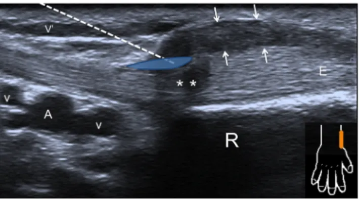
ten back-and-forth passes, while always keeping the needle’s tip in sight to reduce the possibility of damaging Fig. 2 Ultrasonography is used to guide the local anaesthesia over the path of the retinaculum of the first extensor compartment being released. On this longitudinal view of the radial side of the wrist, the dotted line indicates the needle's theoretical path. Care is needed to avoid the small superficial vein (V') located in the path. The needle's path must reach the deep and distal portion of the thickened retinaculum (arrows) and position itself superficial to the extensor pollicis brevis tendon (E). The anisotropic area in the tendon (**) at the distal portion of the first compartment extensor is evidence of the sudden change in direction of the tendon fibres where the most stenosis exists. The anaesthetic agent will be injected along the needle's path and in the sheath of the first extensor compartment tendons. This injection may be difficult and generally only involves the distal recess of the tendon sheaths (blue oval shape). Deep to the tendons, the radial artery (A) and satellite veins (V) are visible at the anatomical snuffbox. R = radial styloid
Fig. 3 Photograph of a US guided release for de Quervain's tenosynovitis (simulation). The ulnar side of the hand rests on a small bolster made of a sterile drape which places the wrist in slight ulnar deviation; this makes it easier to approach the radial side of the first compartment without interference from the thumb column. The procedure is carried out in sterile conditions: sterile drapes (removed for the purpose of this photo), skin disinfection, use of sterile gel and sterile probe cover, sterile gloves. A pad of sterile gel (**) is placed between the probe and the patient's skin; this allows the operator to accurately locate the needle's path before it breaches the patient's skin
neighbouring structures. In doing so, an experienced radi-ologist feels a slight resistance consistent with a transected fibrous structure. To release an intertendinous septum ef-fectively, a 90° rotation of the needle was required to orient the bevel perpendicular to the fibres being cut (see Online
Supplemental Material, Fig.3).
When the radiologist felt a significant reduction in the retinaculum's resistance upon sliding the needle, 0.5 cc xylocaine was injected through the 21-G needle left in
place (Movie1[Online Supplemental Material]). The fluid
then easily diffuses in the EPB and APL sheath proximal and distal to the retinaculum. Next, 0.5 cc (or 3.5 mg) betamethasone (MSD, Courbevoie, France) was injected inside the tendon sheath.
Assessment of clinical outcomes
On the day of the procedure (D0), before starting, the radiol-ogist performed a clinical examination with a Finkelstein test and asked the patients to rate their highest pain level in the
prior 7 days (visual analogue scale (VAS) pain, 0–10) and
their function using two questionnaires: Quick DASH (Quick Disabilities of the Arm, Shoulder and Hand outcome measure) and PRWE (Patient Rated Wrist Evaluation). US was also performed to analyse the location and thickness of the retinaculum, presence of a thickened intertendinous sep-tum and presence of a hyperintense signal in Doppler mode.
Fig. 5 Various types of de Quervain's tenosynovitis were found in our cohort, which had to be targeted differently in the release procedure. Here, the three types have drawings of an axial slice of the wrist and the corresponding ultrasonography images. Type 1 consists of overall thickening (arrows on US image, thick red line on drawing) of the retinaculum surrounding the extensor pollicis brevis (E) and abductor pollicis longus (A). The ‘X’ marks the ideal area for partial transection of the retinaculum at its zenith, close of the surface of the underlying tendons. In Type 2a, there is overall thickening of the retinaculum
(arrows on US image, thick red line on drawing) and a thickened septum is present (arrow head on US image, red line on drawing). Release (XX) will occur in the common retinaculum where it joins with the septum and if possible, in the septum itself. In Type 2b, the retinaculum around the extensor pollicis brevis is thickened (arrows on US image, thick red line on drawing) and a thickened septum is present (arrow head on US image, red line on drawing). The theoretical areas for partial release are marked with‘XX’.
Fig. 4 Drawing of the position of the 21 G needle used in release of de Quervain's tenosynovitis. This is a dorsal view with the wrist resting on its ulnar side. The tendons of the first extensor compartment are surrounded by their retinaculum (light green), which has semicircular fibres (curved red lines) perpendicular to the long axis of the tendons. The 21 G needle enters the retinaculum on its distal side with the bevel angled laterally (close up of needle in dotted circle). In this way, the bevel is perpendicular to the long axis of its fibres and is better able to transect them. A translation movement along the path of the retinaculum's apex will be applied to the needle. A = APL; E = EPB; R1 = extensor carpi radialis longus; R2= extensor carpi radialis brevis
This US assessment was used to classify patients into one of three categories:
– Type 1: overall thickening of retinaculum in first com-partment, without intertendinous septum
– Type 2a: overall thickening of retinaculum in first com-partment and presence of thickened intertendinous septum
– Type 2b: EPL retinaculum is thickened in combination with a thickened intertendinous septum; the retinaculum around the APL has a normal appearance.
US was also used to evaluate the appearance of the RNSB, EPB and APL before and after the release procedure to detect any complications.
One month (M1) and 6 months (M6) after the procedure, the patients were seen again by the surgeon who then per-formed a new clinical examination and evaluated the highest pain level in the prior 7 days (VAS), QuickDASH and PRWE. Between these two planned visits, patients had the option to call the surgeon if the disease symptoms worsened.
Patients were contacted by telephone at the end of the study, an average of 12.1 months after the procedure (SD 7
months, range 6–24 months]), by a radiology resident to again
determine the VAS pain level, QuickDASH, PRWE and eval-uate the patient's satisfaction with their care: very satisfied, satisfied, somewhat satisfied, not satisfied.
Statistical analysis
The following tests were used in the statistical analysis: – McNemar’s test to evaluate changes in the Finkelstein test – Paired Student's t-test for changes over time in the VAS
pain, Quick DASH and PRWE assessments
– Logistic regression to look for relationships between variables.
Results
Cadaver study
The following observations were made after dissecting the ten cadaver wrists that underwent US-guided release:
– every retinaculum is thin (average thickness of 0.5 mm) and has no intertendinous septum;
– the retinaculum was not completely released in all cases. Instead the result is partial thickness cuts over a portion of the retinaculum’s length (Online Supplemental Material,
Fig.4);
– there were no dislocations or tendon damage of the ex-tensors in the first compartment;
– no damage to the sensory branches of the radial nerve occurred;
– no vascular damage occurred.
Clinical study
Status of patients on D0
The clinical characteristics of these patients were somewhat
heterogeneous (Table1). The patients did not receive exactly
the same conservative treatment initially. The patients were typically referred to the study’ surgeon once non-operative
treatments had failed (Table2).
US performed on D0 found the retinaculum was thickened in every patient (mean 1.9 mm; SD 0.4 mm). Doppler mode revealed a hyperintense signal in the retinaculum and tendon sheath in 71.4 % of cases (n=25/35). The breakdown of de Quervain's types in the study cohort was: 42.9 % Type 1 (n=15/35), 20 % Type 2a (n=7/35) and 37.1 % Type 2b (n=13/35). Thus, 57.1 % of patients (20/35) had an intertendinous septum.
Fig. 6 Example of a release being performed in the left wrist of a 57 year old female patient with type 1 de Quervain's tenosynovitis. (a) Longitudinal view of the radial side of the wrist showing the thickened retinaculum over the tendons of the first extensor compartment (arrows) and the 21 G needle (arrow with dotted line) on the retinaculum's path
superficial to the extensor pollicis brevis. (b) Transverse axial view in the small axis of the tendons of the first compartment (E extensor pollicis brevis,A abductor pollicis longus): here, the needle is a hyperechogenic spot (arrow with dotted line) deep to the apex of the thickened retinaculum (arrows).
After the release procedure, US showed no damage to the RNSB, no complete transection of the retinaculum, and all the more, no tendon dislocation.
Outcomes
At the 1-month follow-up with the surgeon, the Finkelstein test was negative in 91.4 % of cases (n = 32/35), which was a highly significant improvement (p < 0.001). The 32 patients who had a negative Finkelstein test at 1 month had a clear improvement 7 days after the procedure on average (SD = 6.3). The three other patients (all of whom were subsequently operated on) felt they had improved after 1 week, but their pain reappeared in 2–3 weeks.
The VAS for pain, Quick DASH and PRWE all improved
significantly within the first month (Table3) (p < 0.001).
At 6 months and at the end of the study, these outcomes were maintained, as there were no differences relative to the 1-month outcomes (0.5 < p < 0.9).
When asked if they were satisfied with the outcome of the procedure:
- 77.1 % (27/35) of patients were satisfied or very satisfied; among these 27 patients, seven had previously undergone one or two unsuccessful corticosteroid injections;
– 14.3 % (5/35) of patients were somewhat satisfied; three of these five patients had some improvement but had persistent background pain and did not want further treat-ment; the two other patients mentioned having significant pain for 3 days after the procedure.
– 8.6 % (3/35) of patients were not satisfied and subse-quently underwent surgery: 4 months after the procedure for two patients and 1 year for one patient. The appear-ance found during surgery was the same as the one found during the initial US examination: thickened septum and retinaculum (mean 1.9 mm) around the EPB in two cases (type 2b) and both tendons in one case (type 2a). There was no visible evidence of the US-guided percutaneous release.
Among the three patients who underwent secondary sur-gery, two of them were on sick leave for work. The surgical procedure was successful in two cases. In the third case, pain persisted and made it impossible for this patient to return to work.
The logistic regression analysis found no statistically sig-nificant relationship between patient satisfaction or need for surgical treatment and:
– retinaculum thickness
– type of de Quervain's disease (1, 2a or 2b) – hyperintense signal in Doppler mode.
Conversely, a patient being on sick leave from work was a predictor of poor prognosis. The risk of undergoing surgery was increased 14 times (odds ratio (OR) = 14; p < 0.05).
Complications and costs
The average procedure time was 15 min (SD = 1.4); once the local anaesthesia was in place, the procedure was painless. Table 1 Clinical features of the
35 patients in our study cohort Feature Number of patients (%)
Manual labour (iron worker, waiter, nurse's aide, aerospace engineer) n=11/35 (31.4 %)
Retired n=11/35 (31.4 %)
Manual hobby (gardening, do it yourself) n=8/35 (23 %)
Household chores (cooking, cleaning) n=6/35 (17 %)
Women caring for newborns (3 mothers, 2 grandmothers) n=5/35 (14 %)
Sports involving upper limb (weight training, rugby, gold, petanque) n=4/35 (11.4 %) Patient who received aromatase inhibitor for breast related condition n=1/35 (3 %)
Table 2 Types of conservative (non operative) treatment performed in the study patients
Type of treatment used before the release procedure Number of patients (%)
Wrist splint n=35/35 (100 %)
NSAIDs (per os, topical) n=35/35 (100 %)
Relative rest (alteration of job duties, stoppage of recreational activities) n=32/35 (91 %)
Physical therapy with different modalities n=9/35 (26 %)
Work stoppage (average of 3 months) n=7/35 (20 %)
1 or 2 standard percutaneous corticosteroid injections n=7/35 (20 %)
Subcutaneous bruising appeared the day after the procedure in two patients (6 %) and had resolved after 1 week. The primary complaint after the procedure was an increase in pain on the evening of the procedure in six patients (17 % of cases) which lasted an average of 3.3 days (SD = 2).
These minor incidents led us to adopt the following course of action after the procedure: application of a compressive dressing, recommendation to wear a brace and to take analge-sics in case of pain, and 3 days off work. No other clinical complications were observed:
– no tendon dislocation during dynamic movements – no elective pain in the radial nerve's sensory territory – no skin atrophy at the needle entry point.
The cost of this US-guided release procedure is less than the cost of standard surgery for de Quervain's disease (Online
Supplementary Material, Table1). Standard surgical care
in-volves a visit with the surgeon, ultrasonography, nerve block by an anaesthesiologist, surgical release, 3 weeks away from work and nursing care at home for the wound. The US-guided release procedure involves a diagnostic US examination, US-guided release with corticosteroid injection, and 3 days away from work. Nursing care at home was not required since there was no wound.
Discussion
Overview of study
The purpose of our study was to evaluate the effectiveness of simultaneous corticosteroid injection and partial US-guided release using a 21-G (0.8-mm) needle in patients with de Quervain's tenosynovitis. In our study, 91.4 % of patients (32/35) avoided surgery and were able to resume their activities with significantly less pain within 1 month.
Among these patients, five were only‘somewhat satisfied’
with the outcome: two because of pain in the days imme-diately after the procedure and three because of residual pain (less than initial pain levels), probably due to an in-sufficient partial release. Our procedure was effective, fast and low-cost. The 0.8-mm needle entry point means that
nursing care at home is unnecessary and it allows patients to return to work after 3 days.
None of the typical complications of corticosteroid injec-tion or surgery were observed. The context of sick leave from work was the only risk factor identified for a poor outcome (OR = 14). The presence of an intertendinous septum was not associated with treatment failure in our study.
Our good outcomes may appear contradictory to the results of our cadaver study in which only partial release was accom-plished. In our clinical study after the release procedure, US showed no complete transection of the retinaculum. During surgery in the three operated patients there was no visible evidence of the US-guided percutaneous release, likely due to an incomplete release associated with well-organized scar tissue.
However, we can presume that partial release of the APL and EPB compartments with simultaneous corticosteroid in-jection might be sufficient to eliminate a patient’s symptoms.
Comparison with other studies and techniques
US-guided percutaneous release with a needle has been per-formed successfully with another common type of stenosing tenosynovitis: trigger finger with A1 pulley transection
[28–31]. In the context of trigger finger [28], the same
tech-nique induced partial anatomical release that was clinically effective. To our knowledge, there are few studies on percu-taneous release of the first extensor tendon compartment with
a needle. Guleç et al. [11] described a feasibility study in 48
cadaver wrists with an 18-G needle but no US guidance. Complete release was achieved in only 52.1 % of cases (25/ 48). Several complications occurred in the tendons, including a 39.6 % (19/48) laceration rate. The absence of tendon-related complications in our study can likely be attributed to the US-guided nature of our procedure. In a 2013 case report
[27], Peck described good clinical outcomes after performing
tenotomy with an 18G needle and injecting platelet-rich plasma.
In the literature, simple corticosteroid injections are often described as being effective for de Quervain's disease. The most optimistic studies report a success rate between 82.5 %
(52/63) [26] and 90 % (47/52) [32]. However, upon closer
scrutiny of these studies, the first corticosteroid injection is Table 3 Change in the VAS for pain, QuickDASH and PRWE after release of the retinaculum of the first extensor compartment. All three outcome measures improved significantly (p < 0.001) 1 month after the procedure. At 6 months and the end of the study, the improvement is still present but did not change significantly, suggesting the initial improvement at 1 month is stable for at least 6 months (mean follow up of 12.1 months)
D0 M1 M6 End of study
VAS pain mean (standard deviation) 7,2 (1.9) 1 (1.8) 0,8 (1.3) 0,8 (1.3)
QuickDASH mean (standard deviation) 64,6 (17,1) 11,2 (21) 5,9 (11) 5,2 (10,2)
effective in only 58 % (30/52) [32] to 71 % of cases (45/63)
[26]. Multiple injections are required to attain the reported 80–
90 % success rate; however, the tradeoff is skin-related com-plications: cutaneous atrophy and melting of hypodermic fat
in 31 % of cases (16/52) in the study by Anderson et al. [32].
The presence of an intertendinous septum is typically a risk factor for failure of this injection, as it limits diffusion of the
injected product [25,33]. This was not the case in our study.
After the release in every patient, homogeneous diffusion of xylocaine and then cortisone in the sheath of both tendons was achieved, thereby optimizing the injection and limiting the subcutaneous diffusion of corticosteroids.
The historical gold standard treatment– surgery – is now
reserved for failures of conservative treatment [32]. Its success
rate ranges from 81 % to 100 % [20,32]. However, surgical
treatment implies a relatively long time away from work (3 weeks) and can result in various complications, such as volar
dislocation of extensor tendons in the first compartment [22],
wound dehiscence [34] and damage to the radial nerve's
sen-sory branches [34]. None of these complications occurred in
our study.
Kazmers et al. [35] showed that certain psychosocial
fac-tors were associated with a higher rate of surgical treatment. In
our study,‘being on sick leave’ was identified as a predictor of
poor outcomes, as it was associated with more frequent surgi-cal treatment (OR = 14).
Limitations
Our study has limitations. The primary one is that a single radiologist performed all the release procedures. The operator-dependent nature of US-guided release was not evaluated.
Perspectives
Conclusion
US-guided release of the retinaculum of the first extensor compartment in the context of de Quervain's disease is feasi-ble with a 21-G (0.8-mm) needle. This procedure is fast, not very painful, low risk and low cost. Return to work is possible after 3 days, even in manual laborers. The outcome is satis-factory in 77.1 % of cases, and 91.4 % of patients avoided surgical treatment. The effectiveness of this partial anatomical
release is potentiated by concurrent injection of corticoste-roids. The patients' symptoms and function improve within 1 month and are stable for at least 6 months.
Funding The authors state that this work has not received any funding.
Compliance with ethical standards
Guarantor The scientific guarantor of this publication is Dr Franck Lapègue.
Conflict of interest The authors of this manuscript declare no relation ships with any companies whose products or services may be related to the subject matter of the article.
Statistics and biometry One of the authors has significant statistical expertise.
Informed consent Written informed consent was obtained from all sub jects (patients) in this study.
Ethical approval Institutional Review Board approval was obtained. Methodology
• prospective
• diagnostic or prognostic study • performed at one institution
References
1. de Quervain F (1997) On a form of chronic tendovaginitis by Dr. Fritz de Quervain in la Chaux de Fonds. 1895. Am J Orthop Belle Mead NJ 26:641 644
2. Finkelstein H (1930) Stenosing tendovaginitis at the radial styloid process. J Bone Joint Surg Am:509 540
3. Petit Le Manac’h A, Roquelaure Y, Ha C et al (2011) Risk factors for de Quervain’s disease in a French working population. Scand J Work Environ Health 37:394 401
4. Walker Bone K, Palmer KT, Reading I et al (2004) Prevalence and impact of musculoskeletal disorders of the upper limb in the general population. Arthritis Rheum 51:642 651
5. Wolf JM, Sturdivant RX, Owens BD (2009) Incidence of de Quervain’s tenosynovitis in a young, active population. J Hand Surg 34:112 115
6. Anderson SE, Steinbach LS, De Monaco D et al (2004)BBaby wrist^: MRI of an overuse syndrome in mothers. AJR Am J Roentgenol 182:719 724
7. Kwon BC, Choi S J, Koh SH et al (2010) Sonographic
Identification of the intracompartmental septum in de Quervain’s disease. Clin Orthop 468:2129 2134
8. Volpe A, Pavoni M, Marchetta A et al (2010) Ultrasound differen tiation of two types of de Quervain’s disease: the role of retinacu lum. Ann Rheum Dis 69:938 939
9. Rousset P, Vuillemin Bodaghi V, Laredo J D, Parlier Cuau C (2010) Anatomic variations in the first extensor compartment of the wrist: accuracy of US. Radiology 257:427 433
10. Kutsumi K, Amadio PC, Zhao C et al (2005) Gliding resistance of the extensor pollicis brevis tendon and abductor pollicis longus tendon within the first dorsal compartment in fixed wrist positions. J Orthop Res Off Publ Orthop Res Soc 23:243 248
Our study suggests that significant improvement after US-guided release is achieved within the first month. Based on this finding, the treatment strategy could be adjusted at the 1-month follow-up visit: this same procedure could be repeat-ed if the outcome is not satisfactory or the patient could be scheduled for a standard surgical release procedure.
11. Güleç A, Türkmen F, Toker S, Acar MA (2016) Percutaneous Release of the First Dorsal Extensor Compartment: A Cadaver Study. Plast Reconstr Surg Glob Open 4:e1022.https://doi.org/10. 1097/GOX.0000000000001022
12. Mahakkanukrauh P, Mahakkanukrauh C (2000) Incidence of a sep tum in the first dorsal compartment and its effects on therapy of de Quervain’s disease. Clin Anat 13:195 198
13. Nagaoka M, Matsuzaki H, Suzuki T (2000) Ultrasonographic ex amination of de Quervain’s disease. J Orthop Sci Off J Jpn Orthop Assoc 5:96 99
14. Daenen B, Houben G, Bauduin E et al (2004) Sonography in wrist tendon pathology. J Clin Ultrasound JCU 32:462 469
15. Lane LB, Boretz RS, Stuchin SA (2001) Treatment of De
Quervain’s Disease: Role of Conservative Management. J Hand Surg 26:258 260
16. Woods TH (1964) de Quervain’s Disease: a plea for early operation. a report on 40 cases. Br J Surg 51:358 359
17. Codega G (1987) Tecnica chirurgica nella malattia di de Quervain. Patol, Polso
18. Kapandji AI (1990) Enlargement plasty of the radio styloid tunnel in the treatment of De Quervain tenosynovitis. Ann Chir Main Memb Superieur Organe Off Soc Chir Main Ann Hand Up Limb Surg 9:42 46
19. Le Viet D, Lantieri L (1992) De Quervain’s tenosynovitis. Transversal scar and fixation of the capsular flap. Rev Chir Orthop Reparatrice Appar Mot 78:101 106
20. Bakhach J, Sentucq Rigal J, Mouton P et al (2005) The Omega BOmega^ pulley plasty. A new technique to increase the diam eter of the annular flexor digital pulleys. Ann Chir Plast Esthet 50:705 714
21. Yuasa K, Kiyoshige Y (1998) Limited surgical treatment of de quervain’s disease: Decompression of only the extensor pollicis brevis subcompartment. J Hand Surg 23:840 843
22. White GM, Weiland AJ (1984) Symptomatic palmar tendon sub luxation after surgical release for de Quervain’s disease: a case report. J Hand Surg 9:704 706
23. Sakai N (2002) Selective corticosteroid injection into the extensor pollicis brevis tenosynovium for de Quervain’s disease. Orthopedics 25:68 70
24. Sawaizumi T, Nanno M, Ito H (2007) De Quervain’s disease: effi cacy of intra sheath triamcinolone injection. Int Orthop 31:265 268 25. Zingas C, Failla JM, Van Holsbeeck M (1998) Injection accuracy and clinical relief of de Quervain’s tendinitis. J Hand Surg 23:89 96 26. Harvey FJ, Harvey PM, Horsley MW (1990) De Quervain’s dis
ease: surgical or nonsurgical treatment. J Hand Surg 15:83 87 27. Peck E, Ely E (2013) Successful treatment of de Quervain teno
synovitis with ultrasound guided percutaneous needle tenotomy and platelet rich plasma injection: a case presentation. PM R 5: 438 441. d
28. Lapègue F, André A, Meyrignac O et al (2016) US guided Percutaneous Release of the Trigger Finger by Using a 21 gauge Needle: A Prospective Study of 60 Cases. Radiology 280:493 499 29. Rajeswaran G, Lee JC, Eckersley R et al (2009) Ultrasound guided percutaneous release of the annular pulley in trigger digit. Eur Radiol 19:2232 2237
30. Zhao J G, Kan S L, Zhao L et al (2014) Percutaneous first annular pulley release for trigger digits: a systematic review and meta analysis of current evidence. J Hand Surg 39:2192 2202 31. Jou IM, Chern TC (2006) Sonographically assisted percutaneous
release of the a1 pulley: a new surgical technique for treating trigger digit. J Hand Surg Edinb Scotl 31:191 199
32. Anderson BC, Manthey R, Brouns MC (1991) Treatment of De Quervain’s tenosynovitis with corticosteroids. A prospective study of the response to local injection. Arthritis Rheum 34:793 798 33. Mirzanli C, Ozturk K, Esenyel CZ et al (2012) Accuracy of
intrasheath injection techniques for de Quervain’s disease: a cadav eric study. J Hand Surg Eur Vol 37:155 160
34. Mellor SJ, Ferris BD (2000) Complications of a simple procedure: de Quervain’s disease revisited. Int J Clin Pract 54:76 77 35. Kazmers NH, Liu TC, Gordon JA et al (2017) Patient and Disease
Specific Factors Associated With Operative Management of de Quervain Tendinopathy. J Hand Surg.https://doi.org/10.1016/j. jhsa.2017.07.017