Lame Ducks or Fierce Creatures? - The Role
of Oligodendrocytes in Multiple Sclerosis
T. Zeis
&N. Schaeren-Wiemers
Received: 10 January 2008 / Accepted: 11 January 2008 / Published online: 16 February 2008
# Humana Press Inc. 2008
Abstract In the pathogenesis of multiple sclerosis (MS),
oligodendrocytes and its myelin sheaths are thought to be
the primary target of destruction. The mechanism leading to
oligodendrocyte injury and demyelination is still elusive.
Oligodendrocytes are maintaining up to 50 internodes of
myelin, which is an extraordinary metabolic demand. This
makes them one of the most vulnerable cell types in the
central nervous system (CNS), and even small insults can
lead to oligodendrocyte impairment, demyelination, and
axonal dysfunction. For this reason, oligodendrocytes are
viewed as more or less the
“lame ducks” of the CNS who
can easily become victims. However, recent data
demon-strate that this perception possibly needs to be revised. The
latest data suggest that oligodendrocytes may also act as
“fierce creatures,” influencing the surrounding cells in
many ways to preserve its own, as well as their function,
allowing sustained functionality of the CNS upon an attack.
In this review, the concept of
“reactive or activated
oligodendrocyte
” is introduced, describing alterations in
oligodendrocytes which are either protective mechanisms
allowing survival in otherwise lethal environment or
influence and possibly modulate the ongoing inflammation.
Although
“harnessed”, oligodendrocytes might actively
modulate and shape their environment and be part of the
immune privilege of the brain.
Keywords Oligodendrocyte . Oligodendrocyte pathology .
Normal-appearing white matter . Multiple sclerosis .
Inflammation . Innate immunity
Introduction
Multiple sclerosis (MS) is an inflammatory, demyelinating
disease of the central nervous system (CNS). The
patho-logical hallmark of the disease is the inflammatory plaque.
Studies of its histopathology have revealed a wide
heterogeneity at the cellular and molecular levels, which
might partially reflect the diversity of the clinical disease
course (Lucchinetti et al.
2000
). There are several
hypoth-eses to explain the immunological injury in MS. In the most
prominent and most widely accepted hypothesis, MS is
driven by a T cell-mediated immune response leading to
secondary macrophage and microglia activation and
demy-elination (Compston et al.
2006
). In a majority of MS cases,
this immune response is further accompanied by antibodies
or complement deposition (Lucchinetti et al.
2000
). Other
hypotheses implicate a viral pathogenesis to be the origin of
MS (Kennedy and Steiner
1994
) or intrinsic
oligodendro-cyte damage leading to subsequent MS disease (Lucchinetti
et al.
2000
).
One of the major features of an inflammatory plaque is
demyelination and the loss of oligodendrocytes (Ozawa et
al.
1994
). Because of the fact that oligodendrocytes are
highly specialized and have a high metabolic demand
maintaining many myelin sheaths, oligodendrocytes are one
of the most vulnerable cells in the CNS. There are many
ways which lead to oligodendrocyte impairment and injury
(for review see Ludwin
1997
; Raine
1997
; Merrill and
Scolding
1999
). Still, oligodendrocyte apoptosis and loss is
not the major feature in MS, implicating that the major
target of the destructive process is the myelin sheath
(Ozawa et al.
1994
). In particular cases, however,
oligo-dendrocyte apoptosis might be a primary cause (Lucchinetti
et al.
2000
; Barnett and Prineas
2004
). Studies from animal
models showed that T cell infiltration and subsequent
T. Zeis
:
N. Schaeren-Wiemers (*) Neurobiology, Department of Biomedicine, University Hospital Basel,Pharmacenter 7007, Klingelbergstrasse 50/70, CH-4056 Basel, Switzerland
inflammation in the CNS per se does not necessarily lead to
extensive demyelination (for review see Gold et al.
2000
;
Gold et al.
2006
). Furthermore, oligodendrocytes are able
to resist at least to some extent to autoimmune-mediated
demyelination (Ozawa et al.
1994
). An important question
arises: which mechanisms lead to or protect from potential
harmful oligodendrocyte injury?
Oligodendrocytes - Lame Ducks?
Until now, many cell types have been shown to be
potentially able to damage oligodendrocytes. In the first
part of this review, we discuss some of these cell types and
their mediators leading to oligodendrocyte injury or death.
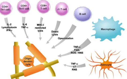
Figure
1
shows a schematic view of these cells and their
possible oligodendrocyte harming mediators.
Oligodendrocyte Injury Mediated by Immune Cells
In acute MS lesions, CD4
+and CD8
+T lymphocytes are
present. These cells can recognize their antigen if presented
by major histocompatibility complex (MHC) molecules
expressed on target cells, and be subsequently activated.
Under normal conditions, MHC expression in the CNS
does either not occur or is below detection levels (Redwine
et al.
2001
). In vitro experiments showed, however, that
oligodendrocytes can be induced to express MHC class I
(Grenier et al.
1989
; Kim
1985
) and MHC class II
molecules (Bergsteindottir et al.
1992
). Also in vivo, it
has been shown that oligodendrocytes are expressing MHC
class I molecules in a murine model of CNS inflammation
and demyelination (Redwine et al.
2001
) and in MS lesions
(Hoftberger et al.
2004
). This suggests that under
patho-logical conditions, oligodendrocytes induce MHC I
expres-sion and can thereby directly activate T cells and
consequently be damaged by them.
CD8
+T Lymphocytes
By the interaction of the CD8
+T cell receptor together with
the MHC class I peptide complex, CD8
+T cells are
activated and are directly cytotoxic to cells presenting their
specific antigen (Parkin and Cohen
2001
). The activation of
CD8
+T cell by recognition of their specific antigen is then
followed by clonal expansion. In MS, this was shown by
analyzing lesions, blood, and cerebral spinal fluid (CSF) for
clonal composition and T cell receptor repertoire (Babbe
et al.
2000
; Skulina et al.
2004
). These results suggested
that CD8
+T cells might have recognized their specific
antigen within the lesion and might have been activated. It
Figure 1 Cells mediating oligodendrocyte injury in the course of MS. Many different cell types have the potential to damage oligodendro-cytes. In this figure, some of these cells and their potential oligodendrocyte-damaging mediators are summarized. CD4+ Th1 T cells have been shown to induce oligodendrocyte damage among others through IL-2, LT, and IFN-g, whereas oligodendrocyte-damaging mechanisms of Th17 T cells involve IL-6 and TNF-a. CD8+T cells can induce oligodendrocyte damage directly by MHC
class I-restricted cell lysis. Furthermore, g/δ T cells were also shown to have the potential of damaging oligodendrocytes by direct lysis. By secreting antibodies, B cell-mediated damage to oligodendrocytes through opsonization was demonstrated. Macrophages are one of the main cell types inducing oligodendrocyte damage by TNF-a, FasL, ROS/RNS, and other mechanisms. Furthermore, astrocytes were shown to be potentially harmful to oligodendrocytes by mechanisms involving TNF-a, LT, and RNS
has been shown that oligodendrocytes are susceptible to
cytolysis by CD8
+T lymphocytes (Jurewicz et al.
1998
;
Ruijs et al.
1990
). Furthermore, an involvement of CD8
+cytotoxic T lymphocytes in autoimmune demyelination was
shown in experimental autoimmune encephalomyelitis
(EAE) (Huseby et al.
2001
; Sun et al.
2001
). Altogether,
this suggests that CD8
+T lymphocytes might contribute to
oligodendrocyte injury in MS.
CD4
+T Lymphocytes
CD4
+T helper cells recognize their cognate antigen
exclusively in the context of MHC class II molecules. In
contrast to MHC class I molecules, the expression of MHC
class II molecules by oligodendrocytes could not be
demonstrated in MS (Lee and Raine
1989
). MHC class II
expression is restricted to professional antigen-presenting
cells such as microglia/macrophages and dendritic cells
(Becher et al.
2000
; Greter et al.
2005
). It is easily
conceivable that CD4
+T helper cells induce oligodendrocyte
damage by secreting cytokines and promoting activation of
nearby macrophages and microglia. Studies in EAE suggest
that CD4
+T cells of the Th1 and Th17 lineage play a major
role in disease pathology (Gutcher et al.
2006
; Langrish et al.
2005
; Lassmann and Ransohoff
2004
; Sospedra and Martin
2005
; Weaver et al.
2006
). Th1 cells are characterized by the
predominant secretion of IFN-g whereas Th17 cells are
shown to secrete IL-17A, IL-17F, and IL-22 (Iwakura and
Ishigame
2006
; Kreymborg et al.
2007
; McGeachy et al.
2007
). It was shown that oligodendrocytes express TNF-a
receptors (Cannella et al.
2007
; Raine et al.
1998
) and other
cytokine receptors such as IFN-g receptor (Cannella and
Raine
2004
), and treatment of oligodendroglial cell lines
with IFN-g induces apoptosis (Buntinx et al.
2004
).
Oligodendrocytes were also shown to be susceptible to
TNF-a-induced cell death (D’Souza et al.
1996
; Jurewicz
et al.
2005
; Selmaj and Raine
1988
). Taken together,
activated CD4
+T lymphocytes do contribute to some extent,
directly or indirectly, to oligodendrocyte injury in MS.
γ/δ T Lymphocytes
Another cell type found in MS lesions are g/δ T
lymphocytes (Wucherpfennig et al.
1992
). g/δ T cells are
a T cell subpopulation showing a different T cell receptor
structure than a/β T cells (Li et al.
1998
). The role of g/δ T
lymphocytes in MS is still unclear. Nevertheless, depletion
of g/δ T lymphocytes during EAE has been shown to
ameliorate disease severity during the acute phases of the
disease (Rajan et al.
1996
). Furthermore, g/
δ T cells were
shown to enhance adoptive transfer of EAE by promoting
antigen presentation and IL-12 production (Odyniec et al.
2004
). As lysis of oligodendrocytes by g/
δ T cells has been
demonstrated in vitro (Freedman et al.
1991
), a possible
impact on oligodendrocyte injury in MS might not be ruled
out.
B Cells and Antibodies
In the CSF of MS patients, abnormal oligoclonal
immuno-globulin bands are detected, which supports the clinical
diagnosis of MS (Compston et al.
2006
). Autoantibodies
against myelin components were reported to be present in
the serum, CSF, and lesions of MS patients (Genain et al.
1999
; Reindl et al.
1999
). In line with this, IgG isolated
from inflamed CNS tissue from MS patients were shown to
recognize MOG (O’Connor et al.
2005
). Recently,
menin-geal B cell follicles were reported to associate with early
onset of disease and severe cortical pathology in secondary
progressive MS (Magliozzi et al.
2007
). Therefore,
anti-body-producing B cells may have potential impact on
oligodendrocyte injury and demyelination. For example,
injection of antibodies augmented demyelination during the
course of a T cell-mediated transfer EAE (Linington et al.
1988
). Further, it has been shown that by opsonizing the
myelin - oligodendrocyte surface, antibodies can stimulate
oligodendrocyte lysis of macrophages through their Fc
receptors (Scolding and Compston
1991
). Also, another
demyelinating mechanism by antibodies was shown to
involve membrane attack complex (MAC) deposition,
which finally leads to complement-mediated cytolysis
(Mead et al.
2002
; for review see Sospedra and Martin
2005
). Altogether, direct antibody-mediated injury of
oligodendrocytes in MS might play an important role,
although its impact on MS pathogenesis could not be
determined yet.
Oligodendrocyte Injury Mediated by Activated
Macrophages/Microglia
Activated macrophages and microglia may play an
impor-tant role in inducing oligodendrocyte injury during acute
inflammation in MS. It has been shown that disease
severity in EAE correlates best with macrophage infiltration
(Berger et al.
1997
). Activated macrophages and microglia
were shown to have incorporated myelin products and express
a large variety of different oligodendrocyte-deleterious
com-pounds, such as TNF-a, reactive oxygen species (ROS),
reactive nitrogen species (RNS), and Fas ligand (FasL).
TNF-a is a potent cytotoxic molecule capable of inducing
oligodendrocyte cell death (D
’Souza et al.
1996
; Jurewicz
et al.
2005
; Selmaj and Raine
1988
). The production of
ROS and RNS by activated macrophages and microglia
can lead to various types of damage such as lipid
peroxidation, tyrosine nitrosylation, and DNA strand
breaks (van der Veen and Roberts
1999
; Willenborg et al.
1999
; Zhang et al.
1994
). High expression of inducible
nitric oxide synthase (iNOS) and neuronal nitric oxide
synthase (nNOS) has been reported in activated
macro-phages and microglia within active lesions in MS (De
Groot et al.
1997
; Hill et al.
2004
), and RNS-mediated
damage in oligodendrocytes has also been demonstrated
(Jack et al.
2007
; Li et al.
2005
; Merrill et al.
1993
).
Oligodendrocytes were also reported to express Fas in MS
lesions (D’Souza et al.
1996
). FasL was shown to induce
oligodendrocyte damage (Li et al.
2002
), and as microglia
express FasL in MS lesions (Becher et al.
1998
), they
might, therefore, induce oligodendrocyte apoptosis.
Fur-thermore, it has been shown that activated macrophages
and microglia are capable of damaging oligodendrocytes in
an antibody-dependent mechanism (Griot-Wenk et al.
1991
). Altogether, activated macrophages and microglia
might be one of the major mediators of oligodendrocyte
injury in MS.
Oligodendrocyte Injury Mediated by Astrocytes
Astrocytes are known to maintain physiological glutamate
levels in the brain. Therefore, malfunctioning or too slow
glutamate uptake might lead to an enhancement of
oligo-dendrocyte excitotoxic damage (Newcombe et al.
2007
). In
addition, astrocytes are also known to express TNF-a and
LT-a. Thus, astrocytes might also be a potential inducer of
oligodendrocyte injury via TNF-a- and LT-a-dependent
mechanisms (for review see Williams et al.
2007
). An
expression of all three isoforms of NOS by astrocytes was
also reported (for review see Gibson et al.
2005
). In MS
plaques, high levels of constitutively expressed NOS were
detected to be expressed by astrocytes and macrophages
(De Groot et al.
1997
). In contrast to astrocytes,
oligoden-drocytes are shown to be much more susceptible to
NO-induced oxidative stress (Mitrovic et al.
1995
). This is
explained by the high iron load stored in oligodendrocytes
(Connor and Menzies
1995
; Roskams and Connor,
1994
;
Thorburne and Juurlink
1996
) and their low content of
reduced glutathione (GSH) (Juurlink et al.
1998
; Thorburne
and Juurlink
1996
). Iron (Fe
2+) was reported to be involved
in the formation of hydroxyl radicals (Gutteridge and
Halliwell
1989
), whereas glutathione peroxidase activity,
using GSH as an electron donor, scavenges hydrogen
peroxide and thus inhibits hydroxyl radical formation
(Juurlink et al.
1998
). A production of NO through NOS
expressed by astrocytes might, therefore, lead to oxidative
stress and damage in oligodendrocytes. Taken together,
activated astrocytes might also be involved in damaging
oligodendrocytes during the disease course of MS.
Reactive or Activated Oligodendrocytes - Pure Defensive
or Even Fierce Creatures?
As discussed before, immune cells and brain resident cells
are able to produce a variety of potentially harmful factors
for oligodendrocytes. These
“attacks” are occurring either
directly via lysis or indirectly via toxic mediators or via an
imbalance of the surrounding environment. As
demyelin-ation is a major feature in MS and loss of oligodendrocyte
during the chronic disease process is also evident,
oligo-dendrocytes can be regarded as
“poor victims” in the
pathogenic process of MS. Still, the question arises if
oligodendrocytes are really
“lame ducks” passively
allow-ing disease progression or if they attempt to defend
themselves in one way or another, which could even
influence disease progression?
Studies characterizing oligodendrocytes in MS lesions, in
primary oligodendrocyte cultures, and in analysis of
normal-appearing white matter (NAWM) MS tissue, which is mostly
devoid of immune infiltrates - therefore suitable to study
prelesional activities of oligodendrocytes - have recently
disclosed a view of oligodendrocytes being potential
immune-modulating in MS. Furthermore, oligodendrocytes
were shown to successfully protect themselves during
pathogenesis of Balo
’s concentric sclerosis (Stadelmann
et al.
2005
). Altogether, these findings might lead to a view
of oligodendrocytes being at least capable to defend
themselves or even be a reactive - to some extent active
cell type - part of the immune privilege of the brain. In this
study, we discuss the capacity of oligodendrocytes to react
against certain insults for their own protection, and how they
might modulate their environment by influencing disease
progression.
Activation of Endogenous Protective Mechanisms
In the last few years, growing evidence suggest an
involve-ment of hypoxia-like pathogenic mechanisms in MS
(Lassmann
2003
). Especially, in the so-called pattern III of
the lesion patterns identified recently (Lucchinetti et al.
2000
), hypoxia-like tissue injury may play a pathogenetic
role (Aboul-Enein et al.
2003
). Hypoxic tissue injury can be
induced in many ways. As already mentioned above, ROS
and RNS are known to induce cellular damages and
proposed to be involved in the demyelinating processes
(Smith et al.
1999
). For example, NO can impair respiratory
chain function in mitochondria, and by that can cause axon
conduction block (Redford et al.
1997
). In particular,
oligodendrocytes are vulnerable to NO-mediated damage
(Smith et al.
1999
; Smith and Lassmann
2002
), and
therefore, activation of mechanisms protecting
oligodendro-cytes from oxidative stress inducing damage would be highly
beneficial. A recent study of subcortical NAWM from MS
cases has shown the upregulation of several genes involved
in ischemic preconditioning (Graumann et al.
2003
). In
particular, HIF-1a has been shown to be an important
regulator of hypoxic preconditioning (Bergeron et al.
2000
;
Bernaudin et al.
2002
; Sharp et al.
2001
) and is activated by
hypoxia, growth factors, NO, and others (for review see
Brune and Zhou
2007
; Semenza
2002
). HIF-1a and some of
its downstream genes were shown to be elevated in MS
NAWM (Graumann et al.
2003
) and in situ hybridization
experiments of MS NAWM (Zeis et al.
2008
), and
examinations of Balo’s concentric sclerosis identified
oligo-dendrocytes expressing this transcription factor (Stadelmann
et al.
2005
), suggesting that oligodendrocytes mount
ischemic protective mechanisms during the disease course
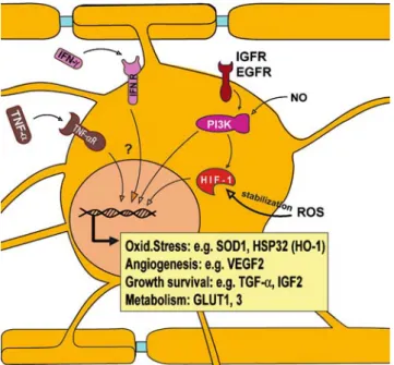
(Fig.
2
). Furthermore, oligodendrocytes were also shown to
express heat shock protein 70 (HSP70) (Stadelmann et al.
2005
) and HSP32 (Stahnke et al.
2007
). In the case of
HSP70, a protective role has been shown in brain ischemia
(for review see Christians et al.
2002
), whereas HSP32 was
shown to exert a protective role against oxidative stress in an
oligodendroglial cell line (Stahnke et al.
2007
).
It is interesting to note that sublethal doses of
inflam-matory cytokines such as IFN-g and TNF-a were reported
to induce protective mechanisms in target cells (Fig.
2
). The
induction of HSP70 in oligodendrocytes was shown in vitro
by treatment of oligodendrocyte cultures with a mix of
cytokines (D
’Souza et al.
1994
). Furthermore, treatment of
oligodendrocyte cultures with IFN-g led to an increase in
the expression of genes involved in protection against
oxidative stress (Balabanov et al.
2007
). In line with this,
treatment of mice with IFN-g before the onset of EAE led
to an amelioration of the disease through activating the
integrated stress response (Lin et al.
2007
). Altogether,
oligodendrocytes are able to induce and express
endoge-nous protective mechanisms allowing them to survive in an
otherwise potentially lethal environment.
Growth Factors
Changes in growth factors and growth factor receptors
expression were demonstrated in MS. Several growth
factors such as nerve growth factor (NGF), insulin-like
growth factor (IGF), and transforming growth factor
β
(TGF-β) were reported to be expressed by
oligodendro-cytes (for review see Du and Dreyfus
2002
). By expression
of these factors, oligodendrocytes might influence the
survival and/or function of neighboring cells. NGF can
bind to the tyrosine kinase receptor A (TrkA) and to the
low-affinity nerve growth factor receptor (p75
NTR). By
binding to TrkA, NGF promotes cell survival whereas
binding to p75
NTRunder some circumstances might also
modulate susceptibility to programmed cell death or
apoptosis (Casaccia-Bonnefil et al.
1999
; Yoon et al.
1998
). In EAE, the expression of TrkA was detected on
neurons, astrocytes, and oligodendrocytes
(Oderfeld-Nowak et al.
2003
; Oderfeld-Nowak et al.
2001
), whereas
p75
NTRwas detected on neurons, microglia, astrocytes, and
oligodendrocytes (Nataf et al.
1998
; Villoslada et al.
2000
).
In EAE, NGF was shown to have beneficial effects, as
NGF-deprived rats display more severe neurological
defi-cits during disease course. Furthermore, treatment of
marmoset monkeys with NGF prevented the full
develop-ment of EAE lesions and delayed the onset of clinical EAE
(Micera et al.
2000
; Villoslada et al.
2000
). Another growth
factor expressed by oligodendrocytes is IGF-1, which was
reported to ameliorate TNF-a-induced demyelination in
transgenic mice (Ye et al.
2007
). Furthermore, IGF-1 was
also reported to reduce demyelination in EAE (Liu et al.
1995
), although this beneficial effect is still under debate
(Cannella et al.
2000
). The expression of TGF-β by
oligodendrocytes was also reported, which is discussed in
the next chapter. Altogether, by expressing several growth
factors, oligodendrocytes are able to influence their own
function and survival, and also the function and survival of
nearby cells.
Figure 2 Ischemic preconditioning pathways in oligodendrocytes. Recent studies showed that oligodendrocytes can mount ischemic preconditioning mechanisms upon different stimuli. Treatment of oligodendrocytes with sublethal doses of IFN-g and TNF-a led to the upregulation of genes involved in ischemic tolerance. Protective genes were also shown to be upregulated in oligodendrocytes after stimulation with growth factors. Furthermore, low levels of RNS/ ROS were reported to lead to a stabilization of HIF-1a, which in turn activates the transcription of protective genes such as for example VEGFR, GLUT1, and GLUT3
Potential Immune-modulating Ability
of Oligodendrocytes
Immunohistochemical analysis of proteins expressed by
oligodendrocytes revealed that oligodendrocytes are able to
express cytokine receptors and members from the JAK/
STAT family (Cannella and Raine
2004
; Zeis et al.
2008
).
In a recent study, we have shown that genes from the
STAT6 signaling pathway are upregulated in MS NAWM
and that STAT6 and its members JAK1/3, 4R, and
IL-13R are expressed by oligodendrocytes (Figs.
3
and
4
)
(Zeis et al.
2008
). The STAT6 signaling pathway is known
from CD4
+T helper cells type 2, and it has been shown that
STAT6 is critically required for differentiation into Th2
cells (Kaplan et al.
1996
). Although still debated, cytokines
of the Th2 type such as IL-4 and IL-10 are thought to be
mostly beneficial in MS and EAE (Cannella and Raine
2004
; Sospedra and Martin
2005
). In EAE, it has been
shown that STAT6 knockout mice develop a more severe
disease than wild-type mice (Chitnis et al.
2001
). This
might be because of the lack of Th2 cells and of the
inability of oligodendrocytes to modulate their environment
in an anti-inflammatory way. The expression and activation
of an anti-inflammatory response by oligodendrocytes
might be crucial for them to compensate for the upregulated
proinflammatory environment and to limit the inflammatory
response and damage (Zeis et al.
2008
). The expression of
different cytokine receptors on oligodendrocytes in active
and silent lesions may further suggest an active role in
innate immunity of the CNS (Cannella and Raine
2004
).
Oligodendrocytes were also shown to express TGF-β in
vitro (da Cunha et al.
1993
; McKinnon et al.
1993
), which
can suppress immune and inflammatory responses (for
review see Pratt and McPherson
1997
), and might promote
myelination and remyelination (Setzu et al.
2006
).
In vitro experiments suggested that, upon stimulation by
INF-γ, oligodendrocytes express protective genes against
oxidative stress and a number of chemokines, including
CXCL10, CCL2, CCL3, and CCL5 (Fig.
4
) (Balabanov
et al.
2007
). CXCL10, CCL2, and CCL5 were also found to
be upregulated in MS NAWM (Graumann et al.
2003
).
Furthermore, mice with oligodendrocytes with suppressed
responsiveness to IFN-g showed higher oligodendrocyte
apoptosis in EAE and an accelerated disease onset, but
milder perivascular inflammation and minimal parenchymal
infiltration and demyelination (Balabanov et al.
2007
). This
effect of IFN-g on oligodendrocytes demonstrates that
oligodendrocytes are capable to react on external immune
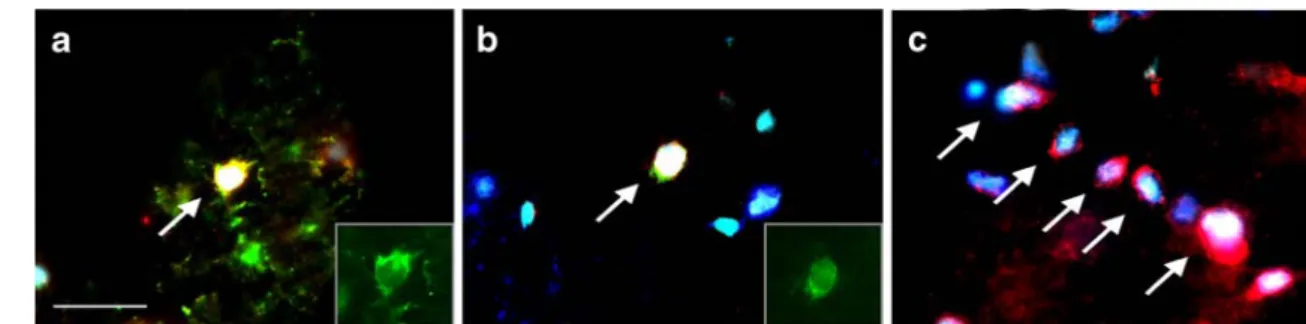
Figure 3 STAT6 signaling pathway expression in oligodendrocytes. Recently, oligodendrocytes were shown to be able to express immune mechanism relevant genes. Immunofluorescence colocalization anal-ysis of proteins from the STAT6 signaling pathway in MS patients revealed the expression of IL-4R (a), IL-13R (b), and STAT6 (c) in oligodendrocytes (Olig2 positive) in subcortical normal-appearing white matter brain tissue. a Colocalization of STAT6 (red), IL-4R
(green, inset), OLIG2 (blue), and DAPI (cyan); b colocalization of STAT6 (red), IL-13R (green, inset), OLIG2 (blue), and DAPI (cyan); c colocalization of STAT6 (red), OLIG2 (blue), and DAPI (cyan) in oligodendrocytes arranged in interfascicular rows, which is typical for myelinating oligodendrocytes (for more detailed pictures see Zeis et al.2008). Scale bar=25μm
Figure 4 Immune response-mediating pathways in oligodendrocytes. Analysis of proteins expressed by oligodendrocytes revealed that oligodendrocytes are able to express immune mechanisms-related proteins. Members of the STAT6 signaling pathway, such as IL-4R, IL13R, JAK1, and STAT6, were shown to be expressed by oligodendrocytes. This might indicate an anti-inflammatory “Th-2”-like response by oligodendrocytes. Furthermore, treatment of oligo-dendrocytes with a sublethal dose of IFN-g and TNF-a led to the secretion of chemokines such as CXCL10 (IP-10), CCL2 (MCP-1), CCL3 (MIP-1a), and CCL5 (Rantes). Altogether, this indicates that oligodendrocytes might play an immune-modulating role MS
challenges by induction of protective mechanisms and that
they can modulate inflammatory responses. The
expres-sion of cytokine receptors and members from the
anti-inflammatory STAT6 signaling pathway and the possibility
of chemokine expression might point to oligodendrocytes
playing a role in the innate immunity by actively modulating
their environment and interacting with cells of the immune
system.
Conclusions
Oligodendrocytes as the myelinating cell type in the CNS
are the major targets in MS. Many studies have shown that
oligodendrocytes are easily damaged by various
mecha-nisms. Therefore, oligodendrocytes might be seen as
“lame
ducks” of the CNS. However, growing evidence indicate
that oligodendrocyte are far more than a passive presence in
the CNS during MS. Oligodendrocytes are either
constitu-tively expressing or inducing various molecules able to
influence inflammatory reactions and prevent cell death to
conserve the functionality of the CNS. It seems that
oligodendrocytes in MS have a rather active or reactive
phenotype, preventing fatal damage and modulating their
surrounding. Therefore, oligodendrocytes may even act as
“fierce creatures,” influencing innate immunity and being
an active part in the formation of the immune privilege of
the brain.
Acknowledgements We thank Prof. Dr. Burkhard Becher (Division of Neuroimmunology, University Hospital Zürich) and Dr. Anna Stalder (Neurobiology, Department of Biomedicine, University Hospital Basel) for the critical reading of the manuscript. This work was supported by the National Multiple Sclerosis Societies of Switzerland, France (ARSEP), United Kingdom, and United States of America.
References
Aboul-Enein, F., Rauschka, H., Kornek, B., Stadelmann, C., Stefferl, A., Bruck, W., et al. (2003). Preferential loss of myelin-associated glycoprotein reflects hypoxia-like white matter damage in stroke and inflammatory brain diseases. Journal of Neuropathology and Experimental Neurology, 62, 25–33.
Babbe, H., Roers, A., Waisman, A., Lassmann, H., Goebels, N., Hohlfeld, R., et al. (2000). Clonal expansions of CD8(+) T cells dominate the T cell infiltrate in active multiple sclerosis lesions as shown by micromanipulation and single cell polymerase chain reaction. Journal of Experimental Medicine, 192, 393–404. Balabanov, R., Strand, K., Goswami, R., McMahon, E., Begolka, W.,
Miller, S. D., et al. (2007). Interferon-gamma-oligodendrocyte interactions in the regulation of experimental autoimmune encephalomyelitis. Journal of Neuroscience, 27, 2013–2024. Barnett, M. H., & Prineas, J. W. (2004). Relapsing and remitting
multiple sclerosis: Pathology of the newly forming lesion. Annals of Neurology, 55, 458–468.
Becher, B., Barker, P. A., Owens, T., & Antel, J. P. (1998). CD95-CD95L: Can the brain learn from the immune system? Trends in Neurosciences, 21, 114–117.
Becher, B., Prat, A., & Antel, J. P. (2000). Brain-immune connection: Immuno-regulatory properties of CNS-resident cells. Glia, 29, 293–304.
Berger, T., Weerth, S., Kojima, K., Linington, C., Wekerle, H., & Lassmann, H. (1997). Experimental autoimmune encephalomy-elitis: The antigen specificity of T lymphocytes determines the topography of lesions in the central and peripheral nervous system. Laboratory Investigation, 76, 355–364.
Bergeron, M., Gidday, J. M., Yu, A. Y., Semenza, G. L., Ferriero, D. M., & Sharp, F. R. (2000). Role of hypoxia-inducible factor-1 in hypoxia-induced ischemic tolerance in neonatal rat brain. Annals of Neurology, 48, 285–296.
Bergsteindottir, K., Brennan, A., Jessen, K. R., & Mirsky, R. (1992). In the presence of dexamethasone, gamma interferon induces rat oligodendrocytes to express major histocompatibility complex class II molecules. Proceedings of the National Academy of Sciences of the United States of America, 89, 9054–9058. Bernaudin, M., Tang, Y., Reilly, M., Petit, E., & Sharp, F. R. (2002).
Brain genomic response following hypoxia and re-oxygenation in the neonatal rat. Identification of genes that might contribute to hypoxia-induced ischemic tolerance. Journal of Biological Chemistry, 277, 39728–39738.
Brune, B., & Zhou, J. (2007). Hypoxia-inducible factor-1alpha under the control of nitric oxide. Methods in Enzymology, 435, 463–478. Buntinx, M., Gielen, E., Van Hummelen, P., Raus, J., Ameloot, M., Steels, P., et al. (2004). Cytokine-induced cell death in human oligodendroglial cell lines. II: Alterations in gene expression induced by interferon-gamma and tumor necrosis factor-alpha. Journal of Neuroscience Research, 76, 846–861.
Cannella, B., Gaupp, S., Omari, K. M., & Raine, C. S. (2007). Multiple sclerosis: Death receptor expression and oligodendrocyte apopto-sis in established lesions. Journal of Neuroimmunology, 188, 128–137.
Cannella, B., Pitt, D., Capello, E., & Raine, C. S. (2000). Insulin-like growth factor-1 fails to enhance central nervous system myelin repair during autoimmune demyelination. American Journal of Pathology, 157, 933–943.
Cannella, B., & Raine, C. S. (2004). Multiple sclerosis: Cytokine receptors on oligodendrocytes predict innate regulation. Annals of Neurology, 55, 46–57.
Casaccia-Bonnefil, P., Gu, C., Khursigara, G., & Chao, M. V. (1999). p75 neurotrophin receptor as a modulator of survival and death decisions. Microscopy Research and Technique, 45, 217–224. Chitnis, T., Najafian, N., Benou, C., Salama, A. D., Grusby, M. J.,
Sayegh, M. H., et al. (2001). Effect of targeted disruption of STAT4 and STAT6 on the induction of experimental autoimmune enceph-alomyelitis. Journal of Clinical Investigation, 108, 739–747. Christians, E. S., Yan, L. J., & Benjamin, I. J. (2002). Heat shock
factor 1 and heat shock proteins: Critical partners in protection against acute cell injury. Critical Care Medicine, 30, S43–S50. Compston, A., McDonald, I., Noseworthy, J., Lassmann, H., Miller, D.,
Smith, K., et al. (2006). McAlpine’s multiple sclerosis. New York: Churchill Livingstone.
Connor, J. R., & Menzies, S. L. (1995). Cellular management of iron in the brain. Journal of the Neurological Sciences, 134(Suppl), 33–44. D’Souza, S. D., Alinauskas, K. A., & Antel, J. P. (1996). Ciliary neurotrophic factor selectively protects human oligodendrocytes from tumor necrosis factor-mediated injury. Journal of Neuro-science Research, 43, 289–298.
D’Souza, S. D., & Antel, J. P. (1994). Freedman MS. Cytokine induction of heat shock protein expression in human oligoden-drocytes: An interleukin-1-mediated mechanism. Journal of Neuroimmunology, 50, 17–24.
da Cunha, A., Jefferson, J. A., Jackson, R. W., & Vitkovic, L. (1993). Glial cell-specific mechanisms of TGF-beta 1 induction by IL-1 in cerebral cortex. Journal of Neuroimmunology, 42, 71–85. De Groot, C. J., Ruuls, S. R., Theeuwes, J. W., Dijkstra, C. D., & Van
der Valk, P. (1997). Immunocytochemical characterization of the expression of inducible and constitutive isoforms of nitric oxide synthase in demyelinating multiple sclerosis lesions. Journal of Neuropathology and Experimental Neurology, 56, 10–20. Du, Y., & Dreyfus, C. F. (2002). Oligodendrocytes as providers of
growth factors. Journal of Neuroscience Research, 68, 647–654. Freedman, M. S., Ruijs, T. C., Selin, L. K., & Antel, J. P. (1991). Peripheral blood gamma-delta T cells lyse fresh human brain-derived oligodendrocytes. Annals of Neurology, 30, 794–800. Genain, C. P., Cannella, B., Hauser, S. L., & Raine, C. S. (1999).
Identification of autoantibodies associated with myelin damage in multiple sclerosis. Natural Medicines, 5, 170–175.
Gibson, C. L., Coughlan, T. C., & Murphy, S. P. (2005). Glial nitric oxide and ischemia. Glia, 50, 417–426.
Gold, R., Hartung, H. P., & Toyka, K. V. (2000). Animal models for autoimmune demyelinating disorders of the nervous system. Molecular Medicine Today, 6, 88–91.
Gold, R., Linington, C., & Lassmann, H. (2006). Understanding pathogenesis and therapy of multiple sclerosis via animal models: 70 years of merits and culprits in experimental autoimmune encephalomyelitis research. Brain, 129, 1953–1971.
Graumann, U., Reynolds, R., Steck, A. J., & Schaeren-Wiemers, N. (2003). Molecular changes in normal appearing white matter in multiple sclerosis are characteristic of neuroprotective mecha-nisms against hypoxic insult. Brain Pathology, 13, 554–573. Grenier, Y., Ruijs, T. C., Robitaille, Y., Olivier, A., & Antel, J. P.
(1989). Immunohistochemical studies of adult human glial cells. Journal of Neuroimmunology, 21, 103–115.
Greter, M., Heppner, F. L., Lemos, M. P., Odermatt, B. M., Goebels, N., Laufer, T., et al. (2005). Dendritic cells permit immune invasion of the CNS in an animal model of multiple sclerosis. Natural Medicines, 11, 328–334.
Griot-Wenk, M., Griot, C., Pfister, H., & Vandevelde, M. (1991). Antibody-dependent cellular cytotoxicity in antimyelin antibody-induced oligodendrocyte damage in vitro. Journal of Neuroimmunology, 33, 145–155.
Gutcher, I., Urich, E., Wolter, K., Prinz, M., & Becher, B. (2006). Interleukin 18-independent engagement of interleukin 18 recep-tor-alpha is required for autoimmune inflammation. Nature Immunology, 7, 946–953.
Gutteridge, J. M., & Halliwell, B. (1989). Iron toxicity and oxygen radicals. Baillieres Clinics in Haematology, 2, 195–256. Hill, K. E., Zollinger, L. V., Watt, H. E., Carlson, N. G., & Rose, J. W.
(2004). Inducible nitric oxide synthase in chronic active multiple sclerosis plaques: Distribution, cellular expression and association with myelin damage. Journal of Neuroimmunology, 151, 171–179. Hoftberger, R., Aboul-Enein, F., Brueck, W., Lucchinetti, C., Rodriguez, M., Schmidbauer, M., et al. (2004). Expression of major histocompatibility complex class I molecules on the different cell types in multiple sclerosis lesions. Brain Pathology, 14, 43–50.
Huseby, E. S., Liggitt, D., Brabb, T., Schnabel, B., Ohlen, C., & Goverman, J. (2001). A pathogenic role for myelin-specific CD8 (+) T cells in a model for multiple sclerosis. Journal of Experimental Medicine, 194, 669–676.
Iwakura, Y., & Ishigame, H. (2006). The IL-23/IL-17 axis in inflammation. Journal of Clinical Investigation, 116, 1218–1222. Jack, C., Antel, J., Bruck, W., & Kuhlmann, T. (2007). Contrasting potential of nitric oxide and peroxynitrite to mediate oligoden-drocyte injury in multiple sclerosis. Glia, 55, 926–934. Jurewicz, A., Biddison, W. E., & Antel, J. P. (1998). MHC class
I-restricted lysis of human oligodendrocytes by myelin basic protein
peptide-specific CD8 T lymphocytes. Journal of Immunology, 160, 3056–3059.
Jurewicz, A., Matysiak, M., Tybor, K., Kilianek, L., Raine, C. S., & Selmaj, K. (2005). Tumour necrosis factor-induced death of adult human oligodendrocytes is mediated by apoptosis inducing factor. Brain, 128, 2675–2688.
Juurlink, B. H., Thorburne, S. K., & Hertz, L. (1998). Peroxide-scavenging deficit underlies oligodendrocyte susceptibility to oxidative stress. Glia, 22, 371–378.
Kaplan, M. H., Schindler, U., Smiley, S. T., & Grusby, M. J. (1996). Stat6 is required for mediating responses to IL-4 and for development of Th2 cells. Immunity, 4, 313–319.
Kennedy, P. G., & Steiner, I. (1994). On the possible viral aetiology of multiple sclerosis. QJM, 87, 523–528.
Kim, S. U. (1985). Antigen expression by glial cells grown in culture. Journal of Neuroimmunology, 8, 255–282.
Kreymborg, K., Etzensperger, R., Dumoutier, L., Haak, S., Rebollo, A., Buch, T., et al. (2007). 22 is expressed by Th17 cells in an IL-23-dependent fashion, but not required for the development of autoimmune encephalomyelitis. Journal of Immunology, 179, 8098–8104.
Langrish, C. L., Chen, Y., Blumenschein, W. M., Mattson, J., Basham, B., Sedgwick, J. D., et al. (2005). IL-23 drives a pathogenic T cell population that induces autoimmune inflammation. Journal of Experimental Medicine, 201, 233–240.
Lassmann, H. (2003). Hypoxia-like tissue injury as a component of multiple sclerosis lesions. Journal of the Neurological Sciences, 206, 187–191.
Lassmann, H., & Ransohoff, R. M. (2004). The CD4-Th1 model for multiple sclerosis: A critical [correction of crucial] re-appraisal. Trends in Immunology, 25, 132–137.
Lee, S. C., & Raine, C. S. (1989). Multiple sclerosis: Oligodendrocytes in active lesions do not express class II major histocompatibility complex molecules. Journal of Neuroimmunology, 25, 261–266. Li, J., Baud, O., Vartanian, T., Volpe, J. J., & Rosenberg, P. A. (2005).
Peroxynitrite generated by inducible nitric oxide synthase and NADPH oxidase mediates microglial toxicity to oligodendro-cytes. Proceedings of the National Academy of Sciences of the United States of America, 102, 9936–9941.
Li, H., Lebedeva, M. I., Llera, A. S., Fields, B. A., Brenner, M. B., & Mariuzza, R. A. (1998). Structure of the Vdelta domain of a human gammadelta T-cell antigen receptor. Nature, 391, 502–506. Li, W., Maeda, Y., Ming, X., Cook, S., Chapin, J., Husar, W., et al.
(2002). Apoptotic death following Fas activation in human oligodendrocyte hybrid cultures. Journal of Neuroscience Research, 69, 189–196.
Lin, W., Bailey, S. L., Ho, H., Harding, H. P., Ron, D., Miller, S. D., et al. (2007). The integrated stress response prevents demyelin-ation by protecting oligodendrocytes against immune-mediated damage. Journal of Clinical Investigation, 117, 448–456. Linington, C., Bradl, M., Lassmann, H., Brunner, C., & Vass, K. (1988).
Augmentation of demyelination in rat acute allergic encephalomy-elitis by circulating mouse monoclonal antibodies directed against a myelin/oligodendrocyte glycoprotein. American Journal of Pathology, 130, 443–454.
Liu, X., Yao, D. L., & Webster, H. (1995). Insulin-like growth factor I treatment reduces clinical deficits and lesion severity in acute demyelinating experimental autoimmune encephalomyelitis. Multiple Sclerosis, 1, 2–9.
Lucchinetti, C., Bruck, W., Parisi, J., Scheithauer, B., Rodriguez, M., & Lassmann, H. (2000). Heterogeneity of multiple sclerosis lesions: Implications for the pathogenesis of demyelination. Annals of Neurology, 47, 707–717.
Ludwin, S. K. (1997). The pathobiology of the oligodendrocyte. Journal of Neuropathology and Experimental Neurology, 56, 111–124.
Magliozzi, R., Howell, O., Vora, A., Serafini, B., Nicholas, R., Puopolo, M., et al. (2007). Meningeal B-cell follicles in secondary progressive multiple sclerosis associate with early onset of disease and severe cortical pathology. Brain, 130, 1089–1104.
McGeachy, M. J., Bak-Jensen, K. S., Chen, Y., Tato, C. M., Blumenschein, W., McClanahan, T., et al. (2007). TGF-beta and IL-6 drive the production of IL-17 and IL-10 by T cells and restrain T(H)-17 cell-mediated pathology. Nature Immunology, 8, 1390–1397.
McKinnon, R. D., Piras, G., Ida Jr., J. A., & Dubois-Dalcq, M. (1993). A role for TGF-beta in oligodendrocyte differentiation. Journal of Cell Biology, 121, 1397–1407.
Mead, R. J., Singhrao, S. K., Neal, J. W., Lassmann, H., & Morgan, B. P. (2002). The membrane attack complex of complement causes severe demyelination associated with acute axonal injury. Journal of Immunology, 168, 458–465.
Merrill, J. E., Ignarro, L. J., Sherman, M. P., Melinek, J., & Lane, T. E. (1993). Microglial cell cytotoxicity of oligodendrocytes is mediat-ed through nitric oxide. Journal of Immunology, 151, 2132–2141. Merrill, J. E., & Scolding, N. J. (1999). Mechanisms of damage to myelin and oligodendrocytes and their relevance to disease. Neuropathology & Applied Neurobiology, 25, 435–458. Micera, A., Properzi, F., Triaca, V., & Aloe, L. (2000). Nerve growth
factor antibody exacerbates neuropathological signs of experi-mental allergic encephalomyelitis in adult lewis rats. Journal of Neuroimmunology, 104, 116–123.
Mitrovic, B., Ignarro, L. J., Vinters, H. V., Akers, M. A., Schmid, I., Uittenbogaart, C., et al. (1995). Nitric oxide induces necrotic but not apoptotic cell death in oligodendrocytes. Neuroscience, 65, 531–539. Nataf, S., Naveilhan, P., Sindji, L., Darcy, F., Brachet, P., & Montero-Menei, C. N. (1998). Low affinity NGF receptor expression in the central nervous system during experimental allergic enceph-alomyelitis. Journal of Neuroscience Research, 52, 83–92. Newcombe, J., Uddin, A., Dove, R., Patel, B., Turski, L., Nishizawa, Y.,
et al. (2007). Glutamate receptor expression in multiple sclerosis lesions. Brain Pathology, 18(1), 52–61.
O’Connor, K. C., Appel, H., Bregoli, L., Call, M. E., Catz, I., Chan, J. A., et al. (2005). Antibodies from inflamed central nervous system tissue recognize myelin oligodendrocyte glycoprotein. Journal of Immunology, 175, 1974–1982.
Oderfeld-Nowak, B., Zaremba, M., Lipkowski, A. W., Kwiatkowska-Patzer, B., Triaca, V., & Aloe, L. (2003). High-affinity NGF receptor in the rat spinal cord during acute and chronic phases of experimental autoimmune encephalomyelitis: A possible function-al significance. Archives Itfunction-aliennes de Biologie, 141, 103–116. Oderfeld-Nowak, B., Zaremba, M., Micera, A., & Aloe, L. (2001).
The upregulation of nerve growth factor receptors in reactive astrocytes of rat spinal cord during experimental autoimmune encephalomyelitis. Neuroscience Letters, 308, 165–168. Odyniec, A., Szczepanik, M., Mycko, M. P., Stasiolek, M., Raine, C. S.,
& Selmaj, K. W. (2004). Gammadelta T cells enhance the expression of experimental autoimmune encephalomyelitis by promoting antigen presentation and IL-12 production. Journal of Immunology, 173, 682–694.
Ozawa, K., Suchanek, G., Breitschopf, H., Bruck, W., Budka, H., Jellinger, K., et al. (1994). Patterns of oligodendroglia pathology in multiple sclerosis. Brain, 117(Pt 6), 1311–1322.
Parkin, J., & Cohen, B. (2001). An overview of the immune system. Lancet, 357, 1777–1789.
Pratt, B. M., & McPherson, J. M. (1997). TGF-beta in the central nervous system: Potential roles in ischemic injury and neurode-generative diseases. Cytokine and Growth Factor Reviews, 8, 267–292.
Raine, C. S. (1997). The Norton lecture: A review of the oligodendro-cyte in the multiple sclerosis lesion. Journal of Neuroimmunology, 77, 135–152.
Raine, C. S., Bonetti, B., & Cannella, B. (1998). Multiple sclerosis: Expression of molecules of the tumor necrosis factor ligand and receptor families in relationship to the demyelinated plaque. Revista de Neurología (Paris), 154, 577–585.
Rajan, A. J., Gao, Y. L., Raine, C. S., & Brosnan, C. F. (1996). A pathogenic role for gamma delta T cells in relapsing-remitting experimental allergic encephalomyelitis in the SJL mouse. Journal of Immunology, 157, 941–949.
Redford, E. J., Kapoor, R., & Smith, K. J. (1997). Nitric oxide donors reversibly block axonal conduction: Demyelinated axons are especially susceptible. Brain, 120(Pt 12), 2149–2157.
Redwine, J. M., Buchmeier, M. J., & Evans, C. F. (2001). In vivo expression of major histocompatibility complex molecules on oligodendrocytes and neurons during viral infection. American Journal of Pathology, 159, 1219–1224.
Reindl, M., Linington, C., Brehm, U., Egg, R., Dilitz, E., Deisenhammer, F., et al. (1999). Antibodies against the myelin oligodendrocyte glycoprotein and the myelin basic protein in multiple sclerosis and other neurological diseases: A comparative study. Brain, 122(Pt 11), 2047–2056.
Roskams, A. J., & Connor, J. R. (1994). Iron, transferrin, and ferritin in the rat brain during development and aging. Journal of Neurochemistry, 63, 709–716.
Ruijs, T. C., Freedman, M. S., Grenier, Y. G., Olivier, A., & Antel, J. P. (1990). Human oligodendrocytes are susceptible to cytolysis by major histocompatibility complex class I-restricted lymphocytes. Journal of Neuroimmunology, 27, 89–97.
Scolding, N. J., & Compston, D. A. (1991). Oligodendrocyte-macrophage interactions in vitro triggered by specific antibodies. Immunology, 72, 127–132.
Selmaj, K. W., & Raine, C. S. (1988). Tumor necrosis factor mediates myelin and oligodendrocyte damage in vitro. Annals of Neurology, 23, 339–346.
Semenza, G. (2002). Signal transduction to hypoxia-inducible factor 1. Biochemical Pharmacology, 64, 993–998.
Setzu, A., Lathia, J. D., Zhao, C., Wells, K., Rao, M. S., Ffrench-Constant, C., et al. (2006). Inflammation stimulates myelination by transplanted oligodendrocyte precursor cells. Glia, 54, 297–303. Sharp, F. R., Bergeron, M., & Bernaudin, M. (2001).
Hypoxia-inducible factor in brain. Advances in Experimental Medicine and Biology, 502, 273–291.
Skulina, C., Schmidt, S., Dornmair, K., Babbe, H., Roers, A., Rajewsky, K., et al. (2004). Multiple sclerosis: Brain-infiltrating CD8+ T cells persist as clonal expansions in the cerebrospinal fluid and blood. Proceedings of the National Academy of Sciences of the United States of America, 101, 2428–2433. Smith, K. J., Kapoor, R., & Felts, P. A. (1999). Demyelination: The role
of reactive oxygen and nitrogen species. Brain Pathology, 9, 69–92. Smith, K. J., & Lassmann, H. (2002). The role of nitric oxide in
multiple sclerosis. Lancet Neurology, 1, 232–241.
Sospedra, M., & Martin, R. (2005). Immunology of multiple sclerosis. Annual Review of Immunology, 23, 683–747.
Stadelmann, C., Ludwin, S., Tabira, T., Guseo, A., Lucchinetti, C. F., Leel-Ossy, L., et al. (2005). Tissue preconditioning may explain concentric lesions in Balo’s type of multiple sclerosis. Brain, 128, 979–987. Stahnke, T., Stadelmann, C., Netzler, A., Bruck, W., & Richter-Landsberg,
C. (2007). Differential upregulation of heme oxygenase-1 (HSP32) in glial cells after oxidative stress and in demyelinating disorders. Journal of Molecular Neuroscience, 32, 25–37.
Sun, D., Whitaker, J. N., Huang, Z., Liu, D., Coleclough, C., Wekerle, H., et al. (2001). Myelin antigen-specific CD8+T cells are encephali-togenic and produce severe disease in C57BL/6 mice. Journal of Immunology, 166, 7579–7587.
Thorburne, S. K., & Juurlink, B. H. (1996). Low glutathione and high iron govern the susceptibility of oligodendroglial precursors to oxidative stress. Journal of Neurochemistry, 67, 1014–1022.
van der Veen, R. C., & Roberts, L. J. (1999). Contrasting roles for nitric oxide and peroxynitrite in the peroxidation of myelin lipids. Journal of Neuroimmunology, 95, 1–7.
Villoslada, P., Hauser, S. L., Bartke, I., Unger, J., Heald, N., Rosenberg, D., et al. (2000). Human nerve growth factor protects common marmosets against autoimmune encephalomyelitis by switching the balance of T helper cell type 1 and 2 cytokines within the central nervous system. Journal of Experimental Medicine, 191, 1799–1806.
Weaver, C. T., Harrington, L. E., Mangan, P. R., Gavrieli, M., & Murphy, K. M. (2006). Th17: An effector CD4 T cell lineage with regulatory T cell ties. Immunity, 24, 677–688.
Willenborg, D. O., Staykova, M. A., & Cowden, W. B. (1999). Our shifting understanding of the role of nitric oxide in autoimmune encephalomyelitis: A review. Journal of Neuroimmunology, 100, 21–35.
Williams, A., Piaton, G., & Lubetzki, C. (2007). Astrocytes—friends or foes in multiple sclerosis? Glia, 55, 1300–1312.
Wucherpfennig, K. W., Newcombe, J., Li, H., Keddy, C., Cuzner, M. L., & Hafler, D. A. (1992). Gamma delta T-cell receptor repertoire in acute multiple sclerosis lesions. Proceedings of the National Academy of Sciences of the United States of America, 89, 4588–4592. Ye, P., Kollias, G., D, , & Ercole, A. J. (2007). Insulin-like growth factor-I
ameliorates demyelination induced by tumor necrosis factor-alpha in transgenic mice. Journal of Neuroscience Research, 85, 712–722. Yoon, S. O., Casaccia-Bonnefil, P., Carter, B., & Chao, M. V. (1998).
Competitive signaling between TrkA and p75 nerve growth factor receptors determines cell survival. Journal of Neuroscience, 18, 3273–3281.
Zeis, T., Graumann, U., Reynolds, R., & Schaeren-Wiemers, N. (2008). Normal-appearing white matter in multiple sclerosis is in a subtle balance between inflammation and neuroprotection. Brain, 131, 288–303.
Zhang, J., Dawson, V. L., Dawson, T. M., & Snyder, S. H. (1994). Nitric oxide activation of poly(ADP-ribose) synthetase in neurotoxicity. Science, 263, 687–689.