HAL Id: hal-01070952
https://hal.archives-ouvertes.fr/hal-01070952
Submitted on 10 Oct 2014HAL is a multi-disciplinary open access
archive for the deposit and dissemination of sci-entific research documents, whether they are pub-lished or not. The documents may come from teaching and research institutions in France or abroad, or from public or private research centers.
L’archive ouverte pluridisciplinaire HAL, est destinée au dépôt et à la diffusion de documents scientifiques de niveau recherche, publiés ou non, émanant des établissements d’enseignement et de recherche français ou étrangers, des laboratoires publics ou privés.
Morphological description of four new species of
Nyctotherus (Ciliophora: Nyctotheridae:
Heterotri-chida), commensal ciliates of the digestive tract of a
terrestrial Oligochaete (Megascolecidae) from the
northwest region of Cameroon
Zéphyrin Fokam, Paul Alain Nana, Pierre Ngassam, Geneviève Bricheux,
Philippe Bouchard, Bernard Vigues, Télesphore Sime-Ngando
To cite this version:
Zéphyrin Fokam, Paul Alain Nana, Pierre Ngassam, Geneviève Bricheux, Philippe Bouchard, et al.. Morphological description of four new species of Nyctotherus (Ciliophora: Nyctotheridae: Heterotri-chida), commensal ciliates of the digestive tract of a terrestrial Oligochaete (Megascolecidae) from the northwest region of Cameroon. Protistology, 2014, 8 (2), pp.62-70. �hal-01070952�
© 2014 The Author(s)
Protistology © 2014 Protozoological Society Affiliated with RAS
Morphological description of four new species of
Nyctotherus (Ciliophora: Nyctotheridae:
Heterotri-chida), commensal ciliates of the digestive tract of
a terrestrial Oligochaete (Megascolecidae) from the
northwest region of Cameroon
Z
é
phyrin Fokam
1, Paul Alain Nana
2, Pierre Ngassam
2,
Genevi
è
ve Bricheux
3,4, Philippe Bouchard
3,
Bernard Vigues
3and T
é
lesphore Sime-Ngando
3,41 Department of Biology, Higher Teacher Training College, The University
of Bamenda, Cameroon
2 Laboratory of General Biology, Faculty of Science, University of Yaoundé
I, Cameroon
3 Laboratoire de Microorganismes : Génome et Environnement, Université
Blaise Pascal, Clermont-Ferrand, France
4 Centre National de Recherche Scientifique, Aubière Cedex, France
Summary
The morphology and ciliature of four Nyctotherus species (Nyctotherus orthostomatus n. sp., Nyctotherus ndoumeleleensis n. sp., Nyctotherus ngassami n. sp. and Nyctotherus
atunibaensis n. sp.), isolated from the mid and hindgut of a Megascolecidae
annelid of the genus Eupolytoreutus from the northwest region of Cameroon, were investigated using living observation (methylene blue) and silver impregnation methods (Fernandez-Galiano, 1994). Nyctotherus orthostomatus n. sp. is recognized by a pear-shaped body (128–152 × 95–104 µm); a conspicuous buccal apparatus with the peristome and the infundibulum perpendicular to each other; about 41–46 somatic kineties covering each side of the cell; one transversally elongated massive macronucleus located in the anterior third of the cell, surmounted by a very small spherical micronucleus. Nyctotherus ndoumeleleensis n. sp. is distinguished by the combination of the following characters: ovoid body (151–163 × 87–92 µm) with rounded poles; about 52–68 somatic kineties uniformly covering the cell body; an anterior, transversal and trapezoidal macronucleus carrying laterally a small micronucleus (2–3.5 µm in diameter). Nyctotherus ngassami n. sp. can be distinguished from its congeners by the following set of features: size about 153–269 × 102–148 µm; body ovoid, with 64–86 ciliary rows covering its surface; a small and globular micronucleus hidden by a transversal macronucleus located slightly above the equatorial part of the cell. Nyctotherus atunibaensis n. sp. is characterized by size about 145–253 × 95–162 µm, ovoid cell body, 108–122 somatic kineties,
·
63Protistology
a transversal and arc-shaped macronucleus hiding a small micronucleus. In the present study, new locality (northwest region of Cameroon) and new species of the genus Nyctotherus are explored.
Key words: Cameroon, megascolecid earthworms, Nyctotherus, N. orthostomatus n. sp., N. ndoumeleleensis n. sp., N. ngassami n. sp., N. atunibaensis n. sp., systematics
Introduction
Ciliates are ubiquitous eukaryotic unicellular microorganisms which have adapted to a wide range of habitats (terrestrial, marine and fresh waters), using different morphological and physiological strategies; some ciliates are symbiotic (Sauvadet, 2010). The symbiotic ciliates could be parasitic, mutualistic and mostly commensal of the digestive tract of vertebrates and invertebrates, particularly annelids and amphibians (de Puytorac, 1994). Thus, the digestive tract of megascolecid annelids can be considered as a microhabitat suitable for the development of diverse microorganisms, notably many types of ciliates including nyctotherans. These ciliates have been targeted for studies by many researchers (Albaret, 1975; Albaret and Njiné, 1975; Ngassam, 1983; Affa’a, 1991; Affa’a and Amiet, 1994; Finler and Yildiz, 2000). The Nyctotherans that live endocommensally only in the mid and hindgut of Megascolecidae annelids of the genus
Eupolytoreutus collected in the central Cameroon
are presently classified basing on their ciliary pattern. The one characterized by an apical secant system on the right side of the cell, belongs to the genus
Nyctotherus Leidy, 1849; another, with an apical
and a postoral secant system, is typical of the genus
Paranyctotherus Ngassam, 1983. So far there is no
information concerning these ciliates from other regions of Cameroon.
In this paper, we describe four new nyctotheran species obtained from Megascolecidae earthworms of the genus Eupolytoreutus which distribution is likely to expand from the central region where they have been studied (Albaret and Njiné, 1975; Ngassam, 1983) up to the Northwest region of Cameroon.
Material and methods
The samples were collected in August-November 2012 in Bambui and Bambili (Tubah Subdivision of the north-west region of Cameroon). This study area is located in the southern part of the meridional Niger basin (5° 59’ 00’’ N and 10° 15’ 00’’ E), at an
altitude of 1350 m (Fig. 1). Worms were identified as belonging to the genus Eupolytoreutus according to the key proposed by Sims and Gerard (1999).
Fragments of the digestive tract pertaining to each of the three main portions (fore-, mid-, and hindgut) were opened under a binocular dissecting microscope Wild M5 (Heerbrugg, Germany) at 250× magnification. This was done in a Petri dish containing either physiological Ringer’s solution or mineral water (Volvic™ in France or Supermont™ in Cameroon). The ciliates were removed from the ingesta using micropipettes, washed and observed
in vivo before fixation for cytology. Their shape and
mobility were registered. Cell staining was made with the ammoniacal pyridinated silver carbonate technique of Fernandez-Galiano (1976, 1994). All cell measurements were made with a calibrated ocular micrometer. The following parameters were calculated: arithmetic mean; standard deviation; minimum and maximum values. Morphometric data were obtained by examination of 30 cells of each species. Drawings of these cells were performed with the aid of a camera lucida attached to a Wild M20 microscope. A digital camera was used for light micrographs. Identification and classification was done according to the key of Albaret (1975).
Results
NYCTOTHERUSORTHOSTOMATUS FOKAM, NANA,
NGASSAMETAL. N. SP.
This species occurs in the mid and hindgut of
Eupolytoreutus sp., where it lives in cohabitation with
many other species of nyctotherans and astomes, specifically Eudrilophrya complanata de Puytorac and Dragesco, 1970 and Anoplophrya vugaris Ngassam, 1883.
Diagnosis. The body is pear-shaped. Both poles are rounded. The peristome starts far from the apex and joins the infundibulum orthogonally in the equatorial region. The infundibulum ends with a short and slender cytopharynx. The macronucleus is transversal, surmounted by an eccentric micro-nucleus and located in the anterior half of the cell.
There is one preoral suture line, but no caudal secant system. One to two contractile vacuoles are located posteriorly.
Type host. Eupolytoreutus sp.
Type locality. Bambili and Bambui, Tubah Subdivision, Bamenda, Cameroon.
Habitat. Mid and hindgut.
Type specimens. Permanent preparations belon-ging to this species are kept in the Department of Biology (Higher Teacher Training College, The University of Bamenda, Cameroon).
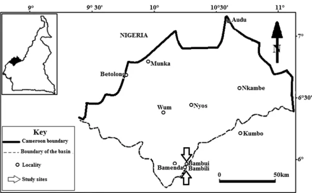
Description. The cell is elongated and laterally flattened into concave left and convex right sides. The posterior end is wider than the anterior end conferring a pear shape on the specimen. The anterior end is slightly pointed (Fig. 2).
Morphometric characteristics are: 143.5 ± 7.1 µm in length and 94.8 ± 37 µm in width (Table 1). The macronucleus (20.03 ± 1.6 µm long and 47.7 ± 1.4 µm across) is massive and kidney shaped (Fig. 2), lodged in the anterior portion of the ciliate, set horizontally and very close to the peristome. The micronucleus (2.7 ± 0.6 µm in diameter) is spherical, situated to the left side on top of the macronucleus. No caryophore surrounding the nuclear apparatus has been observed. One to two contractile vacuoles are located in the posterior zone of the cell.
The body is covered by 82-91 bipolar somatic kineties that extend longitudinally from a prominent
Fig. 1. The study area (modified from Olivry, 1986).
anterior preoral suture line to the posterior part of the cell without converging into a suture line (Fig. 3). The kineties of both the left and the right sides of the cell are evenly distributed. Below the preoral secant system there is an area devoid of kineties that is occupied by the buccal apparatus. No proliferation of kineties by delineation at the level of secant systems has been observed. The boundaries of the somatic kineties are marked by the position occupied by the buccal aperture. The preoral suture line delimits anteriorly the left and the right sides of the cell (Fig. 3). The buccal apparatus consists of two conspicuous parts: the peristome, situated anteriorly at the left lateral margin of the cell, and the infundibulum. The peristomial part is roughly perpendicular to the infundibular zone.
NYCTOTHERUSNDOUMELELEENSIS FOKAM, NANA,
NGASSAMETAL. N. SP.
This species colonizes the median and posterior intestine of Eupolytoreutus sp. collected in Bambili (Ndoumelele quarters) and Bambui in the North West region. It develops there, concomitantly with the previous species.
Diagnosis. Commensal of the digestive tract of Eupolytoreutus sp. Ovoid shape. Anterior and posterior poles rounded. Size 151–163 × 87–92 µm. Transversal and trapezoidal macronucleus.
·
65Protistology
Fig. 2. General morphology of Nyctotherus
ortho-stomatus n. sp. A – Light micrograph after silver
staining (×400); B – drawing. Abbreviations: Cv – contractile vacuoles, Cy – cytopyge, Cyp – cytopharynx, Inf – infundibulum, Ma – macronuc-leus, Mi – micronucmacronuc-leus, Pe – peristome, PrSL – preoral suture line.
Fig. 3. Ciliary pattern of Nyctotherus orthostomatus n. sp. Abbreviations: PrSL – preoral suture line.
Table 1. Morphometric characters of Nyctotherus orthostomatus n. sp.
Cell length (µm) Cell width (µm) Mn length (µm) Mn width (µm) Mi diam. (µm) Kin. Pe length (µm) I length (µm) A I P (°) Max 152 104 28 51 3 91 60 51 98 Mean 143.5 94.8 20.3 47.7 2.7 64.4 53.1 43.6 91.3 Min 128 85 21 40 1 82 41 32 87 SD 7.1 3.7 1.6 1.4 0.6 4.2 3.2 5.5 10.4
Notes. AIP – angle infundibulum-peristome in degree, Kin – kineties, Max – maximum, Min – minimum, Mi diam. – micronucleus diameter, Mn –
macronucleus, Pe – peristome, SD – standard deviation; N=30. Micronucleus spherical. Peristome vertical, starting far from the apex. Infundibulum curved, ending with a short cytopharynx. One preoral suture line, but no caudal secant system.
Type host. Eupolytoreutus sp.
Type locality. Bambili and Bambui, Tubah Subdivision, Bamenda, Cameroon.
Habitat. Mid and hindgut.
Type specimens. Permanent preparations belon-ging to this species are kept in the Department of Biology (Higher Teacher Training College, The University of Bamenda, Cameroon).
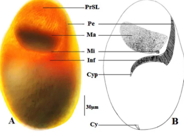
Description. The ciliate lives in the middle and posterior intestine of a megascolecid earthworms
Eupolytoreutus sp., collected in the moist soil in
Bambili and Bambui. The cell is ovoid (Fig. 4), with the anterior end narrow and the posterior end more or less rounded. It measures 151–163 µm long and 87–92 µm across. The macronucleus (27–35 × 54–60 µm) is transversally elongated and
has a trapeze shape. The micronucleus (2–3.5 µm in diameter) lies on the dorsal posterior side of the macronucleus (Fig. 4, Table 2).
There are 52–68 somatic kineties covering the two sides regularly (Fig. 5). On the right side anteriorly, there is an axial suture line deriving from the convergence of kineties. The left side is devoid of secant system.
The buccal apparatus is composed of a vertical peristome (42–58 µm long) and a curved infun-dibulum (38–52 µm) linking equatorially at an average angle of 103.3˚ (Table 2).
NYCTOTHERUSNGASSAMI FOKAM, NANA, NGASSAMET AL. N. SP.1
This ciliate co-inhabits the mid and hindgut of Eupolytoreutus sp. with the previously described species.
Diagnosis. Commensal of the digestive tract of Eupolytoreutus sp. Ovoid shape. Size 153–269 × 102–148 µm. Longitudinally extended macro-nucleus with a crescent shape. Curved peristome
Table 2. Morphometric characters of Nyctotherus ndoumeleleensi n. sp. Cell length (µm) Cell width (µm) Mn length (µm) Mn width (µm) Mi diam. (µm) Kin. Pe length (µm) I length (µm) A I P (°) Max 163 92 35 60 3.5 68 58 52 111 Mean 158.0 89.7 33.1 58.3 2.7 55.1 53.4 46.9 103.2 Min 151 87 27 54 2 52 42 38 89 SD 2.4 1.8 2.1 1.6 1.1 5.3 1.7 2.0 8.3
Notes. AIP – angle infundibulum-peristome in degree, Kin – kineties, Max – maximum, Min – minimum, Mi diam. – micronucleus diameter, Mn –
macronucleus, Pe – peristome, SD – standard deviation; N=30. Fig. 4. General morphology of Nyctotherus
ndou-meleleensis n. sp. A – Light micrograph after silver
staining (×400); B – drawing. Abbreviations: Cy – cytopyge, Cyp – cytopharynx, Inf – infundibulum, Ma – macronucleus, Mi – micronucleus, Pe – peristome, PrSL – preoral suture line.
Fig. 5. Ciliary pattern of Nyctotherus
ndoumele-leensis n. sp. A – Drawing of the right side; B – light
micrograph of the left side after silver staining (×400); C – drawing of the left side.
starting far from the apex. Rectilinear infundibulum at an angle of 150° with the peristome; 64–86 somatic kineties with a preoral and acaudal secant systems.
Type host. Eupolytoreutus sp.
Type locality. Bambili and Bambui, Tubah Subdivision, Bamenda, Cameroon.
Habitat. Mid and hindgut.
Type specimens. Permanent preparations belon-ging to this species are kept in the Department of Biology (Higher Teacher Training College, The University of Bamenda, Cameroon).
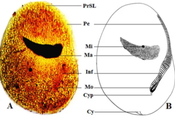
Description. Cells are ovoid and rounded at both ends; they are flattened on the lower side and slightly convex on the upper side. Cell measurements are: length, 153–269 µm, and width, 102–148 µm. The crescent-shaped macronucleus (16–31 × 36–53 µm) is located transversally in the anterior third of the cell (Fig. 6, Table 3). The spherical micronucleus
(3–5 µm) is found in a concavity of the anterior part of the macronucleus. The peristome begins at the anterior end of the body, is vertically oriented and extends slightly backwards and down towards the posterior end leading to the infundibulum at an angle of 150.8°. The infundibulum, shaped like a long oblique funnel, is supported by a slender monitorium and ends with a short cytopharynx (Fig. 6). The cell is covered by 64–86 meridional somatic kineties. The anterior and posterior ends of the right face (upper surface) kineties converge forming two median suture lines (a preoral suture line and a posterior suture line) (Fig. 7).
NYCTOTHERUSATUNIBAENSIS FOKAM, NANA, NGASSAM ETAL. N. SP.
Diagnosis. Commensal of the digestive tract of Eupolytoreutus sp. Ovoid shape. Average size 194.2 (145–253) × 125.0 (95–162) µm. Arc-shaped laterally extended macronucleus. Vertically oriented peristome starting far from the apex. Curved infundibulum; 108–122 somatic kineties with a preoral but not a caudal secant system.
·
67Protistology
Table 3. Morphometric characters of Nyctotherus ngassami n. sp.
Cell length (µm) Cell width (µm) Mn length (µm) Mn width (µm) Mi diam. (µm) Kin. Pe length (µm) I length (µm) A I P (°) Max 269 148 31 53 5 86 48 26 162 Mean 204.3 122.5 23.4 44.2 3.1 76.7 34.6 23.5 150.8 Min 153 102 16 36 3 64 29 18 137 SD 15.3 12.4 1.9 3.4 1.8 8.3 2.0 4.5 8.3
Notes. AIP – angle infundibulum-peristome in degree, Kin – kineties, Max – maximum, Min – minimum, Mi diam. – micronucleus diameter, Mn –
macronucleus, Pe – peristome, SD – standard deviation; N=30. Type host. Eupolytoreutus sp.
Type locality. Bambili and Bambui, Tubah Subdivision, Bamenda, Cameroon.
Habitat. Mid and hindgut.
Type specimens. Permanent preparations belon-ging to this species are kept in the Department of Biology (Higher Teacher Training College, The University of Bamenda, Cameroon).
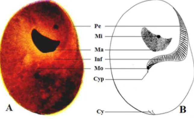
Description. The body is kidney-shaped, 145–253 µm long and 95–162 µm wide, somewhat flattened laterally, and rounded at both ends. The right margin is slightly depressed in the equatorial part of body (Fig. 8). The dual nuclear apparatus includes a transversally curved macronucleus (20–32 × 26–44 µm) located anteriorly; and a tiny micronucleus (3.3 µm in diameter) situated axially in the concave side of the macronucleus (Fig. 8, Table 4). The buccal apparatus comprises a slightly vertical and curved peristome that starts far from the apex of the cell and is in continuity in the equatorial part of the cell with
Fig. 6. General morphology of Nyctotherus ngassami n. sp. A – Light micrograph after silver staining (×400); B – drawing. Abbreviations: Cy – cytopyge, Cyp – cytopharynx, Inf – infundibulum, Ma – macronucleus, Mi – micronucleus, Mo – monitorium, Pe – peristome, PrSL – preoral suture line.
Fig. 7. Ciliary pattern of Nyctotherus ngassami n. sp. Abbreviations: PrSL – preoral suture line, PSL – posterior suture line.
the internal infundibulum. The junction between these two structures forms an obtuse angle of about 102.2° (mean). The infundibulum ends in a short and thin cytopharynx. The cell is evenly covered by 108–122 somatic meridian kineties. These kineties converge in the anterior part of the cell to form an apical suture line (Fig. 9).
Discussion
With respect to morphology and the ciliature pattern (absence of postoral secant system), the four ciliate species described above (Table 5) belong to the genus Nyctotherus, as was originally described by Leidy (1849) and some subsequent authors: Grassé (1928), Puytorac and Öktem (1967), Albaret and Njiné (1975) and Ngassam (1983).
By its general appearance, Nyctotherus
orthosto-matus can be confused at a first glance with Nycto-therus polymorphus found by Ngassam (1983) in
megascolecid earthworms collected in the central region (Yaounde). However, it differs from the latter by the higher number of kineties and the
Table 4. Morphometric characters of Nyctotherus atunibaensis n. sp. Cell length (µm) Cell width (µm) Mn length (µm) Mn width (µm) Mi diam. (µm) Kin. Pe length (µm) I length (µm) A I P (°) Max 253 162 32 44 4 122 40 22 108 Mean 194.2 125.0 18.9 36.1 3.3 96.1 34.0 18.6 102.2 Min 145 95 20 26 2 108 25 14 89 SD 16.9 7.3 1.6 1.4 0.8 9.8 4.3 4.6 6.5
Notes. AIP – angle infundibulum-peristome in degree, Kin – kineties, Max – maximum, Min – minimum, Mi diam. – micronucleus diameter, Mn –
macronucleus, Pe – peristome, SD – standard deviation; N=30. arrangement of the buccal apparatus in which the peristome and the infundibulum are oblique and joint orthogonally. N. polymorphus differs from the other species described by his relatively small size and the pear shape. Additionally, Nyctotherus
orthostomatus also shows some similarity with Nyctotherus dupouyi Ngassam, 1983 (anterior and
posterior ends rounded, with the anterior one third budging from the posterior two thirds of the cell). The distinguishing characteristics of the two species reside in the organization of their buccal and nuclear apparatus: Nyctotherus dupouyi possesses a peristome that begins from the apex of the cell and extends to the equatorial region where it is continuous with the infundibulum entering the endoplasm and ending posteriorly with a long curved cytopharynx. The peri-stome of N. orthostomatus is oblique and starts far from the apex of the cell; slightly above the equatorial plane it joins the infundibulum at a right angle; the infundibulum, in its turn, ends with a short
cytopharynx creating an angle of about 90°. The macronucleus of N. dupouyi is horseshoe-shaped, while that of N. orthostomatus is transversal and kidney-shaped. For all these reasons, we believe that the ciliate described above can be considered as a new species of the genus Nyctotherus Leidy, 1849.
Nyctotherus ndoumeleleensis reminds the
pre-vious species by it general morphology, even though the posterior pole is less wide. Another prominent feature of similarity is the slight torsion of the anterior region, consecutive to a slight spiralisation of the kineties in the anterior part of the cell. The trapezoidal macronucleus resembles that of N. polymorphus, but the location of the micronucleus is different – lateral and posterior. N.
ndoumeleleensis actually presents enough variability
in terms of the shape of the nuclear apparatus and the infundibulum which extends almost to the other side of the cell, ending with a curved hook-like cytopharynx. N. ndoumeleleensis could also be confounded with Nyctotherus inflatus described by Tuzet and Manier, 1858 (see: Albaret, 1975), due to its general morphology and orientation of the nuclear apparatus.
Fig. 8. General morphology of Nyctotherus
atuni-baensis n. sp. A – Light micrograph after silver
staining (×400); B – drawing. Abbreviations: Cy – cytopyge, Cyp – cytopharynx, Inf – infundibulum, Ma – macronucleus, Mi – micronucleus, Mo – monitorium, Pe – peristome.
Fig. 9. Ciliary pattern of Nyctotherus atunibaensis n. sp. Abbreviations: PrSL – preoral suture line.
·
69Protistology
Nyctotherus ngassami differs from the other
species of the ovoid shape by the presence of a caudal secant system (Table 5) and a somewhat rectilinear disposition of the buccal apparatus. Although the general morphology of N. ngassami reminds the one of N. dupouyi Ngassam, 1983 and the ovoid form of
N. polymorphus Ngassam, 1983, the area occupied
by the infundibulum differs in these two later species. Besides, N. dupouyi and N. polymorphus have a massive macronucleus hiding the micronucleus while in N. ngassami the macronucleus is crescent in shape with a micronucleus situated anteriorly in its concavity.
By its general morphology, Nyctotherus
atuni-baensis is similar to Nyctotherus cordiformis Stein,
1862, living in the rectum of anuran amphibians, but it differs from the latter by the relative reduction in size. Moreover, the macronucleus of N. atunibaensis has a crescent shape bearing a micronucleus in a concavity oriented anteriorly, while in N. cordiformis the massive macronucleus is kidney shaped, bearing a micronucleus in a concavity oriented posteriorly. Both species have an apical suture line; however, the caudal suture line is present in N. cordiformis but absent in N. atunibaensis. These particularities, added to the difference in host prompted us to con-sider this specimen as a new representative of the genus Nyctotherus Leidy, 1849.
References
Affa’a F-M. 1991. Observations morpholo-giques sur deux nyctothères (ciliés hétérotriches) commensaux de batraciens anoures du Québec. Can. J. Zool. 69, 2765–2770.
Affa’a F.-M. and Amiet J.-L. 1994. Progrès récents dans la connaissance des Nyctothères (Protozoaires, Ciliés Hétérotriches) associés aux anoures. Alytes. 12 (2), 75–92.
Albaret J.L. 1975. Etude systématique et cyto-logique sur les ciliés hétérotriches endocommen-saux. Mémoires du Museum National d’Histoire Naturelle (Nouvelle Série). 89, 1–114.
Albaret J.L. and Njiné T. 1975. Description de cinq espèces nouvelles de ciliés hétérotriches des genres Metanyctotherus Albaret, Pronyctotherus n. gen. et Plagiotoma Dujardin, endocommensaux d’oligochètes du Cameroun. Protistologica. 11, 305–311.
Fernandez-Galiano D. 1976. Silver impreg-nation of ciliated protozoa: procedure yielding good results with the pyridinated silver carbonate method. Trans. Am. Microsc. Soc. 95, 557–560.
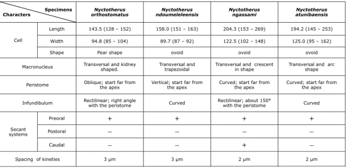
Fernandez-Galiano D. 1994. The ammoniacal silver carbonate method as a general procedure in the study of protozoa from sewage (and other) waters. Water Res. 28, 495–496. Specimens Characters Nyctotherus orthostomatus Nyctotherus ndoumeleleensis Nyctotherus ngassami Nyctotherus atunibaensis Cell Length 143.5 (128 – 152) 158.0 (151 – 163) 204.3 (153 – 269) 194.2 (145 – 253) Width 94.8 (85 – 104) 89.7 (87 – 92) 122.5 (102 – 148) 125.0 (95 – 162)
Shape Pear shape ovoid ovoid ovoid
Macronucleus Transversal and kidney shaped. Transversal and trapezoidal Transversal and crescent in shape Transversal and arc shape
Peristome Oblique; start far from the apex Vertical; start far from the apex Curved; start far from the apex Curved; start far from the apex
Infundibulum Rectilinear; right angle with the peristome Curved Rectilinear; about 150° with the peristome Curved
Secant systems Preoral + + + + Postoral – – – – Caudal – – + – Spacing of kineties 3 µm 3 µm 2 µm 2 µm
Table 5. Comparative characters between the four species of Nyctotherus (N=30)
Fienler N.G. and Yildiz I. 2000. The ciliate fauna in the digestive system of Rana ridibunda (Amphibia: Anura). II. Nyctotherus (Nyctotheridae: Heterotrichida). Turk. J. Zool. 24, 245–252.
Grassé P.P. 1928. Sur quelques Nyctotherus (Infusoires héterotriches) nouveaux ou peu connus. Ann. Protistol. 1, 55–68.
Leidy J. (1849): Nyctotherus, a new genus of Polygastrica, allied to Plescoma. Proc. Acad. Nat. Sci. Philad; 4, 233.
Ngassam P. 1983. Description de quatre espèces nouvelles de ciliés hétérotriches des genres
Nycto-therus Leidy et ParanyctoNycto-therus nov. gen.
endocom-mensaux d’un oligochète terricole au Cameroun. Protistologica. 19, 167–175.
Olivry J.C. 1986. Fleuves et rivières du Came-roun. Monographies hydrologiques ORSTOM No 9. O.R.S.T.O.M., Paris.
Puytorac P. de. 1994. Phylum Ciliophora Doflein, 1901. In : Traité de zoologie, infusoires ciliés. Masson, Paris. 2, pp. 1–15.
Puytorac P. de. and Öktem N. 1967. Observa-tions cytologiques sur les Nyctothères des genres
Nyctotherus Leidy et Prosicuophora n. gen. Ciliés parasites de Batraciens Anoures du Gabon. Biol. Gabon. 3, 223–242.
Sauvadet A.-L. 2010. Interactions entre ciliés et métazoaires dans deux environnements marins contrastés: Les sources hydrothermales et les sédiments anoxiques. Thèse de Doctorat de l’Uni-versité Pierre et Marie Curie, spécialité Biologie marine, Ecole Doctorale Diversité du Vivant, 197p.
Sims R.W. and Gerard B.M. 1999. Earthworm. FSC Publ., London.
Address for correspondence: Zéphyrin Fokam. Department of Biology, Higher Teacher Training College, University of Bamenda, Cameroon, P.O. Box 39, Bamenda, Cameroon; e-mail: fokam2z@yahoo.com