HAL Id: hal-02374336
https://hal.archives-ouvertes.fr/hal-02374336
Submitted on 14 Jan 2020
HAL is a multi-disciplinary open access
archive for the deposit and dissemination of
sci-entific research documents, whether they are
pub-lished or not. The documents may come from
teaching and research institutions in France or
L’archive ouverte pluridisciplinaire HAL, est
destinée au dépôt et à la diffusion de documents
scientifiques de niveau recherche, publiés ou non,
émanant des établissements d’enseignement et de
recherche français ou étrangers, des laboratoires
Chronic hepatitis C virus infection and pathogenesis of
hepatocellular carcinoma
Simonetta Bandiera, C Billie Bian, Yujin Hoshida, Thomas Baumert, Mirjam
Zeisel
To cite this version:
Simonetta Bandiera, C Billie Bian, Yujin Hoshida, Thomas Baumert, Mirjam Zeisel. Chronic hepatitis
C virus infection and pathogenesis of hepatocellular carcinoma. Current Opinion in Virology, Elsevier,
2016, 20, pp.99-105. �10.1016/j.coviro.2016.09.010�. �hal-02374336�
Chronic hepatitis C virus infection and pathogenesis of
hepatocellular carcinoma
Simonetta Bandiera
1,2, C. Billie Bian
3, Yujin Hoshida
3, Thomas F. Baumert
1,2,4, and Mirjam
B. Zeisel
1,21
Inserm, U1110, Institut de Recherche sur les Maladies Virales et Hépatiques, Strasbourg,
France
2
Université de Strasbourg, Strasbourg, France
3
Division of Liver Diseases, Department of Medicine, Liver Cancer Program, Tisch Cancer
Institute, Icahn School of Medicine at Mount Sinai, New York, USA
4
Institut Hospitalo-Universitaire, Pôle hépato-digestif, Nouvel Hôpital Civil, Strasbourg, France
Abstract
Hepatitis C virus (HCV) infection is one of the major causes of advanced liver disease and
hepatocellular carcinoma (HCC) worldwide. While the knowledge about the molecular virology of
HCV infection has markedly advanced, the molecular mechanisms of disease progression leading
to fibrosis, cirrhosis and HCC are still unclear. Accumulating experimental and clinical studies
indicate that HCV may drive hepatocarcinogenesis directly via its proteins or transcripts, and/or
indirectly through induction of chronic liver inflammation. Despite the possibility to eradicate
HCV infection through direct-acting antiviral treatment, the risk of HCC persists although specific
biomarkers to estimate this risk are still missing. Thus, a better understanding of HCV-induced
HCC and more physiological liver disease models are required to prevent cancer development.
Keywords
Hepatitis C virus; hepatocellular carcinoma; fibrosis; cancer hallmarks; direct-acting
antiviral-based therapies
Introduction
Hepatitis C virus (HCV) is single-strand RNA virus from the Flaviviridae family targeting
hepatocytes. Chronic HCV infection induces immune dysfunctions such as impaired T-cell
Corresponding authors: Thomas F. Baumert, MD, and Mirjam B. Zeisel, PharmD, PhD, Inserm, U1110, Institut de Recherche sur les Maladies Virales et Hépatiques, Université de Strasbourg, 3 Rue Koeberlé, F-67000 Strasbourg, France. [email protected] and [email protected].
Declaration of interest
The authors do not have any conflict of interest and did not receive writing assistance.
Publisher's Disclaimer: This is a PDF file of an unedited manuscript that has been accepted for publication. As a service to our customers we are providing this early version of the manuscript. The manuscript will undergo copyediting, typesetting, and review of
HHS Public Access
Author manuscript
Curr Opin Virol
. Author manuscript; available in PMC 2017 October 11.
Published in final edited form as:
Curr Opin Virol. 2016 October ; 20: 99–105. doi:10.1016/j.coviro.2016.09.010.
A
uthor Man
uscr
ipt
A
uthor Man
uscr
ipt
A
uthor Man
uscr
ipt
A
uthor Man
uscr
ipt
functions and inefficient antibody responses, metabolic disorders such as hepatic steatosis,
iron accumulation, and insulin resistance often associated with type 2 diabetes. More
importantly, HCV is one of the major etiologies of chronic hepatitis and progressive liver
fibrosis that lead to development of lethal complications, i.e., cirrhosis and hepatocellular
carcinoma (HCC), the second leading cause of cancer mortality worldwide and the only and
most rapidly increasing cancer death in the U.S. [1,2]. Chronic HCV infection is highly
prevalent globally, including developed countries [3]. In the U.S., more than 1 million
individuals, representing the “baby boomer” population, are estimated to develop
HCV-related liver cirrhosis and/or HCC by 2020. Recently developed direct-acting antivirals
(DAAs) for HCV effectively cure HCV infection, i.e., they enable to achieve sustained
virologic response (SVR), but the high costs will limit their wide-spread use [4]. Of note,
HCC risk remains high for decades even after SVR, and HCV-related HCC is predicted to
increase until 2030 despite improved viral cure by DAAs [5,6].
HCV does not integrate its genetic material into the host genome, and therefore requires
continuous replication to maintain chronic infection. Many host factors, playing essential
roles in the HCV life cycle and immune evasion, have been identified as candidate targets
for antiviral interventions (reviewed in [7]). However, disease pathogenesis that ultimately
causes HCC is still unclear. Experimental studies to date have suggested models of viral
carcinogenesis unique to HCV [8]. Increasing evidence shows that HCV transmits signals
and modulates hepatocyte gene expression following engagement with cellular receptors
[9,10]. Moreover, viral proteins have been involved in disrupting signal transduction
pathways that affect cell survival, proliferation, and transformation [8]. This suggests that
virus-host interactions and signaling during viral infection contribute to cellular
transformation and development of HCC directly via HCV proteins or RNA, and/or
indirectly through induction of chronic inflammation. Additionally, the genetic background
of the host may play a role in HCC pathogenesis. Genetic analyses in HCV-infected patients
have unraveled specific mutation or polymorphisms in MICA/HCP5, LEPR and IFNL3 loci
that are associated with the development of HCC [11–16], indicating that genetic variation
may contribute to individual susceptibility for HCV-driven HCC.
Of note, the persisting risk of HCC development even after viral cure suggests that HCV
leaves molecular imprinting in the host genome that keeps driving carcinogenesis.
Management of post-SVR HCC will be increasingly relevant as more patients achieve SVR
by the DAA treatment in clinic. Here, we review several examples of mechanisms that may
contribute to HCV-induced HCC and discuss the clinical challenges to prevent HCC
development in at-risk patients in the era of DAA-based anti-HCV therapies.
Viral factors directly driving hepatocarcinogenesis
The strong and reproducible association of HCV genotype 3 with development of steatosis
and HCC, genotype 1b with more frequent progression to HCC, and HCV core gene variants
with post-SVR HCC suggests that specific viral factors influence or determine progression
of liver disease [17–19]. The viral genome encodes for three structural (core, E1, E2) and
seven non-structural proteins (p7, NS2, NS3, NS4A, NS4B, NS5A and NS5B). Several in
vivo studies in transgenic mouse models reported direct induction of liver disease by the
A
uthor Man
uscr
ipt
A
uthor Man
uscr
ipt
A
uthor Man
uscr
ipt
A
uthor Man
uscr
ipt
expression of viral proteins (reviewed in [20]). Although none of these models could
faithfully recapitulate the full features of human disease, some of the phenotypes were
consistent with epidemiological data from HCV-infected patients. Interestingly, these studies
highlighted that HCV RNA and proteins can perturb hepatocellular homeostasis by driving
several major cancer hallmarks (Figure 1).
First, metabolic reprogramming including disturbance of lipid metabolism and
mitochondrial dysfunction were shown to play an important role in HCV-driven HCC
(Figure 1). Indeed, chronic HCV infection enhances mitochondrial liver injury together with
oxidative stress in human as well as experimental models [21]. Several studies highlight a
role of the HCV core protein in steatosis and HCC nodule development as well as in insulin
resistance, which is accompanied with intrahepatic lipid accumulation [20,22]. The
alteration of lipid metabolism is induced by an HCV core-mediated impairment of lipid
β-oxidation, which is associated with a reduced activity of the mitochondrial electron transport
chain [20]. Recently, HCV core protein was also shown to contribute to mitochondrial
damage by impairing mitophagy [23]. The resulting oxidative stress is regarded as a key
trigger of HCC initiation and development (Figure 1). Imbalance of the oxidant/antioxidant
state in the liver was shown to induce HCC in HCV core transgenic mice in the absence of
inflammation [24]. Moreover, generation of reactive oxygen species (ROS) in the course of
HCV infection was associated with genomic instability, a hallmark of cancer cells [20].
Indeed, accumulation of genetic mutations as well as chromosomal alterations crucially
drive the development of HCC in patients [25]. By inducing a
β-Catenin-dependent
upregulation of c-Myc via NS5A, HCV was shown to perturb ROS production in association
with enhanced DNA damage and aberrant cell-cycle arrest (Figure 1) [26]. In addition,
increased telomerase (TERT) activity, a characteristic of transforming or
transformation-prone cells, was observed in HCV core-transfected primary human hepatocytes that acquired
an immortalized phenotype [27]. In line with this observation, somatic mutations in the
TERT promoter that enhance TERT expression were shown to be among the earliest and
most prevalent neoplastic event in HCC associated with all major etiologies including HCV
[28].
Another major hallmark of cancer that is directly affected by HCV is evasion from cell death
and senescence (Figure 1). Although HCV proteins were reported to have both pro-apoptotic
and anti-apoptotic properties [8], HCV is likely involved in evasion from apoptosis in vivo.
A number of studies indicate that Fas-mediated apoptosis is directly inhibited by different
HCV proteins [20,29–31]. Given that the Fas system accounts for T cell-mediated
cytotoxicity, suppression of cell death is not only a mechanism of sustained cell proliferation
but also one strategy that enables HCV to escape immune surveillance by T cells and thus to
establish persistent infection [32].
Finally, recent evidence indicates that viral proteins impact on epithelial mesenchymal
transition (EMT) pathway, which promotes fibrogenesis, tumor development and metastases
(Figure 1). HCV NS5A was shown to activate Twist2, a transcriptional regulator of EMT,
and to cooperate with Ras oncogene to enhance tumor cell invasiveness in xenograft mouse
models [33]. Furthermore, expression of HCV core in transgenic mice enhances intrahepatic
TGF-
β signaling, a key regulator of EMT driving the activation of human stellate cells
A
uthor Man
uscr
ipt
A
uthor Man
uscr
ipt
A
uthor Man
uscr
ipt
A
uthor Man
uscr
ipt
(HSCs) [34]. Further studies showed that induction of EMT by HCV core is mediated by at
least two mechanisms: i) the inhibition of E-cadherin expression by a complex comprising
HCV core, Snail and the histone deacetylases HDAC1/HDAC2 [35]; ii) the HCV
core-induced epigenetic silencing of SFRP1 via DNA methylation and histone modifications that
in turn activates Wnt/
β-catenin signaling [36]. Yet the clinical relevance of these recent
findings remains to be determined.
HCV-induced inflammatory responses indirectly driving
hepatocarcinogenesis
HCV infection can induce chronic hepatic inflammation with varying activity, which causes
progressive liver fibrogenesis and leads to development of cirrhosis (Figure 1). Clinically,
the majority of HCV-related HCC tumors develop in livers with cirrhosis established after
decades of chronic inflammation, underscoring the key role of virus-induced inflammatory
responses, besides the viral materials themselves, in HCC pathogenesis. Several
inflammatory pathways have been implicated in HCC. First, the sensing of HCV infection
by pathogen recognition receptors of the innate immune system activates the NF-
κB
signaling and downstream proinflammatory chemokines and cytokines including type III
interferon (IFN), which is associated with HCC development (Figure 1) [37–39]. Ectopic
lymphoid structure aggregated near the portal tract was reported as a niche of HCC initiation
associated with striking NF-
κB activation in a subset of HCV-infected human livers [40].
Approximately in 70 % of chronic hepatitis C (CHC) patients the immune response fails to
eradicate the virus due to impaired T cell and antibody responses, and little antiviral efficacy
of IFN-stimulated genes (ISGs) [41]. The adaptive immune response mediated by cytotoxic
T cells has been suggested to contribute to liver injury by triggering repeated cycles of
hepatocyte death and regeneration/proliferation. The inflammatory response also exacerbates
oxidative stress in the liver (Figure 1). Cytokines, ROS and apoptotic signals contribute to
HSC activation, which triggers aberrant deposition of extracellular matrix proteins and
progressive fibrosis (Figure 1). As such, the functional liver parenchyma is progressively
replaced by non-functional fibrotic tissue. Overall, this pattern of chronic inflammation,
unresolved wound healing response and increased hepatocellular proliferation in CHC is
thought to generate an environment highly permissive for hepatocarcinogenesis.
Despite the growing knowledge, many open questions remain unanswered. The molecular
bases of the interplay between the innate and adaptive immune responses in the course of
CHC and their relevance for HCC development are still largely unclear [41]. IFN pathway
activation is one of the key components of the host responses to HCV, although cell types
secreting IFN as well as types of secreted IFN stimulating specific subset of ISGs are still
elusive. This is partly because of the lack of a robust immunocompetent in vivo HCV
infection model that mirrors the cell circuits of HCC development as well as the crosstalk
between parenchymal and non-parenchymal cell types driving disease progression under
physiological condition as in human [20]. Transgenic mouse models coupled with
epidemiological data in patients have provided important insights into mechanistic
investigation. This approach was successful in unraveling a pathway of hepatocarcinogenesis
driven by the pro-inflammatory cytokine lymphotoxin (LT)
α and β [42]. By using
A
uthor Man
uscr
ipt
A
uthor Man
uscr
ipt
A
uthor Man
uscr
ipt
A
uthor Man
uscr
ipt
transgenic mice for either LTs expression or NF-
κB signaling components, Haybeack et al.
discovered that LTs overexpression induces chronic hepatitis and HCC by altering NF-
κB
signaling in both hepatocytes and lymphocytes. These observations were corroborated by the
enhanced LT expression in clinical liver specimens from virus-induced chronic hepatitis and
HCC as compared to healthy liver [42]. More recently, HCV NS5B was shown to promote
pro-inflammatory LT
β signaling in liver cells [43]. Likewise, two recent studies casted new
light on novel mechanisms of HSC activation and liver fibrogenesis in CHC. The first
involves the acetylation of HMGB1 by extracellular osteopontin (OPN), a stress sensor
protein that is enhanced in liver disease and elevated in serum of patients who are at risk of
HCC development [44]. Acetylated HMGB1 interacts with HDAC1/HDAC2 to promote
collagen-I expression by HSCs and increase its histological deposition [45]. The second
mechanism relies on the upregulation of the Gas6/Axl pathway in HSC leading to activation
of these cells and liver fibrogenesis upon carbon tetrachloride-induced injury in mice [46].
Importantly, the clinical relevance of both mechanisms was evidenced by the correlation
between the severity of liver injury and increased expression of OPN/HMGB1 or Gas6/Axl,
respectively, in HCV-infected patients [45,46]. However, additional clinical cohort studies
may be required to corroborate the involvement of these processes in HCV pathogenesis.
Treatment of HCV infection and prevention of HCC
Rapidly evolving DAA-based anti-HCV therapies now enable more than 90% of SVR rate
with all-oral regimens even in the cases hard to cure before [47]. In patients previously
treated with older, IFN-based regimens, SVR was significantly associated with reduced but
not eliminated future risk of HCC development over a decade [48]. In the retrospective
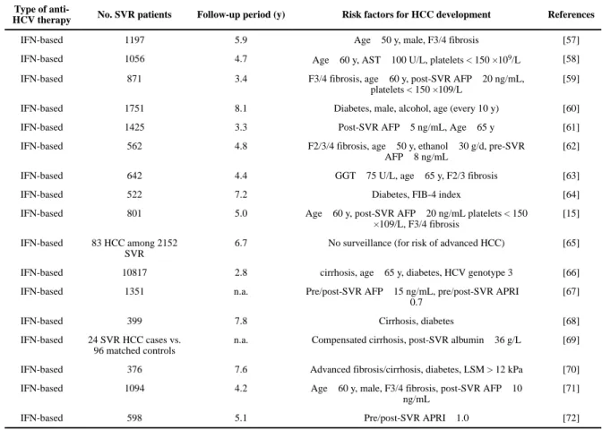
studies, several clinical characteristics such as more advanced liver fibrosis, older age, and
male sex among others have been suggested as predisposing factors for post-SVR HCC
(Table 1). However, estimation of HCC risk in patients newly achieving an SVR is still
infeasible and the mechanisms of carcinogenesis are totally unknown. Given the annual
incidence of post-SVR HCC, which is likely below the threshold that rationalizes regular
HCC surveillance, HCC risk biomarkers or indices will play a critical role to perform
cost-effective and practically feasible HCC surveillance by triaging the patients according to the
predicted HCC risk [21]. Also, such biomarkers may provide clues to targets of HCC
chemopreventive interventions. It is still unanswered question whether HCC risk after
DAA-based or other types of anti-HCV therapies such as viral entry inhibition [49] is comparable
to that of IFN-based therapies. Modulation of cellular signaling pathways such as IFN, EGF,
mTOR, and retinoid X receptor-
α pathways and drugs for metabolic disorder, some of
which have been already clinically evaluated, may serve as alternative options of HCC
chemoprevention for broader etiologies, including post-SVR HCC [50–56]. Experimental
systems that allow mechanistic assessments of the carcinogenic drivers will be critical in
identifying and developing rational molecular-targeted HCC chemoprevention therapies.
Acknowledgments
The authors acknowledge grant support of the European Union (ERC-2014-AdG-671231-HEPCIR (T.F.B, Y.H.), H2020-667273-HEPCAR (T.F.B.), EU-ANR ERA-NET Infect-ERA hepBccc (T.F.B.), ANRS (T.F.B., M.B.Z.), the French Cancer Agency (ARC IHU201301187 (T.F.B.)), the IdEx program of the University of Strasbourg (M.B.Z., T.F.B.), the Foundation University of Strasbourg (T.F.B.), NIH/NIDDK R01 DK099558 (Y.H.), and Irma T. Hirschl
A
uthor Man
uscr
ipt
A
uthor Man
uscr
ipt
A
uthor Man
uscr
ipt
A
uthor Man
uscr
ipt
Trust (Y.H.). This work has been published under the framework of the LABEX ANR-10-LAB-28 and benefits from a funding from the state managed by the French National Research Agency as part of the Investments for the future program.
References
* of special interest
** of outstanding interest
1. El-Serag HB. Hepatocellular carcinoma. N Engl J Med. 2011; 365:1118–1127. [PubMed: 21992124]
2. Ryerson AB, Eheman CR, Altekruse SF, Ward JW, Jemal A, Sherman RL, Henley SJ, Holtzman D, Lake A, Noone AM, et al. Annual Report to the nation on the status of cancer, 1975–2012; featuring the increasing incidence of liver cancer. Cancer. 2016; doi: 10.1002/cncr.29936
3. Hajarizadeh B, Grebely J, Dore GJ. Epidemiology and natural history of HCV infection. Nat Rev Gastroenterol Hepatol. 2013; 10:553–562. [PubMed: 23817321]
4. Chung RT, Baumert TF. Curing chronic hepatitis C--the arc of a medical triumph. N Engl J Med. 2014; 370:1576–1578. [PubMed: 24720678]
5. Morgan RL, Baack B, Smith BD, Yartel A, Pitasi M, Falck-Ytter Y. Eradication of hepatitis C virus infection and the development of hepatocellular carcinoma: a meta-analysis of observational studies. Ann Intern Med. 2013; 158:329–337. [PubMed: 23460056]
6. Harris RJ, Thomas B, Griffiths J, Costella A, Chapman R, Ramsay M, De Angelis D, Harris HE. Increased uptake and new therapies are needed to avert rising hepatitis C-related end stage liver disease in England: Modelling the predicted impact of treatment under different scenarios. J Hepatol. 2014; 61:530–537. [PubMed: 24824282]
7. Zeisel MB, Lupberger J, Fofana I, Baumert TF. Host-targeting agents for prevention and treatment of chronic hepatitis C - perspectives and challenges. J Hepatol. 2013; 58:375–384. [PubMed: 23041307]
8. Mesri EA, Feitelson MA, Munger K. Human viral oncogenesis: a cancer hallmarks analysis. Cell Host Microbe. 2014; 15:266–282. [PubMed: 24629334]
9. Fang X, Zeisel MB, Wilpert J, Gissler B, Thimme R, Kreutz C, Maiwald T, Timmer J, Kern WV, Donauer J, et al. Host cell responses induced by hepatitis C virus binding. Hepatology. 2006; 43:1326–1336. [PubMed: 16729312]
10. Zona L, Lupberger J, Sidahmed-Adrar N, Thumann C, Harris Helen J, Barnes A, Florentin J, Tawar Rajiv G, Xiao F, Turek M, et al. HRas signal transduction promotes hepatitis C virus cell entry by triggering assembly of the host tetraspanin receptor complex. Cell Host Microbe. 2013; 13:302–313. [PubMed: 23498955]
11. Goto K, Kato N. MICA SNPs and the NKG2D system in virus-induced HCC. J Gastroenterol. 2015; 50:261–272. [PubMed: 25270965]
12. Lange CM, Bibert S, Dufour JF, Cellerai C, Cerny A, Heim MH, Kaiser L, Malinverni R, Mullhaupt B, Negro F, et al. Comparative genetic analyses point to HCP5 as susceptibility locus for HCV-associated hepatocellular carcinoma. J Hepatol. 2013; 59:504–509. [PubMed: 23665287] 13. Kumar V, Kato N, Urabe Y, Takahashi A, Muroyama R, Hosono N, Otsuka M, Tateishi R, Omata
M, Nakagawa H, et al. Genome-wide association study identifies a susceptibility locus for HCV-induced hepatocellular carcinoma. Nat Genet. 2011; 43:455–458. [PubMed: 21499248]
14. Ikeda A, Shimizu T, Matsumoto Y, Fujii Y, Eso Y, Inuzuka T, Mizuguchi A, Shimizu K, Hatano E, Uemoto S, et al. Leptin receptor somatic mutations are frequent in HCV-infected cirrhotic liver and associated with hepatocellular carcinoma. Gastroenterology. 2014; 146:222–232. e235. [PubMed: 24055508]
15. Chang KC, Tseng PL, Wu YY, Hung HC, Huang CM, Lu SN, Wang JH, Lee CM, Chen CH, Tsai MC, et al. A polymorphism in interferon L3 is an independent risk factor for development of hepatocellular carcinoma after treatment of hepatitis C virus infection. Clin Gastroenterol Hepatol. 2015; 13:1017–1024. [PubMed: 25460552]
A
uthor Man
uscr
ipt
A
uthor Man
uscr
ipt
A
uthor Man
uscr
ipt
A
uthor Man
uscr
ipt
16. Asahina Y, Tsuchiya K, Nishimura T, Muraoka M, Suzuki Y, Tamaki N, Yasui Y, Hosokawa T, Ueda K, Nakanishi H, et al. Genetic variation near interleukin 28B and the risk of hepatocellular carcinoma in patients with chronic hepatitis C. J Gastroenterol. 2014; 49:1152–1162. [PubMed: 23860735]
17. Goossens N, Hoshida Y. Hepatitis C virus-induced hepatocellular carcinoma. Clin Mol Hepatol. 2015; 21:105–114. [PubMed: 26157746]
18*. Akuta N, Suzuki F, Kobayashi M, Sezaki H, Kawamura Y, Hosaka T, Kobayashi M, Saitoh S, Suzuki Y, Arase Y, et al. Impact of mutations at amino acid 70 in HCV genotype 1b core region on hepatocarcinogenesis following eradication of HCV RNA. J Clin Microbiol. 2015; 53:3039– 3041. Clinical report indicating the oncogenic potential of the mutant HCV core in a large cohort of patients cleared for chronic HCV infection. [PubMed: 26135874]
19. El-Shamy A, Eng FJ, Doyle EH, Klepper AL, Sun X, Sangiovanni A, Iavarone M, Colombo M, Schwartz RE, Hoshida Y, et al. A cell culture system for distinguishing hepatitis C viruses with and without liver cancer-related mutations in the viral core gene. J Hepatol. 2015; 63:1323–1333. [PubMed: 26220749]
20. Lerat H, Higgs M, Pawlotsky JM. Animal models in the study of hepatitis C virus-associated liver pathologies. Expert Rev Gastroenterol Hepatol. 2011; 5:341–352. [PubMed: 21651352]
21. Hoshida Y, Fuchs BC, Bardeesy N, Baumert TF, Chung RT. Pathogenesis and prevention of hepatitis C virus-induced hepatocellular carcinoma. J Hepatol. 2014; 61:S79–90. [PubMed: 25443348]
22. Miyamoto H, Moriishi K, Moriya K, Murata S, Tanaka K, Suzuki T, Miyamura T, Koike K, Matsuura Y. Involvement of the PA28gamma-dependent pathway in insulin resistance induced by hepatitis C virus core protein. J Virol. 2007; 81:1727–1735. [PubMed: 17135326]
23. Hara Y, Yanatori I, Ikeda M, Kiyokage E, Nishina S, Tomiyama Y, Toida K, Kishi F, Kato N, Imamura M, et al. Hepatitis C virus core protein suppresses mitophagy by interacting with parkin in the context of mitochondrial depolarization. Am J Pathol. 2014; 184:3026–3039. [PubMed: 25244949]
24. Moriya K, Nakagawa K, Santa T, Shintani Y, Fujie H, Miyoshi H, Tsutsumi T, Miyazawa T, Ishibashi K, Horie T, et al. Oxidative stress in the absence of inflammation in a mouse model for hepatitis C virus-associated hepatocarcinogenesis. Cancer Res. 2001; 61:4365–4370. [PubMed: 11389061]
25. Tornesello ML, Buonaguro L, Izzo F, Buonaguro FM. Molecular alterations in hepatocellular carcinoma associated with hepatitis B and hepatitis C infections. Oncotarget. 2016; doi: 10.18632/ oncotarget.17837
26. Higgs MR, Lerat H, Pawlotsky JM. Hepatitis C virus-induced activation of beta-catenin promotes c-Myc expression and a cascade of pro-carcinogenetic events. Oncogene. 2013; 32:4683–4693. [PubMed: 23108410]
27. Ray RB, Meyer K, Ray R. Hepatitis C virus core protein promotes immortalization of primary human hepatocytes. Virology. 2000; 271:197–204. [PubMed: 10814584]
28*. Nault JC, Zucman-Rossi J. TERT promoter mutations in primary liver tumors. Clin Res Hepatol Gastroenterol. 2016; 40:9–14. This review provides an overview on the role of TERT promoter mutations in HCC development. [PubMed: 26336998]
29. Machida K, Tsukiyama-Kohara K, Seike E, Tone S, Shibasaki F, Shimizu M, Takahashi H, Hayashi Y, Funata N, Taya C, et al. Inhibition of cytochrome c release in Fas-mediated signaling pathway in transgenic mice induced to express hepatitis C viral proteins. J Biol Chem. 2001; 276:12140–12146. [PubMed: 11278624]
30. Machida K, Tsukamoto H, Liu JC, Han YP, Govindarajan S, Lai MM, Akira S, Ou JH. c-Jun mediates hepatitis C virus hepatocarcinogenesis through signal transducer and activator of transcription 3 and nitric oxide-dependent impairment of oxidative DNA repair. Hepatology. 2010; 52:480–492. [PubMed: 20683948]
31. Zemel R, Gerechet S, Greif H, Bachmatove L, Birk Y, Golan-Goldhirsh A, Kunin M, Berdichevsky Y, Benhar I, Tur-Kaspa R. Cell transformation induced by hepatitis C virus NS3 serine protease. J Viral Hepat. 2001; 8:96–102. [PubMed: 11264729]
A
uthor Man
uscr
ipt
A
uthor Man
uscr
ipt
A
uthor Man
uscr
ipt
A
uthor Man
uscr
ipt
32. Nagao M, Nakajima Y, Hisanaga M, Kayagaki N, Kanehiro H, Aomatsu Y, Ko S, Yagita H, Yamada T, Okumura K, et al. The alteration of Fas receptor and ligand system in hepatocellular carcinomas: how do hepatoma cells escape from the host immune surveillance in vivo? Hepatology. 1999; 30:413–421. [PubMed: 10421649]
33. Akkari L, Gregoire D, Floc’h N, Moreau M, Hernandez C, Simonin Y, Rosenberg AR, Lassus P, Hibner U. Hepatitis C viral protein NS5A induces EMT and participates in oncogenic
transformation of primary hepatocyte precursors. J Hepatol. 2012; 57:1021–1028. [PubMed: 22750466]
34. Benzoubir N, Lejamtel C, Battaglia S, Testoni B, Benassi B, Gondeau C, Perrin-Cocon L, Desterke C, Thiers V, Samuel D, et al. HCV core-mediated activation of latent TGF-beta via
thrombospondin drives the crosstalk between hepatocytes and stromal environment. J Hepatol. 2013; 59:1160–1168. [PubMed: 23928402]
35. Nie D, Shan X, Nie L, Duan Y, Chen Z, Yang Y, Li Z, Tian L, Gao Q, Shan Y, et al. Hepatitis C virus core protein interacts with Snail and histone deacetylases to promote the metastasis of hepatocellular carcinoma. Oncogene. 2015; 35:3626–35. [PubMed: 26549030]
36. Quan H, Zhou F, Nie D, Chen Q, Cai X, Shan X, Zhou Z, Chen K, Huang A, Li S, et al. Hepatitis C virus core protein epigenetically silences SFRP1 and enhances HCC aggressiveness by inducing epithelial-mesenchymal transition. Oncogene. 2014; 33:2826–2835. [PubMed: 23770846] 37. Luedde T, Schwabe RF. NF-kappaB in the liver--linking injury, fibrosis and hepatocellular
carcinoma. Nat Rev Gastroenterol Hepatol. 2011; 8:108–118. [PubMed: 21293511]
38. Yang D, Liu N, Zuo C, Lei S, Wu X, Zhou F, Liu C, Zhu H. Innate host response in primary human hepatocytes with hepatitis C virus infection. PLoS One. 2011; 6:e27552. [PubMed: 22087337] 39. Thomas E, Gonzalez VD, Li Q, Modi AA, Chen W, Noureddin M, Rotman Y, Liang TJ. HCV
infection induces a unique hepatic innate immune response associated with robust production of type III interferons. Gastroenterology. 2012; 142:978–988. [PubMed: 22248663]
40**. Finkin S, Yuan D, Stein I, Taniguchi K, Weber A, Unger K, Browning JL, Goossens N, Nakagawa S, Gunasekaran G, et al. Ectopic lymphoid structures function as microniches for tumor progenitor cells in hepatocellular carcinoma. Nat Immunol. 2015; 16:1235–1244. In vivo
study demonstrating that ectopic lymphoid-like structures within the liver form an immunopathological microenvironment, which possibly serves as niche to promote HCC initiation. [PubMed: 26502405]
41. Heim MH, Thimme R. Innate and adaptive immune responses in HCV infections. J Hepatol. 2014; 61:S14–25. [PubMed: 25443342]
42. Haybaeck J, Zeller N, Wolf MJ, Weber A, Wagner U, Kurrer MO, Bremer J, Iezzi G, Graf R, Clavien PA, et al. A lymphotoxin-driven pathway to hepatocellular carcinoma. Cancer Cell. 2009; 16:295–308. [PubMed: 19800575]
43. Simonin Y, Vegna S, Akkari L, Gregoire D, Antoine E, Piette J, Floc’h N, Lassus P, Yu GY, Rosenberg AR, et al. Lymphotoxin signaling is initiated by the viral polymerase in HCV-linked tumorigenesis. PLoS Pathog. 2013; 9:e1003234. [PubMed: 23555249]
44. da Costa AN, Plymoth A, Santos-Silva D, Ortiz-Cuaran S, Camey S, Guilloreau P, Sangrajrang S, Khuhaprema T, Mendy M, Lesi OA, et al. Osteopontin and latent-TGF beta binding-protein 2 as potential diagnostic markers for HBV-related hepatocellular carcinoma. Int J Cancer. 2015; 136:172–181. [PubMed: 24803312]
45. Arriazu E, Ge X, Leung TM, Magdaleno F, Lopategi A, Lu Y, Kitamura N, Urtasun R, Theise N, Antoine DJ, et al. Signalling via the osteopontin and high mobility group box-1 axis drives the fibrogenic response to liver injury. Gut. 2016; doi: 10.1136/gutjnl-2015-310752
46. Barcena C, Stefanovic M, Tutusaus A, Joannas L, Menendez A, Garcia-Ruiz C, Sancho-Bru P, Mari M, Caballeria J, Rothlin CV, et al. Gas6/Axl pathway is activated in chronic liver disease and its targeting reduces fibrosis via hepatic stellate cell inactivation. J Hepatol. 2015; 63:670–678. [PubMed: 25908269]
47*. Majumdar A, Kitson MT, Roberts SK. Systematic review: current concepts and challenges for the direct-acting antiviral era in hepatitis C cirrhosis. Aliment Pharmacol Ther. 2016; 43:1276–1292. A comprehensive overview of the antiviral effects of DAA treatments in patients with HCV-induced cirrhosis as assessed during the past 3 years. [PubMed: 27087015]
A
uthor Man
uscr
ipt
A
uthor Man
uscr
ipt
A
uthor Man
uscr
ipt
A
uthor Man
uscr
ipt
48. Li DK, Chung RT. Impact of hepatitis C virus eradication on hepatocellular carcinogenesis. Cancer. 2015; 121:2874–2882. [PubMed: 26079399]
49. Lupberger J, Zeisel MB, Xiao F, Thumann C, Fofana I, Zona L, Davis C, Mee CJ, Turek M, Gorke S, et al. EGFR and EphA2 are host factors for hepatitis C virus entry and possible targets for antiviral therapy. Nat Med. 2011; 17:589–595. [PubMed: 21516087]
50. Lok AS, Everhart JE, Wright EC, Di Bisceglie AM, Kim HY, Sterling RK, Everson GT, Lindsay KL, Lee WM, Bonkovsky HL, et al. Maintenance Peginterferon Therapy and Other Factors Associated With Hepatocellular Carcinoma in Patients With Advanced Hepatitis C. Gastroenterology. 2011; 140:840–849. [PubMed: 21129375]
51. Bruix J, Poynard T, Colombo M, Schiff E, Burak K, Heathcote EJ, Berg T, Poo JL, Mello CB, Guenther R, et al. Maintenance therapy with peginterferon alfa-2b does not prevent hepatocellular carcinoma in cirrhotic patients with chronic hepatitis C. Gastroenterology. 2011; 140:1990–1999. [PubMed: 21419770]
52. Hoshida Y, Fuchs BC, Tanabe KK. Prevention of hepatocellular carcinoma: potential targets, experimental models, and clinical challenges. Curr Cancer Drug Targets. 2012; 12:1129–1159. [PubMed: 22873223]
53. Menon KV, Hakeem AR, Heaton ND. Meta-analysis: recurrence and survival following the use of sirolimus in liver transplantation for hepatocellular carcinoma. Aliment Pharmacol Ther. 2013; 37:411–419. [PubMed: 23278125]
54. Emberson JR, Kearney PM, Blackwell L, Newman C, Reith C, Bhala N, Holland L, Peto R, Keech A, Collins R, et al. Lack of effect of lowering LDL cholesterol on cancer: meta-analysis of individual data from 175,000 people in 27 randomised trials of statin therapy. PLoS One. 2012; 7:e29849. [PubMed: 22276132]
55. Singh S, Singh PP, Singh AG, Murad MH, Sanchez W. Anti-diabetic medications and the risk of hepatocellular cancer: a systematic review and meta-analysis. Am J Gastroenterol. 2013; 108:881– 891. quiz 892. [PubMed: 23381014]
56**. Fuchs BC, Hoshida Y, Fujii T, Wei L, Yamada S, Lauwers GY, McGinn CM, Deperalta DK, Chen X, Kuroda T, et al. Epidermal growth factor receptor inhibition attenuates liver fibrosis and development of hepatocellular carcinoma. Hepatology. 2014; 59:1577–1590. This study provides
in vivo evidence supporting that EGFR signaling could be an HCC chemoprevention target in cirrhotic liver. [PubMed: 24677197]
57. Makiyama A, Itoh Y, Kasahara A, Imai Y, Kawata S, Yoshioka K, Tsubouchi H, Kiyosawa K, Kakumu S, Okita K, et al. Characteristics of patients with chronic hepatitis C who develop hepatocellular carcinoma after a sustained response to interferon therapy. Cancer. 2004; 101:1616– 1622. [PubMed: 15378504]
58. Ikeda M, Fujiyama S, Tanaka M, Sata M, Ide T, Yatsuhashi H, Watanabe H. Risk factors for development of hepatocellular carcinoma in patients with chronic hepatitis C after sustained response to interferon. J Gastroenterol. 2005; 40:148–156. [PubMed: 15770398]
59. Chang KC, Hung CH, Lu SN, Wang JH, Lee CM, Chen CH, Yen MF, Lin SC, Yen YH, Tsai MC, et al. A novel predictive score for hepatocellular carcinoma development in patients with chronic hepatitis C after sustained response to pegylated interferon and ribavirin combination therapy. J Antimicrob Chemother. 2012; 67:2766–2772. [PubMed: 22899800]
60. Arase Y, Kobayashi M, Suzuki F, Suzuki Y, Kawamura Y, Akuta N, Sezaki H, Saito S, Hosaka T, Ikeda K, et al. Effect of type 2 diabetes on risk for malignancies includes hepatocellular carcinoma in chronic hepatitis C. Hepatology. 2013; 57:964–973. [PubMed: 22991257]
61. Oze T, Hiramatsu N, Yakushijin T, Miyazaki M, Yamada A, Oshita M, Hagiwara H, Mita E, Ito T, Fukui H, et al. Post-treatment levels of alpha-fetoprotein predict incidence of hepatocellular carcinoma after interferon therapy. Clin Gastroenterol Hepatol. 2014; 12:1186–1195. [PubMed: 24321207]
62. Yamashita N, Ohho A, Yamasaki A, Kurokawa M, Kotoh K, Kajiwara E. Hepatocarcinogenesis in chronic hepatitis C patients achieving a sustained virological response to interferon: significance of lifelong periodic cancer screening for improving outcomes. J Gastroenterol. 2014; 49:1504– 1513. [PubMed: 24317936]
63. Huang CF, Yeh ML, Tsai PC, Hsieh MH, Yang HL, Hsieh MY, Yang JF, Lin ZY, Chen SC, Wang LY, et al. Baseline gamma-glutamyl transferase levels strongly correlate with hepatocellular
A
uthor Man
uscr
ipt
A
uthor Man
uscr
ipt
A
uthor Man
uscr
ipt
A
uthor Man
uscr
ipt
carcinoma development in non-cirrhotic patients with successful hepatitis C virus eradication. J Hepatol. 2014; 61:67–74. [PubMed: 24613362]
64. Toyoda H, Kumada T, Tada T, Kiriyama S, Tanikawa M, Hisanaga Y, Kanamori A, Kitabatake S, Ito T. Risk factors of hepatocellular carcinoma development in non-cirrhotic patients with sustained virologic response for chronic hepatitis C virus infection. J Gastroenterol Hepatol. 2015; 30:1183–1189. [PubMed: 25678094]
65. Toyoda H, Tada T, Tsuji K, Hiraoka A, Tachi Y, Itobayashi E, Takaguchi K, Senoh T, Takizawa D, Ishikawa T, et al. Characteristics and prognosis of hepatocellular carcinoma detected in patients with chronic hepatitis C after the eradication of hepatitis C virus: A multicenter study from Japan. Hepatol Res. 2016; 46:734–742. [PubMed: 26508201]
66**. El-Serag HB, Kanwal F, Richardson P, Kramer J. Risk of hepatocellular carcinoma after sustained virological response in Veterans with hepatitis C virus infection. Hepatology. 2016; 64:130–137. An interesting retrospective cohort study revealing that risk of post-SVR HCC, though markedly decreased by HCV cure, remains relatively high in aged patients with cirrhosis, diabetes or HCV (gen3) infection. [PubMed: 26946190]
67. Wu CK, Chang KC, Hung CH, Tseng PL, Lu SN, Chen CH, Wang JH, Lee CM, Tsai MC, Lin MT, et al. Dynamic alpha-fetoprotein, platelets and AST-to-platelet ratio index predict hepatocellular carcinoma in chronic hepatitis C patients with sustained virological response after antiviral therapy. J Antimicrob Chemother. 2016; 71:1943–1947. [PubMed: 27073265]
68. Hedenstierna M, Nangarhari A, Weiland O, Aleman S. Diabetes and Cirrhosis Are Risk Factors for Hepatocellular Carcinoma After Successful Treatment of Chronic Hepatitis C. Clin Infect Dis. 2016:ciw362. [pii]. doi:310.1093/cid/ciw1362.
69. Zeng QL, Li B, Zhang XX, Chen Y, Fu YL, Lv J, Liu YM, Yu ZJ. Clinical Model for Predicting Hepatocellular Carcinomas in Patients with Post-Sustained Virologic Responses of Chronic Hepatitis C: A Case Control Study. Gut Liver. 2016; doi: 10.5009/gnl15321
70. Wang JH, Yen YH, Yao CC, Hung CH, Chen CH, Hu TH, Lee CM, Lu SN. Liver stiffness-based score in hepatoma risk assessment for chronic hepatitis C patients after successful antiviral therapy. Liver Int. 2016; doi: 10.1111/liv.13179
71. Nagaoki Y, Aikata H, Nakano N, Shinohara F, Nakamura Y, Hatooka M, Morio K, Kan H, Fujino H, Kobayashi T, et al. Development of hepatocellular carcinoma in patients with hepatitis C virus infection who achieved sustained virological response following interferon therapy: A large-scale, long-term cohort study. J Gastroenterol Hepatol. 2016; 31:1009–1015. [PubMed: 26584407] 72. Lee HW, Chon YE, Kim SU, Kim BK, Park JY, Kim do Y, Ahn SH, Jung KS, Park YN, Han KH.
Predicting Liver-Related Events Using Transient Elastography in Chronic Hepatitis C Patients with Sustained Virological Response. Gut Liver. 2016; 10:429–436. [PubMed: 26347515]
A
uthor Man
uscr
ipt
A
uthor Man
uscr
ipt
A
uthor Man
uscr
ipt
A
uthor Man
uscr
ipt
Highlights
•
The molecular mechanisms of HCV-driven HCC are still elusive.
•
HCV perturbs hepatocellular homeostasis by driving several major
cancer hallmarks.
•
HCV-induced inflammatory responses indirectly drive
hepatocarcinogenesis.
•
Biomarkers to predict HCC risk in patients after HCV cure are missing.
•
HCV may leave a cancer-prone molecular imprinting in the host
genome.
•
Novel experimental systems are needed to assess HCC drivers
mechanistically.
A
uthor Man
uscr
ipt
A
uthor Man
uscr
ipt
A
uthor Man
uscr
ipt
A
uthor Man
uscr
ipt
Figure 1. HCV RNA and proteins perturb hepatocellular homeostasis by driving major cancer hallmarks
The diagram (adapted from [8]) shows the HCV-host interactions and signaling upon viral
infection that contribute to cellular transformation and development of HCC. The red arrows
indicate HCV RNA and proteins exerting a direct effect on a specific hallmark. Black arrows
link specific hallmarks to examples of mechanisms of HCV-driven HCC, which were
observed in both clinical and in vivo experimental models. Regarding the tumor promoting
inflammation hallmark (in orange in the diagram), this is activated by the pathogen
recognition receptors that sense HCV infection. Dotted lines indicate examples of
inflammation-driven carcinogenesis. sRNA, small RNA.
A
uthor Man
uscr
ipt
A
uthor Man
uscr
ipt
A
uthor Man
uscr
ipt
A
uthor Man
uscr
ipt
A
uthor Man
uscr
ipt
A
uthor Man
uscr
ipt
A
uthor Man
uscr
ipt
A
uthor Man
uscr
ipt
Table 1Clinical risk factors for post-SVR HCC development
Type of anti-
HCV therapy No. SVR patients Follow-up period (y) Risk factors for HCC development References
IFN-based 1197 5.9 Age ≥ 50 y, male, F3/4 fibrosis [57]
IFN-based 1056 4.7 Age ≥ 60 y, AST ≥ 100 U/L, platelets < 150 ×109/L [58] IFN-based 871 3.4 F3/4 fibrosis, age ≥ 60 y, post-SVR AFP ≥ 20 ng/mL,
platelets < 150 ×109/L
[59]
IFN-based 1751 8.1 Diabetes, male, alcohol, age (every 10 y) [60]
IFN-based 1425 3.3 Post-SVR AFP ≥ 5 ng/mL, Age ≥ 65 y [61]
IFN-based 562 4.8 F2/3/4 fibrosis, age ≥ 50 y, ethanol ≥ 30 g/d, pre-SVR AFP ≥ 8 ng/mL
[62]
IFN-based 642 4.4 GGT ≥ 75 U/L, age ≥ 65 y, F2/3 fibrosis [63]
IFN-based 522 7.2 Diabetes, FIB-4 index [64]
IFN-based 801 5.0 Age ≥ 60 y, post-SVR AFP ≥ 20 ng/mL platelets < 150 ×109/L, F3/4 fibrosis
[15]
IFN-based 83 HCC among 2152 SVR
6.7 No surveillance (for risk of advanced HCC) [65]
IFN-based 10817 2.8 cirrhosis, age ≥ 65 y, diabetes, HCV genotype 3 [66] IFN-based 1351 n.a. Pre/post-SVR AFP ≥ 15 ng/mL, pre/post-SVR APRI ≥
0.7
[67]
IFN-based 399 7.8 Cirrhosis, diabetes [68]
IFN-based 24 SVR HCC cases vs. 96 matched controls
n.a. Compensated cirrhosis, post-SVR albumin ≤ 36 g/L [69]
IFN-based 376 7.6 Advanced fibrosis/cirrhosis, diabetes, LSM > 12 kPa [70] IFN-based 1094 4.2 Age ≥ 60 y, male, F3/4 fibrosis, post-SVR AFP ≥ 10
ng/mL
[71]
IFN-based 598 5.1 Pre/post-SVR APRI ≥ 1.0 [72]
SVR: sustained virologic response, HCC: hepatocellular carcinoma, HCV: hepatitis C virus, y: years, IFN: interferon, AFP: alpha-fetoprotein, GGT: gamma-glutamyl transpeptidase, FIB-4: fibrosis-4, LSM: liver stiffness measurement, APRI: aspartate aminotransferase-to-platelet ratio index, n.a.: not available.