HAL Id: hal-02940175
https://hal.archives-ouvertes.fr/hal-02940175
Submitted on 16 Sep 2020HAL is a multi-disciplinary open access archive for the deposit and dissemination of sci-entific research documents, whether they are pub-lished or not. The documents may come from teaching and research institutions in France or abroad, or from public or private research centers.
L’archive ouverte pluridisciplinaire HAL, est destinée au dépôt et à la diffusion de documents scientifiques de niveau recherche, publiés ou non, émanant des établissements d’enseignement et de recherche français ou étrangers, des laboratoires publics ou privés.
Fibroblast growth factor 23 decreases PDE4 expression
in heart increasing the risk of cardiac arrhythmia;
Klotho opposes these effects
Marta Lindner, Hind Mehel, Amandine David, Christine Leroy, Martine
Burtin, Gérard Friedlander, Fabiola Terzi, Delphine Mika, Rodolphe
Fischmeister, Dominique Prié
To cite this version:
Marta Lindner, Hind Mehel, Amandine David, Christine Leroy, Martine Burtin, et al.. Fibroblast growth factor 23 decreases PDE4 expression in heart increasing the risk of cardiac arrhythmia; Klotho opposes these effects. Basic Research in Cardiology, Springer Verlag, 2020, 115 (5), �10.1007/s00395-020-0810-6�. �hal-02940175�
Fibroblast Growth Factor 23 decreases PDE4 expression in heart increasing the risk of cardiac arrhythmia, Klotho opposes these effects
Marta Lindner*, Hind Mehel*, Amandine David*, Christine Leroy*, Martine
Burtin*, Gérard Friedlander*§†, Fabiola Terzi*, Delphine Mika#, Rodolphe
Fischmeister#, and Dominique Prié*§‡
Running title: FGF23 and Klotho modify cardiomyocyte PDE expression
*INSERM U1151-CNRS UMR8253
§Université de Paris Faculté de Médecine
†Service de Physiologie Explorations Fonctionnelles Hôpital Européen
Georges Pompidou, Assistance Publique- Hôpitaux de Paris
Université Paris-Saclay,Inserm U1180, 92296, Châtenay-Malabry, France
‡Service de Physiologie Explorations Fonctionnelles Hôpital Necker-Enfants
Malades, Assistance Publique- Hôpitaux de Paris
Abstract:
Aims: The concentration of Fibroblast Growth Factor 23 (FGF23) rises
progressively in renal failure (RF). High FGF23 concentrations have been
consistently associated with adverse cardiovascular outcomes or death, in
chronic kidney disease (CKD), heart failure or liver cirrhosis. We identified the
mechanisms whereby high concentrations of FGF23 can increase the risk of
death of cardiovascular origin.
Methods and Results: We studied the effects of FGF23 and Klotho in adult rat
ventricular cardiomyocytes (ARVMs) and on the heart of mice with CKD. We
show that FGF23 increases the frequency of spontaneous calcium waves
(SCWs), a marker of cardiomyocyte arrhythmogenicity, in ARVMs. FGF23
increased sarcoplasmic reticulum Ca2+ leakage, basal phosphorylation of Ca2+
-cycling proteins including phospholamban and ryanodine receptor type 2.
These effects are secondary to a decrease in phosphodiesterase 4B (PDE4B)
in ARVMs and in heart of mice with RF. Soluble Klotho, a circulating form of the
FGF23 receptor, prevents FGF23 effects on ARVMs by increasing PDE3A and
PDE3B expression.
Conclusions: Our results suggest that the combination of high FGF23 and low
sKlotho concentrations decreases PDE activity in ARVMs, which favors the
occurrence of ventricular arrhythmias and may participate in the high death rate
observed in patients with CKD.
Key words: Fibroblast growth factor 23, Klotho, Heart, Phosphodiesterases,
Declarations
Funding sources: This work was supported by grants from the French
agency Agence Nationale de la Recherche (CERF ANR-
13-BSV1-0002-01, EFIKAC ANR-16-CE14-0010) and Laboratory of Excellence GR-Ex
(ANR- 11-LABX-0051, ANR-11-IDEX-0005-02) and LERMIT
(ANR-10-LABX-33, ANR-11-IDEX-0003-01).
Acknowledgements: We are grateful to David Bergerat (INSERM
U1151-CNRS UMR8253) and Florence Lefebvre (Inserm UMR-S 1180, Faculté
de Pharmacie Université Paris-Saclay) for their skillful help in preparing
cell cultures.
The manuscript does not contain clinical studies or patient data.
Conflict of interest: The authors declare that they have no conflict of interest.
1. Introduction
Fibroblast growth factor 23 (FGF23) is a hormone synthesized by osteoblasts
and osteocytes. Its physiological role is to maintain phosphate and calcitriol
concentrations within the normal range. Under physiological conditions the
main target of FGF23 is the kidney. FGF23 increases phosphate excretion in
[9]. FGF23 lowers calcitriol concentration by diminishing its synthesis, and by
stimulating its degradation. The actions of FGF23 on the kidneys require the presence of a FGF receptor (FGFR) and αKlotho. αKlotho is a single pass
trans-membrane glycoprotein of 130 kDa expressed at the cell surface of renal
cells also called membrane klotho (mKlotho). mKlotho associates to a FGFR to form the receptor for FGF23 [26]. A circulating form of αKlotho (soluble klotho
or sKlotho) is released in the plasma. sKlotho is produced by the cleavage of
mKlotho. The physiological role of sKlotho is still unclear.
At the early step of chronic kidney disease (CKD) FGF23 plasma
concentration rises in order to maintain plasma phosphate concentration within
normal values. Thus, blocking FGF23 effects by specific antibodies in animals
with altered glomerular filtration rate augments phosphate, calcium and
calcitriol plasma concentration and results in vascular calcifications [17, 48].
Regardless of the etiology, CKD is associated with an increased risk of
death that augments exponentially as renal function declines [55]. Patients with
CKD have a higher risk of dying prematurely than to progress to end-stage
renal failure (RF). The death is largely due to cardiovascular causes: sudden
death, myocardial infarction, etc. The causes of these cardiovascular disorders
in patients with CKD are multifactorial. Control of blood pressure and lipid
abnormalities has limited effects on cardiovascular mortality in patients with RF
suggesting that other mechanisms are involved. Understanding the
mechanisms that contribute to these outcomes could help identifying new
strategies to lower the risk of death of patients with RF. Elevated concentrations
of FGF23 in RF have been repeatedly associated with cardiac hypertrophy,
studies [30, 47, 52, 58]. High FGF23 concentration is the best predictor of the
risk of death in patients with liver cirrhosis [40]. FGF23 levels are associated
with poorer outcomes in patients with heart insufficiency [15, 20, 21, 25, 29, 39,
53]. All these data suggest that FGF23 could have a deleterious effect on heart.
It has been demonstrated that FGF23 caused pathological hypertrophy of
isolated neonatal cardiomyocytes via activation of the calcineurin-NFAT
pathway [11]. This effect was FGF receptor-dependent but independent of
Klotho. It has been recently reported that FGF23 could favor spontaneous
pro-arrhythmic Ca2+ events and that sKlotho could block this effect [34].While all
these data support a direct role of FGF23 on heart, the effects of FGF23 beyond
the cardiac hypertrophy, and the mechanisms by which high FGF23
concentration could favor ventricular arrhythmias and sudden death are
however largely unknown. The deletion of Klotho gene in mice is associated with an increase in FGF23 plasma concentration and premature death of
uncertain causes [26, 46]. sKlotho concentration also decreases in the plasma
of patients with CKD [26]. The consequences of the combination of high-FGF23
and low-sKlotho plasma concentrations on cardiomyocytes are unknown. The
aim of this study was thus to further characterize the effects of FGF23 on adult
rat ventricular myocytes (ARVMs), the mechanisms involved, and to determine
the role of sKlotho on ARVMs.
2. Methods
A detailed description of the methods is presented in the Electronic
All experiments were carried out according to the European Community guiding
principles in the care and use of animals (2010/63/UE, 22 September 2010),
the local Ethics committee (CEEA26 CAPSud) guidelines and the French
decree n°2013-118, 1st February 2013 on the protection of animals used for
scientific purposes (JORF n°0032, 7 February 2013 p2199, text n°24). Animal
experiments were approved by the French Ministry of Agriculture (prefectural
agreement N°2016-108 and agreement to our animal facility N° C 92-019-01).
Isolation and culture of ARVMs. Male Wistar rats (250-300 mg) were
anesthetized by intraperitoneal injection of dolethal (150 mg/kg) and the hearts
were excised rapidly from anaesthetized male Wistar rats (250-300 mg).
Individual ARVMs were obtained by retrograde perfusion through the
ascending aorta using Langendorff apparatus as described previously[54]. The
method used to isolate ARVM is detailed in Supplementary Material.
Production of FGF23 and sKlotho. Human recombinant FGF23 was
purchased from R&D Systems (Minneapolis USA). FGF23 concentration was
determined using the iFGF23 immuno-chemi- luminescent sandwich assay
developed by DiaSorin (Saluggia, Italy) as previously described[50].
HEK cells were stably transfected with a plasmid encoding amino acid 1
to 986 that represents the circulating or soluble form of Klotho (sKlotho). sKlotho was released in the supernatant, the size of the protein was controlled
by western blot. The concentration of sKlotho was determined with the IBL Elisa kit (Human soluble α-Klotho Assay Kit code No. 27998 – IBL, Japan).
cAMP measurements by FRET. ARVMs were infected with an adenovirus
encoding the Epac-SH187 cAMP-FRET probe (kindly provided by Dr. Kees
Jalink, Cancer Institude, Amsterdam, Netherlands[24]) for 48h. Changes in
cAMP levels were assessed by YFP/CFP (yellow fluorescent protein/cyan
fluorescent protein) emission ratios.
ICa,L current measurements. L type Ca2+ current was recorded in the whole-
cell configuration of the patch-clamp technique as previously described[54].
Currents were not compensated for capacitance and leak currents. All
experiments were performed at room temperature.
Measurements of SR Ca2+ leak and load. Isolated ARVMs were loaded with
1 µM Fura-2-AM (Invitrogen, Carlsbad, CA, USA). Calcium transient was recorded with Fura-2 ratios (IonOptix) as described previously[28]. Myocytes were stimulated at a frequency of 0.5 Hz. SR Ca2+ leak and load were
measured as previously described [49].
Immunoblot Analysis. Protein samples were prepared from ventricular
myocytes. Samples were separated in denaturizing acrylamide gels and
transferred ontro nitrocellulose or PVDF membranes. After blocking the
membranes with skimmed milk (5%) for 1h, the incubation with appropriate
antibodies was performed over night at 4°C. After three washes with TBST,
blots were incubated with appropriate peroxidase-conjugated antibodies for 1
(Thermo Scientific Waltham, MA, USA) and quantified with Quantity One
software (Biorad, Hercules, CA, USA).
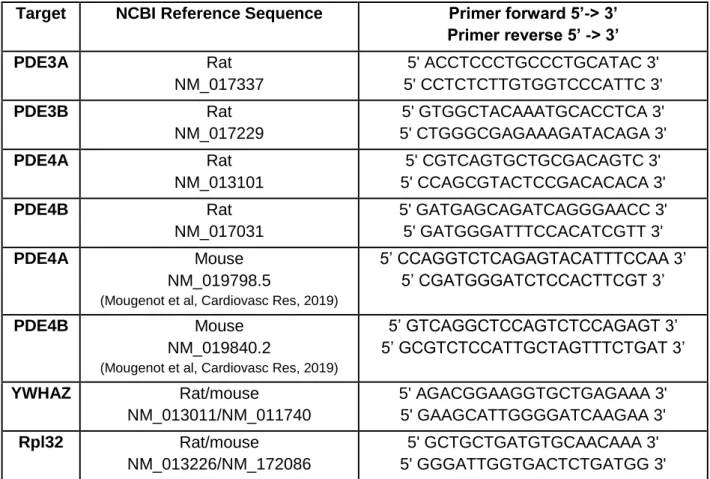
Real time quantitative RT-PCR analysis of mRNA. RNA was extracted from
heart samples with trizol (Invitrogen, Carlsbad, CA, USA), reverse-transcribed
into cDNA (iScript cDNA Synthesis Kit, Biorad Hercules, CA, USA), and mRNA
abundance was analyzed by real-time PCR with SYBR-green (SYBR
Green/ROX qPCR, Master Mix, Fermentas, Waltham, MA, USA). Details of
primers are provided in Supplementary Table 1.
Mouse experiments. All experiments were performed on 9-week-old FVB/N
mice from (Janvier’s Labs, Le Genest-Saint Isle, France) subjected to 75%
nephrectomy or sham-operation and sacrificed 8 weeks after surgery. Surgery
and euthanasia of animals were performed under general anesthesia by
intraperitoneal injection of ketamine (160 mg/kg) and xylazine (8 mg/kg). At the
time of sacrifice urine and plasma were collected and the kidney and heart were
removed and frozen in liquid nitrogen.
Circulating mouse FGF23 concentrations were measured in duplicate using an
ELISA assay (Immutopics International, San Clemente, CA, USA), according
to the manufacturers protocol.
Statistical analysis. The tests used to assess statistical significance are
results are expressed as mean (±SEM). Differences with p-values <0.05 were
considered as statistically significant.
3. Results
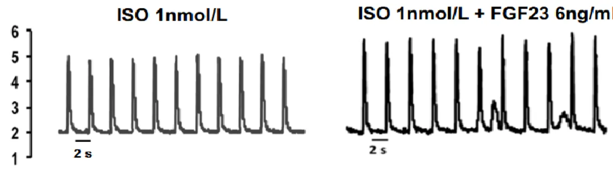
Effects of FGF23 and sKlotho on SCWs. Because ventricular arrhythmia is a
frequent cause of death in patients with RF, we checked if incubation of ARVMs
with FGF23 at a concentration of 6 ng/ml, a level commonly observed in
patients with RF, could promote SCWs, a marker of arrhythmic events. Ca2+
transients were recorded in ARVMs cultivated for 24 h with a control medium
or FGF23 and subsequently loaded with the calcium indicator Fura-2 and
stimulated at a pace of 0.5 Hz in the presence of isoprenaline (ISO 1 or 10 nM;
Table 1 and supplementary Figure 1 A). We assessed the number of cells with
SCWs during a 3-min recording. In the absence of FGF23, 1 nM ISO did not
induce SCWs (Table 1 and supplementary Figure 1). By contrast 45.8% of
ARVMs treated with FGF23 exhibited SCWs in the presence of 1 nM ISO (Table
1 and supplementary Figure 1). Raising ISO concentration to 10 nM induced
SCWs in 62.5% of ARVMs treated with control medium, and treatment with
FGF23 further increased the number of ARVMs presenting SCWs to 95.0%
(Table 1). Incubation of ARVMs with sKlotho decreased the number of cells
with SCWs thus preventing the FGF23 effect (Table 1). Unexpectedly in the
absence of FGF23, sKlotho alone also decreased the number of cells with
Effects of FGF23 on SR Ca2+ leak, SR Ca2+load and phosphorylation of
Ca2+ handling proteins. Because ISO-induced arrhythmias are largely due to
an increased diastolic Ca2+ leak from the SR via RyR2, we measured Ca2+
leakage using a Na+/Ca2+-free Tyrode solution (0Na+/0Ca2+ solution) to prevent
Ca2+ extrusion by the Na+/Ca2+ exchanger (NCX) and tetracaine (1 mM) to block
RyR2. Application of caffeine (10 mM) at the end of the experiment was used
to evaluate SR Ca2+ load. FGF23 treatment drastically increased SR Ca2+ leak
(Figure 1A and B), whereas it did not modify SR Ca2+ load (Figure 1C). To
analyze the presence of SCWs, cells were briefly exposed to 0Na+/0Ca2+
solution and SCWs were recorded during a 20s pacing pause. Cardiomyocytes
treated with FGF23 showed more occurrences of SCWs than control cells
(Figure 1D and 1E) suggesting pro-arrhythmogenic effects of FGF23 on the
heart. To determine the molecular mechanisms of the increased SR Ca2+ leak,
we analyzed the phosphorylation level of two key Ca2+-handling proteins by
immunoblot: PLB and RyR2. A 24h-incubation of ARVMs with FGF23
significantly increased phosphorylation at Serine 16 and Threonine 17 of PLB
and at Serine 2808 and 2814 of RyR (Figures 2A and 2C). These results
indicate that both protein kinase A (PKA) and Ca2+/calmodulin-dependent
protein kinase II (CaMKII) are activated by FGF23 treatment. Co-incubation of
FGF23 and sKlotho prevents the FGF23-induced phosphorylation of PLB and
RyR2 (Figures 2B and 2D).
FGF23 decreases PDE4 expression and increases ISO-induced cAMP levels. Since FGF23 increases and sKlotho decreases SCW frequency
(Figure 2), we checked if FGF23 and sKlotho could affect [cAMP]i accumulation
in ARVMs in response to ISO stimulation. We measured real-time changes of
ISO-induced [cAMP]i in ARVMs infected with an adenovirus expressing the
FRET-based cAMP probe Epac-SH187. ARVMs were incubated for 24 h with
control medium, FGF23, sKlotho alone or FGF23 in combination with sKlotho.
The cells were then challenged with a short application of ISO (30 nM, 15 s,
Figure 3A, B and C). Maximal [cAMP]i accumulation in response to a 15 sec
application of ISO was significantly higher (Figure 3A and C) and the decay
kinetic, which reflects cAMP hydrolysis by PDEs[22], was slower (Figure 3B) in
FGF23 treated than in control ARVMs. ARVMs express FGR1, FGFR3 and
FGFR4 (supplementary Figure 3D). In heart FGF23 can stimulate the
calcineurin/NFAT pathway. To determine if this pathway could mediate FGF23
effect on [cAMP]i in response to ISO we inhibited calcineurin stimulation with
CspA. CspA alone did not modify ISO-induced [cAMP]i level (Figure 3A and C).
However, in the presence of CspA FGF23 did not enhance ISO-induced
[cAMP]i accumulation (Figure 3A and C). Next we determined the effect of
sKlotho alone or in combination with FGF23 on β-AR induced [cAMP]i. sKlotho
prevented the amplification of ISO-induced [cAMP]i accumulation by FGF23
(Figure 3A and C).
To further characterize the mechanisms by which FGF23 could interfere
with ISO-induced cAMP accumulation we assessed by western blotting the
expression of the main cAMP-specific PDE isoforms expressed in ARVMs.
FGF23 specifically decreased PDE4B expression (Figure 3D and E). PDE3A,
PDE3B and PDE4A protein expression were unchanged by FGF23 treatment
reverse-transcription polymerase chain reaction (qRT-PCR) and we found
about 5-fold lower level of PDE4B in ARVMs treated with FGF23 compared with
cells treated with control medium, indicating that changes in PDE4B expression
induced by FGF23 take place at the transcriptional level (Figure 3F). To
determine if the decrease in PDE4B expression was specific of FGF23 or
secondary to cardiac hypertrophy we used FGF2 as a control. FGF2 alone,
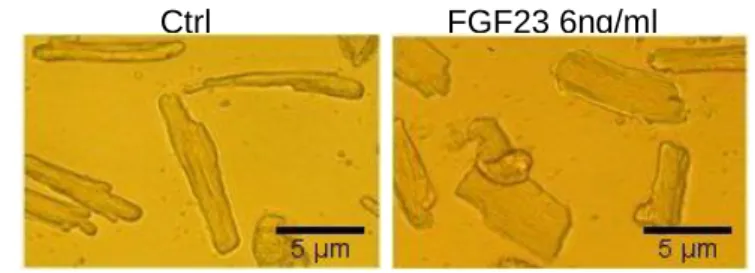
which increased ARVMs surface area (Supplementary Figure 2B), did not
modify the expression of PDE4B nor that of PDE3A, 3B and 4A (Figure 4A).
This confirms that the decrease in PDE4B expression was specifically induced
by FGF23 and not a consequence of ARVMs hypertrophy. Incubation of
ARVMs with sKlotho or CspA in combination with FGF23 completely prevented
the effect of FGF23 on PDE4B expression (Figure 4B, 4C and 4D).
To determine the contribution of different pathways to PDE4B regulation by
FGF23 in ARVMs, we analyzed the phosphorylation pattern of Extracellular Signal-regulated Kinase (ERK), Phospholipase Cγ (PLCγ) and serine/threonine
kinase (Akt) after acute (15 min) or 24h stimulation with FGF23. We used ARVMs treated for 10 min with phenylephrine (Phe, 50 μM) as positive control.
Exposition of ARVMs with FGF23 for 15 min did not modify Akt phosphorylation
(Figure 5A) while a 24h incubation significantly decreased the phosphorylation level of Akt (Figure 5B). Moreover, FGF23 increased phosphorylation of PLCγ1
within 15 min of treatment without changing overall PLCγ1 expression (Figure
5C) but a 24h-exposition with FGF23 did not affect PLCγ1 phosphorylation
(Figure 5D). FGF23 did not change the level of ERK phosphorylation after acute
Effects of FGF23 on excitation-contraction coupling. We tested whether the
decrease in PDE4B expression induced by FGF23s modified the β-AR
regulation of excitation-contraction coupling. First, we analyzed the L-type
calcium current amplitude (ICa,L) by the whole cell patch clamp technique.
FGF23 significantly increased basal ICa,L density (Figure 7 C, 7D and 7F). The
response to a pulse application of ISO (30 nM, 15 s) was markedly increased
by FGF23 treatment (Figure 7A, 7B and 7E). However, co-incubation of
cardiomyocytes with FGF23 and either CspA, or FGFR antagonist PD173074,
or sKlotho restored a normal response to ISO (Figure 7).
Effects of sKlotho on PDEs expression levels and Ca2+-handling proteins.
We further characterized the effects of sKlotho on ARVMs. We first verified by qRT-PCR and western blot that ARVMs did not express αKlotho (data not
shown). Since sKlotho alone decreases the percentage of ARVMs exhibiting
SCWs in response to ISO (Table 1) and prevents the actions of FGF23 on basal
phosphorylation level of PLB and RyR2 (Figure 2B), [cAMP]i accumulation
(Figure 3A, B and C) and ICa,L amplitude (Figure 7E and 7F), we checked if
sKlotho could modify the expression of PDEs. Incubation of ARVMs with
sKlotho (0.95ng/ml, a concentration within the normal range in human[45],[38]
for 24 h) increased the expression of PDE3A and PDE3B at the protein level
(respectively by ~60% and ~30%, western blot Figure 8A) as well as at the
mRNA level (respectively by ~7-fold and 9-fold, qRT-PCR Supplementary
Figure 3B). sKlotho did not modify the expression of PDE4A and PDE4B
FGF23 and sKlotho did not modify the expression level of PDEs (Figure 8B).
sKlotho alone decreased the phosphorylation level of PLB and RyR2 at both
PKA and CAMKII phosphorylation sites (Figure 8C) resulting in a decreased
number of cells exhibiting SCWs in response to ISO (Table 1).
sKlotho did not modify transient receptor potential cation channels
(TRPC6) or SR Ca2+-ATPase (SERCA2a) expressions (Supplementary Figure
3C) nor phosphorylation level of ERK (Figure 6C).
Subtotal nephrectomy induces cardiac hypertrophy and modifies PDE4B expression. To determine if the FGF23-induced modifications of PDE
expression observed in ARVMs in culture were relevant in a model of CKD we
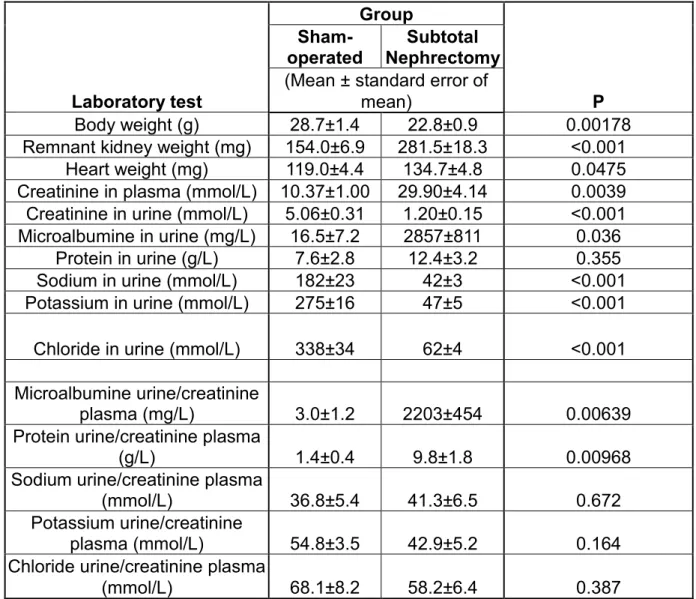
studied mice submitted to 75% excision of total renal mass (Nx). Mean values
of body, kidney, or heart weight, creatinine and proteinuria levels and urine
excretion of Na+, K+ and Ca2+ in sham-operated and Nx mice are presented in
Supplementary Table 2. As expected, plasma creatinine concentration and
urinary protein, two markers of renal function were significantly increased in Nx
mice as compared to the Sham group (Figure 9A, Supplementary Table 2)
indicating glomerular injury. Nx animals also exhibited cardiac hypertrophy
(Figure 9B). Plasma FGF23 concentration measured six weeks after surgery
was higher in Nx mice than in Sham animals (Figure 9C). To test whether
elevated FGF23 plasma concentrations observed in CKD could modify cardiac
PDEs levels, we assessed by western blotting the expression of the main
cAMP-specific PDE isoforms in the heart. We observed that PDE4B expression
was specifically reduced in Nx mice as compared to the Sham group (~2 fold,
Nx animals than in Sham mice (Figure 9D). The impact of the Nx on PDE4B
was specific since neither the mRNA, nor the protein level of PDE3A, PDE3B
and PDE4A changed in the heart after the Nx.
4. Discussion
While it is known that a high concentration of FGF23 can induce hypertrophy
and have deleterious effects on heart function, the mechanisms by which
FGF23 alters cardiomyocyte physiology remain largely unknown [14]. Our
results unveil some of these mechanisms. We show that treatment with FGF23
increases the number of SCWs in ARVMs in response to ISO. We observed
that FGF23 increased RyR2 and PLB phosphorylation at PKA and CaMKII
sites, modifications that are known to augment SR Ca2+ leak. There is strong
evidence that increased diastolic Ca2+ leak from the SR via RyR2, increases
occurrence of SCWs via activation of the electrogenic Na+/Ca2+ -exchanger,
causing delay after depolarizations which are able to trigger action potentials,
increasing the risk of ventricular arrhythmia [3]. Increased CaMKII
phosphorylation of S2814 on RyR2 was shown to play a critical role in the
development of ventricular arrhythmias even in the absence of structural heart
disease [37, 41]. Our findings that FGF23 increases the number of SCWs in cardiomyocytes by acute β-AR stimulation due to increased SR Ca2+ leak and
higher RyR2-S2814 phosphorylation are in agreement with these observations.
Recently Novarro-Garcia et al reported similar results [34]. They observed that
FGF23 induced RyR phosphorylation at the CaMKII site resulting in an increase
in Ca2+ spark frequency that could trigger pro-arrhythmogenic events.Our data
We report for the first time that FGF23 specifically inhibits PDE4B
expression. PDE4B is a cAMP-specific PDE and one of the main isoforms of
PDEs expressed in rodent cardiomyocytes. It plays a central role in the control of cAMP signaling in response to β-AR stimulation and is associated with the
L-type calcium channels [8, 31, 54]. PDE4 is also expressed in human heart
and is involved [33] in the generation of cAMP compartments by forming
barriers for cAMP diffusion or acting as local sink for cAMP [23, 27, 42, 43] .
We observed that a 24 h exposure of ARVMs to FGF23 specifically decreased
the expression of PDE4B, and that this effect was abolished when the
calcineurin/NFAT pathway was inhibited by CspA. Abi-Gerges et al. reported a
decreased expression and activity of PDE4B in cardiac hypertrophy [1].
However, the decrease in PDE4B expression induced by FGF23 was not due
to ARVMs hypertrophy per se since FGF2, which had similar effects on ARVMs
size as FGF23, did not modify PDE expression. The decrease in PDE4B
expression resulted in an increase in ISO-induced [cAMP]i accumulation, PLB
and RyR2 phosphorylations, ICa,L amplitude, calcium transients and sarcomere
shortening. These results parallel those obtained by Bobin et al, who showed
similar effects in ARVMs with a selective PDE4 inhibitor [4]. Our findings also
mimic those obtained in cardiomyocytes from PDE4B-/- mice where Ca2+
transients, cell contraction, and spontaneous Ca2+ release events under
β-adrenergic stimulation were increased by comparison with wild-type
cardiomyocytes [28]. Leroy et al. also reported in in vivo experiments that the
percentage of mice with ventricular tachycardia after ISO infusion and a burst
pacing was significantly higher in PDE4B-/- mice than in wild type. We observed
shortening and Ca2+ transient in the absence of ISO stimulation. These results
are similar to the results obtained by Cilvik et al., who showed that ventricular
myocytes from FGFR1-transgenic mice have enhanced contraction suggesting
that activation of FGF signaling pathway in cardiomyocytes increases
contractility and results in a hypertrophic cardiomyopathy [7]. In addition, we
showed that PDE4B is decreased at protein and transcriptional level in mice
subjected to subtotal nephrectomy. Altogether, our data demonstrate that the
reduction in PDE4B expression induced by FGF23 has functional
consequences which may contribute to the increased risk of ventricular
arrhythmia responsible for sudden death in patients with RF. Blocking these
effects of FGF23 could decrease the death rate in CKD patients.
Our data show that the effects of FGF23 on cardiomyocytes are
mediated by a FGFR. Indeed, FGFR antagonists prevented all the effects of
FGF23 on cardiomyocytes. Although the affinity of FGF23 for FGFR is low, at
the high concentrations as observed in patients with RF, FGF23 is able to stimulate the calcineurin/NFAT pathway via a FGFR in the absence of αKlotho
[11]. We showed that a short exposition of ARVMs to FGF23 increases phosphorylated PLCγ level. This finding is supported by studies of Grabner et
al and Han et al [13][16] who showed that FGF23 activates cardiotoxic PLCγ
signaling pathway and induces cardiac hypertrophic growth in vivo and in vitro
via FGFR4 in the absence of co-expression of α-Klotho. A 24h FGF23
treatment decreased the amount of phosphorylated Akt. FGF23-induced changes in PLCγ and/or Akt activation could mediate other FGF23 effects on
The mechanism by which FGF23 decreases PDE4B expression and the role of PLCγ and Akt signaling will require further investigations.
We also studied the role of sKlotho on ARVMs in the presence or in the absence of FGF23. Although the role of αKlotho as a FGF23 co-receptor is
established, the role of the circulating form of Klotho is still largely unclear. We
report here that sKlotho. can modify cardiomyocytes functions via two
mechanisms: by interfering with FGF23 action, it prevents FGF23-induced
PDE4B decreases, and by acting alone in the absence of FGF23, it increases
PDE3A and 3B expression. Klotho can modulate TRPC6 activity in the absence
of FGF23 or FGFR in cardiomyocytes or in a myoblast cell line [56] [57]. Klotho
also modifies KCNQ1/KCNE1 K+ channels stability when expressed in
Xenopus oocytes [2]. sKlotho can modify the activity of various ion channels
and Na-phosphate cotransporters [18], and can bind to Wnt ligands [5, 19, 57].
We report here that sKlotho can also prevent the decrease in PDE4B
expression induced by FGF23. Several mechanisms can be involved. It has
been recently reported that sKlotho can form a complex with a FGFR and
FGF23 that can activate the phosphorylation of the ERK pathway in HEK293
cells[6]. While the possibility of formation of a complex between sKlotho, FGF23
and a FGFR in ARVMs would require further investigation, we did not observe
phosphorylation of ERK in the presence of FGF23 and sKlotho at
concentrations that prevent FGF23 effects on ARVMs. Alternatively, sKlotho
could prevent the association of FGF23 to FGFR preventing the activation of
the NFAT pathway.
We also show that not only sKlotho interferes with FGF23 actions, but it
It has been reported that sKlotho can interfere with phosphoinositide 3-kinase (PI3K) signaling triggered by IGF1 in cardiomyocytes [57]. PI3Kγ can associate
with PDE3 and increase its activity but not its expression [12]. The mechanisms
by which sKlotho increases PDE3 expression and the potential role of PI3K
signaling in this matter will require further investigations.
PDE3 is a major regulator of cAMP in cardiomyocytes and was shown
to regulate cardiac contractility by modulating PLB-SERCA2 activity and
subsequent sarcoplasmic reticulum Ca2+ uptake[44]. Moreover PDE3 might be
also important in pathological cardiac remodeling, for example decreased
PDE3A expression has been found in diseased hearts in rodents [1]. Some
studies showed that the inhibition of PDE3 activity or knockdown of PDE3
expression was associated with myocyte apoptosis [10]. Recent study
demonstrated that myocardial overexpression of PDE3A in transgenic mice
leads to a decrease in heart rate and myocardial contractile function through
attenuating PKA-mediated troponin I phosphorylation and can protect the heart
against cardiac ischemia/reperfusion injury [36]. Our findings suggest that a
decrease in PDE3 expression could participate in the enhanced ISO-induced
hypertrophy observed in Klotho deficient mice.
Low levels of circulating Klotho concentration are inconstantly
associated to the risk of atrial fibrillation in patients with end stage renal disease
[35][32]. Our results could give an explanation for these differences between
studies. High concentrations of FGF23 markedly decrease PDE4B expression
that favor arrhythmogenic effects as reported here. Observing an additional
the characteristics of the populations studied such as FGF23 levels, the distribution and levels of sKlotho concentration…
Klotho is not present in ventricular myocytes but is detected in the
sinoatrial node [51]. It is unknown if this form of Klotho is similar to sKlotho. It
has been reported that Isolated sino-atrial nodes of mice with Klotho gene
disruption are dysfunctional. The mechanisms by which Klotho expressed in
nodal cells modifies their function remain to be elucidated.
5. Conclusions
This study unveils the mechanisms by which the high concentrations of FGF23
associated to low sKlotho concentration in the plasma have deleterious effects
on the heart of patients with CKD. FGF23 decreases PDE4B by stimulating the
calcineurin/NFAT pathway via the activation of a FGFR. This results in an
increase in Ca2+ leakage and basal phosphorylation levels of Ca2+-cycling
proteins including PLB and RyR2 leading to the higher occurrence of SCWs.
We show that FGF23 increases [cAMP]i levels, basal and ISO-induced ICa,L
amplitude, and calcium transients. All these effects are prevented in the
presence of sKlotho that increases PDE3A and PDE3B expressions. PDE4 is
expressed in human myocardium and its inhibition has been shown to induce
arrhythmias in human atrium. Our data suggest that inhibition of PDE4
expression[33] could also participate in the increase rate of sudden death
mainly due to ventricular arrhythmia in disorders with high FGF23 concentration
6. References
1. Abi-Gerges A, Richter W, Lefebvre F, Mateo P, Varin A, Heymes C,
Samuel JL, Lugnier C, Conti M, Fischmeister R, Vandecasteele G
(2009) Decreased expression and activity of cAMP phosphodiesterases in cardiac hypertrophy and its impact on β-Adrenergic cAMP signals.
Circ Res 105:784–792. doi: 10.1161/CIRCRESAHA.109.197947
2. Almilaji A, Pakladok T, Muñoz C, Elvira B, Lang F, Almilaji A, Pakladok
T, Muñoz C, Elvira B, Almilaji A, Pakladok T, Muñoz C, Elvira B, Sopjani
M, Lang F (2014) Upregulation of KCNQ1 / KCNE1 K channels by
Klotho Upregulation of KCNQ1 / KCNE1 K + channels by Klotho. 6950.
doi: 10.4161/chan.27662
3. Bers DM (2014) Cardiac Sarcoplasmic Reticulum Calcium Leak: Basis and Roles in Cardiac Dysfunction. Annu Rev Physiol 76:107–127. doi:
10.1146/annurev-physiol-020911-153308
4. Bobin P, Varin A, Lefebvre F, Fischmeister R, Vandecasteele G, Leroy
J (2016) Calmodulin kinase II inhibition limits the pro-Arrhythmic
Ca2+waves induced by camp-phosphodiesterase inhibitors. Cardiovasc Res 110:151–161. doi: 10.1093/cvr/cvw027
5. Buendía P, Ramírez R, Aljama P, Carracedo J (2016) Klotho Prevents Translocation of NFκB. In: Vitamins and Hormones. pp 119–150
6. Chen G, Liu Y, Goetz R, Fu L, Jayaraman S, Hu M-C, Moe OW, Liang G, Li X, Mohammadi M (2018) α-Klotho is a non-enzymatic molecular
scaffold for FGF23 hormone signalling. Nature 553:461–466. doi:
Weinheimer CJ, House SL, Kovacs A, Nichols CG, Ornitz DM (2013)
Fibroblast growth factor receptor 1 signaling in adult cardiomyocytes
increases contractility and results in a hypertrophic cardiomyopathy. PLoS One 8:1–17. doi: 10.1371/journal.pone.0082979
8. Conti M, Richter W, Mehats C, Livera G, Park JY, Jin C (2003) Cyclic
AMP-specific PDE4 phosphodiesterases as critical components of cyclic AMP signaling. J Biol Chem 278:5493–5496. doi:
10.1074/jbc.R200029200
9. Courbebaisse M, Lanske B (2017) Biology of Fibroblast Growth Factor 23 : From physiology to pathology. Cold Spring Harb Perspect Med
10. Ding B, Abe JI, Wei H, Huang Q, Walsh RA, Molina CA, Zhao A,
Sadoshima J, Blaxall BC, Berk BC, Yan C (2005) Functional role of
phosphodiesterase 3 in cardiomyocyte apoptosis: Implication in heart failure. Circulation 111:2469–2476. doi:
10.1161/01.CIR.0000165128.39715.87
11. Faul C, Amaral AP, Oskouei B, Hu M-CC, Sloan A, Isakova T, Gutiérrez
OM, Aguillon-Prada R, Lincoln J, Hare JM, Mundel P, Morales A, Scialla
J, Fischer M, Soliman EZ, Chen J, Go AS, Rosas SE, Nessel L,
Townsend RR, Feldman HI, St. John Sutton M, Ojo A, Gadegbeku C, Di
Marco GS, Reuter S, Kentrup D, Tiemann K, Brand M, Hill JA, Moe
OW, Kuro-o M, Kusek JW, Keane MG, Wolf M, Gutierrez OM,
Aguillon-Prada R, Lincoln J, Hare JM, Mundel P, Morales A, Scialla J, Fischer M,
Soliman EZ, Chen J, Go AS, Rosas SE, Nessel L, Townsend RR,
Feldman HI, St John Sutton M, Ojo A, Gadegbeku C, Di Marco GS,
Kusek JW, Keane MG, Wolf M, Guti??rrez OM, Aguillon-Prada R,
Lincoln J, Hare JM, Mundel P, Morales A, Scialla J, Fischer M, Soliman
EZ, Chen J, Go AS, Rosas SE, Nessel L, Townsend RR, Feldman HI,
Sutton MSJ, Ojo A, Gadegbeku C, Di Marco GS, Reuter S, Kentrup D,
Tiemann K, Brand M, Hill JA, Moe OW, Kuro-o M, Kusek JW, Keane
MG, Wolf M (2011) FGF23 induces left ventricular hypertrophy. J Clin Invest 121:4393–4408. doi: 10.1172/jci46122
12. Ghigo A, Perino A, Mehel H, Zahradníková A, Morello F, Leroy J,
Nikolaev VO, Damilano F, Cimino J, De Luca E, Richter W,
Westenbroek R, Catterall WA, Zhang J, Yan C, Conti M, Gomez AM,
Vandecasteele G, Hirsch E, Fischmeister R (2012) Phosphoinositide 3-kinase γ protects against catecholamine-induced ventricular arrhythmia
through protein kinase A-mediated regulation of distinct phosphodiesterases. Circulation 126:2073–2083. doi:
10.1161/CIRCULATIONAHA.112.114074
13. Grabner A, Amaral AP, Schramm K, Singh S, Sloan A, Yanucil C, Li J,
Shehadeh LA, Hare JM, David V, Martin A, Fornoni A, Di Marco GS,
Kentrup D, Reuter S, Mayer AB, Pavenstädt H, Stypmann JJ, Kuhn C,
Hille S, Frey N, Leifheit-Nestler M, Richter B, Haffner D, Abraham R,
Bange J, Sperl B, Ullrich A, Brand M, Wolf M, Faul C, Pavenst??dt H,
Stypmann JJ, Kuhn C, Hille S, Frey N, Leifheit-Nestler M, Richter B,
Haffner D, Abraham R, Bange J, Sperl B, Ullrich A, Brand M, Wolf M,
Faul C (2015) Activation of Cardiac Fibroblast Growth Factor Receptor 4 Causes Left Ventricular Hypertrophy. Cell Metab 22:1020–1032. doi:
14. Grabner A, Faul C (2016) The role of fibroblast growth factor 23 and Klotho in uremic cardiomyopathy. 25:314–324. doi:
10.1097/MNH.0000000000000231
15. Gruson D, Lepoutre T, Ketelslegers J-M, Cumps J, Ahn SA, Rousseau
MF (2012) C-terminal FGF23 is a strong predictor of survival in systolic heart failure. Peptides 37:258–262. doi: 10.1016/j.peptides.2012.08.003
16. Han X, Cai C, Xiao Z, Quarles LD (2020) Journal of Molecular and
Cellular Cardiology FGF23 induced left ventricular hypertrophy
mediated by FGFR4 signaling in the myocardium is attenuated by soluble Klotho in mice. J Mol Cell Cardiol 138:66–74. doi:
10.1016/j.yjmcc.2019.11.149
17. Hasegawa H, Nagano N, Urakawa I, Yamazaki Y, Iijima K, Fujita T,
Yamashita T, Fukumoto S, Shimada T (2010) Direct evidence for a
causative role of FGF23 in the abnormal renal phosphate handling and
vitamin D metabolism in rats with early-stage chronic kidney disease. Kidney Int 78:975–980. doi: 10.1038/ki.2010.313
18. Hu MC, Shi M, Zhang J, Pastor J, Nakatani T, Lanske B, Razzaque MS, Rosenblatt KP, Baum MG, Kuro-o M, Moe OW Klotho : a novel
phosphaturic substance acting as an autocrine enzyme in the renal
proximal tubule. doi: 10.1096/fj.10-154765
19. Huang C (2010) Regulation of ion channels by secreted Klotho :
mechanisms and implications. Kidney Int 77:855–860. doi:
10.1038/ki.2010.73
20. Isakova T, Houston J, Santacruz L, Schiavenato E, Somarriba G,
between fibroblast growth factor 23 and cardiac characteristics in pediatric heart failure. Pediatr Nephrol 28:2035–2042. doi:
10.1007/s00467-013-2515-7
21. Ix JH, Katz R, Kestenbaum BR, De Boer IH, Chonchol M, Mukamal KJ,
Rifkin D, Siscovick DS, Sarnak MJ, Shlipak MG (2012) Fibroblast
growth factor-23 and death, heart failure, and cardiovascular events in
community-living individuals: CHS (Cardiovascular Health Study). J Am Coll Cardiol 60:200–207. doi: 10.1016/j.jacc.2012.03.040
22. Jérôme L, Aniella A-G, O. NV, Wito R, Patrick L, Jean-Luc M, Marco C, Rodolphe F, Grégoire V (2008) Spatiotemporal Dynamics of
β-Adrenergic cAMP Signals and L-Type Ca2+ Channel Regulation in Adult Rat Ventricular Myocytes. Circ Res 102:1091–1100. doi:
10.1161/CIRCRESAHA.107.167817
23. Jurevičius J, Skeberdis VA, Fischmeister R (2003) Role of cyclic
nucleotide phosphodieterase isoforms in cAMP compartmentation following β2-adrenergic stimulation of ICa,L in frog ventricular
myocytes. J Physiol 551:239–252. doi: 10.1113/jphysiol.2003.045211
24. Klarenbeek JB, Goedhart J, Hink MA, Gadella TWJ, Jalink K (2011) A
mTurquoise-based cAMP sensor for both FLIM and ratiometric read-out has improved dynamic range. PLoS One 6:2–7. doi:
10.1371/journal.pone.0019170
25. Koller L, Kleber ME, Brandenburg VM, Goliasch G, Richter B,
Sulzgruber P, Scharnagl H, Silbernagel G, Grammer TB, Delgado G,
Tomaschitz A, Pilz S, Berger R, Mörtl D, Hülsmann M, Pacher R, März
and Specific Predictor of Mortality in Patients With Heart Failure and Reduced Ejection Fraction. Circ Hear Fail 8:1059–1067. doi:
10.1161/CIRCHEARTFAILURE.115.002341
26. Kuro-o M (2019) The Klotho proteins in health and disease. Nat Rev
Nephrol 15. doi: 10.1038/s41581-018-0078-3
27. Leineweber K, Böhm M, Heusch G (2006) Cyclic Adenosine
Monophosphate in Acute Myocardial Infarction With Heart Failure Slayer or Savior ? Circulation 114:365–367. doi:
10.1161/CIRCULATIONAHA.106.642132
28. Leroy J, Richter W, Mika D, Castro LR V, Abi-gerges A, Xie M,
Scheitrum C, Lefebvre F, Schittl J, Mateo P, Westenbroek R, Catterall
WA, Charpentier F, Conti M, Fischmeister R, Vandecasteele G (2011)
Phosphodiesterase 4B in the cardiac L-type Ca 2 + channel complex
regulates Ca 2 + current and protects against ventricular arrhythmias in mice. J Clin Invest 121:2651–2661. doi: 10.1172/JCI44747.association
29. Lutsey PL, Alonso A, Selvin E, Pankow JS, Michos ED, Agarwal SK,
Loehr LR, Eckfeldt JH, Coresh J (2014) Fibroblast growth factor-23 and
incident coronary heart disease, heart failure, and cardiovascular
mortality: the atherosclerosis risk in communities study. J Am Hear
Assoc 3:e000936. doi: 10.1161/JAHA.114.000936
30. Mehta R, Cai X, Lee J, Scialla JJ, Bansal N, Sondheimer JH, Chen J,
Hamm LL, Ricardo AC, Navaneethan SD, Deo R, Rahman M, Feldman
HI, Go AS, Isakova T, Wolf M, Appel LJ, He J, Kusek JW, Lash JP, Ojo
A, Townsend RR (2016) Association of fibroblast growth factor 23 With
Insufficiency Cohort Study. JAMA Cardiol 1:548–556. doi:
10.1001/jamacardio.2016.1445
31. Mika D, Bobin P, Pomérance M, Lechêne P, Westenbroek RE, Catterall
WA, Vandecasteele G, Leroy J, Fischmeister R (2013) Differential
regulation of cardiac excitation-contraction coupling by cAMP phosphodiesterase subtypes. Cardiovasc Res 100:336–346. doi:
10.1093/cvr/cvt193
32. Mizia-Stec K, Wieczorek J, Polak M, Wybraniec MT,
Woźniak-Skowerska I, Hoffmann A, Nowak S, Wikarek M, Wnuk-Wojnar A, Chudek J, Więcek A (2018) Lower soluble Klotho and higher fibroblast
growth factor 23 serum levels are associated with episodes of atrial fibrillation. Cytokine 111:106–111. doi: 10.1016/j.cyto.2018.08.005
33. Molina CE, Leroy J, Richter W, Xie M, Scheitrum C, Lee IO, Maack C,
Rucker-Martin C, Donzeau-Gouge P, Verde I, Llach A, Hove-Madsen L,
Conti M, Vandecasteele G, Fischmeister R (2012) Cyclic adenosine
monophosphate phosphodiesterase type 4 protects against atrial arrhythmias. J Am Coll Cardiol 59:2182–2190. doi:
10.1016/j.jacc.2012.01.060
34. Navarro-García JA, Delgado C, Fernández-Velasco M, Val-Blasco A,
Rodríguez-Sánchez E, Aceves-Ripoll J, Gómez-Hurtado N, Bada-Bosch
T, Mérida-Herrero E, Hernández E, Praga M, Salguero R, Solís J,
Arribas F, Delgado JF, Bueno H, Kuro-O M, Ruilope LM, Ruiz-Hurtado
G (2019) Fibroblast growth factor-23 promotes rhythm alterations and
contractile dysfunction in adult ventricular cardiomyocytes. Nephrol Dial
35. Nowak A, Bjorn F, Artunc F, Serra AL, Breidthardt T, Twerenbold R,
Peter M, Mueller C (2014) Prognostic Value and Link to Atrial Fibrillation of Soluble Klotho and FGF23 in Hemodialysis Patients. PLoS One 9:1–
11. doi: 10.1371/journal.pone.0100688
36. Oikawa M, Wu M, Lim S, Knight WE, Miller CL, Cai Y, Lu Y, Blaxall BC,
Takeishi Y, Abe J ichi, Yan C (2013) Cyclic nucleotide
phosphodiesterase 3A1 protects the heart against ischemia-reperfusion injury. J Mol Cell Cardiol 64:11–19. doi: 10.1016/j.yjmcc.2013.08.003
37. Van Oort RJ, McCauley MD, Dixit SS, Pereira L, Yang Y, Respress JL,
Wang Q, De Almeida AC, Skapura DG, Anderson ME, Bers DM,
Wehrens XHT (2010) Ryanodine receptor phosphorylation by
calcium/calmodulin-dependent protein kinase II promotes
life-threatening ventricular arrhythmias in mice with heart failure. Circulation 122:2669–2679. doi: 10.1161/CIRCULATIONAHA.110.982298
38. Pavik I, Jaeger P, Ebner L, Poster D, Krauer F, Kistler AD, Rentsch K,
Andreisek G, Wagner CA, Devuyst O, Wuthrich RP, Schmid C, Serra
AL (2012) Soluble klotho and autosomal dominant polycystic kidney disease. Clin J Am Soc Nephrol 7:248–257. doi:
10.2215/CJN.09020911
39. Plischke M, Neuhold S, Adlbrecht C, Bielesz B, Shayganfar S,
Bieglmayer C, Szekeres T, Hörl WH, Strunk G, Vavken P, Pacher R,
Hülsmann M (2012) Inorganic phosphate and FGF-23 predict outcome in stable systolic heart failure. Eur J Clin Invest 42:649–656. doi:
10.1111/j.1365-2362.2011.02631.x
D, Lebrec D, Durand F, Friedlander G (2013) Plasma Fibroblast Growth
Factor 23 Concentration Is Increased and Predicts Mortality in Patients
on the Liver-Transplant Waiting List. PLoS One 8:e66182. doi:
10.1371/journal.pone.0066182
41. Respress JL, Van Oort RJ, Li N, Rolim N, Dixit SS, Dealmeida A, Voigt
N, Lawrence WS, Skapura DG, Skårdal K, Wisløff U, Wieland T, Ai X,
Pogwizd SM, Dobrev D, Wehrens XHT (2012) Role of RyR2
phosphorylation at S2814 during heart failure progression. Circ Res 110:1474–1483. doi: 10.1161/CIRCRESAHA.112.268094
42. Rochais F, Abi-Gerges A, Horner K, Lefebvre F, Cooper DMF, Conti M,
Fischmeister R, Vandecasteele G (2006) A specific pattern of
phosphodiesterases controls the cAMP signals generated by different
Gs-coupled receptors in adult rat ventricular myocytes. Circ Res 98:1081–1088. doi: 10.1161/01.RES.0000218493.09370.8e
43. Rochais F, Vandecasteele G, Lefebvre F, Lugnier C, Lum H, Mazet JL,
Cooper DMF, Fischmeister R (2004) Negative feedback exerted by
cAMP-dependent protein kinase and cAMP phosphodiesterase on
subsarcolemmal cAMP signals in intact cardiac myocytes: An in vivo
study using adenovirus-mediated expression of CNG channels. J Biol Chem 279:52095–52105. doi: 10.1074/jbc.M405697200
44. Sanja B, Faiyaz A, Weixing S, Jie L, Samy M, Nazari P, Junhui S,
Steven H, Wook CY, Matthew M, Elizabeth M, Vincent M, H. BP (2013)
Phosphodiesterase Type 3A Regulates Basal Myocardial Contractility
Through Interacting With Sarcoplasmic Reticulum Calcium ATPase Type 2a Signaling Complexes in Mouse Heart. Circ Res 112:289–297.
doi: 10.1161/CIRCRESAHA.111.300003
45. Sari F, Inci A, Dolu S, Ellidag HY, Cetinkaya R, Ersoy FF (2017) High serum soluble α-Klotho levels in patients with autosomal dominant
polycystic kidney disease. J Investig Med 65:358–362. doi:
10.1136/jim-2016-000193
46. Segawa H, Yamanaka S, Ohno Y, Onitsuka A, Shiozawa K, Aranami F,
Furutani J, Tomoe Y, Ito M, Kuwahata M, Imura A, Nabeshima Y,
Miyamoto KI (2007) Correlation between hyperphosphatemia and type
II Na-Pi cotransporter activity in klotho mice. Am J Physiol - Ren Physiol 292:769–779. doi: 10.1152/ajprenal.00248.2006
47. Seiler S, Rogacev KS, Roth HJ, Shafein P, Emrich I, Neuhaus S,
Floege J, Fliser D, Heine GH (2014) Associations of FGF-23 and
sklotho with cardiovascular outcomes among patients with CKD stages 2–4. Clin J Am Soc Nephrol 9:1049–1058. doi: 10.2215/CJN.07870713
48. Shalhoub V, Shatzen EM, Ward SC, Davis J, Stevens J, Bi V, Renshaw
L, Hawkins N, Wang W, Chen C, Tsai M-M, Cattley RC, Wronski TJ, Xia
X, Li X, Henley C, Eschenberg M, Richards WG (2012) FGF23 neutralization improves chronic kidney disease–associated
hyperparathyroidism yet increases mortality. J Clin Invest 122:2543–
2553. doi: 10.1172/jci61405
49. Shannon TR, Ginsburg KS, Bers DM (2002) Quantitative assessment of the SR Ca2+ leak-load relationship. Circ Res 91:594–600
50. Souberbielle JC, Prié D, Piketty ML, Rothenbuhler A, Delanaye P,
Chanson P, Cavalier E (2017) Evaluation of a New Fully Automated Assay for Plasma Intact FGF23. Calcif Tissue Int 101:510–518. doi:
10.1007/s00223-017-0307-y
51. Takeshita K, Fujimori T, Kurotaki Y, Honjo H, Yasui K, Lee J, Kamiya K,
Kitaichi K, Ito M, Kondo T, Iino S, Inden Y, Takeshita K, Fujimori T,
Kurotaki Y, Honjo H (2004) Sinoatrial Node Dysfunction and Early
Unexpected Death of. doi: 10.1161/01.CIR.0000124224.48962.32
52. Tanaka S, Fujita S, Kizawa S, Morita H, Ishizaka N (2016) Association between FGF23, α-Klotho, and Cardiac Abnormalities among Patients
with Various Chronic Kidney Disease Stages. PLoS One 11:e0156860.
doi: 10.1371/journal.pone.0156860
53. Udell JA, Morrow DA, Jarolim P, Sloan S, Hoffman EB, O’Donnell TF,
Vora AN, Omland T, Solomon SD, Pfeffer MA, Braunwald E, Sabatine
MS (2014) Fibroblast growth factor-23, cardiovascular prognosis, and
benefit of angiotensin-converting enzyme inhibition in stable ischemic heart disease. J Am Coll Cardiol 63:2421–2428. doi:
10.1016/j.jacc.2014.03.026
54. Verde I, Vandecasteele G, Lezoualc’h F, Fischmeister R (1999)
Characterization of the cyclic nucleotide phosphodiesterase subtypes
involved in the regulation of the L-type Ca2+ current in rat ventricular myocytes. Br J Pharmacol 127:65–74. doi: 10.1038/sj.bjp.0702506
55. Webster AC, Nagler E V, Morton RL, Masson P (2016) Chronic Kidney Disease. Lancet 6736:1–15. doi: 10.1016/S0140-6736(16)32064-5
56. Wright JD, An SW, Xie J, Lim C, Huang CL (2019) Soluble klotho
regulates TRPC6 calcium signaling via lipid rafts, independent of the FGFR-FGF23 pathway. FASEB J 33:9182–9193. doi:
57. Xie J, Cha SK, An SW, Kuro-O M, Birnbaumer L, Huang CL (2012)
Cardioprotection by Klotho through downregulation of TRPC6 channels in the mouse heart. Nat Commun 3:1211–1238. doi:
10.1038/ncomms2240
58. Yang H, Luo H, Tang X, Zeng X, Yu Y, Ma L, Fu P (2016) Prognostic
value of FGF23 among patients with end-stage renal disease: a systematic review and meta-analysis. Biomark Med 10:547–556. doi:
10.2217/bmm.16.11
Isoproterenol Control FGF23 sKlotho FGF23 +
sKlotho
p-value
1 nM 0 45.8% 0 6% 0.000075
10 nM 62.5% 95% 41.1 % 56.6% 0.002723
Table1. Percent of cells with spontaneous Ca2+ waves in the presence of ISO
(1 or 10 nM) after treatment with vehicle (CON), FGF23 (6 ng/ml), sKlotho
(0.95 ng/ml) or FGF23+sKlotho. Statistical significance was determined by Fisher’s exact test to compare occurrence of events. (N=3 rats, n=16-20 cells
Figure legends:
Fig. 1. FGF23 potentiates the effect of submaximal β-AR stimulation on SR
Ca2+ leak while kept normal SR Ca2+ load. A: Representative traces of SR Ca2+
leak and load measurements in Fura-2 loaded ARVMs treated with control solution or FGF23 (6 ng/ml) for 24h upon β-AR stimulation by ISO (10 nM).
Tetracaine (1 mM) was used to measure SR Ca2+ leak and caffeine (10 mM)
was used to measure SR Ca2+ load. B: Mean amplitude (±SEM) of SR Ca2+
leak. C: Mean amplitude (±SEM) of SR Ca2+ load. D: Representative traces of
spontaneous Ca2+ waves in Fura-2 loaded ARVMs upon β-AR stimulation with
ISO (10 nM). E: Average occurence of spontaneous Ca2+ waves (±SEM)
recorded during a 20s pacing pause. Statistical significance was determined by Student’s t test to compare the 2 groups, *p<0.05, ***p<0.001 vs. control (N=4
rats, n=12 cells per group).
Fig. 2. Phosphorylation of Ca2+ handling SR proteins is increased in
FGF23-treated cardiomyocytes and is prevented by sKlotho. Lysates prepared from
ARVMs treated with vehicle (Ctrl), FGF23 (6 ng/ml) or in combination of FGF23
(6 ng/ml) with sKlotho (0.95 ng/ml) were analyzed by immunoblot with the
indicated specific antibodies. Protein phosphorylation was normalized to the
respective total protein. Immunoblots are shown in A and B. C: quantification
of immunoblots shown in A upper panel, D: quantification of immunoblots
Data are presented as a fold change from the control (Ctrl) levels (±SEM), statistical significance was determined by Student’s t test, *p<0.05, **p=0.001,
***p=0.0002 vs. control (N=4 rats per group).
Fig. 3. Intracellular cAMP concentration and PDE isoform expression in ARVMs
treated with vehicle, FGF23, sKlotho and cyclosporine A. A,C: Normalized
average time course of [cAMP]i in response to a 15 s application of ISO (30 nM)
in ventricular myocytes infected with an adenovirus expressing the FRET (Fӧrster Resonance Energy Transfer)-based cAMP probe Epac-SH187. Maximal
[cAMP]i was significantly higher in FGF23-treated (6ng/ml) cells than in control
conditions (Anova, ***p<0.0001, Dunnet’s t-test ***p=0.0006 FGF23 vs.
control). sKlotho (0.95 ng/ml) and cyclosporine A (CspA, 1 µM) prevented the
effect of FGF23 on cAMP accumulation (N=3 rats, n=15-18 cells per group). B:
Decay kinetics (t1/2off) of cAMP response shown in A and C (Anova,
***p<0.0001, Dunnetts’s t test, ***p<0.0001 vs. control). sKlotho (0.95 ng/ml)
and CspA (1 µM prevented the effect of FGF23 on decay kinetics of cAMP. All
data are presented as mean (±SEM, N=3 rats, n=15-18 cells per group) D:
Representatiove immunoblot showing expression of PDE3A, 3B, 4A and 4B in
ARVMs in control condition (Ctrl) or after treatment with FGF23 (6ng/ml) for
24h. E: Quantification of immunoblot shown in D, FGF23-treated ARVMs show
a lower expression of PDE4B isoform expression, data are presented as a fold change from the control (Ctrl) levels (±SEM), Student’s t test, **p<0.01 vs.
control (N=5 rats per group). F: RT-PCR showing the mRNA expression of
(6ng/ml). FGF23-treated ARVMs show a lower PDE4B mRNA level. All data are shown as mean (±SEM), Student’s t test, ***p<0.001 vs. control (N=4 rats
per group).
Fig. 4. FGF2 did not modify the expression of PDE4B, sKlotho and cyclosporine
A completely prevent FGF23-induced inhibition of PDE4B. Lysates prepared
from ARVMs treated with control solution, FGF2 (6 ng/ml) or in combination of
FGF23 (6ng/ml) with sKlotho (0.95 ng/ml) or CspA (1 µM) were analyzed by
immunoblot with the indicated specific antibodies. Protein expression was
normalized to calsequestrin (CSQ). Immunoblots are shown in A, B and C left
panels and the quantifications in A right panels andD. Data are presented as a
fold change from the control (Ctrl) levels (±SEM), statistical significance was determined by Anova and Tukey Kramer’s test, ***p<0.001 vs. control (N=2-4
rats per group).
Fig. 5. Acute stimulation of ARVMs with FGF23 activates phosphorylated PLCγ
and FGF23 treatment within 24h decreases phosphorylation level of Akt.
Lysates prepared from ARVMs treated with control solution, phenylephrine (50 μM) during 10 min (A), FGF23 (6 ng/mL) during 15 min (A,C) or with FGF23
(6ng/ml) during 24h (B,D) were analyzed by immunoblot with the indicating
specific antibodies. Immunoblots are shown in A,B,C,D left panels and their
quantification in A,B,C,D right panels. Data are presented as a fold change from
the control (Ctrl) levels (±SEM). A: Statistical significance was determined by One-way ANOVA followed by Tukey’s multiple comparison test, *p<0.05,
***p<0.001 vs. control (N=3-4 rats per group); B: Statistical significance was determined by Student’s T test, **p<0.01 vs. control (N=4 rats per group), C:
Statistical significance was determined by One-way ANOVA followed by Tukey’s multiple comparison test, **p<0.01, ***p<0.001 vs. control, $$ p<0.01
vs. Phe(N=3-4 rats per group); D: No difference was observed after treatment
with FGF23 compared to control group (N=4 rats per group).
Fig 6. FGF23 and sKlotho did not modify the phosphorylation level of ERK.
Lysates prepared from ARVMs treated with control solution, phenylephrine (50 μM) during 10 min (A), FGF23 (6 ng/mL) during 15 min (A) or with FGF23
(6ng/ml) during 24h (B,C) were analyzed by immunoblot with the indicating
specific antibodies. Immunoblots are shown in A,B,C left panels and their
quantification in A,B,C right panels. Data are presented as a fold change from
the control (Ctrl) levels (±SEM). A: Statistical significance was determined by One-way ANOVA followed by Tukey’s multiple comparison test, *p<0.05 vs.
control and FGF23 group (N=3-4 rats per group). B: No difference was
observed after treatment with FGF23 compared to control group (N=4 rats per
group). C: The phosphorylation of ERK was not different between the groups
(N=2 rats per group).
Fig. 7. Ventricular myocytes treated with FGF23 showed increased
β-adrenergic response of L-type Ca2+ current and is prevented by sKlotho,
cyclosporine A or FGFR antagonist. Normalized average time course of ICa,L
amplitude after ISO pulse stimulation (30 nM) in control and FGF23 treated
or sKlotho (E). Each square represents the amplitude of ICa,L recorded every 8s
during a depolarization from -50 to 0 mV. C, D, F: Averaged current densities
recorded under basal conditions. All data are presented as mean (±SEM), statistical significance was determined by Anova and Tukey Kramer’s test,
***p<0.0001 vs. control (N=4 rats, n=16-52 cells per group).
Fig. 8. Effects of sKlotho on PDE3 expression and the phosphorylation of Ca2+
handling SR proteins. Lysates prepared from ARVMs treated with control
solution, sKlotho (0.95 ng/ml) or with a combination of sKlotho (0.95 ng/ml) and
FGF23 (6ng/ml) were analyzed by immunoblot with the indicating specific
antibodies. Protein expression was normalized to calsequestrin (CSQ). Protein
phosphorylation was normalized to the respective total protein. Immunoblots
are shown in A, B and C left panels and the quantifications in A, B and C right
panels. A: sKlotho increases PDE3A and PDE3B expression, B: FGF23 in
combination with sKlotho did not modify the expression of PDE isoforms, C:
sKlotho decreases RyR and PLB phosphorylations. Data are presented as a
fold change from the control (Ctrl) levels (±SEM), statistical significance was determined by Student’s t test, **p<0.01, ***p<0.001 vs. control (N=4 rats per
group).
Fig. 9. Subtotal nephrectomy induces cardiac hypertrophy and modifies PDE4B
expression. A: Changes in serum creatinine level measured in sham-operated
weight was increased after Nx C: FGF23 serum level was increased in Nx
animals, D: Nx surgery decreases PDE4B mRNA level measured by
quantitative RT-PCR, E: PDE4B protein expression is decreased in Nx animals
without any change in PDE3A, 3B and 4A expression level. Left panel shows
immunoblot and right panel its quantification. Protein expression was
normalized to calsequestrin (CSQ) Data are presented as a fold change from
the control (Ctrl) levels (±SEM). Statistical significance was determined by Student’s t test (Nephrectomy vs. Sham-operated), **p<0.01, ***p<0.001 vs.
E
FGF23 0 Na+/0Ca2+ 0 Na+/0Ca2+ tetracaine caffeine 10s Ctrl FGF23 2s ISO ISO Δ Fura -2 ra tio 2s 0 Na+/0Ca2+ 0 Na+/0Ca2+ SCWs SCWsA
ISO Δ Fura -2 ra tio Ctrl 0 Na+/0Ca2+ 0 Na+/0Ca2+ tetracaine caffeine 10sD
0 0,05 0,1 0,15 0,2 0,25 0,3 0,35 0,4 Δ Fura -2 ra tio SR Ca leak (ISO 10nM) ctrl FGF23 50 60 70 80 90 100 ΔFura -2 ra tio,(% ) SR Ca load ctrl FGF23B
C
***
0 2 4 6 8 10 me a n number of S CW s /c e ll /2 0 s*
E
p-PLB S16 p-PLB T17 PLB p-RyR S2808 p-RyR S2814 RyR2
B
Ctrl FGF23+sKlotho p-PLB S16 p-PLB T17 PLB p-RyR S2808 p-RyR S2814 RyR2 Ctrl FGF23A
—11kDa— —11kDa— —11kDa— —300kDa— —300kDa— —300kDa— 0 1 2 3 4 5 6 7 8 9 vs. Co nt rol Ctrl FGF23 P-PLB S16 P-PLB T17 P-RyR S2808 P-RyR S2814C
**
***
*
*
0 0,2 0,4 0,6 0,8 1 1,2 1,4 1,6 1,8 v s . C ont rol Ctrl FGF23+sKlothoD
0 20 40 60 80 100 120 140 0 2 4 6 CF P/ YF P (% in cr ea se vs .b as al ) 0 20 40 60 80 100 120 0 60 120 180 240 CF P /Y FP (% of inc re a s e v s . bas al ) Time (s) Ctrl FGF23 sKlotho FGF23+sKlotho CspA FGF23+CspA ISO
A
PDE3A 98kDa PDE3B 135kDa PDE4A 98kDa PDE4B 98kDa CSQ 55kDa Ctrl FGF23 ******
***D
F
10 20 30 40 50 60 0 1 2 3 4 5 6 7 t1/2 o ff (s )B
***
Ctrl FGF23 sKlotho FGF23 + sKlotho CspA FGF23 + CspA Ctrl FGF23 sKlotho FGF23 + CspA FGF23C
PDE3A PDE3B PDE4A PDE4B
**
0 0,2 0,4 0,6 0,8 1 1,2 1,4 vs. C on trol Ctrl FGF23E
0 0,2 0,4 0,6 0,8 1 1,2 1,4 1,6 1,8 vs. C on trol Ctrl FGF23PDE4A 98kDa PDE4B 98kDa PDE3A 98kDa PDE3B 135kDa CSQ 55kDa Ctrl FGF2
A
PDE4B 98kDa CSQ 55kDa Ctrl FGF23 FGF23+sKlothoB
PDE4B 98kDa CSQ 55kDa Ctrl FGF23 FGF23+CspAC
0
0.2
0.4
0.6
0.8
1
1.2
1.4
1.6
0
2
4
6
8
V
s
C
o
n
tr
o
l
Ctrl FGF23 FGF23 + sKlothoPDE4B
***
FGF23 + CspAD
0 0,2 0,4 0,6 0,8 1 1,2 1,4 1,6 vs . C on trol Ctrl FGF2p-Akt S473 60 kDa p-Akt T308 60 kDa Total Akt 60 kDa 0 1 2 3 4 5 v s . Co nt rol Ctrl FGF23 15min Phe Ctrl Phe FGF23 p-Akt S473 p-Akt T308
***
*
p-Akt S473 60 kDa p-Akt T308 60 kDa Total Akt 60 kDa 0 0,2 0,4 0,6 0,8 1 1,2 1,4 vs. Co nt rol Ctrl FGF23 FGF23 Ctrl p-Akt S473 p-Akt T308**
**
p-PLCγ1 155 kDa Total PLCγ1 155 kDa Ctrl Phe FGF23 0 1 2 3 4 5 6 vs . Co nt rol Ctrl Phe FGF23 15min***
**,$$
p-PLCγ1 155 kDa Total PLCγ1 155 kDa 0,2 0,4 0,6 0,8 1 1,2 1,4 vs . Co nt rol Ctrl FGF23 CtrlB
C
A
D
Ctrl FGF23 Ctrl FGF23 sKlotho FGF23+ sKlotho p-ERK Total ERK