HAL Id: tel-02000983
https://tel.archives-ouvertes.fr/tel-02000983
Submitted on 1 Feb 2019HAL is a multi-disciplinary open access archive for the deposit and dissemination of sci-entific research documents, whether they are pub-lished or not. The documents may come from teaching and research institutions in France or abroad, or from public or private research centers.
L’archive ouverte pluridisciplinaire HAL, est destinée au dépôt et à la diffusion de documents scientifiques de niveau recherche, publiés ou non, émanant des établissements d’enseignement et de recherche français ou étrangers, des laboratoires publics ou privés.
Pathophysiology and gene therapy of the optic
neuropathy in Wolfram Syndrome
Jolanta Jagodzinska
To cite this version:
Jolanta Jagodzinska. Pathophysiology and gene therapy of the optic neuropathy in Wolfram Syndrome. Human health and pathology. Université Montpellier, 2016. English. �NNT : 2016MONTT057�. �tel-02000983�
!
Délivré par l’Université de Montpellier
Préparée au sein de l’école doctorale
Sciences Chimiques et Biologiques pour la Santé
et de l’unité de recherche
INSERM U1051
Institut des Neurosciences de Montpellier
Spécialité : Neurosciences
Présentée par Jolanta JAGODZINSKA
Soutenue le 22/12/2016 devant le jury composé de
Timothy BARRETT, Pr, University of Birmingham Président Sulev KOKS, Pr, University of Tartu Rapporteur Marisol CORRAL-DEBRINSKI, DR2 CNRS, UPMC Paris 06 Examinateur Benjamin DELPRAT, CR1 INSERM, INM, Montpellier Examinateur Agathe ROUBERTIE, PH, CHRU Montpellier Examinateur Cécile DELETTRE-CRIBAILLET, CR1 INSERM, INM, Montpellier Directeur de thèse Christian HAMEL, Pr, PU-PH, CHRU Montpellier Co-directeur de thèse
Pathophysiology and gene therapy
of the optic neuropathy in Wolfram Syndrome
1
ACKNOWLEDGEMENTS
I would like to express deep gratitude to my research supervisor, Dr Cécile Delettre-Cribaillet for her constant advice, trust, motivation, kindness and patience. Big thanks for the knowledge you passed on me and for the possibility to work with you! I would like to show my appreciation to Prof. Christian Hamel for creating constructive environ-ment, his inquiring approach, and countless possibilities for scientific progress. Especially, thank youfor the invaluable advice and mentorship! I am immensely grateful to all the members of our research group for their help and consultation, especially Dr Marie Péquignot, Dr Emmanuelle Sarzi, Camille Mégy, and Mélanie Quiles. Also, I thank all the PhD students, employees and interns from INM, for the moral support and creating great atmosphere. It was a pleasure working with you! I would like to thank the animal facility staff for housing, Dr Volker Becker for his assistance with ImageJ macros, as well as CRIC and RHEM platform staff for the invaluable advice on TEM and his-tology; especially Dr Chantal Cazevieille. I am also tremendously grateful to Prof. Sulev Koks and Dr
Benjamin Delprat for providing the animals used in my experiments. I would like to thank the following organizations for their generosity, which made my research
pos-sible: L’Association Syndrome de Wolfram, La Retina France, AFM Telethon, La Fondation Maladie Rares, La Fondation Recherche Médicale, Inserm Transfer, and Labex EpiGenMed. I truly appreciate the input of Prof. Tim Barrett and Prof. Sulev Koks, the reviewers of this thesis, as
well as Dr Marisol Corral-Debrinski, Dr Benjamin Delprat, and Dr Agathe Roubertie, the examiners. Thank you for your time and feedback! And last but not least, my dear family and fiancée, thank you for bearing with me during the PhD!
2
3
Table of Contents
1. ABBREVIATIONS ... 6 2. ABSTRACT ... 9 3. SUMMARY IN FRENCH ... 11 4. INTRODUCTION ... 21 4.1. Wolfram Syndrome ... 214.1.1. Clinical features: diabetes... 22
4.1.2. Clinical features: optic atrophy ... 23
4.1.3. Other clinical features ... 24
4.1.4. Treatment for Wolfram Syndrome ... 25
4.1.5. Final conclusions ... 26
4.2. Wolfram Syndrome-related genetic disorders ... 27
4.3. Gene associated with Wolfram Syndrome: WFS1... 28
4.3.1. Structure ... 28
4.3.2. Subcellular localization of the protein ... 30
4.3.3. Expression pattern: human ... 30
4.3.4. Expression pattern: mouse ... 31
4.3.5. WFS1 in stress response ... 34
4.3.6. WFS1 in cell death and protein degradation ... 38
4.3.7. WFS1 in calcium homeostasis ... 39
4.3.8. WFS1 in anabolism, proliferation and cell cycle ... 40
4.3.9. WFS1 in protein biosynthesis and secretion ... 41
4.3.10. WFS1 in development ... 43
4.3.11. Final conclusions ... 43
4.4. Gene associated with Wolfram Syndrome: WFS2... 44
4.5. Wolfram Syndrome mouse models ... 45
4.6. Gene therapy for retinal ganglion cell disease ... 47
4.6.1. Ocular gene therapy ... 47
4.6.2. Non-viral vectors ... 49
4.6.3. Viral vectors ... 52
4
4.6.5. AdV and LV in optic neuropathy ... 57
4.6.6. Final remarks ... 58
5. AIM OF THE STUDY ... 59
6. MATERIALS AND METHODS ... 60
6.1. Mice ... 60
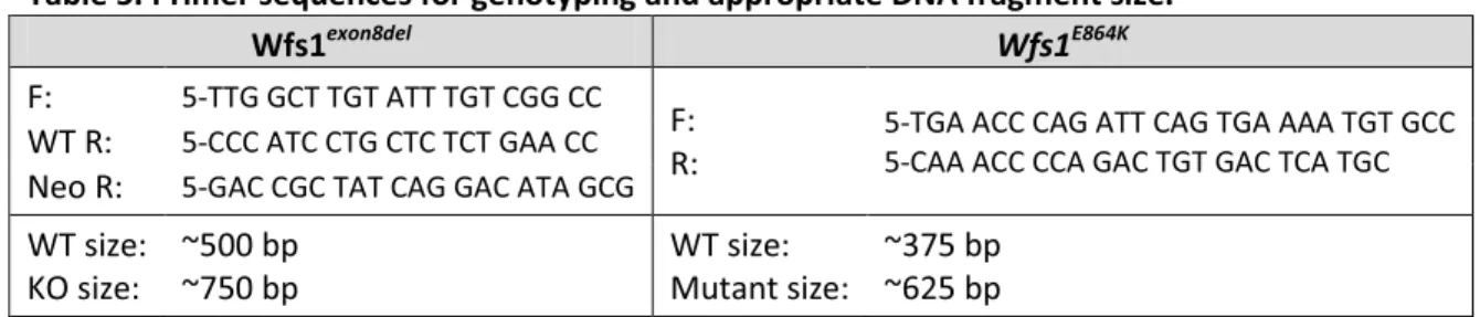
6.1.1. Genotyping ... 61
6.2. Intraocular Pressure ... 62
6.3. Blood Glucose ... 62
6.4. Visual acuity and contrast sensitivity ... 63
6.5. Electrophysiology ... 65
6.6. Eye fundus ... 68
6.7. Optical Coherence Tomography ... 69
6.8. Transmission Electron Microscopy ... 70
6.9. Immunohistochemistry ... 71
6.10. Q-PCR assay ... 72
6.11. Gene Therapy ... 73
6.12. Statistical analysis ... 74
7. RESULTS ... 75
7.1. Part I: Phenotyping Wfs1exon8del line ... 75
7.1.1. Normal IOP ... 75
7.1.2. Hyperglycemia ... 76
7.1.3. Progressive loss of visual acuity and contrast sensitivity ... 77
7.1.4. Changes in RGC function... 81
7.1.5. Changes in photoreceptor, ON-bipolar and Müller cell function ... 83
7.1.6. Somewhat slower conductivity towards the visual cortex ... 86
7.1.7. Optic disc pallor ... 87
7.1.8. Thinning of RNFL/GCL and GC complex layers ... 89
7.1.9. Severe axonal damage ... 90
7.1.10. No RGC loss ... 92
7.1.11. No ER stress in the retina ... 93
7.2. Part II: Phenotyping Wfs1E864K ... 95
5
7.2.1. Abnormal behavior and hyperglycemia ... 95
7.2.2. Early loss of RGC function ... 96
7.2.3. Changes in photoreceptor, ON-bipolar and Müller cell function ... 98
7.2.4. Damaged optic disc ... 100
7.2.5. GC complex layer thinning ... 102
7.2.6. No RGC loss ... 103
7.3. Part III: Gene therapy against Wolfram Syndrome ... 105
7.3.1. WFS1 in mouse retina ... 105
7.3.2. Rescue of the visual acuity ... 107
7.3.3. Rescue of the optic disc pallor ... 110
7.3.4. Rescue of the optic nerve damage ... 112
8. DISCUSSION... 117
8.1. Wfs1KO mice have a strong Wolfram Syndrome ophthalmic phenotype ... 117
8.2. Wfs1E864K mice are an alternative Wolfram Syndrome model ... 131
8.3. Gene Therapy partially rescues Wolfram Syndrome ophthalmic phenotype ... 135
9. FINAL CONCLUSIONS AND PERSPECTIVES ... 140
10. REFERENCES ... 141
11. ANNEX I: Supplementary results for Wfs1exon8del ... 159
12. ANNEX II: Supplementary results for Wfs1E864K ... 161
13. ANNEX III: Supplementary results for gene therapy in Wfs1exon8del line ... 168
6
1.!ABBREVIATIONS
aa Aminoacids
AATF Apoptosis Antagonizing Transcription Factor AAV Adeno-Associated Virus
AdV Adenovirus
AdCy8 Adenylyl Cyclase 8
ATF6 Activating Transcription Factor-!" bGT Bilateral Gene Therapy
BIP Binding Immunoglobulin Protein C4b Complement Component 4b
CaM Calmodulin
cAMP Cyclic Adenosine Monophosphate CASP3 Caspase-3
CHM Choroideremia
CHOP C/EBP Homology Protein
Cip1 Increased Cyclin-Dependent Kinase Inhibitor Protein-1 CMV Cytomegalovirus
CNS Central Nervous System CS Contrast Sensitivity DOA Dominant Optic Atrophy
ds Double Strand
E Embryonic day
EIAV Equine Infectious Anemia Virus eIF Eukaryotic Initiation Factor ER Endoplasmic Reticulum ERAD ER-Associated Degradation ERG electroretinogram
ERSE ER Stress Response Element FBLN3 Fibulin-3
FIV Feline Immunodeficiency Virus
GA Golgi Apparatus
GCL Ganglion Cell Layer
GCSH Glycine Cleavage System Protein H
GFP Green Fluorescent Protein; or: Eyes Injected with AAV-2/2-CMV-GFP
GH Growth Hormone
GT Gene Therapy
HSPG Heparin Surface Proteoglycan HSV Herpes Simplex Virus
IC Index of Circularity
IGF1 Insulin-Like Growth Factor-1 IOP Intraocular Pressure
7 IP Intraperitoneal
IP3 Inositol Triphosphate
IPL Inner Plexiform Layer IRE1 Inositol Requiring Enzyme-1 ITR Inverted Terminal Repeats INL Inner Nuclear Layer KO Wfs1exon8del Knock-Out
LCA Leber Congenital Amaurosis LHON Leber Hereditary Optic Neuropathy
LV Lentivirus
m Months of age
MAM Mitochondria-Associated ER Membranes Nef3 Neurofilament-3
RNFL Nerve Fiber Layer NI Eyes Non-Injected Nrn1 Neuritin-1
OA Optic Atrophy
OCT Optical Coherence Tomography
OD Optic Disc
OMR Optomotor Reflex
ON Optic Nerve
OPN Optic Neuropathy
P Post-natal day
PARP Poli-ADP-Ribose Polymerase PBS Eyes injected with PBS PC-1 Plasma Cell Glycoprotein-1 PC2 Prohormone Convertase-2 PEI Polyethylenimine
PERK PKR-Like ER Kinase PFA Paraformaldehyde
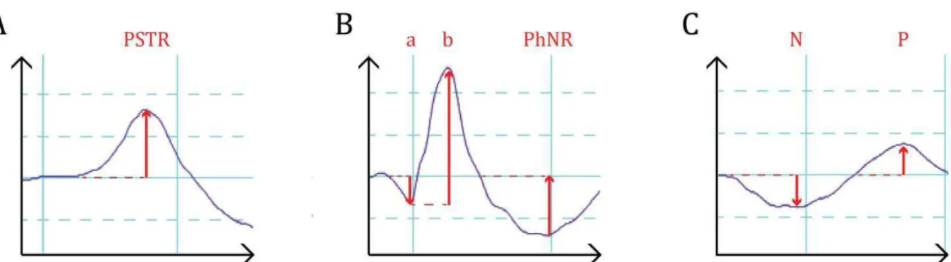
PhNR Photopic Negative Response
PhNRN Photopic Negative Response Normalized To B-Wave
PR Photoreceptor
PSTR Positive Scotopic Threshold Response qPCR Quantitative PCR
RGC Retinal Ganglion Cell RGS4 Regulator of G Protein-4 RNFL Retinal Nerve Fiber Layer RP Retinitis Pirmentosa RPE Retinal Pigment Epithelium
8 sc Self-complementary
SEM Standard Error of the Mean SERT Serotonin Transporter shRNA Short-hairpin RNA siRNA Small Interfering RNA
Smurf HECT-Type Ubiquitin Ligase (E3) Smad Ubiquitination Regulatory Factor 1
ss Single Strand
T1D Type 1 Diabetes Mellitus T2D Type 2 Diabetes Mellitus
TEM Transmission Electron Microscopy Thy1 Thymocyte Antigen-1, CD90 uGT Unilateral gene therapy UPR Unfolded Protein Response UTR Untranslated Regions
V Volt
VEP Visual Evoked Potentials
VG Vector Genomes
VPA Valproic Acid
WFS1 Wolfram Syndrome Protein-1, Wolframin; or: eyes injected with AAV-2/2-CMV-WFS1, depending on the context
WS Wolfram Syndrome
WT Wfs1exon8del wild-type
XBP1 X-Box Binding Protein
9
2.!ABSTRACT
Wolfram Syndrome (WS; OMIM #222300, prevalence 1-9 / 1 000 000) in its classical form has a juvenile onset and incorporates diabetes insipidus, diabetes mellitus, optic atrophy (OA), and deafness; leading to death in middle age. OA is its first neurological symptom, starting in adoles-cence and ending with blindness 8 years later. Unfortunately, a suitable WS mouse model compris-ing ophthalmic symptoms has not yet been found, therefore the search for its treatment is de-layed. In this thesis, I studied visual impairment in two WS mouse models along with a success of a gene therapy (GT) approach with the human WFS1 gene.
Firstly, 3 and 6 months old Wfs1exon8del mice were examined for the visual acuity (VA) and
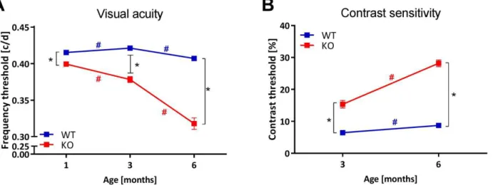
contrast sensitivity (CS) via changes in the optomotor reflex (OMR), the neural retinal function via electroretinogram (ERG), as well as the eye physiology via fundoscopy and optical coherence to-mography (OCT). Also, the proportion of retinal ganglion cells (RGC) and the axonal loss at the age of 7 months were determined with anti-Brn3a immuno-labeling of retinal sections and electron microscopy of optic nerve (ON) sections, respectively. There was a progressive loss of VA and CS in Wfs1exon8del-/- mice, starting already at 1 month of age. It was accompanied by optic disc pallor,
reti-nal thinning as well as axoreti-nal damage. However, there was no RGC loss and the endoplasmic retic-ulum (ER) stress in the retina was at a normal level. It suggested a presence of another cause for the reported degeneration in KO mice; in opposition to what was proposed in the literature. I brief, KO mice exhibit significant WS ophthalmic phenotype.
Secondly, in search for another model, visual functions of Wfs1E864K mouse line were
inves-tigated. This line was originally a model of Wolfram-like Syndrome, characterized by dominant mu-tations in WFS1 leading to congenital progressive hearing impairment, diabetes mellitus and OA. Only homozygous mutants, however, showed expected visual impairment. Already at 1 month of age, Wfs1E864K mice had drastic loss of RGC function, albeit keeping the cell number at a normal
level. This was accompanied by retinal thinning and a severe ON damage, as shown with OCT and fundoscopy, respectively. In contrast, the RGC function in Wfs1E864K/+ mice dropped slightly only at
10
mice is different than in humans. Therefore, Wfs1E864K mice, with their strong ophthalmic
pheno-type, could potentially serve as a model of the classical WS.
Finally, to investigate future treatment options, 1 month old Wfs1exon8del+/+ (WT) and
Wfs1exon8del-/- (KO) mice underwent a uni- and bilateral intravitreal gene therapy (GT) with
AAV-2/2-CMV-WFS1. Exams at 3 and 6 months of age showed improved VA, as well as optic pallor and ax-onal damage rescue in KO mice. Also, no adverse effects related to either GT or sham injections were noted. Following this idea, the Wfs1E864K mice were also subjected to intravitreal GT,
deliv-ered at P14, but without success.
In conclusion, Wfs1exon8del mouse line is a reliable model of WS, including the visual aspects.
I propose the Wfs1E864K
model as an alternative, especially to investigate Wfs1 function in the eye. Finally, the intravitreal AAV-driven GT with WFS1 has a potential to partially rescue the ophthalmic phenotype, paving the wave towards the treatment for WS patients.
11
3.!SUMMARY IN FRENCH
Le Syndrome de Wolfram (OMIM #222300) est une maladie neurodégénérative qui, dans sa forme classique se présente avec un début juvénile, intégrant le diabète insipide, diabète sucré, l’atrophie optique, et la surdité (Table S1). Il entraîne la mort vers 40 ans en raison de l'atrophie du tronc cérébral. L’atrophie optique est généralement son premier symptôme neurologique, com-mençant à l’âge de 11 ans et se terminant par la cécité 8 ans plus tard. Ceci est le symptôme le plus effrayant pour les patients. Malheureusement, un modèle murin du Syndrome de Wolfram appro-prié aux symptômes ophtalmologiques n'a pas encore été trouvé, donc la recherche d’une thérapie pour préserver la vision en est à ces débuts. Dans cette thèse j’ai étudié l’atteinte visuelle de deux modèles de souris mutantes pour le Syndrome de Wolfram et le succès d’une approche de théra-pie génique avec le gène humain WFS1.
Table S1. Les symptômes du Syndrome de Wolfram. Modifié de Urano 2016 (Urano, 2016) avec la
bibliographie citée dans le texte; a – ans d’âge, AV – acuité visuelle, CFN – couche des fibres ner-veuses, CGR – cellules ganglionnaires de la rétine, DO – disque optique, DT1 – diabète sucré de type 1, EPR – épithélium pigmentaire rétinien, m – mois d’âge, s – semaines d’âge.
Symptômes
typiques Détails Début
Diabète insipide Partiel central (51-87%) 14 a (3 m – 40 a) Diabète sucré Perte #$%%&%$'(')'besoins en insuline quotidienne
infé-rieure à DT1 6 a (3 s – 16 a)
Atrophie optique Bilatéral. L’AV, vision des couleurs et champs visuels diminués; pâleur du DO, grand DO, amincissement de la CFN, perte des CGRs, défauts pupillaires affé-rences, strabisme, nystagmus, cataracte (29,6 à 66,6%), rétinopathie pigmentaire (30%), rétinopathie diabétique (7,6 à 34,6%)
11 a (6 s – 19 a), cata-ractes parfois plus tôt; cécité légale dans les 8 a après le diagnostic ini-tial
Surdité Perte auditive neurosensorielle des hautes fré-quences, progression lente (62%)
65% des patients, appa-rition de l'enfance à l'adolescence Ataxie La plupart des symptômes neurologiques communs:
ataxie du tronc
60% des patients, appa-rition chez jeunes adultes
12 Complications
des voies uri-naires
Vessie neurogène, incontinence urinaire, infections des voies urinaires
60 – 90% des patients
Symptômes
communs Détails
Généraux Fatigue, hypersomnie
Neurologiques Apnée (cause de mortalité), dysphagie, maux de tête, odorat et goût altérés Psychiatriques Anxiété, attaques de panique, dépression, sautes d'humeur
Dysfonction autonomiques
Régulation de la température affaiblie, vertiges en position debout, constipa-tion, diarrhée, transpiration excessive
Endocrines Hypogonadisme, hyponatrémie
Le Syndrome de Wolfram est une maladie récessive autosomique causée par des mutations dans le gène WFS1. Pour la première fois, il a été rapporté en 1938 par les Professeurs Wolfram et Wagner chez 4 patients atteints de diabète sucré et d’atrophie optique (Wolfram and Wagner, 1938). Aujourd'hui, il est considéré comme affectant 1 personne sur 160 000 et 770 000 dans le monde entier (Barrett et al., 1995, 1997; Urano, 2016), ce qui signifie entre 10 et 46 milles patients en 2016. Le diabète sucré est le plus souvent le premier symptôme de la maladie, commençant déjà aux alentours de 6 ans, en moyenne (Barrett et al., 1995). Il est associé à la perte de cellules (' dans le pancréas, problèmes avec la conversion de la pro-insuline à l’insuline et sa sécrétion, ainsi que l’intolérance au glucose (Barrett et al., 1995; Hatanaka et al., 2011; Hilson et al., 2009; Ro-hayem et al., 2011; Urano, 2016; Viswanathan et al., 2008). Le deuxième symptôme majeur est l’ atrophie optique, ce qui affecte grandement leur qualité de vie. Autour de 11 ans, les patients dé-veloppent une gêne visuelle, c’est-à-dire une perte de la perception des couleurs et la vision péri-phérique (Barrett et al., 1995; Hoekel et al., 2014). Ceci est accompagné par une diminution de l’acuité visuelle et des champs visuels, une pâleur du disque optique, l’élargissement du disque optique, un amincissement de la couche des fibres nerveuses (CFN) ainsi que les couches plexiformes, une perte dramatique des cellules ganglionnaires de la rétine (CGR) avec leurs axones, un déficit pupillaire afférent, un strabisme, un nystagmus, et également une cataracte (Bababeygy et al., 2012; Grenier et al., 2016; Hilson et al., 2009; Hoekel et al., 2014; Zmyslowska et al., 2015). Dernièrement, l’amincissement de la rétine est devenu un indicateur fiable de la progression du
13
Syndrome de Wolfram (Hoekel et al., 2014; Zmyslowska et al., 2015). Les anomalies affectent en particulier les fibres visuelles (Barrett et al., 1997; Hershey et al., 2012), mais les CGRs et l’épithélium pigmentaire rétinien (EPR) sont également fortement endommagés (Al-Till et al., 2002; Hilson et al., 2009; Schmidt-Kastner et al., 2009; Yamamoto et al., 2006). Ils affichent une perte de la myélinisation et la mort cellulaire, ce qui pourrait être lié au stress du réticulum endoplasmique (RE), l’excès du glucose (Rohayem et al., 2011), ou la perturbation de l’homéostasie du Ca2+. Certainement, la conductivité du nerf optique est également altérée (Ito et al., 2007). De plus, le développement des principaux symptômes, comme les diabètes, l’atrophie optique avec la sur-dité, est variable et souvent accompagné par des complications des voies urinaires, ainsi qu’une ataxie, des anomalies cérébrales et des troubles psychologiques (Aloi et al., 2012; Barrett et al., 1995; Hershey et al., 2012; Kinsley et al., 1995; Marshall et al., 2013; Takeda et al., 2001; Zmyslowska et al., 2014). D’autres aspects de la vie humaine sont également touchés par le Syn-drome de Wolfram. Par exemple, les lymphocytes provenant de patients ont un stress du RE élevé (Fonseca et al., 2010), ce qui peut potentiellement réduire l'immunité. De plus, certains patients sont affectés par un hypogonadisme, conjointement avec l'infertilité, un dysfonctionnement érec-tile et menstruel peut être associé (Medlej et al., 2004; Noormets et al., 2009; Urano, 2016).
Jusqu’à présent, seul l’Idebenone, un médicament contre des troubles cognitifs, a été propo-sé comme traitement ophtalmique pour le Syndrome de Wolfram. Ce produit chimique est simi-laire au coenzyme Q10, un transporteur d'électrons dans les mitochondries, et a été testé unique-ment chez un seul patient du Syndrome de Wolfram (Bababeygy et al., 2012). En 6 mois, l’Idebenone a amélioré la vision mais la tomographie par cohérence optique et l’ophtalmoscopie restent inchangées. La re-myélinisation axonale ainsi qu’une amélioration significative des fonc-tions visuelles sont attendues plus long terme, mais cela nécessite des études supplémentaires. En parallèle, il a été montré que l'un des médicaments contre les atteintes bipolaires, l'acide val-proïque (AVP), influe sur les cellules par la régulation de WFS1 (Kakiuchi et al., 2009), restaurant la tolérance au glucose (Terasmaa et al., 2011). C’est pourquoi, l’AVP a été proposé comme un médi-cament contre la neurodégénérescence liée au Syndrome de Wolfram et est protégé par un brevet (Appl. No. PCT/GB2013/052523). Des essais cliniques sont en cours, au Royaume-Uni. Une autre option de traitement serait le Dantrolène (Dantrolen). A l'origine un myorelaxant en hypothermie
14
maligne, le Dantrolen combat l’augmentation du Ca2+ cytosolique caractéristique au Syndrome de Wolfram ; et par conséquent sauve les cellules de l’apoptose (Lu et al., 2014). L’essai clinique de phase I/II a été enregistré en Juillet 2016 et recrute actuellement des participants aux États-Unis d'Amérique (identificateur sur clinicaltrials.gov : NCT02829268). Cependant à ce jour, aucun de ces traitements n’a prouvé une efficacité sur l’atrophie optique, d’où la demande restante pour la thérapie génique.
Des mutations de WFS1 sont responsables non seulement du Syndrome de Wolfram à trans-mission autosomique récessif, mais il existe aussi des mutations à transtrans-mission autosomiques do-minantes. Certaines d'entre elles sont responsables de « Wolfram-like Syndrome » (OMIM #614296) et d’autres de surdités comme DFNA6/14/38 (OMIM #600965). Le Syndrome Wolfram-like est caractérisé par une perte progressive de l’audition, une atrophie optique et/ou une diminu-tion de la réguladiminu-tion du glucose (Eiberg et al., 2006; Fujikawa et al., 2010; Gürtler et al., 2005; Hogewind et al., 2010; Rendtorff et al., 2011), tandis que DFNA6 est caractérisé par une perte audi-tive non-syndromique au niveau des basses fréquences (Bai et al., 2014; Bramhall et al., 2008; Chaussenot et al., 2015; Gonçalves et al., 2014; Kunz et al., 2003; Lesperance et al., 2003; Noguchi et al., 2005; Sun et al., 2011; Young et al., 2001). De plus, des mutations de WFS1 sont également responsables à l’état autosomique dominant de rares cas de diabète non-syndromique (Bon-nycastle et al., 2013; Zalloua et al., 2008), de diabète associé à une surdité congénitale (Valéro et al., 2008), ainsi que de cataracte congénitale (Berry et al., 2013). Enfin, tel que publié par Grenier et al. (Grenier et al., 2016), certains patients avec une atrophie optique non-syndromique isolée ont des mutations autosomiques récessives dans WFS1, comme les patients du Syndrome de Wol-fram. En conclusion, des mutations dans WFS1, récessives ou dominantes, conduisent toujours à un dysfonctionnement neuronal et / ou endocrinien.
Le gène WFS1 réside dans le chromosome 4p16.1 et code la protéine WFS1, également ap-pelée la wolframine (Collier et al., 1996; Inoue et al., 1998). WFS1 est une protéine localisée dans la membrane du RE (Hofmann et al., 2003; Inoue et al., 1998; Rigoli et al., 2011). Les 250 kb de WFS1 (Inoue et al., 1998; Rigoli et al., 2011) génèrent 8 exons, avec l’exon-1 non-codant et l’exon-8 con-tenant l’information pour 2/3 de la protéine (Hofmann et al., 2003). Elle se compose de 890 acides
15
aminés avec une masse moléculaire de 100 kDa (Cryns et al., 2003; Hofmann et al., 2003; Takeda et al., 2001), étant généralement hydrophobe, mais avec les extrémités hydrophiles (Takeda et al., 2001). Par conséquent, WFS1 crée 3 domaines structuraux liés à ces fonctions : le N-terminale drophile dans le cytoplasme, un corps hydrophobe dans la membrane du RE, et le C-terminale hy-drophile dans le lumen du RE (Inoue et al., 1998). Il y réside comme un oligomère qui est capable de former des canaux (Hofmann et al., 2003) et interagit avec d’autres protéines. WFS1 est une protéine multifonctionnelle, jouant un rôle dans la réponse au stress, la survie cellulaire, la prolifé-ration et le cycle cellulaire, dans la biosynthèse, la sécrétion et la dégradation de protéines, dans le développement et, le plus important, dans l’homéostasie du Ca2+. Par conséquent, le système vi-suel est fragilisé en cas de carence de WFS1.
La thérapie génique est une approche novatrice et prometteuse dans la médecine moderne, découverte par le professeur Waclaw Szybalski et son équipe en 1962 (Szybalska and Szybalski, 1962; Szybalski, 2013). Depuis les premiers essais cliniques lancés en 1990 par Anderson et Blaese (Anderson et al., 1990), il a suscité un grand intérêt parmi les scientifiques et les cliniciens. En Fé-vrier 2016, il y avait 2356 essais cliniques dans le monde entier impliquant la thérapie génique (John Wiley & Sons, Ltd., 2016). Sa popularité provient du fait de traiter la cause de la maladie au lieu de simplement les symptômes, et une possibilité de personnalisation. Le transfert peut être stable ou transitoire, en fonction de la méthode utilisée, mais porte toujours un risque d'effets secondaires. Ces effets secondaires sont liés à sa nature instable et éphémère, ce qui conduit sou-vent à la ré-administration de médicaments. De plus, il existe un risque de réponse immunitaire, de toxicité génétique, l’expression non-contrôlée, la non-spécificité et la tumorigenèse. Malgré tout, le nombre d'essais cliniques impliquant la thérapie génique augmente chaque année.
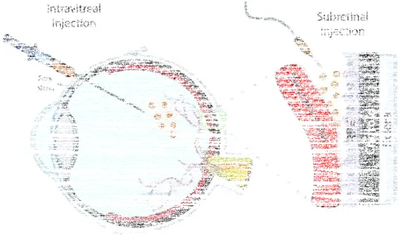
L'œil comme une extension du système nerveux central est un candidat idéal pour la thérapie génique (Sahel and Roska, 2013; Trapani et al., 2014; Yu-Wai-Man, 2016). Il est relativement petit et isolé des autres tissus, ce qui permet de ciblage local spécifique avec une faible quantité de mé-dicament. De plus, les cellules de la rétine sont assez différenciées et par conséquent, il existe un considérable potentiel d'une expression génique soutenue ; en raison de l'absence de mitose. Par ailleurs, grâce à la structure étanche de l'EPR, la barrière hémato-rétinienne et
l'immunosuppres-16
sion locale naturelle, le risque de réponse immunitaire est minime (Caspi, 2010; Willett and Ben-nett, 2013). Enfin, l'œil est exposé et transparent. Cela rend la transduction de gènes ainsi que le suivi des effets bénéfiques beaucoup plus facile, en utilisant des méthodes non-invasives comme l’ERG, fondoscopie et la tomographie par cohérence optique. Il existe deux méthodes principales pour la thérapie génique intraoculaire : les injections intravitréennes et sous-rétiniennes (Liang et al., 2001). Le premier signifie l’injection du médicament dans le corps vitré et la transduction de la rétine intérieure, donc essentiellement les CGRs ainsi que les cellules de Müller (Ali et al., 1996, 1998). Le second signifie l'injection sous l’EPR.
Les vecteurs les plus populaires contre les maladies ophtalmiques sont basés sur des virus adéno-associés (AAV), la «norme d'or», mais aussi des lentivirus (LV) et adénovirus (AdV) (Kumar-Singh, 2008; Lipinski et al., 2013). Déjà en 1996, il a été montré que le vecteur AAV peut efficace-ment transduire les CGRs dans la rétine de la souris, après l'injection intravitréenne (Ali et al., 1996, 1998). Le sérotype AAV2/2 persiste à être choisi le plus souvent dans les traitements actuels contre la dégénérescence des CGRs. Il semble efficace dans les essais cliniques contre l’Amaurose Congé-nitale de Leber, la Neuropathie Optique Héréditaire de Leber, ainsi que la rétinite pigmentaire (Hellström and Harvey, 2011; Yu-Wai-Man, 2016), qui également indique son potentiel dans le Syndrome de Wolfram.
Les souris Wfs1exon8del avec suppression de l’exon-8 du gène Wfs1 sont récemment devenues
le modèle de choix de la maladie. En cohérence avec les symptômes du Syndrome de Wolfram si-gnalés (Swift and Swift, 2005; Swift et al., 1990, 1991), elles développent le diabète (Hatanaka et al., 2011; Ishihara et al., 2004), ainsi que l’anxiété (Luuk et al., 2009; Raud et al., 2009, 2015; Sütt et al., 2015) et la dépression (Visnapuu et al., 2013a). En outre, elles ont un phénotype métabo-lique fort caractérisé par plusieurs dysfonctionnements physiologiques, tels que la consommation de nourriture et d'eau, la température corporelle, la consommation d’O2, ainsi que l’effluence du
CO2 et la chaleur (Ehrlich et al., 2016; Noormets et al., 2014). Leur durée de vie est fortement
rac-courcie aussi, ce qui correspond entièrement au dysfonctionnement autonome observé chez les patients (Urano, 2016). Les souris ont également manifesté des fonctions neuroendocrines pertur-bées (Ivask et al., 2016; Luuk et al., 2009) avec une infertilité partielle (Noormets et al., 2009).
17
Néanmoins, leur fonction visuelle n’a pas été encore étudiée. Par conséquent, je me suis concen-trée sur son évaluation par des études ante et post mortem, aussi bien que sur la recherche d'un traitement contre la perte de vision.
Les expériences avec la lignée Wfs1exon8del ont duré jusqu’à 7 mois en raison du mal-être des
souris Wfs1exon8de-/- (KO) ainsi que le développement de l’exophtalmie à cet âge (Table S2). La
pres-sion intraoculaire était normale. La présence de l'hyperglycémie a été confirmée, comme chez les patients atteints de Syndrome de Wolfram, mais n'atteint pas le niveau diabétique. Ensuite, à 1, 3 et 6 mois les souris ont été examinées pour l’acuité visuelle et la sensibilité aux contrastes via des changements dans le reflexe optomoteur (ROM). Elles ont montré une baisse dans les deux aspects commençant à l’âge de 1 mois, et qui progresse avec l’âge. Leur fonction rétinienne a été exami-née par électrorétinogramme (ERG), qui n’a pas montré de disfonctionnement des CGRs, mais plu-tôt dans les cônes. En parallèle, il y avait une pâleur du disque optique, visible déjà à 3 mois, ac-compagné par l’amincissement de la rétine comprenant la couche des CGRs, comme chez les pa-tients. Au contraire, l’immunohistologie et l’histochimie sur des coupes de rétine de 7 mois n’ont montré aucune perte cellulaire. Cependant, les axones ont été fortement endommagés, comme illustré par microscopie électronique. De plus, le stress du RE dans la rétine était à un niveau nor-mal, contrairement à ce qui a été proposé dans les publications. Sur ce modèle, notre équipe a aussi trouvé une perte d’audition (résultats non présentés), caractéristique pour le Syndrome de Wolfram. Brièvement, les souris KO présentent un phénotype ophtalmique du Syndrome de Wol-fram significatif et peuvent servir comme modèle.
A la recherche d'un autre modèle du Syndrome de Wolfram, les fonctions visuelles de la li-gnée Wfs1E864K de la souris ont été étudiées (Table S2). Cette ligne était à l'origine un modèle du
« Wolfram-like Syndrome » entraîné par la mutation faux-sens E864K dans l’exon-*'+#,-./012345' au caractère autosomique dominant. Cette mutation a été décrite pour la première fois en 2006 (Eiberg et al., 2006) et provoque une perte auditive neurosensorielle de basse fréquence, une atrophie optique et un diabète sucré. La surdité a une apparition juvénile mais l’atrophie optique varie. Certains de ces patients développent des complications psychiatriques aussi (Eiberg et al., 2006; Fukuoka et al., 2007; Rigoli and Di Bella, 2012; Valéro et al., 2008). En voyant que les souris
18
Wfs1E864K/+ ne présentaient pas de symptômes, il a été décidé d’élever les souris Wfs1E864K, donc
homozygotes bi-alleliques pour la mutation. Un tel cas n'existe pas chez l'homme. Car il n'y a pas de copie de Wfs1 fonctionnelle, il a donc été proposé de les utiliser comme un autre modèle du Syndrome de Wolfram au lieu de la maladie d'origine. Leurs examens ont été effectués seulement jusqu'à 3 mois en raison du mal-être général ainsi que les blessures liées à leur comportement an-xieux. Il convient de noter de nombreux problèmes avec la reproduction des souris Wfs1E864K, semblable à l'autre lignée. La descendance était peu viable en raison d'avortements spontanés des femelles Wfs1E864K. C’est pourquoi, la reproduction a été ajustée de manière
appro-priée, puisque les meilleurs résultats ont été obtenus en croisant deux femelles Wfs1E864K/+ avec un
mâle Wfs1E864K. Contrairement à la lignée Wfs1exon8del, la fertilité masculine Wfs1E864K semblait
in-tacte. Déjà à 1 mois, les souris Wfs1E864K avait une perte drastique de la fonction des CGRs, mais en
gardant le nombre de cellules à un niveau normal. Ceci a été accompagné par un amincissement de la rétine et d’un sévère dommage du nerf optique, comme montré par la tomographie par cohé-rence optique et le fond d’œil, respectivement. En revanche, la fonction des CGRs chez souris Wfs1E864K/+ est légèrement diminuée à l'âge de 7 et 12 mois, ce qui indique une pathologie diffé-rente entre la souris et l’homme liée à cette mutation. En conséquence, les souris Wfs1E864K, avec
leur fort phénotype ophtalmique, pourraient servir de modèle du Syndrome de Wolfram classique. Enfin, pour enquêter sur les futures options de traitement contre le Syndrome de Wolfram, les souris sauvage (WT) et KO de la lignée Wfs1exon8del à 1 mois ont subi une thérapie génique
in-travitréenne uni- ou bilatérale avec 3 · 108 VG du AAV-2/2-CMV-WFS1 (Table S2). Les examens à 3 et 6 mois ont montré une amélioration de l’acuité visuelle, ainsi que de la pâleur papillaire et la réduction des lésions axonales chez la souris KO. En outre, aucun effet indésirable lié à des injec-tions de thérapie génique n’a été noté. Suivant cette idée, les souris Wfs1E864K ont également été
soumis à la thérapie génique intravitréenne, délivrée à P14, mais sans succès.
Pour la première fois dans les publications, je signale la découverte d'un modèle du Syn-drome de Wolfram de la souris avec un phénotype ophtalmique approprié. Les changements fonc-tionnels et physiologiques chez les souris KO avec l’exon-8 supprimé du gène Wfs1 montrent une perte progressive de l’acuité visuelle, une pâleur du disque optique, et l'amincissement de la rétine
19
liée à une dégénérescence axonale, typique pour le Syndrome de Wolfram. En outre, puisque la manifestation de la maladie chez les souris Wfs1E864K a un début précoce et est accompagnée d'un
fort phénotype ophtalmique, je propose de l'utiliser comme un autre modèle. Enfin et surtout, la thérapie génique testée contre le Syndrome de Wolfram a prouvé son efficacité sans effets secon-daires. Cette approche thérapeutique devrait être explorée plus loin avec les deux modèles de sou-ris, en utilisant des approches adaptées et un titrage élevé du vecteur. Les résultats préliminaires prometteurs de la thérapie génique ouvrent la voie vers un traitement futur contre la cécité chez les patients atteints de Syndrome de Wolfram.
Table S2. Les résultats du phénotypage et de la thérapie génique. AV – acuité visuelle, AO -
atro-phie optique, CFN/CG – couche des fibres nerveuses et cellules ganglionnaires, CGR – cellules gan-glionnaires de la rétine, DO – disque optique, ERG – électrorétinogramme, m – mois d’âge, NO – nerf optique, PEV - potentiel évoqué visuel, RE – réticulum endoplasmique, ROM – reflexe opto-moteur, TCO – tomographie par cohérence optique.
LE PHENOTYPAGE
La souris L’examen Résultats Signification
Wfs1exon8del KO
La pression intraoculaire Normale Une autre origine d’exophtalmie La glycémie périphérique Hyperglycémie Traitement du
glu-cose altéré L’AV et la sensibilité au
contraste ; ROM
Diminution progressive de l’AV et de la sensibilité au contraste
Indication de l’AO L’électrophysiologie ; ERG
et PEV
Seule la réponse des cônes est perturbée ?
Pas de changement fonctionnel des CGRs ?
La fondoscopie La pâleur du DO Indication de l’AO
La TCO Amincissement de la CFN/CG et
de la couche du complexe des CGRs
Indication de l’AO
La microscopie électro-nique du NO
Les lésions et changement de forme axonale
Indication de l’AO L’histologie de la rétine Normale Pas de perte des
20 L’expression des gènes
dans la rétine
Normale Pas de stress du RE
Wfs1E864K
La glycémie périphérique Hyperglycémie Traitement du glu-cose altéré
L’électrophysiologie ; ERG et PEV
Perte précoce drastique de la réponse des CGRs
Indication de l’AO La fondoscopie Endommagement du DO Indication de l’AO
La TCO Amincissement de la CFN/CG et
de la couche du complexe des CGRs
Indication de l’AO
L’histologie de la rétine Normale Pas de perte des CGRs
LA THERAPIE GENIQUE AVEC AAV-CMV-WFS1
La souris L’examen Résultats Signification
Wfs1exon8del KO
L’AV et la sensibilité au contraste ; ROM
Evolution plus lente de la dimi-nution de l’AV et de la sensibili-té au contraste
Sauvetage partiel de l’AO
L’électrophysiologie ; ERG Pas de changement -
La fondoscopie Pas de la pâleur du DO Sauvetage partiel de l’AO
La TCO Pas de changement -
La microscopie électro-nique du NO
Beaucoup moins des lésions et du changement de la forme axonale
Sauvetage partiel de l’AO
21
4.! INTRODUCTION
4.1.! Wolfram Syndrome
Wolfram Syndrome (WS; Online Mendelian Inheritance in Man [OMIM] #222300) is an auto-somal recessive disorder caused by mutations in WFS1 gene. It was first reported in 1938 by Wolf-ram and Wagner in 4 patients with diabetes mellitus and optic atrophy (OA) (WolfWolf-ram and Wagner, 1938). Nowadays, it is considered to affect 1 in between 160 000 and 770 000 people worldwide (Barrett et al., 1995, 1997; Urano, 2016), which means 10 - 46 thousand WS patients in 2016. The syndrome is most commonly described as DIDMOAD, which stands for diabetes insipidus, diabetes mellitus, (bilateral) OA and deafness. However, development of these symptoms is variable and often accompanied by urinary tract complications as well as psychological disorders (Aloi et al., 2012; Barrett et al., 1995; Hershey et al., 2012; Kinsley et al., 1995; Marshall et al., 2013). Other common WS symptoms are listed in Table 1. At the age of 25-49 years, the patients pass away prematurely, most commonly due to brain stem atrophy (median: 30) (Barrett and Bundey, 1997; Barrett et al., 1995). So far, the disease is incurable.
Table 1. Symptoms of Wolfram Syndrome. Modified from Urano 2016 (Urano, 2016) with bibliography cited in the text; m – months of age, OD – optic disc, RGC – retinal ganglion cells, RNFL – retinal nerve fiber layer, RPE – retinal pigment epithelium, T1D – type 1 diabetes mellitus, VA – visual acuity, w – weeks of age, y – years of age.
Typical
symptoms Details Onset
Diabetes
Insipidus Partial central (51-87%) 14 y (3 m – 40 y)
Diabetes
Mellitus (-cell loss; lower daily insulin requirement than T1D 6 y (3 w – 16 y) Optic Atrophy Bilateral. Diminished VA, color vision, visual fields; OD
pallor, large OD, RNFL thinning, RGC loss, afferent pupil-lary defects, strabismus, nystagmus, cataracts (29.6-66.6%), pigmentary retinopathy (30%), diabetic retinopa-thy (7.6-34.6%)
11 y (6 w – 19 y), cataracts sometimes earlier; legal blindness within 8 y after the initial diagnosis
Deafness Sensorineural high frequency hearing loss, slowly pro-gressing (62%)
65% of patients, onset from infancy to adolescence Ataxia Most common neurological symptom: a. of the trunk 60% of patients, onset in
early adulthood Urinary Tract
Complications
Neurogenic bladder, bladder incontinence, urinary tract infections
22 Common
symptoms Details
General Fatigue, hypersomnia
Neurological Apnea (cause for mortality), dysphagia, headaches, impaired smell and taste Psychiatric Anxiety, panic attacks, depression, mood swings
Autonomic dysfunction
Impaired temperature regulation, dizziness when standing up, constipation, diarrhea, excessive sweating
Endocrine Hypogonadism, hyponatremia
4.1.1.!Clinical features: diabetes
Diabetes in WS patients is most commonly the first symptom of the disease, commencing at 6 years of age (y) (Barrett et al., 1995). This is sooner than the type 1 diabetes mellitus (T1D) and, furthermore, WS is less frequently correlated with ketoacidosis, like type 2 diabetes (T2D) is hayem et al., 2011). Simultaneously, hypoglycemia and neuropathies are accelerated in WS (Ro-hayem et al., 2011), what altogether makes its pathology more severe than T1D. On the other hand, the patients do not exhibit T2D-characteristic insulin resistance, but do lose a significant 678&9:'8;'(-cells (Barrett et al., 1995; Hilson et al., 2009; Rohayem et al., 2011; Urano, 2016; Vis-wanathan et al., 2008). Of note, diabetes starts earlier in boys, and a similar tendency was also found in murine models (Noormets et al., 2011). This phenomenon was attributed to problems with converting pro-insulin to insulin and its release from secretory granules, which appears more acute in males (Hatanaka et al., 2011; Ivask et al., 2016; Noormets et al., 2011). That places the WS <=6>$:$?'@=:A'=:?'#A6B6#:$B=?:=#'(-cell loss between T1D and T2D.
So what evokes diabetes in WS? Insulin-producing human and rodent cells deficient in WFS1 showed uncontrolled endoplasmic reticulum (ER) stress (Fonseca et al., 2005, 2010, 2012; Riggs et al., 2005; Shang et al., 2014; Yamada et al., 2006) and deformations in ER structure (Riggs et al., 2005), what tempered with its functions. Therefore, production and secretion of insulin were im-paired; and the damage in response to ER stress led to cell cycle arrest along with cell death (Fon-seca et al., 2012; Hatanaka et al., 2011; Ishihara et al., 2004; Marshall et al., 2013; Noormets et al., 2011; Riggs et al., 2005; Shang et al., 2014; Yamada et al., 2006). Those processes manifested as the aforementioned reduction in (-cell mass (Ivask et al., 2016; Riggs et al., 2005). Furthermore,
23
the observed DNA fragmentation seems to also be a result of high glucose exposure (Ishihara et al., 2004), and could be diminished through ER stress reduction (Terasmaa et al., 2011). In the same time5'8:A$B'<C?;&9#:=896%'(-cells, insulinoma cells, expressed high levels of WFS1 (Hofmann et al., 2003). They did so probably to soothe the damages caused by excessive glucose in the microenvi-ronment. To complement the story, a significant down-regulation of the plasma cell glycoprotein-1 (PC-1) gene was found in fibroblasts from WS patients (Philbrook et al., 2005). Normally, this gene is related to the type 2 diabetes through an inhibition of the insulin receptor’s activity. Here, for the first time, its underflow was shown to co-exist @=:A'(-cell loss. Furthermore, an in vitro study with BRIN-DB11 cell line (McBain and Morgan, 2003) suggested that Wfs1 is necessary for normal (-cell proliferation. Hence a conclusion that in the absence o;'DEFG5'(-cell biology is abnormal, the cells are unable to proliferate and prone to stress-induced death.
4.1.2.!Clinical features: optic atrophy
Wolfram Syndrome involves series of neurological problems, together with hearing and sion loss. Around 11 y, patients develop visual discomfort, namely loss of color and peripheral vi-sion (Barrett et al., 1995; Hoekel et al., 2014), what greatly affects their life quality. This is accom-panied by decreased visual acuity (VA) and visual fields, presence of the optic disc (OD) pallor, OD enlargement, retinal nerve fiber layer (RNFL) and plexiform layers thinning, dramatic loss of RGCs with their axons, afferent pupillary defects, strabismus, nystagmus, and also cataracts (Bababeygy et al., 2012; Grenier et al., 2016; Hilson et al., 2009; Hoekel et al., 2014; Zmyslowska et al., 2015). Lately, the retinal thinning has become a reliable indicator for the disease progression (Hoekel et al., 2014; Zmyslowska et al., 2015). As such, the robust expression of WFS1 in the visual system along with its neural nature suggests blaming WFS1 absence-related stress for the progressing blindness. The reported abnormalities especially affect optic radiations (Barrett et al., 1997; Her-shey et al., 2012), but retinal ganglion cells (RGC) and retinal pigment epithelium (RPE) are also heavily damaged (Al-Till et al., 2002; Hilson et al., 2009; Schmidt-Kastner et al., 2009; Yamamoto et al., 2006). RGCs display loss of myelination and cell death, what could be related to ER stress, ex-cessive glucose (Rohayem et al., 2011), or distorted Ca2+ homeostasis. Certainly, optic nerve (ON) conductivity is compromised as well (Ito et al., 2007). However, more research is necessary to
de-24
termine the causality of OA in WS. Complementary information about the nature of optic neuropa-thy (OPN), present in WS, is included in ANNEX IV.
4.1.3.!Other clinical features
Regarding the brain, most adult patients suffer from ataxia and imbalance (Barrett et al., 1995; Hershey et al., 2012; Zmyslowska et al., 2014). This is associated with smaller intracranial volumes as well as anomalies in the brainstem, limbic system and cerebellum, even in the early stages of the disease (Hershey et al., 2012; Takeda et al., 2001). Furthermore, WFS1-deficient neu-ronal cells harbor endless ER stress-induced damage (Marshall et al., 2013; Schmidt-Kastner et al., 2009). In coherence, gene expression profiling in Wfs1exon8del KO mouse hypothalamus
demonstrat-ed an excess of the complement component 4b (C4b), what is associatdemonstrat-ed with neurodegenerative syndromes (Kõks et al., 2011). Also, a concurrent shortage in presence of the regulator of G pro-tein-4 (RGS4) was found, what is linked to schizophrenia and bipolar disease. Some WS patients do develop these psychiatric conditions, but their association with WFS1 is still to be explained (Koido et al., 2005; Swift and Swift, 2005; Swift et al., 1990, 1991; Xavier et al., 2016). According to the current state of knowledge, they may be driven by undermined serotonin and noradrenaline me-tabolism, combined with abolished serotonin transporter (SERT) levels (Visnapuu et al., 2013a), but also by faulty dopamine metabolism (Matto et al., 2011; Visnapuu et al., 2013b). Consistently with the reported WS symptoms (Swift and Swift, 2005; Swift et al., 1990, 1991), Wfs1exon8del KO mice
also displayed anxiety (Luuk et al., 2009; Raud et al., 2009, 2015; Sütt et al., 2015) and depression (Visnapuu et al., 2013a). However, circadian rhythms were unaffected (Luuk et al., 2012), in con-trast to Wfs1exon2del KO mice where expression of genes regulating these rhythms as well as late
stage of insulin secretion were altered (Nakabayashi et al., 2013). Taken together, it is safe to as-sume a strong correlation between the lack of WFS1 and neurodegeneration, along with its influ-ence on emotional behavior.
Other aspects of human life are equally affected by Wolfram Syndrome. For instance, patient-derived lymphocytes showed continuous ER stress (Fonseca et al., 2010), what may potentially lower the immunity. Also, some patients are affected with hypogonadism, together with infertility, erectile dysfunction, and defective menstruation (Medlej et al., 2004; Noormets et al., 2009;
Ura-25
no, 2016). This is consistent with studies on Wfs1exon8del KO mice, which showed abolished
sper-matogenic cell number, namely spermatogonia and Sertoli cells (Noormets et al., 2009). Their sperm was deformed, but they were not infertile. In addition, they displayed a strong metabolic phenotype characterized by several physiological dysfunctions, such as food and water intake, body temperature, O2 consumption, as well as CO2 and heat effluence (Ehrlich et al., 2016;
Noor-mets et al., 2014). Their lifespan was strongly abolished, too, what altogether matches the auto-nomic dysfunction observed in WS patients (Urano, 2016). Moreover, it was recently discovered that pro-hormone convertase-2 (PC2), native to neuroendocrine cells, is more active in Wfs1exon8del
KO mouse hippocampus (Tein et al., 2015). The main role of PC2 is to process neuropeptide and peptide hormone precursors (Brakch et al., 1995), what ties it to the reported altered peptide pro-cessing in WS patients (Gabreëls et al., 1998). Coherently, the Wfs1exon8del KO mice displayed
strong, abnormal corticosterone hormone release in stress response, coupled with general behav-ioral adaptation issues; indicating neuroendocrine imbalance (Luuk et al., 2009). Finally, pathways related to endocrine system development and function in these mice, together with tissue mor-phology and molecular transport, were induced as well (Ivask et al., 2016). All this sheds light on the origin of the human neuroendocrine symptoms, but needs further investigation.
4.1.4.!Treatment for Wolfram Syndrome
Although the symptoms can be managed to a certan degree with adaptive treatment, there is no cure for WS. Moreover, so far only Idebenone, a drug against cognitive disorders, was proposed as medication for OA in WS. This chemical, similar to coenzyme Q10, is a carrier in the mitochon-drial electron transportation and was tested only in one WS patient (Bababeygy et al., 2012). With-in 6 months, it improved vision along with unchanged OCT and ophthalmoscopy. Doctors hope for axonal re-myelination and significant improvement with longer time, but that requires further studies. There is a need for much larger number of patients as well as the support of proper basic research. In parallel, it was shown that one of medicines against bipolar disease, the valproic acid (VPA), influences cells through regulation of WFS1 (Kakiuchi et al., 2009), providing glucose toler-ance restoration (Terasmaa et al., 2011). Therefore, VPA was proposed as a medicine against WS-related neurodegeneration, was patent-protected (Appl. No. PCT/GB2013/052523), and is awaiting
26
clinical trials in the United Kingdom. Another treatment option would be dantrolene sodium (Dan-trolen). Originally a muscle-relaxant in malignant hypothermia, it fights the WS-characteristic in-creased cytosolic Ca2+, essentially blocking calpain hyper-activation; and therefore rescuing cells from apoptosis (Lu et al., 2014). The phase I/II clinical trial with Dantrolen was registered in July 2016 and is currently recruiting participants in the United States of America (clinicaltrials.gov iden-tifier: NCT02829268). To date however, neither of these treatments was proven successful in OA rescue, hence the remaining demand for gene therapy (GT).
4.1.5.!Final conclusions
As explained above, Wolfram Syndrome is a rare but complex, multidimensional systemic disorder. It has frightening ophthalmic, endocrinological and neurological aspects; in addition to the lack of cure. The complexity of the disease is strictly related to the function of the WFS1 pro-tein, which is still under investigation. Once this is established, there will be a better chance for developing cause-eliminating medical care.
27
4.2.! Wolfram Syndrome-related genetic disorders
Mutations in WFS1 are found not only in WS with its autosomal recessive inheritance, but al-so in a variety of autoal-somal dominant conditions. Some of these are classified under the name of Wolfram-like Syndrome (OMIM #614296) and others as DFNA6/14/38 (OMIM #600965). The for-mer is characterized by progressive hearing loss, OA and/or impaired glucose regulation (Eiberg et al., 2006; Fujikawa et al., 2010; Gürtler et al., 2005; Hogewind et al., 2010; Rendtorff et al., 2011), while the latter by non-syndromic low frequency hearing loss (Bai et al., 2014; Bramhall et al., 2008; Chaussenot et al., 2015; Gonçalves et al., 2014; Kunz et al., 2003; Lesperance et al., 2003; Noguchi et al., 2005; Sun et al., 2011; Young et al., 2001). An example of Wolfram-like Syndrome is a condition driven by the E864K missense mutation in exon-*'+#,-./01234,'H:'@6?';=B?:'B$I8B:$<'=9' 2006 (Eiberg et al., 2006) and provokes low-frequency sensorineural hearing loss, OA and diabetes. Deafness has a juvenile onset, but OA varies. Some of these patients develop psychiatric complica-tions as well (Eiberg et al., 2006; Fukuoka et al., 2007; Rigoli and Di Bella, 2012; Valéro et al., 2008). Furthermore, WFS1 mutations are also responsible for rare cases of non-syndromic autosomal dominant diabetes (Bonnycastle et al., 2013; Zalloua et al., 2008), autosomal dominant diabetes and congenital hearing loss (Valéro et al., 2008), as well as autosomal dominant congenital cataract (Berry et al., 2013). Finally, as published by Grenier et al. (Grenier et al., 2016), some patients with isolated autosomal recessive non-syndromic OA have bi-allelic mutations in WFS1, like WS patients do. In conclusion, mutations in WFS1, recessive or dominant, consistently lead to neuronal and/or endocrine dysfunction.
28
4.3.! Gene associated with Wolfram Syndrome: WFS1
The WFS1 deficiency affects mostly ER stress-sensitive tissues, where the ER is well devel-oped. This concerns especially endocrine and neuronal cells; and most probably is associated with the accumulation of ER stress-related injuries. In this case, Ca2+ homeostasis, protein folding and redox regulation are distorted. Furthermore, WFS1 mutation type seems to determine the onset of the disease (Rohayem et al., 2011) and its severity, which is the highest when the exon-4 is affect-ed (Aloi et al., 2012). As will be explainaffect-ed later, in the absence of functional WFS1, ER homeostasis is broken. The nonsense transcripts are unstable and break down (Hofmann et al., 2003), but what happens to the missense polypeptide? In simian cells, for example, mutant WFS1 does not become a part of ER, but creates insoluble complexes in the lumen (Fonseca et al., 2005). However, such aggregates were not present in human subjects (Fonseca et al., 2005; Hofmann and Bauer, 2006), probably due to effective proteasome degradation. Of note, another probable cause might be a malformation advanced enough to prevent detecting the polypeptide with immunohistology. In fact, it appears that in patient cells, there is less of the steady-state WFS1 protein and the polypep-tide has a low half-life (Hofmann and Bauer, 2006; Hofmann et al., 2003). Therefore, it looks like the cause for the disease is the lack of function of WFS1.
WFS1 is a highly multifunctional protein. It plays a role in stress response, cell survival, prolif-eration and cell cycle, in protein biosynthesis, secretion and degradation, in Ca2+ homeostasis and in development. In this part of introduction I will summarize the research done in the subject so far and briefly discuss the popular ideas. Since the function of WFS1 is related to its structure, localiza-tion and distribulocaliza-tion, it will be discussed as first.
4.3.1.!Structure
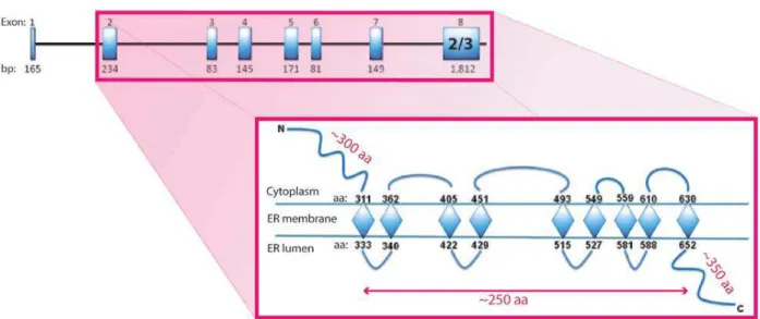
WFS1 gene codes for the WFS1 protein, also called wolframin (Collier et al., 1996; Inoue et al., 1998). It resides on the 4p16.1 chromosome, which is associated with ER membrane-embedded proteins, stretching over 33.4 kb of genomic DNA (Hofmann et al., 2003; Inoue et al., 1998; Rigoli et al., 2011). The 3.7 kb of WFS1 mRNA (Inoue et al., 1998; Rigoli et al., 2011) generate 8 exons, with the exon-1 non-coding and the exon-8 comprising information for 2/3 of the WFS1 protein
29
(Hofmann et al., 2003) (Figure 1). The latter consists of 890 amino acids (aa) with approximately 100 kDa of molecular mass (Cryns et al., 2003; Hofmann et al., 2003; Takeda et al., 2001), being generally hydrophobic, but with hydrophilic ends (Takeda et al., 2001). Hence, WFS1 creates 3 structural domains: a hydrophilic N-terminus of 300 aa, a hydrophobic body of 240 aa, and a hy-drophilic C-terminus of 350 aa, approximately (Inoue et al., 1998). After the translation, WFS1 does not undergo proteolytic processing, as some proteins do, but is N-glycosylated and, certainly, local-izes to ER (Fonseca et al., 2005; Hatanaka et al., 2011; Hofmann et al., 2003; Philbrook et al., 2005). It resides there as an oligomer, most probably a tetramer, stabilized by the N-glycosylation (Hof-mann et al., 2003). Now, experiments with human cells, after constructing polyclonal antibodies against the N- and C-termini, showed that each WFS1 contains 9 trans-membrane domains, with the N-terminus in the cytosol and the C-terminus in the ER lumen (Hofmann et al., 2003). However, AC<B8IA8>=#=:C'8;'DEFG'=9<=#6:$?':A6:'=;':A$'<876=9?'#89?=?:'89%C'8;'"-helical structures, it is more likely to be 10 of them instead of 9 (Inoue et al., 1998). In other words, WFS1 is an oligomeric ER integral protein, exposed to its lumen and to the cytosol.
Figure 1. Structure of the WFS1 gene and WFS1 protein. WFS1 consists of 250 kb, which constitute 8 exons. Transcription starts from exon-2, while exon-8 codes for 2/3 of the WFS1 protein. WFS1 is built of 890 aa and divided into two hydrophilic domains, the N- and C-terminus, with a hydrophobic core that pierces through the ER membrane 9 times. Modified from Rigoli et al. (Rigoli et al., 2011).
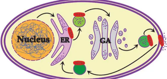
30 4.3.2.!Subcellular localization of the protein
WFS1 is an ER protein. However, it is not strictly devoted to ER (Figure 2), since it has been found in membranes of secretory granules in human neuroblastoma (Gharanei et al., 2013) and mouse pa9#B$6:=#'(-cells (Hatanaka et al., 2011). Also, it resides in the canalicular reticulum, a spe-cialized ER variant, as evidenced by immunohistochemical and in situ hybridization studies in mouse inner ear (Cryns et al., 2003). The amount of literature reporting its presence in the ER ex-cludes the possibility of misfolding or overexpression-driven placement. It is so especially since it co-locates with ER markers, but neither Golgi apparatus (GA) nor mitochondria markers (Takeda et al., 2001; Takei et al., 2006; Ueda et al., 2005), as would be implied by the WS pathology. Eventual-ly, it was demonstrated that WFS1 is sensitive to H-endoglycosidase in rodents (Osman et al., 2003; Takeda et al., 2001), and, even over-expressed, does not change its ER localization (Takeda et al., 2001; Takei et al., 2006).
Figure 2. Subcellular localization of WFS1. WFS1 can be found in the ER, secretory granule and cell mem-branes.
4.3.3.!Expression pattern: human
WFS1 activity changes during human development. At fetal stage (14-16 weeks), its expres-sion is low, and gradually increases in time to reach a plateau after attaining sexual maturity (De Falco et al., 2012). When it comes to distribution in the body, WFS1 is omnipresent (in adults), with the most robust expression in the brain, the pancreas and the heart (Hofmann et al., 2003).
Re-31
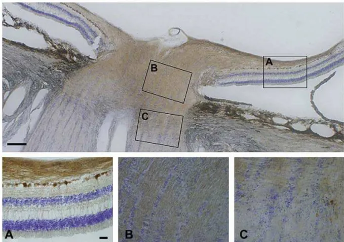
garding the eye, only RPE, RGC and ON glial cells (cynomolgus monkey (Yamamoto et al., 2006)), but not other cell populations, are rich in WFS1 (Schmidt-Kastner et al., 2009; Yamamoto et al., 2006) (Figure 3). Its presence is also highly notable in many simple and stratified epithelia throughout the body, but only moderate in the liver and the endocrine part of the pancreas (De Falco et al., 2012). In conclusion, the pattern of WFS1 expression investigated so far in primates seems to correlate with the WFS1-deficiency symptoms.
Figure 3. WFS1 in human retina. Human retina with the optic nerve (ON) was immunostained against WFS1 (brown) and counterstained with cresyl violet. Boxes in the upper panel mark the enlarged sections in the lower panel. Magnification bar equals 250 mm. A) Retinal ganglion cells (RGC) and the ON layer in the reti-na, but not other layers, are strongly positive for WFS1. B) RGC axons in the ON head are positive for WFS1. C) Transition to ON is accompanied by reduced WFS1 expression at the interface of myelination. Magnifica-tion bar for A-C equals 40 mm. Modified from Schmidt-Kastner et al. (Schmidt-Kastner et al., 2009).
4.3.4.!Expression pattern: mouse
What is the expression in rodents like? WfsG'6II$6B?'&>=J&=:8&?'=9'I69#B$6:=#'(-cells (Fonse-ca et al., 2005; Hatanaka et al., 2011; Ishihara et al., 2004; Philbrook et al., 2005; Ueda et al., 2005),
32
the limbic system (Luuk et al., 2008; Philbrook et al., 2005) and its accompanying structures (Ka-wano et al., 2008; Luuk et al., 2008; Osman et al., 2003). Further experiments on pancreas exposed somatostatin-IB8<&#=9K'L-cells as Wfs1-positive, as well (Fonseca et al., 2005; Ishihara et al., 2004; Philbrook et al., 2005; Ueda et al., 2005). In the same time however, neither glucagon-IB8<&#=9K'"-cells nor polypeptide-producing PP-glucagon-IB8<&#=9K'"-cells shared the trait. This was investigated with mRNA expres-sion, immunochemistry, cleavage with endoglycosidase H, and subcellular fractionation; providing similar results. Regarding the brain, other CNS parts than the limbic system were enriched in Wfs1, too. They belong to forebrain, diencephalon, brainstem, cerebellum, medulla spinalis and circum-ventricular organs; including the gray matter and the visual cortices (Kawano et al., 2008; Luuk et al., 2008, 2012). Finally, dopaminergic nerves were strongly Wfs1-positive, too (Visnapuu et al., 2013b), as shown with immunohistology. Here it should be emphasized that analogous brain struc-tures in humans were linked to psychiatric and behavioral deviations often reported in WS (Takeda et al., 2001), as well as vision and hearing process. The same applies to the pancreatic cells.
Following on that, much attention was brought to murine retina, where the Wfs1 protein was detected in RGCs, Müller cells, amacrine cells and the RPE (Schmidt-Kastner et al., 2009) (Figure 4). In another study, high Wfs1 mRNA levels were reported in amacrine and Müller cells, mild in pho-toreceptors (PR) and horizontal cells, while faint in RGC and bipolar cells (Kawano et al., 2008). The optic nerve was also found Wfs1-positive, especially the supportive astroglia, but this was not re-ported for the thither optic pathway, namely the optic chiasm or the optic tract (Kawano et al., 2008).
Time-dependent Wfs1 expression was also investigated in mice, with regards to the inner ear (Cryns et al., 2003), brain (Kawano et al., 2009; Tekko et al., 2014), and pancreas (Xu et al., 2009). Using in situ hybridization and immunochemistry against the N-terminus of Wfs1, its presence was demonstrated in diverse inner ear cells, without assorting across the cochlea. Furthermore, until p35, when the inner ear is already mature, there was no evident time-dependence, but some dif-ferences in expression were visible (Cryns et al., 2003). In the case of developing brain however, three expression patterns emerged (mRNA-based studies (Kawano et al., 2009)). In the first, the expression was faint or missing in infants but present in young adults. This was valid for hippocam-pal CA1 part, parasubiculum and entorhinal cortex. In the second, the expression was steady
33
throughout lifetime, what was characteristic for some limbic structures and brainstem nuclei. Final-ly, in the third, the expression climaxed at the second week of age, what was seen for the thalamic reticular nucleus. Tekko et al., however, showed on Wfs1exon8del forebrain that Wfs1 expression
starts in late embryonic development, in the dorsal striatum and amygdala (Tekko et al., 2014). Its expression spread throughout other parts after birth, and was strictly correlated with neuronal differentiation. Altogether, these data indicate great importance of Wfs1 in neural tissue develop-ment.
Figure 4. Wfs1 in mouse retina. Mouse retina was immunostained against Wfs1. Cells in RGC, RPE and to some extent in INL were Wfs1-positive. INL, among others, contains amacrine cells; INL – inner nuclear lay-er, IPL – inner plexiform laylay-er, ONL – outer nuclear laylay-er, IS – layer of inner segments, RGC – retinal ganglion cell layer, RPE – retinal pigment epithelium. Magnification bar equals 50 mm. Modified from Schmidt-Kastner et al. (Schmidt-Schmidt-Kastner et al., 2009).
34
The Wfs1 expression pattern in relation to development was also investigated on rat pancre-as (Xu et al., 2009), and it appears that Wfs1 is involved in organ maturation. The highest mRNA amount was reported at E18.5, lowering with age and reaching the minimum in adulthood, similar-ly to humans. On the contrary, the protein levels were much more dynamic, being very low at E18.5, raising after birth to reach the maximum at P14, and dropping again very low at P21. This fashion suggests a time-related presence of an mRNA processing mechanism; or accumulation of the protein as long as its degradation (or secretion) is impaired; what stops after the second week of age.
To summarize, the Wfs1 expression in rodents is tissue-specific and, in certain cases, time-dependent. In general, it resembles the expression pattern in humans and correlates with WFS1-deficiency symptoms.
4.3.5.!WFS1 in stress response
What about WFS1 activation and degradation? An interaction between WFS1 and GRP94 chaperone protein, a component of the unfolded protein response (UPR) was reported (Kakiuchi et al., 2009). This was resolved upon ER stress, suggesting that in normal conditions, WFS1 is main-tained dormant, at least in some part, and activates to lower UPR. When the job is done, WFS1 seems to be degraded by the HECT-type ubiquitin ligase (E3) Smadubiquitination regulatory factor 1 (Smurf) (Guo et al., 2011). Smurf associates with C-terminus of WFS1 to encourage its ubiquitina-tion as well as degradaubiquitina-tion by proteasome. However, upon ER stress Smurf is destroyed, just as GRP94 is detached, what essentially elevates the active WFS1 level.
As described above, WFS1 is localized somewhere in the ER and its activation and degrada-tion rely on ER stress. Therefore, it is only logical to investigate its involvement in ER funcdegrada-tions, especially the ones related to cell survival. This of course includes protein assembly and trafficking, ER stress, UPR and Ca2+ flux.