HAL Id: hal-03096337
https://hal.archives-ouvertes.fr/hal-03096337
Submitted on 22 Mar 2021
HAL is a multi-disciplinary open access
archive for the deposit and dissemination of
sci-entific research documents, whether they are
pub-lished or not. The documents may come from
teaching and research institutions in France or
abroad, or from public or private research centers.
L’archive ouverte pluridisciplinaire HAL, est
destinée au dépôt et à la diffusion de documents
scientifiques de niveau recherche, publiés ou non,
émanant des établissements d’enseignement et de
recherche français ou étrangers, des laboratoires
publics ou privés.
Deciphering the Role of CD1e Protein in Mycobacterial
Phosphatidyl- myo -inositol Mannosides (PIM)
Processing for Presentation by CD1b to T Lymphocytes
Diane Cala-de Paepe, Emilie Layre, Gaëlle Giacometti, Luis Garcia-Alles,
Lucia Mori, Daniel Hanau, Gennaro de Libero, Henri de la Salle, Germain
Puzo, Martine Gilleron
To cite this version:
Diane Cala-de Paepe, Emilie Layre, Gaëlle Giacometti, Luis Garcia-Alles, Lucia Mori, et al..
Decipher-ing the Role of CD1e Protein in Mycobacterial Phosphatidyl- myo -inositol Mannosides (PIM)
Process-ing for Presentation by CD1b to T Lymphocytes. Journal of Biological Chemistry, American Society
for Biochemistry and Molecular Biology, 2012, 287 (37), pp.31494-31502. �10.1074/jbc.M112.386300�.
�hal-03096337�
Deciphering the Role of CD1e Protein in Mycobacterial
Phosphatidyl-myo-inositol Mannosides (PIM) Processing for
Presentation by CD1b to T Lymphocytes
*
□SReceived for publication, May 30, 2012, and in revised form, June 29, 2012Published, JBC Papers in Press, July 10, 2012, DOI 10.1074/jbc.M112.386300
Diane Cala-De Paepe‡§1, Emilie Layre‡§1,2, Gaëlle Giacometti¶储3, Luis F. Garcia-Alles‡§, Lucia Mori**‡‡,
Daniel Hanau¶储, Gennaro de Libero**‡‡, Henri de la Salle¶储, Germain Puzo‡§, and Martine Gilleron‡§4
From‡CNRS, IPBS (Institut de Pharmacologie et de Biologie Structurale), 205 route de Narbonne, 31077 Toulouse, France, §Université de Toulouse, UPS, IPBS, 31077 Toulouse, France,¶INSERM U725, Etablissement Français du Sang-Alsace, Strasbourg, France, the储Université de Strasbourg, 67000 Strasbourg, France, the **Division of Experimental Immunology, Department of Biomedicine, University Hospital, CH-4031 Basel, Switzerland, and the‡‡Singapore Immunology Network Agency for Science, Technology, and Research, 138648 Singapore
Background:To become antigenic, mycobacterial hexamannosylated phosphatidyl-myo-inositol (PIM6) undergo
CD1e-assisted␣-mannosidase processing.
Results:Measuring membrane-to-membrane PIM transfer, CD1e selectively transferred diacylated PIM species in accordance with the fact that liposome-inserted di-acylated PIM6 were the only PIM species digested into antigenic molecules by
␣-mannosidase.
Conclusion:CD1e assists PIM processing through its lipid transfer protein property.
Significance:This study reveals the molecular mechanisms by which CD1e contributes to lipid immunoediting.
Lipids are important antigens that induce T cell-mediated specific immune responses. They are presented to T lympho-cytes by a specific class of MHC-I like proteins, termed CD1. The majority of the described CD1-presented mycobacterial anti-gens are presented by the CD1b isoform. We previously demon-strated that the stimulation of CD1b-restricted T cells by the hexamannosylated phosphatidyl-myo-inositol (PIM6), a family of mycobacterial antigens, requires a prior partial digestion of the antigen oligomannoside moiety by␣-mannosidase and that CD1e is an accessory protein absolutely required for the gener-ation of the lipid immunogenic form. Here, we show that CD1e behaves as a lipid transfer protein influencing lipid immu-noediting and membrane transfer of PIM lipids. CD1e selec-tively assists the␣-mannosidase-dependent digestion of PIM6
species according to their degree of acylation. Moreover, CD1e transfers only diacylated PIM from donor to acceptor liposomes and also from membranes to CD1b. This study provides new insight into the molecular mechanisms by which CD1e contributes to lipid immunoediting and CD1-restricted presentation to T cells.
CD1 proteins are membrane proteins specialized in the pres-entation of endogenous and exogenous lipid antigens to T lym-phocytes (1). A large fraction of the known exogenous antigens are components of the Mycobacterium tuberculosis envelope and induce specific T cell responses (2). Human antigen-pre-senting cells express five CD1 isoforms, CD1a, CD1b, CD1c, CD1d, and CD1e, that traffic in different endocytic compart-ments (3). During recycling, CD1a, -b, -c, and -d molecules bind and form stable complexes with lipid antigens, which are then exposed on the cell surface for T cell stimulation (4). CD1e displays very distinct characteristics. In contrast to other CD1 isoforms, it is expressed only in dendritic cells. CD1e accumu-lates in the Golgi compartments of immature dendritic cells, and upon dendritic cell maturation, CD1e is transported to late endosomes/lysosomes where it is cleaved and accumulates as a soluble form (sCD1e)5(5, 6). CD1e is never expressed on the
cell surface, thus preventing any direct interaction with T cells, but is implicated in the processing of the complex mycobacte-rial lipid antigens hexamannosylated phosphatidyl-myo-inosi-tol (PIM6)6 into dimannosylated phosphatidyl-myo-inositol
*This work was supported by the Centre National de la Recherche Scien-tifique, the Agence Nationale de la Recherche Emergence (ANR-05-MIIM-006), the Etablissement Français du Sang-Alsace, European Com-munity’s Seventh Framework Program Grant 241745, and Swiss National Foundation Grants 3100AO-122464/1 and Sinergia CRS133-124819.
□S
This article containssupplemental Table S1 and Figs. S1–S5.
1Both authors contributed equally to this work.
2Present address: Harvard Medical School, Brigham and Women’s Hospital, 1
Jimmy Fund Way, Smith 514, Boston, MA 02115.
3Supported by an Association de Recherche et de De´veloppement en
Me´de-cine et Sante´ Publique grant.
4To whom correspondence should be addressed: Institut de Pharmacologie
et de Biologie Structurale, CNRS UMR5089, Dept. of Molecular Mechanisms of Mycobacterial Infections, 205 route de Narbonne, 31077 Toulouse, Cedex 4, France. Tel.: 33-561-175-919; Fax: 33-561-175-994; E-mail: martine.gilleron@ipbs.fr.
5The abbreviations used are: sCD1e, soluble form of CD1e; rsCD1e,
recombi-nant soluble form of CD1e; GM2AP, GM2-activator protein; LTP, lipid transfer protein; LUV, large unilamellar vesicle(s); myo-Ins, myo-inositol; PIM, phosphatidyl-myo-inositol mannoside(s); PIM3,
phosphatidyl-myo-inositol trimannoside(s); Sap, saposin; SUV, small unilamellar vesicle(s); PI, phosphatidyl-myo-inositol; POPS, 1-palmitoyl-2-oleyl-sn-glycero-3-phosphoserine.
6PIM is used to describe the global family of PIM that carries one to four fatty
acids and one to six Manp residues. In AcXPIMY, X refers to the number of
acyl groups esterified to available hydroxyls on the Manp or myo-inositol residues, y refers to the number of Manp residues, e.g. Ac1PIM6
corre-sponds to triacylated PIM6, the phosphatidyl-myo-inositol hexamannoside
PIM6carrying two acyl groups attached to the glycerol (the diacylglycerol
substituent) and one acyl group esterified to the Manp residue. Inherent in the abbreviation for PI is the diacylglycerol unit. Accordingly, natural acyl form mixtures of PIM2or PIM6will be designated by mixPIM2and mixPIM6.
(PIM2) (7). In addition to this role, CD1e may positively or negatively affect lipid presentation by CD1b, CD1c, and CD1d, due to the capacity of CD1e to facilitate rapid formation of CD1-lipid complexes and also to accelerate their turnover (8). Thus, CD1e is an important modulator of both group 1 and group 2 CD1-restricted responses influencing the lipid antigen availability as well as the generation and persistence of CD1-lipid complexes (8). The crystal structure of human recombi-nant sCD1e (rsCD1e) at 2.90 Å resolution revealed that CD1e displays a main portal wider than that of other CD1 molecules as well as a less intricate groove, allowing accommodation of a variety of lipid antigens and facilitating the rapid exchange of bound lipids (9).
Recent studies have implicated saposins in CD1 lipid antigen loading and presentation. To date, five activator proteins of sphingolipid catabolism have been described, saposin-A to saposin-D (Sap-A to Sap-D) and the GM2 activator protein (GM2AP). These are non-enzymatic cofactors involved in the degradation of sphingolipids by lysosomal hydrolases (10), assisting membrane-inserted substrate digestion. They medi-ate the interaction between the wmedi-ater-soluble exohydrolase and their membrane-embedded glycolipid substrates at the lipid-water interface. Saposins may act via two distinct mechanisms, including membrane destabilization (11) and direct binding and transferring of monomeric lipids (12). In the context of CD1 proteins, saposins facilitate binding of␣-GalCer to CD1d (13), with Sap-B involved in the loading of CD1d with lipid antigens (14, 15), and Sap-C involved in the loading of CD1b with mycobacterial lipoarabinomannan antigen (16).
Similarly to saposins, CD1e participates in lipid antigen edit-ing. Processing of the PIM6oligosaccharide requires the
pres-ence of CD1e and ␣-mannosidase in lysosomes to produce PIM2, which are the antigenic forms recognized by CD1b-re-stricted T cells (7). In the absence of CD1e, PIM6are digested to
phosphatidyl-myo-inositol trimannosides (PIM3), which are
not able to stimulate the PIM-specific T cells.
Both the multiple effects of CD1e on antigen presentation (8) and its structural characteristics (9) suggest that CD1e might have evolved to mediate lipid exchange/editing processes. Although CD1e/PIM association has been shown by isoelectric focusing experiments (7, 9), the mechanisms by which CD1e protein participates in PIM processing and influences the immune responses remains to be precisely determined. PIM exist naturally in the mycobacterial envelope as a mixture of different acyl forms, including mono-, di-, tri-, and tetra-acy-lated forms (17). Using liposome-inserted PIM and membrane-to-membrane transfer assays, we find that CD1e is an LTP, transferring only diacylated PIM species. This finding is in accordance with the fact that only liposome-inserted diacylated PIM6species can be digested by ␣-mannosidase to generate
PIM2.
EXPERIMENTAL PROCEDURES
Preparation of Mycobacterial PIM2 and PIM6 Mixtures—
PIM2 and PIM6 mixtures were purified from Mycobacterium
smegmatistotal lipid extract. Briefly, total lipids were extracted according to the Folch method (18). Acetone was then added to the concentrated organic phase to precipitate acetone-insoluble
lipids (overnight at 4 °C). After centrifugation (20 min, 2000⫻ g, 4 °C), the acetone-insoluble fraction was dried under nitrogen flow and loaded on a silica column (25⫻ 2.3 cm; KG60, 230–400 mesh, Merck), successively eluted with 300 ml of chloroform, 300 ml of chloroform/methanol/water 9:1:0.1 (by volume), 200 ml of chlo-roform/methanol/water 8:2:0.1 (by volume), 200 ml of chloro-form/methanol/water 7:3:0.2 (by volume), 300 ml of chloroform/ methanol/water 7:3:0.4 (by volume), 300 ml of chloroform/ methanol/water 60:35:6 (by volume), and 300 ml of chloro-form/methanol/water 60:35:8 (by volume). PIM2 were eluted
using 7:3:0.2 and 7:3:0.4 proportions and PIM6using 60:35:6 and
60:35:8.
Purification of PIM2and PIM6Acyl Forms—Different PIM2
and PIM6acyl forms were purified as described previously (19). PIM2- or PIM6-enriched fractions (40 mg) were loaded in
equil-ibrating buffer (0.1 Mammonium acetate solution containing
20% propanol-1), on an octyl-Sepharose CL-4B column (20⫻ 1.5 cm; Amersham Biosciences). The elution was performed with a linear gradient from 20 to 50% propanol-1 (135-ml each) at 5 ml/h flow rate. The purification process was monitored by TLC and MALDI-TOF mass spectrometry analysis, and each purified acyl form was controlled by1H NMR spectroscopy as
described previously (20, 21).
Liposome Preparations—Liposomes contained POPC (1-palmitoyl-2-oleyl-sn-glycero-3-phosphocholine; Sigma; 4.55 mol), cholesterol (Sigma; 1.75 mol), POPS (1-palmitoyl-2-oleyl-sn-glycero-3-phosphoserine; Avanti polar lipids; 0.7 mol) and optionally, PIM6 mixture (0.28 mol). PIM and
commercial lipids were solubilized in the solvent systems chlo-roform/methanol/water 60:35:8 (by volume) or chloroform, respectively, mixed in the desired proportions and dried under nitrogen flow. Lipid films were hydrated in appropriate volume of Tris buffer (10 mMTris, pH 7.4, 150 mMNaCl, 1 mMEDTA),
mixed by vortexing and subjected to 10 freeze/thaw cycles by alternative passages in liquid nitrogen and water (25 °C) to obtain large unilamellar vesicles (LUV). For fluorophore-en-capsulated LUV, the entire procedure was performed in the presence of 20 mM calcein. LUV size was homogenized by
sequential extrusion through 0.2- and 0.1-m pore diameter polycarbonate membranes. Liposomes were dialyzed against 50 mMphosphate buffer, pH 6.0, or acetate buffer, pH 4.7. LUV
size (diameter of 120⫾ 10 nm) was assessed by dynamic light scattering and electron microscopy.
Recombinant Soluble Human CD1e Production (rsCD1e)— rsCD1e was expressed in Drosophila S2 cells, previously trans-duced with human2microglobulin, and purified as described (7). Briefly, rsCD1e with the propeptide (amino acids 20 –305 of the pre-␣ chain) was expressed as a fusion protein with the BIP signal peptide and a C-terminal tag containing the V5 epitope followed by a hexahistidine sequence (pMT BIP-V5His, Invitrogen). The recombinant protein was purified by metal chelate chromatography followed by affinity chromatography using 20.6 anti-hCD1e mAb (5).
Recombinant Saposins—Human saposins B and C (Sap-B and Sap-C) cDNA were amplified from human spleen RNA (Clontech), using Platinum TaqDNA polymerase high fidelity (Invitrogen), cloned into pCR2.1-TOPO (Invitrogen) and sequenced (7). After subcloning into the NcoI-BamHI sites of
the procaryotic expression vector pQE-60 (Qiagen), the plas-mids were used to transform Escherichia coli BL21(DE3) LysS (Novagen). Recombinant saposins were purified from bacterial lysates via their C-terminal His6tag with the His-select spin
columns (Sigma) according to standard procedures. Purity of the preparations was estimated by Coomassie staining on SDS-PAGE.
In Vitro␣-Mannosidase Digestion of PIM6—In vitro
␣-man-nosidase digestion assays were carried out as described previ-ously (7). PIM6(27.7 nmol), Ac1PIM6(24.1 nmol), or Ac2PIM6
(21.5 nmol) were sonicated in 100l of mannosidase buffer (0.1
Msodium acetate, pH 4.5, 1 mMZnSO4). The enzymatic
reac-tion was performed in the presence of jack bean␣-mannosidase (Sigma) (0.44 unit) and rsCD1e (1.05 nmol) for 1 h at 37 °C (total volume of 150l). In vitro ␣-mannosidase digestion of sonicated mixPIM6(100g) in the presence of Sap-B or Sap-C
(30g; 3 nmol) instead of rsCD1e (120 g; 3 nmol) were also tested. These assays were also carried out with LUV (18 mMof
total lipids) composed by POPC/cholesterol/PIM6or Ac1PIM6
65:25:10 (by mol): typically, 14l of LUV solution (252 nmol of lipids) were incubated with rsCD1e (80 g; 2.1 nmol) and ␣-mannosidase (20 g; 0.44 unit) in a final volume of 150 l for 8 h at 37 °C.
In all cases, PIM were extracted following the reaction by the Folch method as described previously (7). The organic extracts were dried under nitrogen stream and resuspended in 20l of chloroform/methanol/water 60:35:8 (by volume) for MALDI-TOF MS analysis.
Fluorophore Leakage—Calcein-encapsulated LUV were pre-pared as described above and dialyzed against acetate buffer, pH 4.7, to remove free calcein and purified on a Sephadex G-75 column (10⫻ 2 cm; Sigma). The elution was performed with acetate buffer at 0.3 ml/min flow rate, and 1-ml fractions were collected. The presence of liposomes within each fraction was determined by spectrophotometry (600-nm wavelength), and the most enriched fractions were used for leakage experiments. Calcein-encapsulated LUV (40 nmol of lipids) were incu-bated with jack bean␣-mannosidase (0.44 unit) and rsCD1e (2.1 nmol) or Sap-C (0.5 nmol), as a positive control, in 2 ml of acetate buffer (0.1Msodium acetate, pH 4.5, 1 mMZnSO4).
Fluorophore leakage was measured by spectrofluorometry (SAFAS) at 37 °C under agitation. Excitation wavelength was 480 nm, and emission was recorded at 517 nm with 4-nm slit widths. For each experiment, the maximum fluorophore leak-age was obtained by final addition of 10% Triton X-100 (10l), which induces disorganization of liposomes.
Dynamic Light Scattering—Dynamic light scattering meas-urements were performed at 20 °C in a microcuvette (90° scattering angle) with a DynaPro-MS/X instrument (Protein-Solution, Inc.) equipped with a laser diode operating at 830 nm wavelength and a digital correlator with 248 exponentially spaced channels. Diluted LUV solutions (1l in 100 l) were centrifuged for 10 min at 30,000 ⫻ g and transferred into a 100-l microcuvette. The laser intensity was set at 10% of its maximum power, and 30 acquisitions of 10 s were cumulated. Each single experiment resulted from the sum of 30 acquired spectra. Data analysis was performed with the cumulative method using Dynamics V6 software (ProteinSolution, Inc.).
Electron Microscopy—For negative staining, 10 l of LUV preparations were applied to carbon-coated copper grids. After 30 s, solution in excess was blotted off with filter paper, and samples were stained with 1% (w/v) uranyl acetate for 1 min. Analysis was performed using a Jeol 120 EX transmission elec-tron microscope, and images were acquired with a digital cam-era (AMT-USA) at 75,000 –120,000⫻ magnification.
Lipid Transfer by CD1e—LUV characterized by different lipid compositions were prepared as described above and used at 25 mM lipid concentration. Acceptor liposomes (LUV-A)
contained POPC (7.5 mol) and cholesterol (2.5 mol), whereas donor liposomes (LUV-D) contained POPC (6.5 mol), cholesterol (2.5 mol), and negatively charged phos-pholipids (1.0mol), including POPS, PI (Sigma), or PIM2acyl forms.
Neutral liposomes (40l, LUV-A; 1.0 mol total lipids) were incubated with an equal amount of negatively charged lipo-somes (LUV-D), in the presence of rsCD1e (2.1 nmol) in 50 mM
phosphate buffer, pH 6.0 (total volume of 160l), for 36 h at 37 °C on agitation. Samples were then loaded on a 1.2 ml of DEAE-Sephadex A-25 (GE Healthcare) column pre-equili-brated with phosphate buffer. The elution was performed with phosphate buffer containing increasing concentrations of NaCl (12.5, 60, and 1M). Three 1-ml fractions were collected for each
NaCl concentration, and liposome elution was monitored by measuring absorbance at 600 nm wavelength. A progressive and selective elution of LUV-A (0% of POPS), LUV-I (5% of POPS), and LUV-D (10% of POPS) from the DEAE column was obtained using increasing NaCl concentrations (0, 60, and 1M,
respectively).
The presence of rsCD1e and lipids in the collected fractions was assessed by SDS-PAGE (15% acrylamide) and silver nitrate staining. Control experiments were performed in the absence of rsCD1e and in the presence of denaturated rsCD1e or BSA. The stability of individual liposome populations over incuba-tion time in the presence of rsCD1e was verified by electron microscopy and analyzing their chromatographic behavior.
CD1b PI Loading Assays—LUV-D (3.6 mMof total lipids),
containing POPC (86.4 nmol), cholesterol (28.8 nmol), and PI (28.8 nmol) were prepared as described above. PI was supple-mented with [3H]PI (PerkinElmer Life Sciences) to obtain a
280⫻ 103dpm/nmol specific activity. LUV-D were incubated
with rsCD1b (19.6 pmol) in the absence or presence of rsCD1e (6.6 and 13.2 pmol) or N Oct-3 transactivation domain (16.7 pmol) in 50 mMphosphate buffer, pH 6.0, 60 mMNaCl (4l of
final volume). The reaction was performed at 37 °C for 5 h under agitation. 2l of each sample were then loaded on an isoelectric focusing gel (PhastGel 4 – 6.5, GE Healthcare) to separate proteins and free lipids. Electrophoresis was per-formed in a PhastGel system (Amersham Biosciences) at 600 accumulated volts/h. After the migration, bands corresponding to rsCD1b were excised from the gel, and the associated [3H]
radioactivity was counted. The capacity of rsCD1e to load PI into rsCD1b molecules was analyzed at different time points (5, 30, and 300 min). Control experiments in the absence of rsCD1b were performed to assess the radioactivity diffusion within the pH range corresponding to rsCD1b isoelectric point. The percentage of rsCD1b loaded with PI was determined by
comparing the amounts of PI detected by radioactivity with the total amounts of rsCD1b loaded on the gel, assuming that 1 mol of rsCD1b is associated to 1 mol of PI.
Matrix-assisted Laser Desorption/Ionization Mass Spectrom-etry—MALDI-TOF MS analysis was carried out using a 4700 proteomics analyzer (with TOF/TOF optics, Applied Biosys-tems, Voyager DE-STR, Framingham, MA) in reflectron mode. Ionization was achieved by irradiation with a Nd:YAG laser (355 nm) operating at 500-ps pulses with a frequency of 200 Hz. PIM were analyzed in negative-ion mode. Spectra from 2000 to 4000 laser shots were summed to obtain the final spectrum. Typically, 2l (2–5 g) of PIM dissolved in the solvent sys-tem chloroform/methanol/water 60:35:8 (by volume) were mixed with 2 l of matrix solution, 2-(4-hydroxyphenylazo)-benzoic dissolved at 5 mg/ml in ethanol/water 1:1 (by volume) (Sigma). 0.4l of this mixture were spotted on the target. RESULTS
CD1e Assists Mannosidase in Processing of Specific Acyl Forms of PIM—Previously, we showed that CD1e acts as an accessory protein to assist␣-mannosidase-mediated process-ing of PIM6into PIM2, which is required for CD1b-restricted T cells activation (7). Using the natural pool of PIM6, containing a
mixture of different acyl forms (mixPIM6) as substrate for in
vitro digestion (Fig. 1A), we noticed that only di-acylated (PIM6) and tri-acylated (Ac1PIM6) forms of PIM6, but not
tetra-acylated PIM6(Ac2PIM6) were digested by␣-mannosidase in
the presence of rsCD1e to generate the respective PIM2acyl forms (7). Moreover, insertion of mixPIM6into liposomes, to
better mimic physiological conditions, resulted in a nearly exclusive digestion of diacylated forms (7). These prior obser-vations suggested that the degree of PIM acylation interferes with their glycosidic processing efficiency. To better under-stand this selective hydrolysis, in vitro digestion assays were performed on individual purified PIM6, Ac1PIM6, and Ac2PIM6
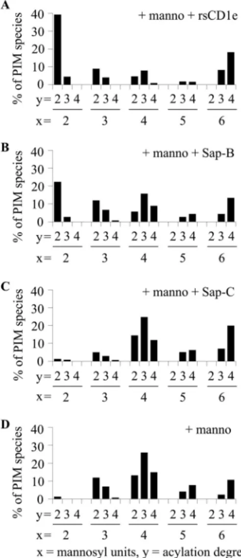
acyl forms (Fig. 1, B–D), in the absence or presence of rsCD1e. The reaction products were analyzed by negative-ion mode MALDI-TOF mass spectrometry (supplemental Fig. S1), and the relative abundances of the generated PIM glycoform were determined (Fig. 2). In the presence of ␣-mannosidase only (⫹ manno), a more effective enzymatic digestion was observed for PIM with a lower acylation degree, as Ac2PIM6, Ac1PIM6,
and PIM6acyl forms were digested to Ac2PIM4, Ac1PIM3, and PIM2glyco-forms, respectively (Fig. 2, A–C). The addition of
rsCD1e to the reaction mixture (⫹ manno ⫹ rsCD1e) did not alter the digestion profile of Ac2PIM6(Fig. 2A), suggesting that CD1e does not assist their digestion by␣-mannosidase. In con-trast, rsCD1e improved the digestion of both Ac1PIM6 and
PIM6, with Ac1PIM6digested to Ac1PIM2(Fig. 2B), and the proportion of the PIM2 glycoform resulting from PIM6 was
greatly increased (Fig. 2C). To get closer to physiological
con-C
D
O O O H O HO HO OH O O O O HOHO OH O O HOHO O P O O R4 O O O HO HO HO OH O O HO HO HO O O HO HO HO O O HO HO OH O O HO HO OH O O O O HOHO OH O O HOHO O P O OA
PIM
6PIM
2B
1823.9 1781.8PIM
6Ac PIM
1 6Ac PIM
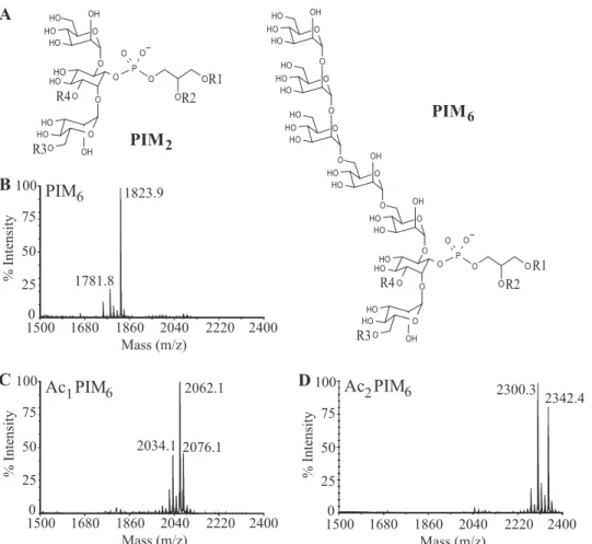
2 6 2034.1 2062.1 2076.1 2300.3 2342.4 1500 1680 Mass (m/z) 1860 2040 2220 2400 25 50 75 100 % Intensity 0 1500 25 50 75 100 % Intensity 0 1680 Mass (m/z) 1860 2040 2220 2400 1500 25 50 75 100 % Intensity 0 1680 Mass (m/z) 1860 2040 2220 2400 R3 R1 R2 R4 R3 R1 R2FIGURE 1. Structure of PIM2and PIM6subfamilies (A) and MALDI-TOF MS spectra in negative-ion mode of purified PIM6(B), Ac1PIM6(C), and Ac2PIM6
(D) acyl forms. In monoacylated PIM (lyso-PIM), R1 represents acyl group; R2–R4 represents H. In diacylated PIM (PIM), R1 and R2 represent acyl group; R3 and
R4 indicate H. In triacylated PIM (Ac1PIM), R1, R2, R3 indicate acyl group; R4 indicates H. In tetraacylated PIM (Ac2PIM), R1 to R4 indicates acyl group.
CD1e in PIM Presentation to T Lymphocytes
ditions, purified PIM6 acyl forms were individually inserted
into liposomes and used in similar digestion experiments. As previously observed with mixPIM6 (7), liposome inclusion greatly modified the type of Ac1PIM6digested products
gener-ated by␣-mannosidase in the presence of rsCD1e ⫹ manno ⫹ rsCD1e as almost no Ac1PIM2were obtained (Fig. 2D). How-ever, PIM6in liposomes⫹manno ⫹ rsCD1e were almost
com-pletely degraded into PIM2(Fig. 2E).
Taken together, these results demonstrate that degree of acy-lation of PIM6determines their in vitro digestion by
␣-manno-sidase and that when PIM6is inserted into liposomes, rsCD1e selectively assists the digestion of diacylated forms. To under-stand this selectivity, we next attempted to uncover the mode of action of CD1e.
Lipid Transfer Protein Assists ␣-Mannosidase Digestion of PIM6—CD1e properties seem to be reminiscent to that of
sapo-sin assistance of the sphingolipid digestion (10). Saposapo-sins have been shown to increase lipid substrate accessibility for enzy-matic digestion by either (i) membrane disorganization as for Sap-C or (ii) lipid transfer properties from membrane-to-mem-brane or from memmembrane-to-mem-brane to protein as for Sap-B (12).
Thus, to get better clues on CD1e activity, we compared it with that of saposins in the in vitro␣-mannosidase digestion of
mixPIM6. In the presence of rsCD1e or Sap-B (Fig. 3A and B),
we observed that mix-PIM6were degraded up to diacylated
PIM2. In contrast, Sap-C had no effect (Fig. 3C and D). These results suggest that the mechanism of CD1e as a lipid transfer protein (LTP) is similar to that of Sap-B but not of Sap-C.
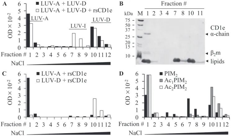
CD1e Selectively Transfers Diacylated PIM—To determine whether rsCD1e is able to promote membrane-to-membrane lipid transfer, we studied the transfer of a negatively charged phospholipid, POPS from POPS-containing donor liposomes (LUV-D) to acceptor liposomes devoid of POPS (LUV-A) in the presence of CD1e (22). Dynamic light scattering analysis revealed homogenous population of both liposomes with apparent hydrodynamic radius of⬃60 nm (supplemental Fig. S2). Accordingly, the images from microscopy allowed deter-mining a diameter of⬃120 nm.
The transfer of POPS from donor to acceptor liposomes was expected to generate a new liposome population with an inter-mediate POPS content (LUV-I) that can be separated from LUV-A and LUV-D by anionic exchange chromatography (DEAE). The sodium chloride gradient was adjusted to provide an effective separation of LUV-A, LUV-D, and potential new LUV-I liposome populations. LUV-A liposomes, devoid of charged POPS, were not retained on the column and were
FIGURE 2. PIM6acylation degree determines their glycosidic processing. The different purified PIM6acyl forms, Ac2PIM6(A), Ac1PIM6(B and D) and PIM6
(C and E), were digested by␣-mannosidase in the absence (⫹manno) or in presence (⫹manno ⫹rsCD1e) of rsCD1e. The relative abundance of the different PIM species generated (PIMX, where X indicates the number of mannosyl units) was determined by negative-ion mode MALDI-TOF MS analysis of the reaction
mixture (mass spectra are shown insupplemental Fig. 1). Ac1PIM6and PIM6were either sonicated (B and C) or inserted in liposomes (D and E). The results
eluted in fractions 1 and 2 (F1 and F2). However, the elution of LUV-D occurred in the presence of 1MNaCl in F10, F11, and
F12. Importantly, co-incubation of rsCD1e with a LUV-A/ LUV-D mixture (ratio 1/1) yielded a new population of lipo-somes, which eluted with 60 mMNaCl in F7 and F8, indicated as
LUV-I (Fig. 4A). This population was not observed when the liposomes were incubated with heat-denaturated rsCD1e or a non-relevant lipid binding protein, BSA (data not shown). The presence of POPS in F7 and F8 was confirmed by TLC analysis (data not shown). Moreover, because the molar ratio between rsCD1e and total phospholipids (POPS and POPC) is 1:714, it is unlikely that LUV-I results from a POPS depletion of LUV-D caused by the formation of CD1e-POPS stable complexes. SDS-PAGE analysis revealed that rsCD1e was present only in the first NaCl-free fractions (Fig. 4B), excluding the possibility that LUV-I resulted from a complex between CD1e and LUV-D. In addition, when LUV-A and LUV-D were individually incubated in the presence of rsCD1e (Fig. 4C), neither a modification of
their respective chromatographic behavior nor the appearance of the intermediate population were noticed. Furthermore, dynamic light scattering measurements and transmission elec-tron microscopy showed that the size of the liposomes was not modified during the experiment, excluding the possibility of fusion events (supplemental Fig. S3). Altogether, these data indicated that rsCD1e was directly responsible for the genera-tion of LUV-I, resulting from the transfer of POPS from LUV-D to LUV-A. Similarly, rsCD1e was able to transfer PI ( supple-mental Fig. S4). However, when LUV-D were prepared using the mixture of PIM2acyl forms (mixPIM2) instead of POPS or
PI, LUV-I were hardly detectable. Because mixPIM2is
com-posed of⬃80% of tetra- and triacylated molecules (Ac2PIM2
and Ac1PIM2, respectively) (19), which might not be efficiently transferred by rsCD1e (Fig. 2), we tested purified PIM2acyl
forms and found that only diacylated PIM2were transferred by
rsCD1e (Fig. 4D). The presence of PIM2in LUV-I was con-firmed by MALDI-TOF MS and TLC analyses of F7 (data not shown). Moreover, LUV-I were not observed in absence of rsCD1e (supplemental Fig. S5).
Altogether, our data support a LTP function for the rsCD1e protein. This activity is specific for diacylated forms of PIM and suggests that in antigen-presenting cells, CD1e might extract PIM6from the membranes and present them to the
␣-manno-sidase for processing of the glycosidic moiety.
CD1e Does Not Disturb Membrane Organization —Mem-brane disorganization properties of rsCD1e were investigated by measuring the leakage of calcein entrapped within liposomes composed of POPC, cholesterol, and POPS in a 65/25/10 ratio, at an acidic pH. As observed by Vaccaro et al. (23), addition of Sap-C (0.5 nmol) disrupted the liposomes and induced calcein leakage, validating our experimental design (Fig. 5A). The addi-tion of 0.21 nmol (data not shown) or 2.1 nmol rsCD1e did not induce any calcein release. We then tested whether PIM, known to bind to CD1e, caused membrane perturbation. Nei-ther the use of liposomes containing 4% of mixPIM6nor the
addition of␣-mannosidase promoted calcein release (Fig. 5B). Moreover, varying experimental parameters, such as liposome size (SUV or LUV), pH (acidic or neutral), rsCD1e concentra-tion or incubaconcentra-tion time (few minutes to four hours) did not make any difference, strongly suggesting that CD1e does not harbor membrane disruption properties, at least in this in vitro assay.
CD1e Loads Liposome-inserted PI onto CD1b—PIM are car-ried by sCD1e during processing but are ultimately presented to T cells by CD1b molecules. To test whether CD1e was able to transfer liposome-inserted PIM onto CD1b, liposome-inserted radiolabeled diacylated phosphatidyl-inositol ([3H]PI) was used
as a lipid antigen model and radiolabeled lipid loading onto rsCD1b was measured in the presence and absence of rsCD1e. After a 5-h incubation, 10% of rsCD1b proteins were loaded by [3H]PI, whereas addition of rsCD1e allowed this proportion to
increase up to 20% (Fig. 6A). PI loading onto CD1b by CD1e was both CD1e concentration- and time-dependent (Fig. 6B). In con-trast, an irrelevant protein (transactivation domain, N Oct-3 trans-activation domain) showing a pI similar to CD1e had no effect (Fig. 6A). These results indicate that CD1e has the capacity to transfer liposome-inserted processed antigen onto CD1b.
FIGURE 3. rsCD1e and Sap-B but not Sap-C promote in vitro
␣-mannosi-dase digestion of purified mixPIM6. Relative abundance of the different
glycoforms deduced from the negative-ion mode MALDI-TOF MS analysis of Ac2PIM6after hydrolysis by␣-mannosidase in the presence of rsCD1e (A),
Sap-B (B), Sap-C (C), or in the absence of any protein (D). x and y indicate mannosyl unit and fatty acid numbers in PIM species, respectively. The results presented here are from one representative experiment of three recorded spectra.
DISCUSSION
Antigen presentation to T cells is a multistep process, includ-ing antigen uptake, editinclud-ing/processinclud-ing, and loadinclud-ing onto pre-senting molecules followed by T cell antigen receptor recogni-tion of the antigenic complex. Although accessory proteins and molecular mechanisms involved in the presentation of peptide antigens have been extensively studied, details on lipid antigen presentation by the CD1 proteins are not yet fully understood. Lipid antigens taken in charge by antigen-presenting cells are embedded in internal vesicles inside lysosomes. Their hydro-phobic nature causes a series of biophysical constraints to their presentation because the different steps preceding CD1-lipid complex formation occur at the interface between hydrophobic and hydrosoluble environments. The stimulation of CD1b-re-stricted T cells by PIM6, a family of mycobacterial antigens,
requires a partial digestion of the PIM oligomannoside moiety by an␣-mannosidase and CD1e is absolutely required for anti-gen processing and T cell stimulation (7). We previously found that rsCD1e binds glycolipid antigens and assists in vitro enzy-matic oligosaccharide hydrolysis of PIM6. In all of these
previ-ous studies, the mechanism by which CD1e mediates these effects remained unknown. Here, we show that CD1e behaves as a LTP capable of facilitating selective transport of unique acylated forms of PIM. Indeed, PIM structure not only contains a variable number of mannosyl residues, but also a variable
number (one to four) of acyl chains (19, 21). Thus, this family of antigens represents an ideal model to investigate the functions of CD1e and its eventual specificity for individual PIM species. Using an in vitro assay in which purified PIM6acyl forms were
digested by ␣-mannosidase in the presence of rsCD1e, we found that the degree of acylation of PIM impacts upon their CD1e-assisted digestion. When the different acyl forms of PIM6 were inserted into liposomes, rsCD1e showed a selective assist-ance in the processing of diacylated forms only. These findings are in apparent contrast with our previous report showing that rsCD1e may form stable complexes with a wide range of pure two- or three-tailed lipids (9) and with the finding that CD1e also facilitates transfer of triacylated PIM. However, in all these previous experiments, the binding and transfer capacity of CD1e was tested with lipids in solution. Here, the selectivity toward the diacylated forms of PIM instead was observed in the more physiological context of lipids embedded in membranes. These important differences highlight the relevance of mem-brane localization of lipid antigens on their processing and transfer to other membranes.
Comparing the activity of rsCD1e with that of saposins in the in vitro␣-mannosidase digestion assay, we observed that CD1e and Sap-B, a LTP expressed in all cell types, display a similar in vitroactivity in terms of assistance of␣-mannosidase digestion of PIM. In contrast, Sap-C was completely ineffective in this in
Ac
1PIM
2Ac
2PIM
2LUV-D + rsCD1e
LUV-A + LUV-D + rsCD1e
75
50
37
25
20
15
10
M 1 2
α-chain
β
2m
lipids
A
B
C
D
NaCl
kDa
LUV-A + LUV-D
LUV-A + rsCD1e
PIM
21 2 3 4 5 6 7 8 9 10 11 12
OD
10
-26
5
4
3
2
1
0
Fraction #
Fraction #
NaCl
1 2 3 4 5 6 7 8 9 10 11 12
0
Fraction #
OD
10
-26
5
4
3
2
1
NaCl
1 2 3 4 5 6 7 8 9 10 11 12
0
Fraction #
OD
10
-26
5
4
3
2
1
3 4 7 8 10 11
CD1e
LUV-A
LUV-I
LUV-D
×
×
×
FIGURE 4. rsCD1e transfers diacylated PIM2. A, LUV-A (1mol of total lipids) and POPS-containing LUV-D (1 mol of total lipids) were incubated with (open squares) or without (black filled squares) rsCD1e (2.1 nmol), and final liposomes populations were separated on DEAE chromatography using increasing concentrations of NaCl (fractions 1–3, no NaCl; 4 – 6, 12.5 mMNaCl; 7–9, 60 mMNaCl; 10 –12, 1MNaCl) and monitored by turbidity measurement at 600 nm of the collected fractions. B, SDS-PAGE analysis of DEAE fractions obtained in A in the presence of rsCD1e. Both proteins and lipids are stained. rsCD1e was eluted in F1 and lipids were present in F1, F2, F7, F8, F10, and F11. C, DEAE chromatography of LUV-A (black filled squares) or LUV-D (open squares) incubated separately with rsCD1e. Fractions 1–12 were eluted using NaCl concentrations as described in A. D, LUV-A and LUV-D containing Ac2PIM2(open squares), Ac1PIM2(gray
filled squares) or PIM2(black filled squares) were incubated with rsCD1e and separated on DEAE chromatography as described in A. One representative
vitrodigestion assay. Recent studies comparing the mode of action of Sap-B and Sap-C showed that the two investigated saposins use different strategies for structurally diverse lipid antigen presentation (24). Although Sap-B forms soluble sapo-sin-lipid complexes and can directly load CD1 proteins, Sap-C inserts itself directly into the membrane bilayer, thereby dis-rupting the tightly packed lipid bilayer and thus facilitating antigen loading onto the presenting CD1 protein. Our findings suggest that the activity of CD1e is more similar to that of Sap-B. Indeed, CD1e selectively transfers diacylated PIM from donor to acceptor liposomes, resembling GM2AP and Sap-B (12, 22, 25) without affecting vesicle structure and membrane fusion, which are instead hallmarks of Sap-C and Sap-D (23, 26). However, CD1e and Sap-B do not have completely over-lapping functions as suggested by the finding that CD1e is abso-lutely required for PIM6antigenicity (7). CD1e can be viewed as an LTP exposing the saccharidic units of PIM to the enzyme for
degradation. Thus, in vivo, Sap-B could not replace per se CD1e in the context of␣-mannosidase-dependent PIM processing. Winau et al. (16) have previously shown that Sap-C extracts lipid antigens from membranes and binds to CD1b, promoting loading of the mycobacterial antigenic lipids on CD1b. There-fore, we cannot exclude a participation of other saposins such as Sap-C in the transfer of processed PIM onto CD1b in vivo.
An important issue is whether CD1e makes cognate interac-tions with other CD1 molecules and with hydrolases involved in glycolipid antigen processing. Following a series of experi-ments, it was not possible to detect contacts between CD1e and other CD1 molecules.7Whether CD1e directly interacts with ␣-mannosidase remains a point to further investigate. A prec-edent is the case of lysosomal degradation of GM2 by -hexo-saminidase A, in which GM2AP was shown to be an essential cofactor (27). GM2AP has an enzyme-binding region (28), which is important for its function, suggesting that GM2AP participates in GM2 degradation by penetrating into the hydro-phobic region of the membrane structure, lifting GM2 out of the lipid plane and interacting specifically with the -hexo-saminidase A. Whether CD1e specifically interacts with the ␣-mannosidase, making the CD1e-PIM complex the true enzyme substrate, will be explored in future studies.
Among all PIM6 acyl forms, CD1e selects only diacylated
PIM. This step may act as a checkpoint in CD1e-dependent antigen processing and subsequent presentation to T cells. Among all PIM6acyl forms, only diacylated PIM might become
antigenic and be presented by CD1b as a consequence of being the only ones transferred by CD1e and efficiently processed by ␣-mannosidase. This possibility raises the question of how tri-and tetra-acylated PIM species, which are the most abundant acyl forms present in the mycobacterial envelope, become anti-genic. One possibility is that they are first processed by lipases that generate diacylated forms and then are assisted by CD1e for the trimming of mannose residues. This hypothesis is sup-ported by current literature showing that all the antigenic
7G. de Libero, L. Mori, and H. de la Salle, unpublished data.
FIGURE 5. rsCD1e does not alter model membrane integrity. LUV were loaded with 20 mMof calcein, and calcein release was measured by recording induced fluorescence at 517 nm. A, calcein release from LUV composed of POPC/cholesterol/POPS, 65/25/10 (40 nmol of total lipids) in the presence of Sap-C (0.5 nmol) at pH 4.7. B, calcein release from LUV composed of POPC/ cholesterol/POPS/PIM6, 65/25/10/4 (40 nmol of total lipids) in the presence of
rsCD1e (2.1 nmol), with or without␣-mannosidase (manno; 0.44 unit) at pH 4.7. The maximum fluorescence, indicating complete liposome disruption, was observed after addition of 10l of Triton X-100 (triton; 10%). A.U., arbi-trary units.
FIGURE 6. rsCD1e promotes loading of liposome-inserted
phosphatidyl-inositol onto rsCD1b. A- [3H]-PI-containing liposomes (LUV-[3H]-PI; 3.6 nmol
total lipids) were incubated for 5 h with rsCD1b (1g; 19.6 pmol) and with or without rsCD1e (0.25 or 0.5g; 6.6 and 13.2 pmol, respectively). rsCD1b and rsCD1e were separated on isoelectric focusing gel, and radioactivity associ-ated to rsCD1b was determined. LUV-[3H]-PI were incubated with rsCD1b and
0.5g of N Oct-3 transactivation domain (TAD) as control. One of three inde-pendent experiments is shown. B, LUV-[3H]-PI were incubated with rsCD1b
and rsCD1e (0.5g) during different periods of time and rsCD1b-associated radioactivity was measured as described in A.
structures presented by CD1b contain a maximum of two fatty acyl chains (2). Several lysosomal glycosidases have been shown to be involved in glycolipid processing (7, 29, 30), but lipases processing multiacylated glycolipids have never been identi-fied. Whether CD1e also facilitates lipase-mediated processing of multiacylated lipid antigens remains an open possibility and PIM antigen recognition may represent an ideal model to inves-tigate the mechanisms of lipolytic processing.
Acknowledgments—We thank S. Mazères (IPBS, Toulouse, France) for fluorescence experiments and N. Benmeradi (IBCG, Toulouse, France) for microscopic analysis of liposomes. We are also grateful to professor E. Clottes for kind gift of transactivation domain protein, Dr. C. Lebrun for discussions, and Drs. J. Nigou and D. Ly for reading the manuscript.
REFERENCES
1. Barral, D. C., and Brenner, M. B. (2007) CD1 antigen presentation: How it works. Nat. Rev. Immunol. 7, 929 –941
2. De Libero, G., Collmann, A., and Mori, L. (2009) The cellular and bio-chemical rules of lipid antigen presentation. Eur. J. Immunol. 39, 2648 –2656
3. Dougan, S. K., Kaser, A., and Blumberg, R. S. (2007) CD1 expression on antigen-presenting cells. Curr. Top Microbiol. Immunol. 314, 113–141 4. Sugita, M., Grant, E. P., van Donselaar, E., Hsu, V. W., Rogers, R. A., Peters,
P. J., and Brenner, M. B. (1999) Separate pathways for antigen presentation by CD1 molecules. Immunity 11, 743–752
5. Angenieux, C., Salamero, J., Fricker, D., Cazenave, J. P., Goud, B., Hanau, D., and de La Salle, H. (2000) Characterization of CD1e, a third type of CD1 molecule expressed in dendritic cells. J. Biol. Chem. 275, 37757–37764
6. Angénieux, C., Fraisier, V., Maître, B., Racine, V., van der Wel, N., Fricker, D., Proamer, F., Sachse, M., Cazenave, J. P., Peters, P., Goud, B., Hanau, D., Sibarita, J. B., Salamero, J., and de la Salle, H. (2005) The cellular pathway of CD1e in immature and maturing dendritic cells. Traffic 6, 286 –302 7. de la Salle, H., Mariotti, S., Angenieux, C., Gilleron, M., Garcia-Alles, L. F.,
Malm, D., Berg, T., Paoletti, S., Maître, B., Mourey, L., Salamero, J., Ca-zenave, J. P., Hanau, D., Mori, L., Puzo, G., and De Libero, G. (2005) Assistance of microbial glycolipid antigen processing by CD1e. Science
310,1321–1324
8. Facciotti, F., Cavallari, M., Angénieux, C., Garcia-Alles, L. F., Signorino-Gelo, F., Angman, L., Gilleron, M., Prandi, J., Puzo, G., Panza, L., Xia, C., Wang, P. G., Dellabona, P., Casorati, G., Porcelli, S. A., de la Salle, H., Mori, L., and De Libero, G. (2011) Fine tuning by human CD1e of lipid-specific immune responses. Proc. Natl. Acad. Sci. U.S.A. 108, 14228 –14233 9. Garcia-Alles, L. F., Giacometti, G., Versluis, C., Maveyraud, L., de Paepe,
D., Guiard, J., Tranier, S., Gilleron, M., Prandi, J., Hanau, D., Heck, A. J., Mori, L., De Libero, G., Puzo, G., Mourey, L., and de la Salle, H. (2011) Crystal structure of human CD1e reveals a groove suited for lipid-ex-change processes. Proc. Natl. Acad. Sci. U.S.A. 108, 13230 –13235 10. Schulze, H., Kolter, T., and Sandhoff, K. (2009) Principles of lysosomal
membrane degradation: Cellular topology and biochemistry of lysosomal lipid degradation. Biochim. Biophys. Acta 1793, 674 – 683
11. Vaccaro, A. M., Tatti, M., Ciaffoni, F., Salvioli, R., Maras, B., and Barca, A. (1993) Function of saposin C in the reconstitution of glucosylceramidase by phosphatidylserine liposomes. FEBS Lett. 336, 159 –162
12. Ciaffoni, F., Tatti, M., Boe, A., Salvioli, R., Fluharty, A., Sonnino, S., and Vaccaro, A. M. (2006) Saposin B binds and transfers phospholipids. J. Lipid Res. 47,1045–1053
13. Kang, S. J., and Cresswell, P. (2004) Saposins facilitate CD1d-restricted presentation of an exogenous lipid antigen to T cells. Nat. Immunol. 5, 175–181
14. Zhou, D., Cantu, C., 3rd, Sagiv, Y., Schrantz, N., Kulkarni, A. B., Qi, X.,
Mahuran, D. J., Morales, C. R., Grabowski, G. A., Benlagha, K., Savage, P., Bendelac, A., and Teyton, L. (2004) Editing of CD1d-bound lipid antigens by endosomal lipid transfer proteins. Science 303, 523–527
15. Yuan, W., Qi, X., Tsang, P., Kang, S. J., Illarionov, P. A., Besra, G. S., Gumperz, J., and Cresswell, P. (2007) Saposin B is the dominant saposin that facilitates lipid binding to human CD1d molecules. Proc. Natl. Acad. Sci. U.S.A. 104,5551–5556
16. Winau, F., Schwierzeck, V., Hurwitz, R., Remmel, N., Sieling, P. A., Mod-lin, R. L., Porcelli, S. A., Brinkmann, V., Sugita, M., Sandhoff, K., Kauf-mann, S. H., and Schaible, U. E. (2004) Saposin C is required for lipid presentation by human CD1b. Nat. Immunol. 5, 169 –174
17. Gilleron, M., Jackson, M., Nigou, J., and Puzo, G. (2008) Structure, biosyn-thesis, and activities of the phosphatidyl-myo-inositol-based lipoglycans in The Mycobacterial Cell Envelope (Daffé, M., and Reyrat, J. M., ed) pp. 75–105, ASM Press, Washington D.C.
18. Folch, J., Lees, M., and Sloane Stanley, G. H. (1957) A simple method for the isolation and purification of total lipides from animal tissues. J. Biol. Chem. 226,497–509
19. Gilleron, M., Ronet, C., Mempel, M., Monsarrat, B., Gachelin, G., and Puzo, G. (2001) Acylation state of the phosphatidylinositol mannosides from Mycobacterium bovis bacillus Calmette Guerin and ability to induce granuloma and recruit natural killer T cells. J. Biol. Chem. 276, 34896 –34904
20. Gilleron, M., Nigou, J., Cahuzac, B., and Puzo, G. (1999) Structural study of the lipomannans from Mycobacterium bovis BCG: Characterization of multiacylated forms of the phosphatidyl-myo-inositol anchor. J. Mol. Biol.
285,2147–2160
21. Gilleron, M., Quesniaux, V. F., and Puzo, G. (2003) Acylation state of the phosphatidylinositol hexamannosides from Mycobacterium bovis bacillus Calmette Guerin and Mycobacterium tuberculosis H37Rv and its implica-tion in Toll-like receptor response. J. Biol. Chem. 278, 29880 –29889 22. Vogel, A., Schwarzmann, G., and Sandhoff, K. (1991) Glycosphingolipid
specificity of the human sulfatide activator protein. Eur. J. Biochem. 200, 591–597
23. Vaccaro, A. M., Ciaffoni, F., Tatti, M., Salvioli, R., Barca, A., Tognozzi, D., and Scerch, C. (1995) pH-dependent conformational properties of sa-posins and their interactions with phospholipid membranes. J. Biol. Chem.
270,30576 –30580
24. León, L., Tatituri, R. V., Grenha, R., Sun, Y., Barral, D. C., Minnaard, A. J., Bhowruth, V., Veerapen, N., Besra, G. S., Kasmar, A., Peng, W., Moody, D. B., Grabowski, G. A., and Brenner, M. B. (2012) Saposins utilize two strategies for lipid transfer and CD1 antigen presentation. Proc. Natl. Acad. Sci. U.S.A. 109,4357– 4364
25. Conzelmann, E., Burg, J., Stephan, G., and Sandhoff, K. (1982) Complexing of glycolipids and their transfer between membranes by the activator pro-tein for degradation of lysosomal ganglioside GM2. Eur. J. Biochem. 123, 455– 464
26. Ciaffoni, F., Salvioli, R., Tatti, M., Arancia, G., Crateri, P., and Vaccaro, A. M. (2001) Saposin D solubilizes anionic phospholipid-containing membranes. J. Biol. Chem. 276, 31583–31589
27. Conzelmann, E., and Sandhoff, K. (1979) Purification and characterization of an activator protein for the degradation of glycolipids GM2 and GA2 by hexosaminidase A. Hoppe-Seylers Z. Physiol. Chem. 360, 1837–1849 28. Wendeler, M., Werth, N., Maier, T., Schwarzmann, G., Kolter, T.,
Schoe-niger, M., Hoffmann, D., Lemm, T., Saenger, W., and Sandhoff, K. (2006) The enzyme-binding region of human GM2-activator protein. FEBS J.
273,982–991
29. Prigozy, T. I., Naidenko, O., Qasba, P., Elewaut, D., Brossay, L., Khurana, A., Natori, T., Koezuka, Y., Kulkarni, A., and Kronenberg, M. (2001) Gly-colipid antigen processing for presentation by CD1d molecules. Science
291,664 – 667
30. Zhou, D., Mattner, J., Cantu, C., 3rd, Schrantz, N., Yin, N., Gao, Y., Sagiv, Y., Hudspeth, K., Wu, Y. P., Yamashita, T., Teneberg, S., Wang, D., Proia, R. L., Levery, S. B., Savage, P. B., Teyton, L., and Bendelac, A. (2004) Lysosomal glycosphingolipid recognition by NKT cells. Science 306, 1786 –1789
![FIGURE 6. rsCD1e promotes loading of liposome-inserted phosphatidyl- phosphatidyl-inositol onto rsCD1b.A- [ 3 H]-PI-containing liposomes (LUV-[ 3 H]-PI; 3.6 nmol total lipids) were incubated for 5 h with rsCD1b (1 g; 19.6 pmol) and with or without rsCD1e](https://thumb-eu.123doks.com/thumbv2/123doknet/14369106.504054/9.891.62.428.87.656/promotes-liposome-inserted-phosphatidyl-phosphatidyl-containing-liposomes-incubated.webp)
