HAL Id: hal-01077983
https://hal.archives-ouvertes.fr/hal-01077983
Submitted on 2 Jan 2017
HAL is a multi-disciplinary open access
archive for the deposit and dissemination of
sci-entific research documents, whether they are
pub-lished or not. The documents may come from
teaching and research institutions in France or
abroad, or from public or private research centers.
L’archive ouverte pluridisciplinaire HAL, est
destinée au dépôt et à la diffusion de documents
scientifiques de niveau recherche, publiés ou non,
émanant des établissements d’enseignement et de
recherche français ou étrangers, des laboratoires
publics ou privés.
Biosynthesis and physiology of coenzyme Q in bacteria.
Laurent Aussel, Fabien Pierrel, Laurent Loiseau, Murielle Lombard, Marc
Fontecave, Frédéric Barras
To cite this version:
Laurent Aussel, Fabien Pierrel, Laurent Loiseau, Murielle Lombard, Marc Fontecave, et al..
Biosyn-thesis and physiology of coenzyme Q in bacteria.. Biochimica biophysica acta (BBA) - Bioenergetics,
Elsevier, 2014, 1837 (7), pp.1004-1011. �10.1016/j.bbabio.2014.01.015�. �hal-01077983�
Review
Biosynthesis and physiology of coenzyme Q in bacteria
☆
Laurent Aussel
a, Fabien Pierrel
b, Laurent Loiseau
a, Murielle Lombard
c, Marc Fontecave
c, Frédéric Barras
a,⁎
a
Laboratoire de Chimie Bactérienne, UMR 7283 Aix-Marseille Université - CNRS, Institut de Microbiologie de la Méditerranée, 31 Chemin Joseph Aiguier 13009 Marseille, France
b
Laboratoire de Chimie et Biologie des Métaux, UMR 5249 CEA - Université Grenoble I - CNRS, 17 Rue des Martyrs, 38054 Grenoble Cedex France
cLaboratoire de Chimie des Processus Biologiques, UMR 8229 CNRS, UPMC, Collège de France, 11 Place Marcellin Berthelot, 75231 Paris Cedex 05 France
a b s t r a c t
a r t i c l e i n f o
Article history:
Received 9 December 2013
Received in revised form 23 January 2014 Accepted 24 January 2014
Available online 28 January 2014 Keywords: Coenzyme Q ubi genes Escherichia coli Q8biosynthesis Aerobic respiration Salmonella
Ubiquinone, also called coenzyme Q, is a lipid subject to oxido-reduction cycles. It functions in the respiratory electron transport chain and plays a pivotal role in energy generating processes. In this review, we focus on the biosynthetic pathway and physiological role of ubiquinone in bacteria. We present the studies which, within a period offive decades, led to the identification and characterization of the genes named ubi and involved in ubi-quinone production in Escherichia coli. When available, the structures of the corresponding enzymes are shown and their biological function is detailed. The phenotypes observed in mutants deficient in ubiquinone biosynthesis are presented, either in model bacteria or in pathogens. A particular attention is given to the role of ubiquinone in respiration, modulation of two-component activity and bacterial virulence. This article is part of a Special Issue entitled: 18th European Bioenergetic Conference.
© 2014 Elsevier B.V. All rights reserved.
1. Introduction
In most living organisms, catalytic reactions involved in cell energi-zation generate electrons which are funneled to the quinone pool. Thereafter, the reduced quinones (quinols) serve as substrates for reduction of the terminal acceptors. In Escherichia coli, three kinds of quinones are involved in this process: ubiquinone, also known as coen-zyme Q (Q), menaquinone (MK) and demethylmenaquinone (DMK)
[1–3]. MK and DMK have low midpoint potentials (E′° = −74 mV and +36 mV, respectively,[4]) and are involved in anaerobic respira-tion while Q, which has a higher midpoint potential (E′° = +100 mV,
[4]), is involved in aerobic respiration[5,6]. The E. coli genome contains gene clusters for three cytochrome oxidase enzymes: cytochrome bo oxidase (cyoABCD), cytochrome bd-I oxidase (cydABX) and cytochrome bd-II oxidase (appCD). The three enzymes function as the major termi-nal oxidases in the aerobic respiratory chain of E. coli catalyzing electron transfer from ubiquinol to oxygen[7].
Ubiquinone is a widespread redox-active lipid which consists of a conserved aromatic ring and a polyprenyl hydrophobic tail, with the number of isoprenyl units varying among species: six in Saccharomyces cerevisiae, eight in E. coli and ten in humans[8–10]. Therefore, E. coli ubiquinone is designated Q8. Its biosynthesis is a highly conserved
path-way, which involves a large number of genes, named ubi, that have been identified from genetic studies[11,12]. Q8is located in the bacterial
plasma membrane and was described to be an essential element for aer-obic respiratory growth, gene regulation, oxidative stress adaptation, and various processes depending upon proton motive force (PMF)
[13–16]. In this review, we present the genes involved in the Q8
biosyn-thetic pathway in bacteria, with a particular attention on those recently identified and on the remaining gaps in current knowledge. We focus on the enzymatic actors, i.e. those involved in the decoration of the aromat-ic ring leading to Q8, as well as the accessory ones. A few structures of
Ubi proteins are presented and the phenotypes associated with ubi mutations are described and discussed.
2. The Q8biosynthetic pathway in bacteria
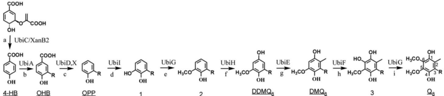
The ubi genes have been extensively studied over a period offive decades since the pioneering work of Cox and Gibson[17]. Biosynthesis of Q8in E. coli requires nine ubi genes, most of them encoding enzymes
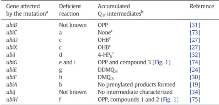
that decorate the aromatic ring of the 4-hydroxybenzoate (4-HB) uni-versal precursor (Fig. 1). Noticeably in E. coli, ubi genes are all scattered around the chromosome (Fig. 2A and B). It is important to stress that in a few cases, the mutation of genes required for Q8production led to the
accumulation of 3-octaprenylphenol (OPP), an early intermediate of Q8
biosynthetic pathway (Fig. 1andTable 1). As a consequence, these
Abbreviations: SAM, S-Adenosylmethionine; Q /Q8, Coenzyme Q /Q8; DMK/DMK8,
Demethylmenaquinone; DDMQ8, Demethyldemethoxy coenzyme Q8; DMQ8, Demethoxy
coenzyme Q8; DMSO, Dimethyl sulfoxide; FAD, Flavine adenine dinucleotide; FMN,
Flavin mononucleotide; H2O2, Hydrogen peroxide; 3-HB, 3-Hydroxybenzoate; 4-HB,
4-Hydroxybenzoate; Fe–S, Iron–sulfur; MK/MK8, Menaquinone; OHB,
3-Octaprenyl-4-hydroxybenzoate; 4-HP8, 3-Octaprenyl-4-hydroxyphenol; OPP, 3-Octaprenylphenol;
PHBH, Para-hydroxybenzoate hydroxylase; PMF, Proton motive force; O2−, Superoxide
anion; TMAO, Trimethylamine N-oxide
☆ This article is part of a Special Issue entitled: 18th European Bioenergetic Conference. ⁎ Corresponding author at: Laboratoire de Chimie Bactérienne, 31 Chemin Joseph Aiguier, 13402 Marseille Cedex 20, France. Tel.: +33 4 91 16 45 79; fax: +33 4 91 71 89 14.
E-mail address:barras@imm.cnrs.fr(F. Barras).
0005-2728/$– see front matter © 2014 Elsevier B.V. All rights reserved. http://dx.doi.org/10.1016/j.bbabio.2014.01.015
genetics studies sometimes failed at identifying precisely the biosyn-thetic step altered by the mutation.
In 1968, a screen based on the selection of mutants unable to grow on malate and examination of the quinone content of these strains
grown on glucose led to the identification of ubiA as the first gene involved in Q8biosynthesis[18]. Poole and colleagues found the ubiA
mutant to be unable to grow aerobically on non-fermentable substrates but able to grow anaerobically on glycerol with alternative electron acceptors such as fumarate[19]. The ubiA gene was predicted to encode a membrane-bound 4-hydroxybenzoate octaprenyltransferase. Within the same operon, ubiA lies downstream the ubiC gene (Fig. 2B). The UbiC protein catalyzes thefirst committed step in the biosynthesis of Q8, the conversion of chorismate to 4-HB (Reaction a,Fig. 1)[20].
Inter-estingly, in Xanthomonas campestris (which does not contain any ubiC gene or homologue), the XanB2 protein was reported to convert chorismate into 3-HB or 4-HB using two distinct catalytic domains not related to UbiC (Reaction a,Fig. 1)[21].
Mutation in the E. coli ubiG gene yields strains unable to grow aero-bically on nonfermentable substrates[22]. UbiG is one of the few Ubi enzymes that has been purified and assayed in vitro. Activity assays showed that UbiG is a S-adenosylmethionine (SAM)-dependent meth-yltransferase, which catalyzes the two O-methylation steps of Q8
biosynthesis (Reactions e and i,Fig. 1)[23].
The ubiE gene encodes the second, also SAM-dependent, meth-yltransferase of the Q8biosynthetic pathway (Reaction g,Fig. 1)
[24]. The ubiE mutant is deficient for growth on succinate and accumulates demethyldemethoxy-coenzyme Q8(DDMQ8) and
demethylmenaquinone (DMK8) as predominant intermediates
(Table 1), leading to the conclusion that ubiE is required for C-methylation in both ubiquinone and menaquinone synthesis[24]. An E. coli ubiD mutant accumulates 3-octaprenyl-4-hydroxybenzoate (OHB) (Reaction c,Fig. 1)[25,26]. It was shown that a partially purified membrane-bound UbiD protein was able to convert OHB into OPP, strongly suggesting UbiD involvement in the decarboxylation step of Q8
biosynthesis[26]. However, a ubiD mutant retains the ability to produce about 25% of the wild-type levels of Q8, consistent with the existence of
a second decarboxylase[26]. Inactivation of the ubiX gene leads to low levels of Q8, a reduced growth on succinate and accumulation of OHB
(Table 1), suggesting a decarboxylase activity for UbiX[27]. A hypothesis is that UbiD and UbiX, which share no sequence similarity, function together during the decarboxylation of OHB (Reaction c,Fig. 1)[27]. Moreover, it might be worth noting that in Salmonella enterica serovar Paratyphi, ubiX and ubiD are organized as a single fusion gene ubiX-ubiD (SPA2778 gene). In E. coli O157:H7, in addition to UbiD and UbiX, a probable aromatic acid decarboxylase called Pad1 was identified but its role in Q8biosynthesis has not been investigated so far.
Hydroxylations represent three of the nine reactions required for Q8
biosynthesis in E. coli. These reactions introduce hydroxyl groups at positions C-5, C-1, and C-6 of the aromatic ring of Q8(Reactions d, f
and h,Fig. 1), and the genes encoding these enzymes were proposed to be ubiB, ubiH, and ubiF, respectively (Fig. 2A and B)[28]. ubiH was thefirst of these genes to be identified and, based on sequence compar-ison, was proposed to be aflavin-containing monooxygenase[29]. A ubiF mutant was shown to accumulate demethoxy coenzyme Q8 Fig. 1. Biosynthetic pathway of ubiquinone in Escherichia coli. The numbering of the aromatic carbon atoms is shown on coenzyme Q8, and the octaprenyl tail is represented by R on C-3 of
the different biosynthetic intermediates. The name of the enzymes catalyzing the reactions (each labeled with a lowercase letter) is indicated. Abbreviations used for 4-hydroxybenzoate (4-HB), 3-octaprenyl-4-hydroxybenzoate (OHB), 3-octaprenylphenol (OPP), coenzyme Q8(Q8), C1-demethyl-C6-demethoxy-Q8(DDMQ8), and C6-demethoxy-Q8 (DMQ8) are
underlined. The XanB2 protein, present in some prokaryotes but not in E. coli, catalyzes the production of 4-HB from chorismate. The biosynthetic intermediates that accumulate in mutants affected in the different steps are listed inTable 1.
ubiB ubiJ ubiE ubiA ubiC fre ubiD ubiF ubiG ubiH pepP ygfB ubiI ubiX purF cvpA 91.62 min 86.58 min 50.39 min 65.81 min 52.34 min 14.97 min 86.74 min
E. coli
100 min.
15 50 52 65 87 92 ubiF ubiG ubiX ubiH-ubiI ubiE-ubiJ-ubiB ubiC-ubiA ubiDA
B
Fig. 2. Genetic organization and location of ubi genes on the Escherichia coli chromosome. A. Location of the eleven ubi genes on the 100 min map of the E. coli chromosome. B. Genetic organization of the ubi genes in E. coli. The location of each gene (or thefirst gene of the operon) is indicated on the left. The genes required for ubiquinone biosynthesis are indicated in bold. Genes encoding monooxygenases are symbolized by black arrows and genes encoding methyltransferases are symbolized by gray arrows.
(DMQ8) (Table 1,Fig. 1) indicating that UbiF, also related to
flavin-containing monooxygenases, was responsible for introducing a hydroxyl group at C-6 of the aromatic ring[30]. UbiB was initially proposed to be involved in the C-5 hydroxylation because a ubiB mutant was shown to accumulate OPP and failed to produce Q8(Table 1)[25,31]. However,
the UbiB protein does not contain any signature sequence for monooxygenases but rather shares identity with a large family of eukaryotic-type protein kinases[31]. Actually, we discovered that the C-5 hydroxylase was the product of the visC gene[32]. Accordingly, the visC gene, locating immediately downstream of ubiH within the same operon, was renamed ubiI (Fig. 2B). A ubiI mutant has a low level of Q8and accumulates 3-octaprenyl-4-hydroxyphenol (4-HP8), an
in-termediate that results from a C5-hydroxylation defect (Table 1)[32]. In fact, 4-HP8is formed by hydroxylation of OPP on C1 by UbiH without
reaction d and e taking place (Fig. 1). Therefore, the UbiH protein is able to perform the C1-hydroxylation in the absence of the methoxyl group on C5. The UbiI protein displays the typical domains of flavin-containing monooxygenases and shares 30 and 39% sequence identity with UbiH and UbiF, respectively[32]. Thus, we proposed to assign to UbiI thefirst hydroxylation step in Q8biosynthesis (Reaction d,Fig. 1)
[32]. The role of the ubiB gene product remains to be investigated. In E. coli, the yigP gene (called ubiJ inFig. 2B) lies in between the ubiE and ubiB genes[31]. Recently, yigP was proposed to encode a small RNA of 252 nucleotides (referred to as esrE), which was proposed to be essential in E. coli[33]. We also characterized the yigP gene in E. coli and Salmonella and our results did not support these conclusions as yigP deletions were obtained in both bacteria. We found yigP to be required for Q8biosynthesis and we changed the name of yigP into
ubiJ[34]. Regarding the small RNA issue, a“scrambled” ubiJ allele including the mutation of 30% of the nucleotides without changing the amino acid sequence restored Q8biosynthesis in Salmonella ubiJ mutant
[34]. We therefore believe that the biological function of ubiJ is mediated by a protein. We also demonstrated that a ubiJ mutant was impaired for growth under aerobic conditions, but did not present any growth defect anaerobically, either with glucose or glycerol supplemented with differ-ent electron acceptors[34].
In conclusion, all enzymatic reactions necessary for Q8biosynthesis in
aerobic conditions have been assigned to a Ubi protein (Fig. 1). However, the role of UbiB and UbiJ proteins remains to be established, as they are also essential for this process.
3. Structural analysis
The E. coli chorismate lyase UbiC structure was solved at 1.4 Å reso-lution[35,36]. This monomeric enzyme is composed of 164 amino acid
Structural information about the UbiX decarboxylase are available from studies carried out in Pseudomonas aeruginosa and E. coli O157: H7. The P. aeruginosa UbiX structure was determined to 1.5 Å resolution
[38]. It shows that the enzyme assembles into a dodecamer, each subunit displaying a typical Rossmann fold, and contains one Flavin MonoNucleotide (FMN) at the interface between two subunits (PDB ID: 3ZQU)[38]. A paralog of UbiX named Pad1 (52% identity) was iden-tified in E. coli O157:H7. Its three-dimensional structure has been deter-mined and refined at 2.0 Å resolution (PDB ID: 1SBZ) (Fig. 4A)[39]. Each Pad1 monomer of the dodecameric assembly consists of a typical Rossmann fold and contains a non-covalently bound molecule of FMN (Fig. 4A)[39]. The FMN cofactor also lies at the interface between two monomers. As expected, the structures of UbiX from P. aeruginosa and Pad1 from E. coli O157:H7 are highly comparable as shown by the root-mean-square deviation of aligned Cα atoms in the range 1.1–1.5 Å (Fig. 4B).
Structural information for the UbiD decarboxylase derived from studies carried out in P. aeruginosa and E. coli. Two genes, PA0254 and PA5237, predicted to encode UbiD homologs arise in P. aeruginosa
[40]. The three-dimensional structure of PA0254 has been determined in two different crystal forms to resolutions of 1.95 and 2.3 Å, respec-tively, showing a dimeric assembly (PDB ID: 4IP2) (Fig. 5A). Strikingly, the quaternary structure of P. aeruginosa PA0254 (25% identity with E. coli UbiD) differs from the hexameric organization of UbiD from E. coli (PDB ID: 2IDB, unpublished,Fig. 5B) and of PA5237 (76% identity with E. coli UbiD)[40]. Each subunit of PA0254, whose overall fold is
ubiE g DDMQ8 [24]
ubiF h DMQ8 [30]
ubiA b No prenylated products formed [19] ubiJ Not known No intermediate characterized [34] ubiH f OPP, compounds 1 and 2 (Fig. 1) [75]
a
The mutations that affect the ubi genes reported in this table vary from point mutation to transposon insertion or complete deletion. See references for details.
b
As detected by the techniques used in the reported studies. For example, OPP is ade-quately detected by using radiolabeled 4-HB but is not evidenced when using electro-chemical detection coupled to HPLC.
c
A low level of Q8is observed in these strains.
Fig. 3. Structure of the chorismate lyase UbiC from Escherichia coli. Crystal structure of the chorismate lyase UbiC from E. coli in complex with the 4-hydroxybenzoate, 1.0 Å resolution (PDB ID: 1TT8)[37]. The core of the protein fold is a 6-stranded antiparallelβ-sheet, with two helix–turn–helix loops colored in yellow and orange. The two internal cavities are colored in gray. The 4-HB product is located in one of these cavities, behind the twoflaps in a hydrophobic pocket.
similar to that of UbiD from E. coli, consists of three domains. The N-terminal part of the molecule is built up of two domains that pack tightly to each other and a C-terminalα/β domain which displays a topol-ogy characteristic for the UbiD protein family[40]. The middle domain shows significant structural similarity to the FMN-binding split barrel from a family offlavoproteins, including the NADH: FMN oxidoreductase from Methylobacillusflagelates (PDB ID: 3E4V), the flavin reductase from Shewanella baltica (PDB ID: 3HMZ), or theflavoredoxin from Desulfovibrio vulgaris (PDB ID: 2D5M). This middle domain also contains a metal bind-ing site, with a magnesium ion coordinated by two histidine and gluta-mate residues, which are conserved in the corresponding metal binding site of some FMN binding proteins. However, no evidence for the incor-poration of aflavin cofactor in PA0254 is available so far.
Recently, we determined the crystal structure of a truncated form of UbiI, involved in the aerobic C5-hydroxylation reaction (Fig. 6A)[32]. Only this form, lacking the 35C-terminal residues, and not the full-length protein, too prone to precipitate, could crystallize. Nevertheless, this structure provides some useful information, in particular as it shares many features with that of the canonical FAD-containing para-hydroxybenzoate hydroxylase (PHBH)[41]. In particular, comparison with PHBH reveals a FAD binding site in UbiI (Fig. 6B) which has been validated by site directed mutagenesis, thus supporting that UbiI and probably UbiH and UbiF are indeed FAD-containing monooxygenases
[32].
As shown from this brief survey, it is obvious that a lot is still missing regarding the structural biology of Q8pathway: few proteins have been
structurally characterized and rather incomplete information has been gained from those which have been crystallized. This opens a large field of research for the future.
4. Physiological role of Q8
4.1. Interplay between Q8and redox sensing two-component systems
The Arc (anoxic redox control) two-component system of E. coli comprises the ArcB transmembrane sensor kinase and the cytosolic ArcA response regulator. It plays a major role in a transcriptional regulatory network that allows facultative anaerobic bacteria to sense various respiratory growth conditions[42]. Georgellis and colleagues showed that quinones act as direct negative signals that inhibit
Fig. 4. Structures of the UbiX decarboxylase from Pseudomonas aeruginosa and the Pad1 paralog from Escherichia coli O157:H7. A. Crystal structure of the dodecameric Pad1 from E. coli O157:H7 complexed with aflavin mononucleotide (FMN), 2.0 Å resolution (PDB ID: 1SBZ)[39]. The view along the threefold axis shows the trimeric arrangement of Pad1. Each subunit is shown in a different color. The FMN cofactor is shown as yellow sticks and its binding site is located at the interface between two monomers. B. Superimposition of one subunit of the dodecameric Pad1 from E. coli O157:H7 (in green) with the corresponding subunit of the dodecameric UbiX from Pseudomonas aeruginosa (PDB ID 3ZQU) (in red). Theflavin cofactors FMN of the two proteins are colored in green in UbiX and in red in Pad1.
Fig. 5. Structures of the UbiD decarboxylases from Pseudomonas aeruginosa and Escherichia coli. A. Crystal structure of the dimeric PA0254 from P. aeruginosa, a putative aromatic acid de-carboxylase bearing a C-terminal domain characteristic of the UbiD protein family, 1.95 Å res-olution (PDB ID: 4IP2)[40]. The three domains of each subunit are shown in blue (N-terminal domain), green (middle domain) and red (C-terminal domain). The magnesium site is indi-cated by a black sphere in the middle domain. B. Crystal structure of the hexameric UbiD from E. coli (PDB ID: 2IDB). Each subunit is shown in a different color.
autophosphorylation of ArcB during aerobiosis[43]. They demonstrated that the oxidative power provided by the Q8pool induced the formation
of two intermolecular cysteine disulfide bonds within the cytosolic do-mains of an ArcB dimer, leading to the inactivation of ArcB kinase activity
[43,44]. Bekker and colleagues provided evidence that additional regula-tion of ArcB kinase activity by the redox state of menaquinone (MK8)
was prevalent under microaerobic to anaerobic conditions[45]. They con-cluded that activation of the ArcBA system was controlled by both MK8
and Q8pools[45]. Moreover, the extent of ArcA phosphorylation was
shown to be modulated by the oxygen supply rate in an ubiE mutant, leading to the conclusion that the oxidized form of DMK8could inactivate
ArcB[46]. However, the authors did not take in consideration the DDMQ8
which also accumulates in a ubiE mutant[24,34]and could play a role in ArcB regulation in the ubiE mutant. A complementary study demonstrat-ed that (D)MK8was required for activation of ArcB upon a shift to anoxic
conditions[4]. The midpoint redox potential of the cysteines of ArcB was determined to be approximately−41 mV, which is in agreement with the proposed model in which the Q8pool can oxidize these cysteines
under aerobic conditions and the disulfide bonds can be reduced by the (D)MK8pool upon a shift to anaerobic conditions[4].
Another example of redox regulation is illustrated by the RegBA two-component system of Rhodobacter capsulatus. RegB is a membrane-spanning sensor kinase that autophosphorylates and transfers the phos-phoryl group to its cognate response regulator RegA[47]. The RegB/ RegA system regulates the synthesis of numerous energy-generating and energy-utilizing systems[48]. Recently, RegB was shown to bind weakly both oxidized and reduced Q8with nearly equal affinity, although
oxidized Q8alone inhibited kinase activity[49]. The current view is that
interaction of RegB with both the reduced and oxidized forms of Q8
allows the kinase to monitor and tune the cellular energy state[49]. In conclusion, in both E. coli and R. capsulatus models, and presum-ably many other bacteria, a connection between the pool, and the redox state, of Q8and two-component systems endows the cell with
the capacity to modulate the expression of multiple genes in response to changes in redox conditions.
4.2. Q8and oxidative stress
The involvement of ubiquinone in oxidative stress resistance was first addressed in eukaryotes, where it was suggested that its reduced
form, i.e. ubiquinol, was able to function as a lipid-soluble anti-oxidant
[50]. Ubiquinol was shown to scavenge lipid peroxyl radicals and there-by prevents a chain reaction causing oxidative damage to polyunsatu-rated fatty acids of biological membranes, a process known as lipid peroxidation[51]. In yeast, the biosynthetic intermediate DMQ6was
concluded to lack antioxidant activity because it failed to protect cells against oxidative stress generated by hydrogen peroxide (H2O2) or
linolenic acid[52]. In prokaryotes, an exhaustive study aimed at investi-gating the role of Q8in resistance against oxidative stress was carried
out by Søballe and Poole[53]. Using a ubiCA mutant deficient for Q8
production, they observed an accumulation of superoxide (O2•−) in
the membranes of the mutant compared to wild-type membranes, reflecting the lack of superoxide-scavenging ubiquinol[53]. Moreover, intracellular H2O2concentration was increased 1.8-fold in the ubiCA
mutant, which was also found to be hypersensitive to an oxidative stress mediated by H2O2[53]. Expression of katG gene, encoding a
cata-lase, and intracellular catalase activity, were both increased in the ubiCA mutant[53]. These observations are consistent with the hypothesis that Q8limits O2•−and H2O2accumulation in scavenging reactive oxygen
species. However, given the multiplicity of catalases, peroxidases and superoxide dismutases present in most bacteria, the relative contribu-tion of Q8to the overall anti-oxidative stress defenses system remains
to be assessed. 4.3. Q8and respiration
The respiratory chain of E. coli contains many enzymes allowing the organism to transfer electrons to oxygen or to use alternative terminal acceptors such as nitrate, dimethyl sulfoxide (DMSO), trimethylamine N-oxide (TMAO) or fumarate[1,2,7]. Q8is generally depicted as the
aerobic quinone since it is more abundant than MK8during aerobic
growth, whereas MK8is essential for anaerobic respiration using
fuma-rate, DMSO and TMAO as electron acceptors[13,14]. In this way, a ubiCA mutant showed severely diminished growth yields aerobically but not anaerobically using nitrate or fumarate as terminal electron acceptors
[54]. Conversely, a ubiE mutant, which contains DMK8but no MK8,
was able to grow with fumarate, TMAO and DMSO, but not with nitrate as electron acceptor[55]. It was then concluded that DMK8(in addition
to MK8) could serve as a redox mediator in fumarate, TMAO and DMSO
respiration, but not in nitrate respiration [55]. Moreover, it was
Fig. 6. Structure of the UbiI monooxygenase from Escherichia coli. A. Crystal structure of the C-terminal truncated form of E. Coli C5-hydroxylase UbiI involved in Q8biosynthesis, 2.0
demonstrated that nitrate reductase A could accept electrons from both Q8and MK8pools, the coupling being more effective with ubiquinol
than with menaquinol[56].
Recently, Bekker and colleagues reported that DMK8played a role
not only in TMAO−, DMSO- and fumarate-dependent respiration, but also in oxidation of succinate[15]. They further concluded that all three quinol oxidases of E. coli accepted electrons from DMK8based
on the residual aerobic respiration observed in a ubiE mutant[15]. As already mentioned, another hypothesis is that DDMQ8might provide
electron towards the quinol oxidases in the ubiE mutant. In the future, it will be interesting to know whether these remaining uncertainties will challenge the classic view associating ubiquinone to aerobic growth and menaquinone to anaerobic growth.
4.4. Q8and antibiotics resistance
The proton-pumping NADH:ubiquinone oxidoreductase, also called complex I, catalyzes the electron transfer from NADH to Q8linked
with a proton translocation from the negative inner to the positive outer side of the membrane[57]. Thus, a proton-motive (PMF) force is generated, which is utilized mainly for ATP synthesis. PMF is instrumen-tal in allowing either import or export of antibiotics[58–60]. As a conse-quence, ubi mutations are likely to modify levels of resistance to antibiotics sustained by bacteria. In fact, resistance to low levels of aminoglycoside antibiotics has been used for long for the isolation of menaquinone-deficient mutants in Bacillus subtilis since the MK deficiency depresses the rate of accumulation of aminoglycosides, which accounts for the resistance phenotype[61]. Thereby, a phenotype of increased aminoglycoside resistance led to the identification of the aarE gene, the ubiA homologue in Providencia stuartii[62]. In E. coli, a ubiF mutation was associated with pleomycin resistance[63]while a ubiD mutation was found to be associated with decreased transport of streptomycin and gentamicin, and increased resistance to those antibi-otics[64]. This mutant also exhibited increased resistance to several other aminoglycoside antibiotics, but not spectinomycin[64]. A series of E. coli mutants located in the vicinity of ubiB and ubiD and consistent with a disruption of Q8synthesis were found to present zwittermicin A
resistance, a novel broad-spectrum antibiotics produced by Bacillus cereus[65]. Rationally, a ubiD mutant was also resistant to zwittermicin A[65]. All together, these studies highlight that connections between Q8production and antibiotics resistance occur in many bacteria.
4.5. Q8and bacterial virulence
In the Caenorhabditis elegans model, Clarke and colleagues revealed that nematodes fed diets of respiratory deficient E. coli lacking Q8
lived significantly longer than worms fed the wild-type parental strain
[66]. Moreover, these E. coli mutants were degraded in the early adult worm and did not accumulate in the intestinal tract[66]. These data led the authors to propose that bacterial respiration might act as a viru-lence factor, influencing the ability of bacteria to colonize – and subse-quently harm– the animal host[66]. As a matter of fact, the role of respiration in host–pathogen interactions has recently been underlined in several studies, including those dedicated to Shigella pathogenicity
[67]and more recently to Salmonella[34].
Indeed, early studies brought in light the requirement of a functional Q8biosynthesis pathway for theflagellation of Salmonella[68].
Interest-ingly, the increase in motility response occurred within a narrow range of the increase in Q8content[68]. More recently, we showed that
Salmonella ubiJ mutant, a strain deficient for Q8production under
aerobic conditions, was killed within macrophages[34]. When macro-phages were infected with an anaerobic inoculum of the ubiJ mutant (in which Q8was still produced), the ubiJ mutant recovered its proliferation
capacity, clearly establishing the requirement for Q8for efficient
intracel-lular proliferation[34]. Several possibilities can be considered to connect Q8defects and virulence attenuation: (i) the necessity for the bacterium
to use aerobic respiration– thus Q8– to grow intracellularly; (ii) the
antioxidant role of Q8to avoid the oxidative burst produced by the
host; (iii) the requirement of a PMF-dependent process for virulence. Thereby, our recent data combined to results from other groups highlighted the importance of Q8biosynthesis in the context of the
host–pathogen interplay and revealed an unexpected link between Q8
and bacterial virulence. 5. Conclusion and perspectives
In this review, we have presented the actors involved in Q8
biosyn-thetic pathway. Phenotypic characterizations of various mutants coupled with the examination of Q8content led to the identification of genes
involved in the decoration of the aromatic ring, from the 4-HB precursor to thefinal ubiquinone. When available, the three-dimensional structures of these enzymes were presented. Last, the role of Q8in diverse cellular
processes was detailed: genetic regulation through the two-component systems ArcBA and RegBA, adaptation to oxidative stress, respiration, antibiotics resistance and bacterial virulence.
Whereas nine ubi genes were found to be directly involved in Q8
biosynthesis, the biological function of ubiB and ubiJ gene products remain unknown. The involvement of UbiB in the C-5 hydroxylation step has to be definitively abandoned. Instead, UbiB might play a regu-latory role through a kinase activity, even though it is still unknown if it displays such an activity and what substrate it acts on. Based on sequence similarities between ubiJ and genes involved in lipid metabo-lism, UbiJ could serve as a carrier of the isoprenoid hydrophobic tail prior the action of monooxygenases and methyltransferases. Alternative-ly, it might chaperone prenylated intermediates during the biosynthetic process.
Intriguingly, we found that the ubiJ mutant synthesizes almost wild-type levels of Q8under anaerobic growth conditions[34]. Actually,
ubiH, ubiF and ubiI mutants also synthesize significant levels of Q8
anaerobically[28,32]. The three latter genes encodeflavin-dependent monooxygenases, which use molecular oxygen to catalyze their respec-tive hydroxylation reactions. In consequence, these proteins will not participate in Q8biosynthesis under anaerobic conditions because of
the absence of molecular oxygen. Therefore, alternative anaerobic hydroxylases must carry out the hydroxylation reactions in anaerobic conditions as proposed by Alexander and Young[28], but so far the identity of these proteins has remained elusive. The involvement of UbiJ only in aerobic Q8biosynthesis is more difficult to rationalize
than that of the aerobic monooxygenases and may reflect a functional link with those enzymes.
It is important to note that with the exception of the E. coli methyl transferase UbiG[23], the partially purified membrane-bound UbiD
[26]and the UbiA octaprenyltransferase[69], none of the other enzymes (the decarboxylases, the putative kinase and the hydroxylases) that participate to Q8biosynthesis have been assayed under in vitro
condi-tions using pure proteins. As a consequence, most of the predicted activ-ities associated with Ubi enzymes remain to be fully established by straight biochemical analyses. This is not surprising considering the difficulties to purify these proteins, their probable association to the membranes and their instability, as well as the difficulties to get access to the substrates, not commercially available.
Another issue is whether Ubi proteins may belong to a large multi-protein complex as suggested by an early report[70]. By a combination of sonication, gelfiltration and equilibrium sedimentation with a sucrose gradient, the author purified a mega complex of at least 12 proteins, rang-ing from 40 to 80 kDa and exhibitrang-ing a molecular weight estimated to 2.106Da. This complex had the capacity to process OPP into Q8in the
presence of NADPH, SAM and O2. It is intriguing that, in spite of this
fascinating observation, no further studies concerning this bacterial com-plex have been reported then. The existence of a high-molecular weight complex has also been documented in Saccharomyces cerevisiae, mostly by genetic and partly by biochemical studies[71,72]. The complex may
Thanks are due to the members of the FB group for fruitful discus-sions. We acknowledge Ludovic Pecqueur for the help in the prepara-tion of molecular graphicsfigures with PyMol. This work was funded by the CNRS, Aix-Marseille Université (AMU), the Institut Universitaire de France (IUF), and the French State Program “Investissements d'Avenir” (Grant “LABEX DYNAMO”, ANR-11-LABX-0011).
References
[1] G. Unden, J. Bongaerts, Alternative respiratory pathways of Escherichia coli: energetics and transcriptional regulation in response to electron acceptors, Biochim. Biophys. Acta 1320 (1997) 217–234.
[2] R.K. Poole, G.M. Cook, Redundancy of aerobic respiratory chains in bacteria? Routes, reasons and regulation, Adv. Microb. Physiol. 43 (2000) 165–224.
[3] I. Belevich, V.B. Borisov, M.I. Verkhovsky, Discovery of the true peroxy intermediate in the catalytic cycle of terminal oxidases by real-time measurement, J. Biol. Chem. 282 (2007) 28514–28519.
[4] A.F. Alvarez, C. Rodriguez, D. Georgellis, Ubiquinone and menaquinone electron carriers represent the yin and yang in the redox regulation of the ArcB sensor kinase, J. Bacteriol. 195 (2013) 3054–3061.
[5] B. Schoepp-Cothenet, C. Lieutaud, F. Baymann, A. Vermeglio, T. Friedrich, D.M. Kramer, W. Nitschke, Menaquinone as pool quinone in a purple bacterium, Proc. Nat. Acad. Sci. U. S. A. 106 (2009) 8549–8554.
[6] A. Nakamura, T. Suzawa, Y. Kato, T. Watanabe, Species dependence of the redox potential of the primary electron donor p700 in photosystem I of oxygenic photo-synthetic organisms revealed by spectroelectrochemistry, Plant Cell Physiol. 52 (2011) 815–823.
[7] V.B. Borisov, R.B. Gennis, J. Hemp, M.I. Verkhovsky, The cytochrome bd respiratory oxygen reductases, Biochim. Biophys. Acta 1807 (2011) 1398–1413.
[8] B. Nowicka, J. Kruk, Occurrence, biosynthesis and function of isoprenoid quinones, Biochim. Biophys. Acta 1797 (2010) 1587–1605.
[9] M. Bentinger, M. Tekle, G. Dallner, Coenzyme Q-biosynthesis and functions, Biochem. Biophys. Res. Commun. 396 (2010) 74–79.
[10] M. Turunen, J. Olsson, G. Dallner, Metabolism and function of coenzyme Q, Biochim. Biophys. Acta 1660 (2004) 171–199.
[11]R. Meganathan, Ubiquinone biosynthesis in microorganisms, FEMS Microbiol. Lett. 203 (2001) 131–139.
[12] U.C. Tran, C.F. Clarke, Endogenous synthesis of coenzyme Q in eukaryotes, Mitochondrion 7 (2007) S62–S71.
[13] B.J. Wallace, I.G. Young, Role of quinones in electron transport to oxygen and nitrate in Escherichia coli. Studies with a ubiA- menA- double quinone mutant, Biochim. Biophys. Acta 461 (1977) 84–100.
[14]B. Søballe, R.K. Poole, Microbial ubiquinones: multiple roles in respiration, gene regulation and oxidative stress management, Microbiology 145 (1999) 1817–1830. [15] P. Sharma, M.J. Teixeira de Mattos, K.J. Hellingwerf, M. Bekker, On the function of the
various quinone species in Escherichia coli, FEBS J. 279 (2012) 3364–3373. [16] S. Grimaldi, B. Schoepp-Cothenet, P. Ceccaldi, B. Guigliarelli, A. Magalon, The
prokaryotic Mo/W-bisPGD enzymes family: a catalytic workhorse in bioenergetic, Biochim. Biophys. Acta 1827 (2013) 1048–1085.
[17] G.B. Cox, F. Gibson, Biosynthesis of vitamin K and ubiquinone. Relation to the shikimic acid pathway in Escherichia coli, Biochim. Biophys. Acta 93 (1964) 204–206.
[18] G.B. Cox, F. Gibson, J. Pittard, Mutant strains of Escherichia coli K-12 unable to form ubiquinone, J. Bacteriol. 95 (1968) 1591–1598.
[19]G. Wu, H.D. Williams, F. Gibson, R.K. Poole, Mutants of Escherichia coli affected in respiration: the cloning and nucleotide sequence of ubiA, encoding the membrane-bound p-hydroxybenzoate:octaprenyltransferase, J. Gen. Microbiol. 139 (1993) 1795–1805.
[20] B.P. Nichols, J.M. Green, Cloning and sequencing of Escherichia coli ubiC and purification of chorismate lyase, J. Bacteriol. 174 (1992) 5309–5316.
[21] L. Zhou, J.Y. Wang, J.H. Wang, A. Poplawsky, S.J. Lin, B.S. Zhu, C.Q. Chang, T.L. Zhou, L.H. Zhang, Y.W. He, Mol. Microbiol. 87 (2013) 80–93.
[22] G. Wu, H.D. Williams, M. Zamanian, F. Gibson, R.K. Poole, Isolation and characterization of Escherichia coli mutants affected in aerobic respiration: the cloning and nucleotide sequence of ubiG. Identification of an S-adenosylmethionine-binding motif in protein, RNA, and small-molecule methyltransferases, J. Gen. Microbiol. 138 (1992) 2101–2112. [23]W.W. Poon, R.J. Barkovich, A.Y. Hsu, A. Frankel, P.T. Lee, J.N. Shepherd, D.C. Myles, C.F. Clarke, Yeast and rat Coq3 and Escherichia coli UbiG polypeptides catalyze both O-methyltransferase steps in coenzyme Q biosynthesis, J. Biol. Chem. 274 (1999) 21665–21672.
[28]K. Alexander, I.G. Young, Alternative hydroxylases for the aerobic and anaerobic biosynthesis of ubiquinone in Escherichia coli, Biochemistry 17 (1978) 4750–4755. [29] K. Nakahigashi, K. Miyamoto, K. Nishimura, H. Inokuchi, Isolation and characterization of a light-sensitive mutant of Escherichia coli K-12 with a mutation in a gene that is required for the biosynthesis of ubiquinone, J. Bacteriol. 174 (1992) 7352–7359. [30]O. Kwon, A. Kotsakis, R. Meganathan, Ubiquinone (coenzyme Q) biosynthesis in
Escherichia coli: identification of the ubiF gene, FEMS Microbiol. Lett. 186 (2000) 157–161.
[31] W.W. Poon, D.E. Davis, H.T. Ha, T. Jonassen, P.N. Rather, C.F. Clarke, Identification of Escherichia coli ubiB, a gene required for thefirst monooxygenase step in ubiquinone biosynthesis, J. Bacteriol. 182 (2000) 5139–5146.
[32] M. Hajj Chehade, L. Loiseau, M. Lombard, L. Pecqueur, A. Ismail, M. Smadja, B. Golinelli-Pimpaneau, C. Mellot-Draznieks, O. Hamelin, L. Aussel, S. Kieffer-Jaquinod, N. Labessan, F. Barras, M. Fontecave, F. Pierrel, A new gene involved in coenzyme Q biosynthesis in Escherichia coli: UbiI functions in aerobic C5-hydroxylation, J. Biol. Chem. 288 (2013) 20085–20092.
[33] Z. Chen, Y. Wang, Y. Li, Y. Li, N. Fu, J. Ye, H. Zhang, Esre: a novel essential non-coding RNA in Escherichia coli, FEBS Lett. 586 (2012) 1195–1200.
[34] L. Aussel, L. Loiseau, M. Hajj Chehade, B. Pocachard, M. Fontecave, F. Pierrel, F. Barras, Coenzyme Q biosynthesis in Escherichia coli and Salmonella typhimurium: ubiJ, a new gene required for aerobic growth and proliferation in macrophage, J. Bacteriol. 196 (2014) 70–79.
[35] C. Stover, M.P. Mayhew, M.J. Holden, A. Howard, D.T. Gallagher, Crystallization and 1.1-A diffraction of chorismate lyase from Escherichia coli, J. Struct. Biol. 129 (2000) 96–99.
[36] D.T. Gallagher, M. Mayhew, M.J. Holden, A. Howard, K.J. Kim, V.L. Vilker, The crystal structure of chorismate lyase shows a new fold and a tightly retained product, Proteins 44 (2001) 304–311.
[37]N. Smith, A.E. Roitberg, E. Rivera, A. Howard, M.J. Holden, M. Mayhew, S. Kaistha, D.T. Gallagher, Structural analysis of ligand binding and catalysis in chorismate lyase, Arch. Biochem. Biophys. 445 (2006) 72–80.
[38]J. Kopec, R. Schnell, G. Schneider, Structure of PA4019, a putative aromatic acid decarboxylase from Pseudomonas aeruginosa, Acta Crystallogr. Sect. F: Struct. Biol. Cryst. Commun. 67 (2011) 1184–1188.
[39]E.S. Rangarajan, Y. Li, P. Iannuzzi, A. Tocilj, L.W. Hung, A. Matte, M. Cygler, Crystal structure of a dodecameric FMN-dependent UbiX-like decarboxylase (Pad1) from Escherichia coli O157: H7, Protein Sci. 13 (2004) 3006–3016.
[40] A. Jacewicz, A. Izumi, K. Brunner, R. Schnell, G. Schneider, Structural insights into the UbiD protein family from the crystal structure of PA0254 from Pseudomonas aeruginosa, PLoS One 8 (2013) e63161.
[41] H.A. Schreuder, P.A. Prick, R.K. Wierenga, G. Vriend, K.S. Wilson, W.G. Hol, J. Drenth, Crystal structure of the p-hydroxybenzoate hydroxylase-substrate complex refined at 1.9 A resolution. Analysis of the enzyme-substrate and enzyme-product complexes, J. Mol. Biol. 208 (1989) 679–696.
[42]A.S. Lynch, E.C. Lin, Escherichia coli and Salmonella: cellular and molecular biology, in: F.C. Neidhardt, R. Curtis III, J.L. Ingraham, E.C.C. Lin, K.B. Low, B. Magasanik, W.S. Reznikoff, M. Riley, M. Schaechter, H.E. Umbarger (Eds.), Am. Soc. Microbiol., 1996, pp. 1526–1538, (Washington, DC).
[43] D. Georgellis, O. Kwon, E.C. Lin, Quinones as the redox signal for the arc two-component system of bacteria, Science 292 (2001) 2314–2316.
[44] R. Malpica, B. Franco, C. Rodriguez, O. Kwon, D. Georgellis, Identification of a quinone-sensitive redox switch in the ArcB sensor kinase, Proc. Nat. Acad. Sci. U. S. A. 101 (2004) 13318–13323.
[45] M. Bekker, S. Alexeeva, W. Laan, G. Sawers, J. Teixeira de Mattos, K. Hellingwerf, The ArcBA two-component system of Escherichia coli is regulated by the redox state of both the ubiquinone and the menaquinone pool, J. Bacteriol. 192 (2010) 746–754. [46]P. Sharma, S. Stagge, M. Bekker, K. Bettenbrock, K.J. Hellingwerf, Kinase activity of ArcB from Escherichia coli is subject to regulation by both ubiquinone and demethylmenaquinone, PLoS One 8 (2013) e75412.
[47] T.H. Bird, S. Du, C.E. Bauer, Autophosphorylation, phosphotransfer, and DNA-binding properties of the RegB/RegA two-component regulatory system in Rhodobacter capsulatus, J. Biol. Chem. 274 (1999) 16343–16348.
[48] S. Elsen, L.R. Swem, D.L. Swem, C.E. Bauer, RegB/RegA, a highly conserved redox-responding global two-component regulatory system, Microbiol. Mol. Biol. Rev. 68 (2004) 263–279.
[49] J. Wu, C.E. Bauer, RegB kinase activity is controlled in part by monitoring the ratio of oxidized to reduced ubiquinones in the ubiquinone pool, MBio 1 (2010) e00272-10. [50] L. Ernster, G. Dallner, Biochemical, physiological and medical aspects of ubiquinone
function, Biochim. Biophys. Acta 1271 (1995) 195–204.
[51] P. Forsmark-Andree, G. Dallner, L. Ernster, Endogenous ubiquinol prevents protein modification accompanying lipid peroxidation in beef heart submitochondrial particles, Free Radic. Biol. Med. 19 (1995) 749–757.
[52] S. Padilla, T. Jonassen, M.A. Jiménez-Hidalgo, D.J. Fernández-Ayala, G. López-Lluch, B. Marbois, P. Navas, C.F. Clarke, C. Santos-Ocaña, Demethoxy-Q, an intermediate of
coenzyme Q biosynthesis, fails to support respiration in Saccharomyces cerevisiae and lacks antioxidant activity, J. Biol. Chem. 279 (2004) 25995–26004.
[53] B. Søballe, R.K. Poole, Ubiquinone limits oxidative stress in Escherichia coli, Microbiology 146 (2000) 787–796.
[54] B. Søballe, R.K. Poole, Requirement for ubiquinone downstream of cytochrome(s) b in the oxygen-terminated respiratory chains of Escherichia coli K-12 revealed using a null mutant allele of ubiCA, Microbiology 144 (1998) 361–373.
[55] U. Wissenbach, D. Ternes, G. Unden, An Escherichia coli mutant containing only demethylmenaquinone, but no menaquinone: effects on fumarate, dimethylsulfoxide, trimethylamine N-oxide and nitrate respiration, Arch. Microbiol. 158 (1992) 68–73. [56]T.H. Brondijk, D. Fiegen, D.J. Richardson, J.A. Cole, Roles of NapF, NapG and NapH,
subunits of the Escherichia coli periplasmic nitrate reductase, in ubiquinol oxidation, Mol. Microbiol. 44 (2002) 245–255.
[57] T. Friedrich, K. Steinmüller, H. Weiss, The proton-pumping respiratory complex I of bacteria and mitochondria and its homologue in chloroplasts, FEBS Lett. 367 (1995) 107–111.
[58]H.W. Taber, J.P. Mueller, P.F. Miller, A.S. Arrow, Bacterial uptake of aminoglycoside antibiotics, Microbiol. Rev. 51 (1987) 439–457.
[59] B.D. Davis, L.L. Chen, P.C. Tai, Misread protein creates membrane channels: an essential step in the bactericidal action of aminoglycosides, Proc. Nat. Acad. Sci. U. S. A. 83 (1986) 6164–6168.
[60]B. Ezraty, A. Vergnes, M. Banzhaf, Y. Duverger, A. Huguenot, A.R. Brochado, S.Y. Su, L. Espinosa, L. Loiseau, B. Py, A. Typas, F. Barras, Fe–S cluster biosynthesis controls uptake of aminoglycosides in a ROS-less death pathway, Science 340 (2013) 1583–1587. [61] H.W. Taber, E.A. Dellers, L.R. Lombardo, Menaquinone biosynthesis in Bacillus
subtilis: isolation of men mutants and evidence for clustering of men genes, J. Bacteriol. 145 (1981) 321–327.
[62] M.R. Paradise, G. Cook, R.K. Poole, P.N. Rather, Mutations in aarE, the ubiA homolog of Providencia stuartii, result in high-level aminoglycoside resistance and reduced expression of the chromosomal aminoglycoside 2′-N-acetyltransferase, Antimicrob. Agents Chemother. 42 (1998) 959–962.
[63] C.M. Collis, G.W. Grigg, An Escherichia coli mutant resistant to phleomycin, bleomycin, and heat inactivation is defective in ubiquinone synthesis, J. Bacteriol. 171 (1989) 4792–4798.
[64] L.E. Bryan, H.M. Van Den Elzen, Effects of membrane-energy mutations and cations on streptomycin and gentamicin accumulation by bacteria: a model for entry of
streptomycin and gentamicin in susceptible and resistant bacteria, Antimicrob. Agents Chemother. 12 (1977) 163–177.
[65] E.V. Stabb, J. Handelsman, Genetic analysis of zwittermicin A resistance in Escherichia coli: effects on membrane potential and RNA polymerase, Mol. Microbiol. 27 (1998) 311–322.
[66] F. Gomez, G.C. Monsalve, V. Tse, R. Saiki, E. Weng, L. Lee, C. Srinivasan, A.R. Frand, C.F. Clarke, Delayed accumulation of intestinal coliform bacteria enhances life span and stress resistance in Caenorhabditis elegans fed respiratory deficient E. coli, BMC Microbiol. 12 (2012) 300.
[67]B. Marteyn, N.P. West, D.F. Browning, J.A. Cole, J.G. Shaw, F. Palm, J. Mounier, M.C. Prévost, P. Sansonetti, C.M. Tang, Modulation of Shigella virulence in response to available oxygen in vivo, Nature 465 (2010) 355–358.
[68]J. Bar-Tana, B.J. Howlett, R. Hertz, Ubiquinone synthetic pathway inflagellation of Salmonella typhimurium, J. Bacteriol. 143 (1980) 637–643.
[69]L.A. Wessjohann, B. Sonntag, Prenylation of benzoic acid derivatives catalyzed by a transferase from Escherichia coli overproduction: method development and substrate specificity, Angew. Chem. Int. Ed. Engl. 35 (1996) 1697–1699. [70]H.E. Knoell, Isolation of a soluble enzyme complex comprising the ubiquinone-8
synthesis apparatus from the cytoplasmic membrane of Escherichia coli, Biochem. Biophys. Res. Commun. 91 (1979) 919–925.
[71]P. Gin, C.F. Clarke, Genetic evidence for a multi-subunit complex in coenzyme Q biosynthesis in yeast and the role of the Coq1 hexaprenyl diphosphate synthase, J. Biol. Chem. 280 (2005) 2676–2681.
[72] C.H. He, L.X. Xie, C.M. Allan, U.C. Tran, C.F. Clarke, Coenzyme Q supplementation or over-expression of the yeast Coq8 putative kinase stabilizes multi-subunit Coq polypeptide complexes in yeast coq null mutants, Biochim. Biophys. Acta (2013), http://dx.doi.org/10.1016/j.bbalip.2013.12.017.
[73]M. Siebert, K. Severin, L. Heide, Formation of 4-hydroxybenzoate in Escherichia coli: characterization of the ubiC gene and its encoded enzyme chorismate pyruvate-lyase, Microbiology 140 (1994) 897–904.
[74]P. Stroobant, I.G. Young, F. Gibson, Mutants of Escherichia coli K-12 blocked in the final reaction of ubiquinone biosynthesis: characterization and genetic analysis, J. Bacteriol. 109 (1972) 134–139.
[75] I.G. Young, P. Stroobant, C.G. Macdonald, F. Gibson, Pathway for ubiquinone biosyn-thesis in Escherichia coli K-12: gene-enzyme relationships and intermediates, J. Bacteriol. 114 (1973) 42–52.