HAL Id: hal-02864335
https://hal.univ-grenoble-alpes.fr/hal-02864335
Submitted on 11 Dec 2020
HAL is a multi-disciplinary open access
archive for the deposit and dissemination of
sci-entific research documents, whether they are
pub-lished or not. The documents may come from
teaching and research institutions in France or
abroad, or from public or private research centers.
L’archive ouverte pluridisciplinaire HAL, est
destinée au dépôt et à la diffusion de documents
scientifiques de niveau recherche, publiés ou non,
émanant des établissements d’enseignement et de
recherche français ou étrangers, des laboratoires
publics ou privés.
Distributed under a Creative Commons Attribution| 4.0 International License
Deletion of the Zinc Transporter Lipoprotein AdcAII
Causes Hyperencapsulation of Streptococcus
pneumoniae Associated with Distinct Alleles of the
Type I Restriction-Modification System
Claire Durmort, Giuseppe Ercoli, Elisa Ramos-Sevillano, Suneeta
Chimalapati, Richard Haigh, Megan de Ste Croix, Katherine Gould, Jason
Hinds, Yann Guerardel, Thierry Vernet, et al.
To cite this version:
Claire Durmort, Giuseppe Ercoli, Elisa Ramos-Sevillano, Suneeta Chimalapati, Richard Haigh, et al..
Deletion of the Zinc Transporter Lipoprotein AdcAII Causes Hyperencapsulation of Streptococcus
pneumoniae Associated with Distinct Alleles of the Type I Restriction-Modification System. mBio,
American Society for Microbiology, 2020, 11 (2), �10.1128/mBio.00445-20�. �hal-02864335�
Deletion of the Zinc Transporter Lipoprotein AdcAII Causes
Hyperencapsulation of Streptococcus pneumoniae Associated
with Distinct Alleles of the Type I Restriction-Modification
System
Claire Durmort,aGiuseppe Ercoli,bElisa Ramos-Sevillano,bSuneeta Chimalapati,bRichard D. Haigh,cMegan De Ste Croix,c
Katherine Gould,dJason Hinds,dYann Guerardel,e Thierry Vernet,aMarco Oggioni,cJeremy S. Brownb
aInstitut de Biologie Structurale (IBS), Univ. Grenoble Alpes, CEA, CNRS, Grenoble, France
bCentre for Inflammation and Tissue Repair, Department of Medicine, Royal Free and University College Medical School, Rayne Institute, London, United Kingdom
cDepartment of Genetics and Genome Biology, University of Leicester, Leicester, United Kingdom
dInstitute for Infection and Immunity, St. George’s University of London, London, United Kingdom
eUniv. Lille, CNRS, UMR 8576 —UGSF-Unité de Glycobiologie Structurale et Fonctionnelle, Lille, France
Giuseppe Ercoli and Elisa Ramos-Sevillano contributed equally to the manuscript.
ABSTRACT The capsule is the dominant Streptococcus pneumoniae virulence factor, yet how variation in capsule thickness is regulated is poorly understood. Here, we describe an unexpected relationship between mutation of adcAII, which encodes a zinc uptake lipoprotein, and capsule thickness. Partial deletion of adcAII in three of five capsular serotypes frequently resulted in a mucoid phenotype that biochemical analysis and electron microscopy of the D39 adcAII mutants confirmed was caused by markedly increased capsule thickness. Compared to D39, the hyperencapsulated ⌬adcAII mutant strain was more resistant to complement-mediated neutrophil killing and was hypervirulent in mouse models of invasive infection. Transcriptome analysis
of D39 and the ⌬adcAII mutant identified major differences in transcription of
the Sp_0505-0508 locus, which encodes an SpnD39III (ST5556II) type I restriction-modification system and allelic variation of which correlates with capsule thickness. A PCR assay demonstrated close linkage of the SpnD39IIIC and F alleles with the
hy-perencapsulated ⌬adcAII strains. However, transformation of ⌬adcAII with fixed
SpnD39III alleles associated with normal capsule thickness did not revert the
hyper-encapsulated phenotype. Half of hyperhyper-encapsulated ⌬adcAII strains contained the
same single nucleotide polymorphism in the capsule locus gene cps2E, which is re-quired for the initiation of capsule synthesis. These results provide further evidence for the importance of the SpnD39III (ST5556II) type I restriction-modification system for modulating capsule thickness and identified an unexpected linkage between
capsule thickness and mutation of ⌬adcAII. Further investigation will be needed to
characterize how mutation of adcAII affects SpnD39III (ST5556II) allele dominance and results in the hyperencapsulated phenotype.
IMPORTANCE The Streptococcus pneumoniae capsule affects multiple interactions with the host including contributing to colonization and immune evasion. During in-fection, the capsule thickness varies, but the mechanisms regulating this are poorly understood. We have identified an unsuspected relationship between mutation of
adcAII, a gene that encodes a zinc uptake lipoprotein, and capsule thickness.
Muta-tion of adcAII resulted in a striking hyperencapsulated phenotype, increased resis-tance to complement-mediated neutrophil killing, and increased S. pneumoniae virulence in mouse models of infection. Transcriptome and PCR analysis linked
the hyperencapsulated phenotype of the ⌬adcAII strain to specific alleles of the
Citation Durmort C, Ercoli G, Ramos-Sevillano
E, Chimalapati S, Haigh RD, De Ste Croix M, Gould K, Hinds J, Guerardel Y, Vernet T, Oggioni M, Brown JS. 2020. Deletion of the zinc transporter lipoprotein AdcAII causes hyperencapsulation of Streptococcus
pneumoniae associated with distinct alleles of
the type I restriction-modification system. mBio 11:e00445-20.https://doi.org/10.1128/mBio .00445-20.
Invited Editor Lance E. Keller, University of
Lausanne
Editor Larry S. McDaniel, University of
Mississippi Medical Center
Copyright © 2020 Durmort et al. This is an
open-access article distributed under the terms of theCreative Commons Attribution 4.0 International license.
Address correspondence to Claire Durmort, claire.durmort@ibs.fr. Received 26 February 2020 Accepted 3 March 2020 Published
crossm
® 31 March 2020on November 19, 2020 at USTL SCD
http://mbio.asm.org/
Downloaded from
SpnD39III (ST5556II) type I restriction-modification system, a system which has previ-ously been shown to affect capsule thickness. Our data provide further evidence for the importance of the SpnD39III (ST5556II) type I restriction-modification system for modulating capsule thickness and identify an unexpected link between capsule
thickness and ⌬adcAII, further investigation of which could further characterize
mechanisms of capsule regulation.
KEYWORDS Streptococcus pneumoniae, capsule expression, virulence, AdcAII,
restriction modification, SpnD39III
S
treptococcus pneumoniae (the pneumococcus) is a Gram-positive bacterialcommen-sal of the human nasopharynx (1) and also a common invasive pathogen causing pneumonia, septicemia, and meningitis (2). S. pneumoniae has multiple virulence factors which facilitate disease pathogenesis (3), the most important of which is the capsule. The capsule is an extracellular polysaccharide layer which plays a crucial role in S. pneumoniae immune evasion by inhibiting complement recognition, phagocytosis, and bacterial entrapment by mucus (4). Variation in S. pneumoniae capsule structure results in multiple different biochemical and antigen structures, with at least 98 distinct capsule polysaccharide serotypes recognized at present (5). This diversity is mainly related to genetic variation in the multigene cps locus (6) and correlates closely with strain phenotypes such as invasive potential, duration of colonization, and ability to evade complement-mediated neutrophil phagocytosis (7, 8). The degree of capsule expression by S. pneumoniae is also affected by phase variation at different sites of infection (9, 10). Opaque-phase S. pneumoniae has increased thickness of the capsule layer and is associated with invasive infections such as septicemia, whereas transparent-phase S. pneumoniae has thinner capsule layers and is associated with colonization and biofilm formation (11–13). Despite the importance of capsule expres-sion during S. pneumoniae interactions with the host, the molecular mechanisms underpinning phase variation and capsule thickness remain relatively poorly under-stood.
One mechanism that has been recently described to control capsule expression is epigenetic regulation by phase-variable control of DNA methylation driven by the type I restriction-modification system SpnD39III (ST5556II) (14). The SpnD39III (ST5556II) system consists of multiple genes that can be shuffled by recombination on inverted repeats to create enzymes capable of methylation at six different recognition sites. Capsule expression and thickness (opaque versus transparent) have been correlated with different SpnD39III alleles (14–16), and this system may be involved in regulating at least some aspects of S. pneumoniae phase variation. As yet, both the environmental conditions influencing allele distribution and how the effects of methylation patterns on gene expression lead to changes in capsule thickness have not been resolved.
Within mammalian hosts, the available concentrations of several cations are strictly controlled. As a consequence, cation ABC transporters of iron, manganese, and zinc are essential for S. pneumoniae growth and survival in the host (17–19). ABC transporters consist of a membrane-attached lipoprotein substrate binding protein and membrane permease(s) and ATPase proteins. Zinc acquisition is mediated by two ABC transporters identified by their lipoprotein components as AdcA and AdcAII (20, 21). Adjacent to
adcAII is phtD, which encodes the surface protein PhtD, a member of the Pht histidine
triad surface protein family that are involved in S. pneumoniae virulence. The histidine triad motifs of Pht proteins have a high affinity for zinc, and these proteins may provide a surface reservoir of zinc for import into S. pneumoniae via AdcA and AdcAII ABC transporters (22–24). We have previously demonstrated that deletion of adcA partially attenuates virulence, and deletion of both adcA and adcAII had a profound effect on S.
pneumoniae physiology under low zinc conditions and strongly attenuated virulence
(19, 25). In contrast, the virulence of the single adcAII deletion mutant was significantly increased. Here, we describe this unexpected consequence of partial deletion of adcAII
in detail and show that the hypervirulence of the D39 ⌬adcAII mutant strains is
Durmort et al. ®
on November 19, 2020 at USTL SCD
http://mbio.asm.org/
associated with a mucoid phenotype and increased capsule expression and is corre-lated closely with specific SpnD39III alleles and a point mutation in the csp2E capsule locus gene.
RESULTS
Deletion of adcAII in the S. pneumoniae D39 strain results in a markedly increased expression of the capsule. During our previous investigation of the
func-tional roles of the AdcA and AdcAII zinc ABC transporter systems, a single deletion mutant of the adcAII gene was made by partial replacement of the adcAII gene with the
chloramphenicol resistance cassette cat (Fig. 1A). The resulting⌬adcAII mutant strains
displayed a visibly increased mucoid colony morphology (Fig. 1B). Capsule thicknesses
FIG 1 Creation and macroscopic phenotype of the ΔadcAII mutant. (A) Gene map of the adcAII locus showing the
bp 201 to 750 deletion and replacement with an antibiotic resistance cassette (cat or kana) present in the ΔadcAII mutant. (B) Colony morphology on Columbia blood agar plates of wild-type (WT) D39 strain and the ΔadcAII mutant. (C) Relative amount of monosaccharides in capsule extracts of WT D39 strain and ΔadcAII mutant determined by GC-MS. All monosaccharide derivatives were identified according to their specific retention times and EI-MS fragmentations, as described in reference 26. (D) Example of measuring the volume of D39 and ΔadcAII bacterial pellets using microcapillary tubes. (E) Height (mm) of bacterial pellets for the WT and mutant strains in the indicated strains measured using microcapillary tubes. Each point represents data for independent clones containing the indicated mutation, and bars represent mean values for independently derived colonies for each
mutant strain. P values were calculated using unpaired t test.**, P⬍ 0.01; ***, P ⬍ 0.001.
on November 19, 2020 at USTL SCD
http://mbio.asm.org/
were compared between the D39 and⌬adcAII mutant strains using a range of assays. Initially, colony volume was assessed by transferring single colonies to a capillary tube and measuring the height of visible bacterial material. This demonstrated an increased
volume of the⌬adcAII strain compatible with a thicker capsule layer (Fig. 1D and E).
Capsule width was then directly visualized for the⌬adcAII and wild-type D39 strains
using electron microscopy (EM), which demonstrated that the bacterial cells of the ⌬adcAII mutant had a considerably enlarged capsule layer compared to D39 (Fig. 2A to F). The mean capsule radius indicated that the ΔadcAII mutant expressed a capsule 5.6
times thicker than the wild-type (WT) D39 (capsule width of 61⫾ 1.8 nm versus 343 ⫾
8.3 nm for the D39 and ΔadcAII strains, respectively; n⫽ 30 for each strain).
Monosac-charide composition of capsule extracts for the ΔadcAII mutant and WT D39 strain extracts were assessed biochemically using gas chromatography-mass spectrometry (GC-MS). Total polysaccharide in capsule extracts demonstrated a 1.5-fold increase in the ΔadcAII mutant compared to the wild-type strain, largely due to a 2-fold increase in rhamnose content (Fig. 1C). Despite these changes in polysaccharide content, the hyperencapsulated ΔadcAII strain was still recognized by serotype-specific antisera (Fig. 2G to I). The small amount of GalNac detected was probably from teichoic acids extracted with the capsular polysaccharide. Overall, these data demonstrated that partial deletion of adcAII modified the polysaccharide content of the capsule with overexpression of rhamnose-containing polysaccharides. To assess whether the ΔadcAII mutant phenotype was serotype specific, additional ΔadcAII mutant strains were obtained in capsular serotype 4, 6A, 6B, and 17F strains. Partial deletion of adcAII in the 6A and 6B serotypes also resulted in a mucoid phenotype suggestive of increased capsule thickness but did not affect capsule thickness in the serotype 4 and 17F strains (Fig. 1E).
FIG 2 Microscopic phenotype of the ΔadcAII mutant. (A, D, and G) Wild type. (B, E, and H) ΔadcAII
mutant. (C and F) ΔcpsD mutant. (I) R6 unencapsulated strain. (A to F) Electron microscopy of WT D39 strain and mutants, showing ultrathin sections of pneumococcus after capsule spring fixation using lysine-acetate-based ruthenium red-osmium protocol. Scale bars and mean capsule width in nm (SD) are given in the closeup views of selected examples of each strain in the right-hand column. (G to I) Confocal microscopy of wild-type D39, R6, and ΔadcAII mutant strains showing the capsule in yellow (anti-type 2 capsule antibody and Alexa Fluor 546 anti-rabbit antibody) and DAPI in blue.
Durmort et al. ®
on November 19, 2020 at USTL SCD
http://mbio.asm.org/
Consistent association of the adcAII mutation with increased capsule expres-sion by D39. To characterize further the relationship between partial deletion of adcAII
and increased capsule thickness, additional transformation and phenotyping
experi-ments were performed. Increased capsule expression was also detected in ⌬adcAII
strains made using the kanamycin resistance cassette kana instead of cat and if the deletion included the immediate downstream gene (phtD) (Fig. 1E and Table 1). Combined deletion of adcA and adcAII did not result in an increased capsule thickness phenotype. When the adcAII mutation was created in an unencapsulated D39 strain (ΔcpsD), colony volumes were similar to the parental strain and markedly lower than
with ⌬adcAII mutations in the WT D39 strain (Fig. 1E). The frequency with which
deletion of adcAII resulted in a strain with an increased capsule thickness was investi-gated using multiple transformants made using the adcAII deletion constructs or by
transformation with genomic DNA extracted from a⌬adcAII strain mutant. Of the 100
transformants, 44% (kana) or 42% (cat) had increased capsule thickness when trans-formed with the PCR construct and 78% (18 out of 23) when transtrans-formed with genomic DNA (Table 1). The remaining mutant clones either had a normal capsule thickness or
were unencapsulated. Growth of the ⌬adcAII strain in chemically defined medium
(CDM) supplemented with 33M cations (Mn2⫹or Zn2⫹), 5% sucrose, or recombinant
PhtD (50g/ml) or in CDM depleted of cations by treatment with 1 mM EDTA did not
reduce increased capsule expression (data not shown; measured using capillary tube colony volume). The increased capsule thickness phenotype was stable, with 100% of 100 colonies retaining a thick capsule after a single mucoid colony was cultured in THY (Todd-Hewitt broth supplemented with yeast extract) liquid medium followed by plating on blood agar plates over five generations. These data show that transforma-tion of the S. pneumoniae D39 strain with a deletransforma-tion construct affecting adcAII fre-quently results in transformants with a marked increase in capsule quantity.
The hyperencapsulated D39 ⌬adcAII strain is resistant to complement-mediated phagocytosis. The capsule is an essential virulence factor that prevents
opsonophagocytosis of S. pneumoniae but at a metabolic cost during S. pneumoniae growth (7, 26). We therefore investigated the phenotypes of the hyperencapsulated
D39⌬adcAII strain in vitro and in murine infection models. Growth of the ⌬adcAII strain
was similar to the WT D39 in complete medium THY and in CDM (supplemented with
33M zinc to overcome effects of loss of adcAII on zinc transport) (Fig. 3A and B). In
contrast, in blood approximately 1 log10more⌬adcAII bacteria were recovered after 4 h
of incubation compared to the D39 WT strain, with large differences in CFU persisting at 6 h (Fig. 3C). Flow cytometry demonstrated increased resistance to opsonization with
complement and macrophage phagocytosis of the D39⌬adcAII strain compared to the
D39 WT strain (Fig. 4A to C). The D39⌬adcAII strain also had increased resistance to
killing by neutrophils compared to the WT strain; these differences were lost if bacteria were opsonized in heat-treated (i.e., complement-deficient) sera or in phosphate-buffered saline (PBS) alone, demonstrating that the differences were largely comple-ment dependent (Fig. 4D). Adhesion assays showed there was no defect for the D39 ⌬adcAII strain in binding to the respiratory epithelium cell line Detroit 562 compared
to the WT strain (Fig. 5A). Hence, increased capsule expression by the⌬adcAII strain was
TABLE 1 ΔadcAII mutant method of construction/source of DNA for the targeted deletion
related to the capsule phenotype for multiple transformants
DNA source for transformation
No. of clones analyzed
Capsule phenotype
Absenta Normal Thick
PCR fragment adcAII::kana 100 14 (1) 32 44
PCR fragment adcAII::cat 100 45 (4) 13 42
Genomic DNA R6 ΔadcAII::cat1 1st 4 0 0 4
Genomic DNA R6 ΔadcAII::cat1 2nd 4 1 0 3
Genomic DNA R6 ΔadcAII::cat2 15 1 3 11
aNumbers in parentheses are numbers of absent capsule strains sequenced all of which contained the Q308
stop codon mutation in cps2E.
on November 19, 2020 at USTL SCD
http://mbio.asm.org/
associated with resistance to complement-mediated phagocytosis but did not inhibit adhesion to a human nasopharyngeal cell line.
The hyperencapsulated ⌬adcAII strain has increased virulence. Both colony
forming units (CFU) in nasal washes at day 5 and competitive infection experiments
demonstrated that the hyperencapsulated D39⌬adcAII strain colonized the
nasophar-ynx to a similar degree as the WT D39 (Fig. 5B; Table 2), results which are consistent with the lack of a difference between the strains for adhesion to Detroit 562 cells. In
contrast, the hyperencapsulated⌬adcAII strain had increased virulence during systemic
or pneumonic infection. In competitive infection experiments using a sepsis model (intraperitoneal [i.p.] inoculation), the D39 adcAII strain strongly outcompeted the WT strain (Table 2), and in a murine sepsis model using pure inocula of each strain, 80% of
FIG 3 Growth phenotype of the WT D39 strain and ΔadcAII mutant. (A and B) Bacteria were inoculated
at an OD595of 0.01 in THY (A) or CDM (B) supplemented with 33M Zn and incubated at 37°C for 8 h.
Black circles, WT D39; white circles, ΔadcAII mutant. Two independent assays were performed using triplicate wells. Each point is the mean (SD) for the results of a representative experiment. (C) Mean (SD)
WT D39 or ΔadcAII mutant CFU after culture in blood (1 ml inoculated with 1⫻ 106CFU) for 4 and 6 h.
P values were calculated using unpaired Student’s t test.*, P⬍ 0.05.
Durmort et al. ®
on November 19, 2020 at USTL SCD
http://mbio.asm.org/
mice infected with the D39 ⌬adcAII strain progressed to fatal infection by 40 h compared to 40% of mice infected with WT D39 (Fig. 6A). Finally, in a pneumonia model higher CFU was recovered in both the lungs and blood from mice infected with the ⌬adcAII mutant compared to wild-type D39 (Fig. 6B and C).
Transcriptome analysis of wild-type and hyperencapsulated⌬adcAII strains. To
investigate mechanisms causing increased capsule production by the⌬adcAII strain, a
transcriptome microarray analysis was performed on WT D39, one hyperencapsulated ⌬adcAII and one ⌬adcAII/phtD strain clone, and one ⌬adcAII::cat unencapsulated clone (Cl44) (Table 3). Three independent RNA extracts for each strain were submitted to transcriptomic analysis. In total, 89 genes showed significant changes in expression
(⬎1.5-fold, P ⬍ 0.05) between the wild-type D39 and hyperencapsulated ⌬adcAII strain
(78 with reduced and 11 with increased expression in the mutant strain including the deleted adcAII and downstream phtD genes), 96% (86/89) of which also showed
comparable changes in expression in the thick-capsule⌬adcAII/phtD strain. In contrast,
11% (10/89) of these genes showed similar changes in expression in the Cl44⌬adcAII
strain without increased capsule expression, suggesting that the gene expression changes were linked to the capsule phenotype. Expression of the D39 capsule locus genes was not significantly different between the strains. Genes showing increased
expression in the hyperencapsulated⌬adcAII and ⌬adcAII/phtD strains included genes
related to zinc uptake (adcR, adcA, phtA, and phtE), suggesting compensatory effects
FIG 4 The ΔadcAII mutant has increased resistance to complement and phagocytosis. (A) Mean fluorescence index
(MFI; measured in arbitrary units) of C3b/iC3b deposition on WT D39 or ΔadcAII mutant measured using flow
cytometry in 25% human serum. Error bars represent SDs. ***, P⬍ 0.001, unpaired t test. (B) Examples of flow
cytometry histograms for C3b/iC3b deposition on WT D39 or ΔadcAII mutant in 100% human serum. Gray shadowing indicates the results for bacteria incubated in PBS alone. (C) Flow cytometry quantification of macrophage (THP-1 cells) phagocytosis of isothiocyanate fluorescein-labeled WT D39, R6 (unencapsulated derivative of D39), and the ΔadcAII mutant for 1 h at 37°C (50 CFU/cell). The percentage of fluorescent macrophages was quantified by flow
cytometry, and the data are expressed as means (SD) of the percentage of the results for the WT D39 strain.**, P⬍
0.01, unpaired Student’s t tests. (D) Mean proportions of WT D39 (white columns) and the ΔadcAII mutant (black columns) surviving incubation with fresh human neutrophils for 45 min (MOI of 500 bacteria/neutrophil). Data are given for bacteria preincubated in PBS, 25% normal human serum, or 25% heat-inactivated human serum (no complement activity). Error bars represent SDs, and P values were obtained using unpaired t tests.
on November 19, 2020 at USTL SCD
http://mbio.asm.org/
due to loss of the AdcAII zinc transporter. The other genes showing increased expres-sion in the hyperencapsulated strains encode proteins of unknown function or con-taining LysM domains predicted to be involved in cell wall metabolism (27). Three of the operons that showed reduced expression in the hyperencapsulated strains are predicted to be involved in pyrimidine synthesis: SPD_0608-09, encoding a predicted orotate decarboxylase and phosphoribosyltransferase and being part of a larger operon encompassing SPD_0608 to SPD_06187 (28); SPD_0851-52, predicted to encode a dihydroorotate dehydrogenase electron transfer subunit (29); and SPD_1131, predicted to encode a carbamoylphosphate synthase large subunit required for pyrimidine synthesis from glutamine (30). Other genes showing reduced expression in the hyper-encapsulated strains have roles in iron uptake (SPD_0224, -0226, and -1650), carbohy-drate uptake (SPD_0279, 0362, 1050-1053, 1501, and 1987-95), and riboflavin synthesis (SPD_0166-69). Of particular interest, the hyperencapsulated strains showed reduced expression of SPD_0450, SPD_0452, and SPD_0453, creX (psrA), hdsS= (hsdS2), and hdsS (hsdS1), from the SpnD39III (ST5556II) type I restriction-modification system,
respec-tively; this is discussed in detail below.
Increased capsule thickness of the ⌬adcAII strains correlated closely with specific hsd alleles. The S. pneumoniae SpnD39III (ST5556II) type I
restriction-modi-fication locus undergoes genetic variation due to recombination within the locus between pairs of inverted repeats, generating six allelic variants which are linked to
FIG 5 Effects of ΔadcAII on adhesion to epithelial cells. (A) Bacterial CFU recovered from the Detroit 562
cell adhesion assay (duration 1 h) with WT D39 or ΔadcAII mutant. There were no statistical differences in CFU recovered for each strain (unpaired t tests). (B) WT D39 or ΔadcAII mutant CFU in nasal washes
recovered from mice 5 days after inoculation of either strain with 107CFU under light halothane general
anesthesia. Each symbol represents data from a single mouse, bars represent medians, and error bars represent the upper interquartile range. There were no statistically significant differences in nasal wash CFU.
TABLE 2 Competitive index data for infection models using a mixed inoculum of 50% WT D39 and 50% D39⌬adcAII hyperencapsulated
strain
Infection model Inoculation route and CFU Sample source (time point) CI (SD) n P value Nasopharyngeal colonization Intranasal, 5⫻ 106CFU Nasal washes (5 days) 1.04 (0.15) 4 0.58
Sepsis Intraperitoneal, 5⫻ 104CFU Blood (24 h) 4.6 (0.62) 7 ⬍0.0001
Durmort et al. ®
on November 19, 2020 at USTL SCD
http://mbio.asm.org/
opaque (increased capsule expression) and transparent (reduced capsule expression) colony morphology (14–16). This suggests that the detected changes in expression of genes within the SpnD39III (ST5556II) locus could reflect differences in the proportions
of the allelic variants between the WT and⌬adcAII strains, and these differences could
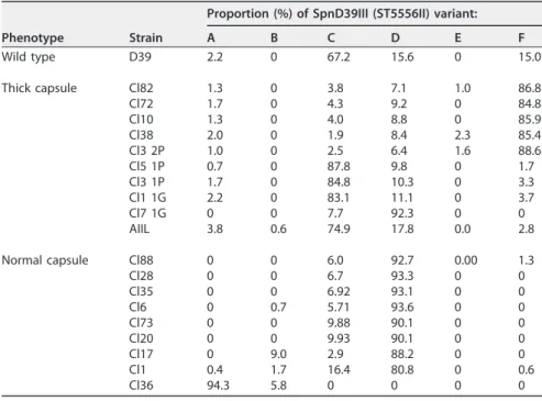
underpin the hyperencapsulated phenotype of the latter. Hence, the proportion of each of the six SpnD39III (ST5556II) variants was obtained for multiple individual ⌬adcAII strains expressing either thick or normal-size capsules using a previously described assay based on PCR followed by restriction digestion of the products (14) (Table 4). This showed a clear correlation between capsule phenotype and the domi-nant SpnD39III (ST5556II) variant. The WT D39 strain contained a mixture of the SpnD39III (ST5556II) variants, mainly SpnD39IIIC with also a significant proportion of the SpnD39IIID and F variants. With one exception, SpnD39IIIC (3 strains) and F (5 strains)
were the dominant variants found in the hyperencapsulated⌬adcAII strains, whereas
SpnD39IIID (7 strains) or A (1 strain) was the dominant variant found in the⌬adcAII
strains with normal capsule thickness. To try to link increased capsule formation by
some⌬adcAII mutants to changes in the dominant alleles of the SpnD39III (ST5556II)
type I restriction-modification locus, the hyperencapsulated⌬adcAII strain was
trans-formed with genomic DNA from D39 mutant strains with locked SpnD39III (ST5556II) alleles due to an inactivated creX gene. Flow cytometry analysis of complement sensitivity was used to rapidly assess capsular phenotype for 10 transformants for each allele (A to F). All transformants retained the complement-resistant phenotype of the
hyperencapsulated ⌬adcAII strain, even those made using the SpnD39III (ST5556II)
alleles associated with a normal capsule width in ⌬adcAII transformants (A and D)
(Fig. 7), suggesting they all remained hyperencapsulated.
Genome sequence data for⌬adcAII strains. Genome sequencing of one ⌬adcAII
and one⌬adcAII/pht strain confirmed they contained the expected partial deletion of
adcAII or adcAII and phtD, respectively, with insertion of the antibiotic resistance FIG 6 The ΔadcAII mutant has increased virulence in mouse models of sepsis and pneumonia. (A) For
the septicemia model, 5⫻ 104CFU of each strain was injected intraperitoneally and the progress of
infection was followed over time. Empty circles represent data for the WT D39 strain, and filled black circles show the ΔadcAII mutant strain. P values were obtained using the log rank test. (B and C) Blood (B) and lung (C) CFU for the pneumonia model determined by plating serial dilutions on Columbia blood
agar recovered 48 h after infection for mice inoculated by intranasal instillation of 5⫻ 106CFU. Each
symbol represents data from a single mouse, bars represent medians, and P values were calculated using
unpaired t tests.*, P⬍ 0.05.
on November 19, 2020 at USTL SCD
http://mbio.asm.org/
TABLE 3 Relative gene expression detected by microarray for genes showing statistically significant⬎1.5-fold differences in expression
for the thick-capsule⌬adcAII AIIL strain compared to the WT D39 straina
Regulation status in hyperencapsulated
strains and gene no. Gene name Predicted/known function
Mutant strain and capsule phenotype
⌬adcAII (AIIL) thick ⌬adcAII/phtD (AIIⴙPcl4) thick ⌬adcAII (Cl44) none Upregulated
SPD_0104 LysM domain protein 1.80 1.93 1.20
SPD_0389 accD Acetyl-CoA carboxylase subunit beta 1.55 1.59 ⫺1.37
SPD_0646 Hypothetical protein 1.74 2.05 ⫺1.18
SPD_0890 phtE Histidine triad protein 3.04 3.18 1.04
SPD_0891 Truncated histidine triad protein 3.25 1.16 1.37
SPD_0892 Truncated histidine triad protein 3.31 1.09 1.43
SPD_0893 Hypothetical protein 3.39 1.04 1.59
SPD_1038 phtA Histidine triad protein 1.98 1.52 ⫺1.05
SPD_1874 LysM domain-containing protein 2.53 2.90 1.19
SPD_1997 adcA Zinc ABC transporter AdcA lipoprotein 1.52 1.41 1.03
SPD_2000 adcR adc operon repressor AdcR 1.55 1.65 ⫺1.06
Downregulated
SPD_0052 purL Phosphoribosylformylglycinamidine synthase ⫺2.45 ⫺2.36 1.24
SPD_0053 purF Amidophosphoribosyltransferase ⫺2.27 ⫺2.34 1.25
SPD_0055 purN Phosphoribosylglycinamide formyltransferase ⫺2.13 ⫺2.18 1.36
SPD_0090 ABC transporter lipoprotein ⫺2.02 ⫺2.19 2.04
SPD_0166 ribH Riboflavin synthase, beta subunit ⫺2.76 ⫺3.44 1.56
SPD_0167 ribB Riboflavin biosynthesis protein RibB ⫺2.51 ⫺3.34 1.71
SPD_0168 ribE Riboflavin synthase subunit alpha ⫺2.52 ⫺3.30 1.66
SPD_0169 ribD Riboflavin biosynthesis protein RibD ⫺2.40 ⫺3.30 1.67
SPD_0224 pitD PitD iron ABC transporter permease ⫺2.28 ⫺2.11 1.46
SPD_0226 pitA PitA iron ABC transporter lipoprotein ⫺2.01 ⫺1.37 1.72
SPD_0265 adhP Alcohol dehydrogenase ⫺1.80 ⫺1.83 1.63
SPD_0279 celB Cellobiose PTS system IIB component ⫺2.28 ⫺1.79 1.76
SPD_0300 Oligohyaluronate lyase ⫺2.49 ⫺1.60 1.32
SPD_0362 mtlF Mannitol PTS system IIA component ⫺2.38 ⫺2.40 1.40
SPD_0364 Amino acid ABC transporter ATPase ⫺3.00 ⫺2.80 1.84
SPD_0444 lytB Endo-beta-N-acetylglucosaminidase ⫺1.55 ⫺1.69 1.38
SPD_0450 creX/psrA Type I restriction-modification system ⴚ3.39 ⴚ4.24 ⴚ1.43 SPD_0452 hsdS= (hsdS2) Type I restriction-modification system ⴚ3.62 ⴚ6.41 1.25
SPD_0453 hsdS (hsdS1) Type I restriction-modification system ⴚ2.01 ⴚ2.36 1.15
SPD_0466 blpT BlpT protein, fusion ⫺1.79 ⫺2.26 1.52
SPD_0472 blpA ABC transporter, ATP-binding protein ⫺2.21 ⫺3.47 1.59
SPD_0473 blpY Immunity protein BlpY ⫺1.50 ⫺2.17 1.44
SPD_0553 Hypothetical protein ⫺1.59 ⫺1.44 1.26
SPD_0595 Hypothetical protein ⫺1.55 ⫺1.72 ⫺0.64
SPD_0608 pyrF Orotidine 5=-phosphate decarboxylase ⫺1.65 ⫺1.60 1.02
SPD_0609 pyrE Orotate phosphoribosyltransferase ⫺1.77 ⫺1.67 1.02
SPD_0610 Hypothetical protein ⫺2.18 ⫺2.26 1.45
SPD_0611 Hypothetical protein ⫺1.76 ⫺1.94 1.19
SPD_0612 Hypothetical protein ⫺2.07 ⫺2.06 1.00
SPD_0613 Hypothetical protein ⫺1.70 ⫺1.83 1.09
SPD_0614 ABC transporter, ATP-binding protein ⫺1.76 ⫺1.77 1.11
SPD_0615 ABC transporter substrate binding protein ⫺1.51 ⫺2.25 1.25
SPD_0616 glnQ Amino acid ABC transporter ATPase ⫺1.56 ⫺2.38 1.13
SPD_0617 glnP Amino acid ABC transporter permease ⫺1.76 ⫺2.64 1.25
SPD_0618 glnP Amino acid ABC transporter permease ⫺1.71 ⫺2.51 1.20
SPD_0851 pyrK Dihydroorotate dehydrogenase II ⫺1.90 ⫺1.90 1.16
SPD_0852 pyrD Dihydroorotate dehydrogenase IB ⫺2.32 ⫺2.28 1.11
SPD_0853 lytB Endo-beta-N-acetylglucosaminidase ⫺1.71 ⫺1.65 1.09
SPD_0888 adcAII Zn2ⴙABC transporter lipoprotein ⴚ3.87 ⴚ3.03 ⴚ5.95 SPD_0889 phtD Hypothetical protein ⴚ1.78 ⴚ2.50 ⴚ4.2
SPD_1009 serB Phosphoserine phosphatase ⫺1.51 ⫺1.24 1.60
SPD_1011 glxK Glycerate kinase ⫺1.63 ⫺1.26 1.58
SPD_1035 PTS system, IIA component ⫺4.70 ⫺4.83 ⫺1.91
SPD_1036 PTS system, IIA component ⫺7.26 ⫺5.84 ⫺2.93
SPD_1050 lacD Tagatose 1,6-diphosphate aldolase ⫺1.61 ⫺1.60 1.44
SPD_1051 lacC Tagatose-6-phosphate kinase ⫺1.62 ⫺1.62 1.49
SPD_1052 lacB Galactose-6-phosphate isomerase LacB ⫺1.58 ⫺1.56 1.46
(Continued on next page)
Durmort et al. ®
on November 19, 2020 at USTL SCD
http://mbio.asm.org/
cassette with no additional mutations elsewhere in the genome. Sequencing of the cps
locus in additional ⌬adcAII strains found that a nonsynonymous single nucleotide
polymorphism (SNP) affecting the capsule locus gene cps2E (E to K at amino acid 322) was present in five out of 10 clones (Table 5), suggesting that this SNP may be relevant
for the hyperencapsulated phenotype at least for a proportion of ⌬adcAII strains.
However, the same SNP was also present in one of eight⌬adcAII strains with normal
capsule thickness. All five of the unencapsulated⌬adcAII strains investigated had cps2E
genes containing a stop codon at amino acid 308, which could explain their unencap-sulated phenotype (Table 5).
DISCUSSION
In this work, we have described that mutation of the zinc transporter lipoprotein gene adcAII in the S. pneumoniae D39 strain leads to an unexpected and striking increase in capsule expression in 42% of the resulting mutants. This phenotype
occurred with⌬adcAII mutations made by transformation either with a PCR construct
or with genomic DNA from another⌬adcAII mutant and was stable over many bacterial
generations. A similar mucoid phenotype was also observed with the⌬adcAII mutation
in two of the four other S. pneumoniae capsular serotypes investigated. The increased capsule quantity was very marked, with EM showing a greater-than-5-fold increase in capsule width and nuclear magnetic resonance (NMR) showing a 60% increase in the quantity of monosaccharides in purified capsule. This level of increase in capsule expression is markedly greater than that seen between opaque and transparent TIGR4
TABLE 3 (Continued) Regulation status in hyperencapsulated
strains and gene no. Gene name Predicted/known function
Mutant strain and capsule phenotype
⌬adcAII (AIIL) thick ⌬adcAII/phtD (AIIⴙPcl4) thick ⌬adcAII (Cl44) none
SPD_1053 lacA Galactose-6-phosphate isomerase LacA ⫺1.61 ⫺1.61 1.51
SPD_1074 metY O-Acetylhomoserine sulfhydrylase ⫺1.64 ⫺1.92 1.51
SPD_1131 carB Carbamoylphosphate synthase subunit ⫺1.60 ⫺1.37 1.06
SPD_1133 pyrB Aspartate carbamoyltransferase subunit ⫺1.51 ⫺1.30 1.01
SPD_1175 Putative membrane protein ⫺1.68 ⫺1.75 ⫺1.43
SPD_1176 ABC transporter, ATP-binding protein ⫺1.69 ⫺1.81 1.38
SPD_1177 Drug efflux ABC transporter ⫺1.73 ⫺1.68 1.43
SPD_1178 ptrB Prolyl oligopeptidase family protein ⫺1.73 ⫺1.78 ⫺1.47
SPD_1179 Hypothetical protein ⫺1.74 ⫺1.80 1.59
SPD_1454 Hypothetical protein ⫺1.56 ⫺1.61 1.17
SPD_1455 Hypothetical protein ⫺1.84 ⫺3.29 1.25
SPD_1498 Oxidoreductase ⫺2.21 ⫺2.23 1.34
SPD_1501 Sugar ABC transporter permease ⫺3.73 ⫺3.58 1.42
SPD_1503 Hypothetical protein ⫺3.25 ⫺4.03 1.09
SPD_1513 Hypothetical protein ⫺1.77 ⫺2.68 ⫺1.40
SPD_1568 GTP cyclohydrolase ⫺1.80 ⫺1.55 1.53
SPD_1584 ABC transporter permease ⫺2.30 ⫺2.16 2.08
SPD_1650 fatC Iron uptake ABC transporter permease ⫺2.84 ⫺2.23 1.30
SPD_1793 Universal stress protein family ⫺1.54 ⫺1.74 2.00
SPD_1865 adh Zinc-containing alcohol dehydrogenase ⫺1.59 ⫺1.28 1.48
SPD_1972 Hypothetical protein ⫺2.38 ⫺2.96 1.75
SPD_1985 adh2 Iron-containing alcohol dehydrogenase ⫺2.05 ⫺1.89 1.59
SPD_1987 Fucolectin-related protein ⫺3.12 ⫺2.96 1.49
SPD_1989 PTS system, IID component ⫺2.19 ⫺1.83 1.88
SPD_1990 PTS system, IIC component ⫺1.94 ⫺1.74 1.60
SPD_1991 PTS system, IIB component ⫺1.83 ⫺1.45 1.52
SPD_1992 PTS system, IIA component ⫺2.03 ⫺1.79 1.68
SPD_1993 fucU Fucose operon FucU protein ⫺2.33 ⫺2.10 1.83
SPD_1994 fucA L-Fuculose phosphate aldolase ⫺2.17 ⫺2.33 1.63
SPD_1995 fcsK L-Fuculose kinase FucK, putative ⫺2.13 ⫺2.07 2.12
SPD_2013 glpK Glycerol kinase ⫺2.76 ⫺2.36 1.25
aFor comparison, the fold differences compared to WT D39 strain for the thick-capsule ΔadcAII/phtD strain (AII⫹ Pcl14) and a normal-capsule-thickness ΔadcAII strain
(Cl144) are provided alongside. The adcAII, phtD, and SpnD39III (ST5556II) type I restriction-modification system genes are indicated in bold, and gene expression profile differences in the ΔadcAII/phtD or the ΔadcAII (Cl144) compared to the ΔadcAII AIIL hyperencapsulated strain are indicated in italics. Abbreviations: CoA, coenzyme A; PTS, phosphotransferase.
on November 19, 2020 at USTL SCD
http://mbio.asm.org/
capsular switched (less than 2-fold) (31) and 6B strains (32), justifying describing the
D39 ⌬adcAII strain as hyperencapsulated. The phenotypic consequence of the
in-creased capsule expression was a high degree of resistance to complement-mediated immunity and hypervirulence in mouse models of pneumonia and sepsis. These
TABLE 4 Proportions of variants (identified by PCR analysis) for the SpnD39III (ST5556II)
type I restriction-modification system for selected⌬adcAII mutant strains divided into those with thick and normal capsule thicknesses
Phenotype Strain
Proportion (%) of SpnD39III (ST5556II) variant:
A B C D E F Wild type D39 2.2 0 67.2 15.6 0 15.0 Thick capsule Cl82 1.3 0 3.8 7.1 1.0 86.8 Cl72 1.7 0 4.3 9.2 0 84.8 Cl10 1.3 0 4.0 8.8 0 85.9 Cl38 2.0 0 1.9 8.4 2.3 85.4 Cl3 2P 1.0 0 2.5 6.4 1.6 88.6 Cl5 1P 0.7 0 87.8 9.8 0 1.7 Cl3 1P 1.7 0 84.8 10.3 0 3.3 Cl1 1G 2.2 0 83.1 11.1 0 3.7 Cl7 1G 0 0 7.7 92.3 0 0 AIIL 3.8 0.6 74.9 17.8 0.0 2.8 Normal capsule Cl88 0 0 6.0 92.7 0.00 1.3 Cl28 0 0 6.7 93.3 0 0 Cl35 0 0 6.92 93.1 0 0 Cl6 0 0.7 5.71 93.6 0 0 Cl73 0 0 9.88 90.1 0 0 Cl20 0 0 9.93 90.1 0 0 Cl17 0 9.0 2.9 88.2 0 0 Cl1 0.4 1.7 16.4 80.8 0 0.6 Cl36 94.3 5.8 0 0 0 0
FIG 7 Flow cytometry analysis of complement sensitivity of the hyperencapsulated ΔadcAII strain after
transformation with locked SpnD39III (ST5556II) alleles (A to F) containing an inactivated creX gene. ΔFP441, ΔFP442, ΔFP443, ΔFP444, ΔMRO559, and ΔMRO560 are all double mutant strains carrying the
adcAII mutation and an extra one in allele SpnIIIB, allele SpnIIIC, allele SpnIIIA, allele SpnIIID, allele SpnIIIE,
and allele SpnIIIF, respectively. (A) Fluorescence index (MFI measured in arbitrary units multiplied by proportion of bacteria positive for C3b/iC3b) of C3b/iC3b deposition on ΔadcAII mutants and ΔadcAII fixed SpnD39III allele transformants (alleles A to F) as a proportion of the fluorescence index for the wild-type normal-capsule-thickness D39 strain. The data were measured using flow cytometry after preincubation in 30% human serum. Error bars represent SDs, and 10 transformants were tested for each
double mutant strain. For all mutant strains, the P value for results compared to D39 was⬍0.001
(unpaired t tests). (B) Examples of flow cytometry histograms for C3b/iC3b deposition on WT D39 (dark gray line) and one ΔadcAII/SpnD39IIID allele (light gray line) double mutant transformant. Gray shading indicates the results for bacteria incubated in PBS alone.
Durmort et al. ®
on November 19, 2020 at USTL SCD
http://mbio.asm.org/
phenotypes are exaggerated versions of the well-described effects of the capsule on S.
pneumoniae evasion of host immunity (7), demonstrating that under a normal level of
expression the capsule effects on immune evasion have not reached maximal potential. Previous data have shown that capsule expression comes at a metabolic cost which inhibits growth when cultured in defined medium and that the capsule prevents adhesion by respiratory epithelium (26, 33, 34). However, surprisingly, these negative
aspects of capsule expression were not identified with the hyperencapsulated⌬adcAII
strain. The serotype 2 S. pneumoniae capsule repeating unit is a hexasaccharide consisting of one glucuronic acid, two glucoses, and three rhamnoses (6, 35). NMR demonstrated that the relative proportion of glucose to rhamnose was altered in the ⌬adcAII strain compared to WT D39, shifting from almost 1 to 1 in the latter to closer to the expected 2-to-3 ratio. This would be compatible with an increased proportion of the total S. pneumoniae glucose pool being used for capsule production. The larger comparative increase in capsule width compared to changes in monosaccharide quan-tity suggests the organization of the capsule may have been altered, perhaps with more
loosely packed but longer capsule strands in the⌬adcAII strain compared to D39.
Why there is increased expression of the capsule in the⌬adcAII strain is not clear.
The close linkage to adcAII suggests a role for disruption of zinc utilization, yet the hyperencapsulated phenotype did not occur with mutation of the other S. pneumoniae zinc uptake lipoprotein gene adcA (19) and was not affected by zinc availability. Combined deletion of adcA and adcAII was also not associated with the hyperencap-sulated phenotype, but the double mutation had major effects on S. pneumoniae
TABLE 5 Mutation construction, capsule phenotype, and (where available) cps2E gene genome sequence data for S. pneumoniae strains Strain/clone Gene deletion
Antibiotic
resistance Mutant construction Capsule ratio/D39 Capsule phenotype Mutation in cps2E
D39 800 None 1 Normal None
D39 WT None 1 Normal None
⌬adcAIIL ⌬adcAII Cm New transformation 3.7 Thick None
Cl10 ⌬adcAII Kana New transformation 3 Thick E for K aaa322
Cl57 ⌬adcAII Kana New transformation 0.5 Unencapsulated Stop codon aa 308
Cl1 1P ⌬adcAII Cm Back-crossing with⌬adcAIIL 0.5 Unencapsulated Not sequenced
Cl1 1G ⌬adcAII Cm Back-crossing with⌬adcAIIL 3 Thick Not sequenced
Cl1 1G ⌬adcAII Cm Back-crossing with⌬adcAIIL 2.9 Thick None
Cl2 1P ⌬adcAII Cm Back-crossing with⌬adcAIIL 0.5 Unencapsulated Not sequenced
Cl2 2P ⌬adcAII Cm Back-crossing with⌬adcAIIL 0.9 Normal Not sequenced
Cl3 1G ⌬adcAII Cm Back-crossing with⌬adcAIIL 3.1 Thick Not sequenced
Cl3 1P ⌬adcAII Cm Back-crossing with⌬adcAIIL 3.2 Thick Not sequenced
Cl3 2P ⌬adcAII Cm Back-crossing with⌬adcAIIL 3.1 Thick None
Cl5 1P ⌬adcAII Cm Back-crossing with⌬adcAIIL 3.8 Thick None
Cl5 1G ⌬adcAII Cm Back-crossing with⌬adcAIIL 3.7 Thick Not sequenced
Cl6 1P ⌬adcAII Cm Back-crossing with⌬adcAIIL 0.5 Unencapsulated Not sequenced Cl6 2P ⌬adcAII Cm Back-crossing with⌬adcAIIL 0.5 Unencapsulated Stop codon aa 308
Cl7 1P ⌬adcAII Cm Back-crossing with⌬adcAIIL 3.5 Thick Not sequenced
Cl7 1G ⌬adcAII Cm Back-crossing with⌬adcAIIL 3.1 Thick None
AIIcl 1 ⌬adcAII Cm New transformation 1 Normal None
AIIcl 17 ⌬adcAII Cm New transformation 1.1 Normal None
AIIcl 20 ⌬adcAII Cm New transformation 1.1 Normal None
AIIcl 28 ⌬adcAII Cm New transformation 1.15 Normal None
AIIcl 31 ⌬adcAII Cm New transformation 0.5 Unencapsulated Stop codon aa 308 AIIcl 35 ⌬adcAII Cm New transformation 0.85 Unencapsulated Stop codon aa 308
AIIcl 36 ⌬adcAII Cm New transformation 1.05 Normal E for K aa 322
AIIcl 38 ⌬adcAII Cm New transformation 2.05 Thick E for K aa 322
AIIcl 44 ⌬adcAII Cm New transformation 0.5 Unencapsulated Stop codon aa 308
AIIcl 72 ⌬adcAII Cm New transformation 2.8 Thick E for K aa 322
AIIcl 73 ⌬adcAII Cm New transformation 1.15 Normal None
AIIcl 75 ⌬adcAII Cm New transformation 1.2 Normal None
AIIcl 78 ⌬adcAII Cm New transformation 1.8 Thick E for K aa 322
AIIcl 82 ⌬adcAII Cm New transformation 2.6 Thick E for K aa 322
AIIcl 88 ⌬adcAII Cm New transformation 1 Normal None
AII⫹Pcl4 ⌬adcAII ⫹ phtD Cm New transformation 2.2 Thick Not sequenced
aaa, amino acid position.
on November 19, 2020 at USTL SCD
http://mbio.asm.org/
physiology (19) which could have obscured or suppressed the mucoid phenotype. Overall regulation of S. pneumoniae capsule expression is poorly understood and is further complicated by the large number of different capsular carbohydrate structures with potentially significant differences in regulatory mechanisms. Factors affecting thickness of the capsule layer include regulation of cps locus gene expression by RitR (an orphan two-component signal transduction component) (36), CpsR (a GntR family regulator) (37), and RegM (38), as well as the conserved 5= cpsABCD (also termed wzg,
wzh, wzd, and wze) genes of the cps locus (39–41). Two S. pneumoniae quorum-sensing
systems (LuxS/AI-2 and the Rgg/small hydrophobic peptide system) increase capsule thickness (42–44), which can also be regulated independently of gene transcription by the supply of capsule monosaccharide precursors (45) or by increased capsule shedding mediated by LytA (12). However, our transcriptome analysis did not identify increased
cps locus gene expression or any effects on the abovementioned known regulators of
capsule expression in the⌬adcAII strain.
Another potential mechanism causing the hyperencapsulated phenotype in the ⌬adcAII mutant was identified by effects on transcription of the SPD_0450-0453 locus. This encodes the SpnD39III (ST5556II) type I restriction-modification system, allelic variants of which correlate with capsule thickness for several serotypes (14–16). We
found that the hyperencapsulated phenotype of⌬adcAII mutants was associated with
a predominance of either the SpnD39IIIC or F allelic variant, whereas SpnD39IIID was
the dominant allele for the majority of⌬adcAII mutants with normal capsule thickness.
This link between the hyperencapsulated phenotype of the⌬adcAII strain and specific
alleles of the SpnD39III (ST5556II) system seems unlikely to be coincidental given the known effects of this restriction-modification system on capsule expression. However, transformation with fixed SpnD39III (ST5556II) alleles, including those associated with normal capsule thickness (A and D), did not alter the hyperencapsulated phenotype of
the ⌬adcAII mutant, showing that any effects of SpnD39III alleles on the capsule
thickness of the ⌬adcAII mutation are not readily reversed by switching alleles. This
situation is further confused by the similarity in allele composition of the wild-type D39
strain and the AIIL⌬adcAII mutant and by differences between our data and published
papers in which SpnD39III alleles are linked to thick or thin capsule phenotypes. Manso et al. found that A, E, and F allele strains were largely opaque but C strains were more transparent, Li et al. found that only E allele strains (termed hsdSa in their paper) were opaque, and Oliver et al. found that the A and B alleles were opaque and the others transparent (14–16). The presumed mechanism of capsule regulation by SpnD39III is differential methylation of genes or regulatory regions (14, 15), but the genes involved remain undetermined. Our transcriptome data have identified multiple additional
genes showing differential expression between hyperencapsulated⌬adcAII strains and
wild-type D39 or a normal-capsule-width ⌬adcAII mutant, some of which could be
involved in mediating increased capsule expression. These include three operons annotated as being involved in pyrimidine metabolism, suggesting a potential role for pyrimidine in controlling capsule expression. Which genes showing differential
expres-sion between the ⌬adcAII strains and WT D39 strains are involved in the capsular
phenotype and whether differential regulation is related to differences in methylation will require considerably more detailed genetic studies.
Interestingly, 50% of independently obtained hyperencapsulated ⌬adcAII strains
contained an identical nonsynonymous SNP affecting the cps locus gene csp2E. The SNP is predicted to affect the cytoplasmic tail of Csp2E, a glucose phosphate transferase that initiates the assembly of capsule components on the cell membrane and is partially conserved among most capsular serotypes (39). Point mutations of cps2E that affect capsule expression have been previously described (32, 39), suggesting a causative role
for this SNP for the⌬adcAII-related capsule phenotypes. However, the same SNP was
not present in one lineage of⌬adcAII with increased capsule thickness (the original
transformant and four back-crossed derivatives) and was also identified in one out of
eight normal-capsule-thickness⌬adcAII strains. All the unencapsulated ⌬adcAII
trans-formants also contained the same SNP in csp2E predicted to introduce a stop codon.
Durmort et al. ®
on November 19, 2020 at USTL SCD
http://mbio.asm.org/
This high frequency of cspE2 stop codon mutations suggests that partial deletion of
adcAII causes significant physiology stress to S. pneumoniae that may induce loss of
capsule production as an escape mutation.
To conclude, we have identified that in the S. pneumoniae D39 strain a hyperen-capsulated phenotype is an unexpected consequence of targeted mutation of adcAII, which encodes a zinc ABC transporter lipoprotein. This strain will be a useful tool for investigating how the capsule affects S. pneumoniae interactions with the host. The hyperencapsulated phenotype partially correlated with both a nonsynonymous SNP in
cps2E and changes in allelic dominance within the SpnD39III (ST5556II)
restriction-modification system. Further investigation of genes showing differential expression between normal and hyperencapsulated D39 strains could help to further identify the underlying mechanism(s) controlling S. pneumoniae capsule thickness.
MATERIALS AND METHODS
Bacterial strains and growth conditions. The ΔadcAII, ΔphtD, ΔadcA/adcAII, and ΔadcAII/phtD
mutant strains were created either in the wild type or in the ΔcpsD D39 strain as well as in wild-type serotype 4 (TIGR4), 6A, 6B (strains 6Aa and 6Ba, respectively, from the work of Hyams et al. [31]), and 17F (46) strains by gene replacement using genomic DNA or PCR-amplified fragments obtained from the corresponding R6 mutants and standard transformation protocols for S. pneumoniae (19). The cat and
kana genes were inserted in the reverse orientation without promoter or terminator sequences to avoid
affecting expression of adjacent genes. Mutant identities were verified by PCR with primers flanking the
cloned regions. S. pneumoniae was grown at 37°C with 5% CO2 in air in THY or on Columbia agar
containing 5% blood. Working stocks grown to an optical density (OD) of 0.4 (⬃108CFU/ml) were made
using THY and stored at⫺80°C in 10% glycerol as single-use aliquots. CFU were confirmed by colony
counting of log10serial dilutions of bacteria cultured overnight on 5% Columbia blood agar. Growth
curves were determined by measuring OD595for bacteria cultured in 2.5 ml of THY or chemically defined
medium (CDM) supplemented with 33M Zn in 24-well plates sealed with a transparent film and
incubated at 37°C in a FLUOstar reader. To measure blood growth, 1⫻ 106CFU/ml of S. pneumoniae was
inoculated into 1 ml of heparinized human blood and incubated at 37°C, with plating of serial dilutions at 0, 4, and 6 h to assess bacterial CFU.
Capsule size measurement and microscopy. An indirect method was developed to measure
capsule size by determining the size of the bacterial pellet. Briefly, 12 ml of culture was centrifuged, the
pellet was resuspended in 120 l of PBS, and 35 l was loaded in a microcapillary tube. After
centrifugation for 15 min at 800⫻ g, the height of the pellet within the tube was measured with a ruler.
Electron microscopy of mid-log-phase S. pneumoniae fixed in 3% paraformaldehyde (PAF) was performed using a ruthenium red and London resin capsule-preserving protocol as previously described (33). Capsule thickness was calculated by direct measurement of the surface layer for 30 randomly chosen S.
pneumoniae bacteria/strain using ImageJ software.
Confocal microscopy on bacteria was performed using an Olympus FV1000 confocal laser scanning
microscope with a 63⫻ objective. Bacteria were fixed for 30 min with 4% PFA (Sigma) on slides (Thermo
Scientific; SuperFrost Plus 10149870) and subsequently stained with anti-serotype 2 antibody (Statens Serum Institute) plus Alexa Fluor 546-conjugated anti-rabbit antibody. DNA was stained with 4=,6-diamidino-2-phenylindole (DAPI).
Capsular polysaccharide extraction and quantification. Capsular polysaccharides were extracted
from 1 liter of culture, and bacteria were resuspended in 10 ml of 0.15 M Tris buffer (pH 8) supplemented with 0.1% deoxycholate and incubated for 10 min at 37°C and then for 35 min at 50°C. Cell debris was removed by centrifugation under acidic condition. Proteins were eliminated from the supernatant by two successive extractions using a 5:1 ratio of chloroform and butanol, before precipitating capsular polysaccharides in 80% ethanol. Pellets were dried, resuspended in 0.1 M phosphate buffer (pH 7.2), and incubated with DNase and RNase for 1 h at 37°C, and then trypsin was added for 2 h at 37°C before purification of capsular polysaccharide by ion exchange on a column of DEAE Sepharose. Monosaccha-ride composition was established by GC and GC-MS as alditol acetate derivatives. Briefly, samples were hydrolyzed in 4 M trifluoroacetic acid (TFA) for 4 h at 100°C and reduced with sodium borohydride in 0.05 M NaOH for 4 h. Reduction was stopped by dropwise addition of acetic acid until pH 6 was reached, and borate salts were codistilled by repetitive evaporation in dry methanol. Peracetylation was performed in acetic anhydride at 100°C for 2 h. All monosaccharide derivatives were identified according to their specific retention times and electron ionization MS (EI-MS) fragmentation patterns (47).
Phagocytosis, neutrophil killing, complement deposition, and adhesion assays. Flow cytometry
phagocytosis and complement deposition assays were performed as previously described (7, 48) using
S. pneumoniae incubated for 30 min with human serum (25%), human heat-inactivated serum (25%), or
just Hanks balanced salt solution (HBSS) medium. For macrophage phagocytosis, THP-1 monocytes cultured in suspension in RPMI medium supplemented with 10% fetal bovine serum (FBS) were treated for 24 h with 10 nM phorbol 12-myristate 13-acetate (PMA) to induce cell adhesion and macrophage differentiation. Flow cytometry was performed using a fluorescence-activated cell sorting (FACS) Verse machine (BD), and the data were analyzed with FlowJo software. For neutrophil killing assays, fresh human neutrophils were purified using a magnetically activated cell sorting (MACS) neutrophil isolation
kit (Miltenyi Biotec) and resuspended in HBSS medium at a concentration of 1⫻ 106 cells/ml. S.
on November 19, 2020 at USTL SCD
http://mbio.asm.org/
pneumoniae previously incubated with human sera was incubated with the neutrophils at a multiplicity
of infection (MOI) of 1:500 (bacteria to neutrophils) in a 48-well plate for 45 min at 37°C. Adhesion assays were performed on Detroit 562 human nasopharyngeal cells (ATCC CCL-138) as previously
described (34, 49) using 3⫻ 105cells/well seeded into 24-well plates, infected with MOIs of 25 and
50, and incubated for 1 h before being washed three times with PBS, followed by addition of Dulbecco’s modified Eagle’s medium (d-MEM)-1% saponin for 10 min and plating of serial dilutions to count bacterial CFU.
Genome sequencing and transcriptional microarray analysis. Mutant strains were genome
sequenced by the Wellcome Trust Centre for Human Genetics (Oxford, United Kingdom) using an Illumina MiSeq sequencer. Sequences were assembled using Velvet, annotated using Prokka, and mapped to the published D39 (R00000036) reference genome. Bases and single-nucleotide variants were identified using the SAMtools “mpileup” command and Bcftools. Sites were filtered to a minimum depth of five reads at each and a single-nucleotide variant quality of 25, and the Integrated Genome Viewer was used to visualize mapping and coverage. Gene transcriptome microarrays were performed as described previously (34). Briefly, RNA was extracted with the RNeasy minikit (Qiagen), and labeled cDNA was prepared using Cy3-dCTP (GE Healthcare, United Kingdom) and SuperScript II reverse transcriptase with random hexamer primers (Life Technologies). Labeled cDNA was hybridized overnight to the BG@S SPv1.4.0 Agilent SurePrint platform (Agilent Technologies) microarray designed by the Bacterial Microar-ray Group at St. George’s, University of London. After hybridization, washed slides were scanned
immediately, using an Agilent high-resolution microarray scanner, at 5-m resolution; scanned images
were quantified using Feature Extraction software v 10.7.3.1 (Agilent Technologies); and statistical analysis of raw intensity data was performed in GeneSpring v14.9.1 (Agilent Technologies). Data for 3 independent biological replicate experiments were analyzed and normalized using a 75th percentile shift plus baseline normalized to the median for the related control sample for each biological replicate.
Statistically significant (P⬍ 0.05) differences between strains were identified in an unpaired t test with
Benjamini and Hochberg false-discovery rate correction.
Assessing allelic variants of SpnD39III. Primers AMRE74L (5= 6-carboxyfluorescein [FAM] label, FAM-GGAAACTGAGATATTTCGTGGTGATGATGGGA) and AMRE59 (CCTGATCGAGCGGAAGAATATTTCTGCC GAGGTTGCC) were used to PCR amplify a 4.2-kb fragment from S. pneumoniae under the following conditions: denaturation at 95°C for 5 min, followed by 40 cycles of 1 min of denaturation at 95°C, 1 min of annealing at 68°C, and 5 min of extension at 68°C, with a final extension of 10 min at 68°C. PCR
products were digested using 1 U DraI, 2 U PleI, and 1⫻ CutSmart Buffer (all from New England Biolabs,
United Kingdom) in a 20-l volume. Each FAM-labeled SpnIII variant has a unique size that can be distinguished through capillary electrophoresis on an ABI Prism gene analyzer (Applied Biosystems, USA) and analyzed using Peak Scanner v1.0 software. Genomic DNA for transformation using locked SpnD39III (ST5556II) alleles due to an inactivated creX gene was obtained from preexisting strains (14).
Animal models of infection. All animal experiments conformed to institutional and United Kingdom
Home Office guidelines and regulations. Outbred CD1 sex-matched white mice were used for infection experiments using established models of infection (50–52). For the nasopharyngeal colonization model,
107CFU of bacteria in 10l was administered by intranasal inoculation under light halothane general
anesthesia, and nasal washes were obtained after 5 days. Mice were inoculated by intraperitoneal (i.p.)
injection of 5⫻ 104 CFU for the sepsis model and by intranasal (i.n.) inoculation under isoflurane
inhalational anesthesia of 5⫻ 106CFU for the pneumonia model. Mice were sacrificed after 24 h (i.p.) or
48 h (i.n.), and serial dilutions of blood and lung homogenates were plated to enumerate target organ CFU. For the sepsis model, disease development was also monitored by observing mice three times a day
(n⫽ 8). For colonization and sepsis competitive infection models, mice were inoculated with a 50/50
ratio of D39 wild type and ΔadcAII strain to determine the competitive index (CI; ratio of mutant to WT strain recovered from mice divided by the ratio of mutant to WT strain in the inoculum).
Statistical analysis. Statistical analyses were conducted using Prism 7 (Graph Pad, USA). Parametric
data are presented as means, and error bars represent standard deviations. Nonparametric date were analyzed using the Mann-Whitney U test. For the disease development model, data were analyzed using the log rank (Mantel-Cox) test.
ACKNOWLEDGMENTS
This work was undertaken at UCLH/UCL, which received a proportion of funding from the Department of Health’s NIHR Biomedical Research Centre’s funding scheme. G.E. was supported by MRC (grant MR/R001871/1), M.O. was supported by grants from the MRC and BBSRC (MR/M003078/1 and BB/N002903/1), and S.C. was supported by the Wellcome Trust (grant WT076442).
REFERENCES
1. Yahiaoui RY, den Heijer C, van Bijnen EM, Paget WJ, Pringle M, Goossens H, Bruggeman CA, Schellevis FG, Stobberingh EE, APRES Study Team. 2016. Prevalence and antibiotic resistance of commensal Streptococcus
pneumoniae in nine European countries. Future Microbiol 11:737–744.
https://doi.org/10.2217/fmb-2015-0011.
2. Gillespie SH1, Balakrishnan I. 2000. Pathogenesis of pneumococcal
in-fection. J Med Microbiol 49:1057–1067. https://doi.org/10.1099/0022
-1317-49-12-1057.
3. Weiser JN, Ferreira DM, Paton JC. 2018. Streptococcus pneumoniae:
trans-mission, colonization and invasion. Nat Rev Microbiol 16:355–367.https://
doi.org/10.1038/s41579-018-0001-8.
4. Nelson AL, Roche AM, Gould JM, Chim K, Ratner AJ, Weiser JN. 2007.
Durmort et al. ®