Compatibility and Toxicity of Polymer-Coated Magnetic
Nanoparticles on Mammalian Cell Systems
by
Kelly M. Kral
B. S. Chemical Engineering Clarkson University, 2001
Submitted to the Department of Chemical Engineering in partialfulfillment of the
requirements for the degree of
Master of Science in Chemical Engineering at the
Massachusetts Institute of Technology
February 2005
© 2005 Massachusetts Institute of Technology. All rights reserved.
Signature ofAuthor ...
Department of Chemical Engineering January 14, 2005
Certified
by ...
...
...
...
Daniel I. C. Wang Institute Professor Thesis SupervisorAccepted
by... ...
.-
...
...
Daniel Blankschtein Professor of Chemical Engineering Chairman, Committee for Graduate StudentsMASSACHUSETTS INSTUTE
OF TECHNOLOGY
Compatibility and Toxicity of Polymer-Coated Magnetic
Nanoparticles on Mammalian Cell Systems
by Kelly M. Kral
Submitted to the Department of Chemical Engineering on January 14, 2005, in partial fulfillment of the requirements for the degree of
Master of Science in Chemical Engineering
Abstract
This thesis focuses on the compatibility of polymer-coated magnetic nanoparticles with mammalian systems. The magnetic particles are designed to increase oxygen transfer in mammalian cell bioreactors. Magnetic nanoparticle A consists of a magnetite core attached to a layer of oleic acid, in which oxygen is four times as soluble as it is in water. The entire particle is coated with an attached layer of a surfactant, hitenol, to stabilize the particle against agglomeration. The entire particle has a diameter of approximately 20 nm. However, particle A was found to be extremely toxic to both y-CHO and hybridoma cells, causing complete cell death within four hours. This is most likely due to the surface surfactant, hitenol.
A more biocompatible nanoparticle, particle B, was created. This particle is synthesized with a brush copolymer consisting of octadecylamine (ODA) and poly(ethylene oxide) (PEO) attached to a poly(acrylic acid) backbone. Once attached to the magnetite core, the ODA forms the inner layer that solubilizes oxygen, while PEO forms the stabilizing coating. Particle B forms nanoclusters about 100 nm in diameter.
Thoroughly cleaning the nanoparticles is very important, as mammalian cells are very sensitive to foreign chemicals. Particles cleaned with dialysis did not remove all impurities, as all y-CHO cells in the presences of these particles were killed within 24 hours. High gradient magnetic separation (HGMS) was used to clean particles, and was found to be a much more effective method. However, sufficient amounts of washing fluid, about sixty column volumes, were needed to ensure proper cleaning. Once properly cleaned, the particles were found to be much less toxic towards the cells. Both y-CHO and hybridoma cells produced normal growth curves in the presence of particles. However, the particles do still exhibit some toxicity towards the cells, as the maximum cell density of cells cultured with particles does not reach that of control cultures.
Both particles were found to increase the oxygen transfer in an aqueous solution. A 1.5% solution of particle A enhanced the oxygen transfer 41.8% over the control, water, while 1.4% particle B enhanced 15.9% over the control. While particle A has a better effect on oxygen transfer, this particle is not suitable to be used with mammalian cell cultures as it is highly toxic.
Thesis Supervisor: Daniel I. C. Wang Title: Institute Professor
Acknowledgements
First and foremost, I would like to thank my thesis advisor, Dr. Wang. You not only helped me to see what it was that I actually wanted, but you helped me achieve it. With the perfect combination of toughness and kindness, but always with unwavering support, you guided me through this journey, and I will never forget the many lessons I have learned. I would also like to acknowledge that the financial support from the National University of Singapore, Singapore-MIT Alliance (SMA) program and MIT's Technology Policy Center is greatly appreciated.
Many individuals in the Wang lab have helped in the course of this thesis. I wish to thank Peng, Fred, and Jin for guiding me through the world of cell culture. You would always answer any questions I had, and were always willing to help whenever I asked for it. Bernat and Andre, I am appreciative of all the guidance you have given me with magnetic particles. Your helpful suggestions and explanations were always useful and valued. Finally, much gratitude to Brian B. I always enjoyed our conversations and they helped to get me through many a day here.
Many thanks to the Hatton lab, especially Simon, Lev, and Harpreet. You were very understanding while I was roaming around your lab, and you would always take the time to answer any questions I had, especially when I could not find something.
Thanks also to the BPEC headquarters for all your help during my time here at MIT. I would especially like to acknowledge Susan, Michelle, Aran, and Dan. Also, thanks to the Chemical Engineering headquarters, especially Suzanne.
None of this would have been possible without the support of my family and friends. Mom and Dad, thanks for encouraging me to be the best I could be. You taught me to reach for the stars, yet always kept me grounded and reminded me that there are more important things. To my sister, Kristi, and my brother, Joey, while I don't get to talk to you as much as I would always like to, you have both inspired me in your own ways. Judy, thanks for sharing your experiences and for being there for me when I needed it most. Also, many thanks to my fellow classmates. Whether it was an
encouraging word in the hallway, or showing interest in my research at a TG, you guys helped me get through more than you will ever know.
Finally, there is one last person to thank, my best friend, John. There are no words strong enough to express my gratitude to you. You have supported me through my journey here at MIT in every way possible. You always believed in me, even when I did
not. This manuscript is just as much your accomplishment as it is mine. I look forward to sharing many other journeys with you.
Table of Contents
Title Page ...
...
Abstract
... ...
3
Acknowledgements
...
5
Table of Contents ...
...
7
List of Figures ...
...
9
1. Introduction 11 2. Literature Review 13 2.1 Oxygen Transfer in Mammalian Cell Cultures ... 132.2 Magnetic Nanoparticles ... 15
3. Materials and Methods
19
3.1 Polymer Coated Magnetic Nanoparticles ... 193.1.1 Materials ... 19
3.1.2 Particle A Synthesis ... 19
3.1.3 Particle B Polymer Synthesis ... 20
3.1.4 Particle B Synthesis ... 20 3.2 Cleaning Procedures ... 21 3.2.1 Dialysis ... 21 3.2.2 HGMS Cleaning ... 21 3.3 Cell Cultures ... 22 3.3.1 Cell Lines ... 22
3.3.2 Culture Medium and Materials ... 22
3.4 Cell Enumeration ... ... 23
3.4.1 Adherent Cells ... 23
3.4.2 Suspension Cells ... 23
3.5 y-CHO Adherent Cell Cultures ... .. ... 23
3.6 y-CHO Suspension Cell Cultures ... 24
3.8 Oxygen Transfer Experiments ... 24
4. Results and Discussion 25 4.1 Cell Compatibility with Particle A ... 25
4.1.1 Synthesis of Particle A ... 25
4.1.2 Compatibility with y-CHO Cells ... 27
4.1.3 Compatibility with Hybridoma Cells ... 28
4.2 Cell Compatibility with Particle B ... 30
4.2.1 Rationale Behind Particle B ... ... 30
4.2.2 Synthesis of Particle B ... ... 30
4.2.3 Compatibility with Adherent y-CHO Cells ... 32
4.2.4 Compatibility with Suspension y-CHO Cells ... ...34
4.2.5 Compatibility with Hybridoma Cells ... 35
4.3 Effect of Particles on Oxygen Transfer ... 37
5. Conclusions 40
Nomenclature ...
41
References ...
... 42
4...
List of Figures
Figure 1. Production of aqueous magnetic particle A. Chemical coprecipitation of
iron salts in the presence of potassium oleate creates a magnetic core attached to oleic acid, the inner hydrophobic layer. Particle size is limited by the binding of the polymer to the Fe304core soon after nucleation begins. The addition of ammonium
persulfate initiates a free radical reaction leading to the formation of a chemical bond between hitenol and the oleic acid. This stabilizes the particle by creating a charged outer hydrophilic coating on the magnetic nanoparticle ... 26
Figure 2. Magnetic nanoparticles A were cleaned using HGMS four times with 50
ml salt water. They were captured in water, concentrated by evaporation in an 80 °C oven, and autoclaved. Suspension hybridoma cells were centrifuged and resuspended in 20 ml of medium containing x % particles. Samples were taken hourly: (El) total cells and (X) viable cells... 29
Figure 3. The graft copolymer PEO/C18 for magnetic nanoparticle B was
synthesized by attaching PEO-NH 2 and ODA to a PAA backbone via an amidation
reaction... 31
Figure 4. Production of aqueous magnetic particle B. Chemical coprecipitation of
iron salts in the presence of the PEO/C1 8 graft copolymer creates the magnetic
nanoparticles. Particle size is limited by the binding of the polymer to the Fe304
core soon after nucleation begins. The hydrophobic portion of the graft copolymer forms the oxygen solubilizing interior, while the hydrophilic PEO coats the entire particle and stabilizes the particle... 32
Figure 5. Magnetic nanoparticles B were cleaned using HGMS twice with 12 ml
salt water then twice with 12 ml water. They were captured in medium and filter sterilized. Adherent y-CHO cells were grown in six-well plates for 18 hours before the medium was removed and replaced with the particle-containing medium at the following concentrations: () No particles, (X) 0.5% Particle B, and (0) 0.25% Particle B ... 33
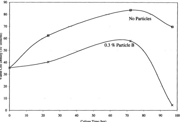
Figure 6. Magnetic nanoparticles B were cleaned using HGMS four times with 50 ml salt water then twice with 10 ml water. They were captured in medium and filter sterilized. Suspension y-CHO cells were centrifuged and resuspended in 40 ml of medium containing (l) no particles and (X) 0.3% Particle B ... 35
Figure 7. Magnetic nanoparticles B were cleaned using HGMS four times with 50
ml salt water then with 20 ml water. They were captured in water and autoclaved for sterilization. Suspension hybridoma cells were centrifuged and resuspended in (El) 10 ml DMEM no particles, (0) 8 ml DMEM plus 2 ml water to act as a control, and (X) 8 ml DMEM plus 2ml 0.75% Particle B for a final concentration of 0.15% particles. The viability of the cell culture is shown above selected data points ... 36
Figure 8. Cylindrical beakers were filled with (0) water, (X) 1.4% particle B, or (0) 1.5% particle A. The beakers were purged of oxygen with nitrogen and allowed to return to saturation via exposure of the surface to the air while the saturation curve was being recorded with a dissolved oxygen polarographic sensor ... 38
Figure 9. Dynamic gassing method was used to determining the oxygen transfer
coefficient in () water, (X) 1.4% particle B, or (0) 1.5% particle A. The slope of each line will correspond to the kLa value in arbitrary units ... 39
1. Introduction
In bioreactors, productivity is usually limited by the transport of substrate or product into or out of the reactor. Due to its low solubility (less than 10 mg/L), oxygen is often the limiting substrate. In bacterial fermentations, the generally accepted method of increasing oxygen transfer to cells is to use agitation and gas sparging in the bioreactor. However, even with agitation and sparging, there can still be oxygen transfer limitations. Thus, other methods have been investigated, such as replacing the sparger with a
chemical means of creating oxygen in the bioreactor (Coulaloglou and Tavlarides 1976) or introducing a new immiscible liquid phase, such as a perfluorocarbon, in which oxygen in more soluble (Junker et al. 1990, Vinke et al. 1993).
The use of mammalian cells has become more popular in industry due to the ability of these cells to properly fold and modify therapeutic proteins post-translationally. Mammalian cell cultures also suffer from low oxygen solubility, especially in industrial scale bioreactors. However, because mammalian cells lack the robust cell wall that protects bacterial cells, sparging with the high gas velocities used in fermentations can cause cell death, which can quickly become a larger problem than oxygen transfer limitations. Many solutions have been put forth, including optimizing the design of the sparger and bioreactor to maximize cell viability (Jobses et al. 1991) and, as was
successful with bacterial fermentations, the addition of perfluorocarbons (Cho and Wang 1988, Ju and Armiger 1992).
This thesis will explore the compatibility and toxicity of polymer-coated magnetic nanoparticles to mammalian cells with the goal of increasing the oxygen transfer
coefficient (kLa). The magnetic nanoparticles, when existing in single domains, consist of a core of magnetite about 10 nm in diameter that can be coated with one or more layers of polymer. The polymer layers will be selected such that they have a high oxygen storage capacity as well as to provide stabilization of the nanoparticle clusters to prevent agglomeration. The particles will form a colloid in an aqueous solvent because their outer layer confers stabilization in water that prevents the cores from coming into contact and prevent aggregation. The magnetic core is important because it will allow the
particles to be easily separated from the culture medium. There are many advantages to using these nanoparticles over perfluorocarbon emulsions. Being approximately three
orders of magnitude smaller than the micron-sized PFC droplets, they present much more surface area for oxygen transfer. They are easily recoverable with magnetic separation, and they are non-volatile.
2. Literature Review
2.1 Oxygen Transfer in Mammalian Cell Culture
With a steadily increasing number of mammalian cell-based therapeutics on the market and in clinical trials, it is highly probable that some of these proteins may be needed in large quantities (hundreds of kg per year) (Varley and Birch 1999). To produce these quantities efficiently, companies will have to increase the size or
volumetric throughput of bioreactors. Some important factors when considering a scale up include: sufficient gas transfer, mixing without causing damage to cells, and
compatibility with upstream and downstream processes (Feder et al. 1985). This thesis concentrates on means in ensuring that there is sufficient oxygen transfer in bioreactors. Since oxygen transfer and mixing are coupled problems in most bioreactors, it is
important to consider both of these behaviors.
Surface aeration, the oxygenation method used commonly for bench-scale spinner and shaker flasks, is sufficient for, but limited to, small volumes (Moreira et al. 1995). This is because the volumetric oxygen transfer coefficient (kLa) depends on surface area per unit volume, and the specific surface area for oxygen transfer decreases as the reactor volume increases. It has been shown that kLa roughly has an inverse square relationship with culture volume (Fleischaker and Sinskey 1981). As this method relies on the degree of mixing at the liquid - gas interface, many methods have been explored to increase this mixing. Increasing agitation will increase oxygen transfer (Fleischaker and Sinskey 1981), however, there is an upper limit to the agitation speed due to the fragility of mammalian cells. Increasing the size of the impeller will increase oxygen transfer (Aunins et al. 1989). Additionally, as placing the impeller within 1 cm of the surface dramatically increases kLa (Aunins et al. 1989), (Hu et al. 1986) created an impeller system with two blades on the shaft, the bottom one used for agitation while the top one was used for surface aeration. This was able to create a four-fold increase in the kLa value. Another method to increase oxygen transfer at the surface is to increase the driving force for oxygen transfer by using either oxygen-enriched air or pure oxygen. However, there have been reports of adverse effects on the growth of various types of mammalian cells when the oxygen partial pressure becomes too great (Oller et al. 1989). To increase the area for oxygen transfer, a membrane aeration system can be used. The
membrane (for example, a silicone or propylene tube) is placed in the bioreactor, and air is pumped through, thus increasing surface area available for oxygen transfer to the liquid phase (Moreira et al. 1995). The main problem with all these methods is scalability. They all work at smaller volumes, but can not scale up to industrial size bioreactors, as
the surface area is just not sufficient. Membranes also present problems because they are difficult to clean, maintain, and sterilize (Moreira et al. 1995).
Sparging can be used in the culture of mammalian cells, but attention needs to be paid to the problems caused by it. The two main drawbacks to sparging are the
vulnerability of mammalian cells to damage and foaming of the culture medium. As most cell death occurs at the top of the bioreactor due to bubble breakup, increasing the
height to diameter ratio of the bioreactor can limit cell damage (Papoutsakis 1991). While a high flow rate is favorable for increasing kLa, it needs to be balanced against the
increased cell death it would cause. Thus, it is suggested that the flow rate be kept to as low as possible, while still keeping the cells in suspension and maintaining a sufficient DO level (Doyle and Griffiths 1998).
There are also chemicals that can be added to the medium to protect cells from shear. The most common are Pluronic surfactants from the BASF Corp. (Parsippany, NJ). These surfactants work by decreasing the surface tension, which causes a decrease in bubble size, which in turn causes bubble break-ups to be less significant to the mammalian cells. While they increase cell viability, they also cause a reduction in the kLa value (Moreira et al. 1995). The second problem with sparging is foaming. The type of sparger used (for example, a porous metal sparger versus a larger multi-hole sparger) can cause large differences in foaming (Chisti 1993). To control foaming, silicon
antifoams are commonly added to the medium. The main disadvantage to this aeration method is that gas flow rate has to be balanced with cell damage, and this could result in sub-optimal oxygenation.
Another method for improving oxygen transfer is to add a second immiscible liquid phase in which oxygen is more soluble. Perfluorocarbons (PFCs) are most commonly used for this purpose. PFCs are organic compounds in which fluorine atoms replace all hydrogen atoms. Oxygen is ten to fifteen times more soluble in PFCs than in water (McMillan 1990) because they are so apolar. There have been many suggested
uses for the oxygen-carrying abilities of PFCs (Lowe et al. 1998, Mattiasson and Adlercreutz 1987), but as this thesis focuses on mammalian cells, so will this review. Cho and Wang (1988) studied the effect of a PFC on a hybridoma cell line in spinner flasks. They found that the initial cell growth and the antibody production rates were faster when PFC was present. They concluded that the PFC did not have any adverse effect on the cells while increasing the rate of oxygen supplied to the cells. In another experiment, it was found that the addition of PFC resulted in longer periods of active hybridoma cell growth in roller flasks (Ju and Armiger 1992). They concluded that this
was due to satisfying the greater oxygen demand of denser cell population. They also reported that the PFC emulsion protected the cells from shear in a column bioreactor. However, this could also be due the surfactants used to create the emulsion, not just to the PFC (Lowe et al. 1998). PFCs have also been used to increase the oxygen transfer to
attached mammalian cells (Rappaport et al. 1996), and is being investigated as a component in three-dimensional scaffolding on which to grow cells (Rappaport 2003). There are some problems associated with using PFCs in bioreactors. There are environmental concerns because PFCs are so inert, yet they have strong infrared
absorption, which could contribute to global warming. They are also hard to recover. As they are heavier than the medium, one could wait for the two layers to separate out due to gravity, but the time scale for this is an issue. A better solution would incorporate the oxygen soluble properties of the PFCs, but have something that would be easier to separate and recycle, as well as being non-volatile.
2.2 Magnetic Nanoparticles
A magnetic fluid is a colloidal dispersion of magnetic nanoparticles in a carrier liquid. Nanoparticles are so small (10 -15 nm) that they do not settle in gravitational or moderate magnetic fields. They have a tendency to aggregate due to large van der Waals interactions, so a surface coating must be added to avoid this. Magnetite, Fe304, is a
commonly used metal oxide for nanoparticles because of its interesting magnetic properties. It is superparamagnetic, which means that it will exhibit no magnetic properties unless placed in a strong magnetic field, where it will become magnetized. However, when the magnetic field is removed, the particles will retain no magnetization
(Rosensweig 1985). This property makes them ideal for easy separations (Moeser et al. 2004a).
An economical and simple way of creating these magnetic particles is
coprecipitation, which is where iron ions are precipitated in the presence of another ion, oxygen. Magnetite, the core of the nanoparticle, is created in the presence of a base at a temperature of 75 - 80 °C according to the following equation:
2 FeC13+ FeC12+ 8 NH40H - Fe304+ 8 NH4Cl + 4 H20 (1)
There are two ways to stabilize magnetic particles so that the cores will not agglomerate: steric hindrance or electrostatic repulsion (Ditsch 2004). Steric hindrance can be a very robust method of stabilization if the correct coating is chosen. Electrostatic repulsion is not as hearty of a method, as it can break down at high ionic strength.
The oldest method of stabilizing the aqueous magnetic particles is to use a surface bi-layer (Shen et al. 1999, Shen et al. 2000). For a bi-layer to form, it is necessary to utilize two surfactants. The inner layer forms when one surfactant bonds to the magnetite core via a carboxylic chelate, leaving the hydrophobic group pointing outward. The outer layer is held in place due to hydrophobic interactions with the inner layer while
presenting its hydrophilic end to the aqueous solution. However, desorption of the outer layer can occur during dilution of the magnetic particles, or if an organic solvent is added into which the outer layer is soluble. Additionally, the mechanism for stabilization is electrostatic repulsion which is not robust in an environment of high ionic strength (Ditsch 2004). To make these more robust, a phospholipid can be used as the outer layer (Bucak et al. 2003). This causes the second layer to be more strongly attracted to the inner layer. However, there are still problems with high ionic strength environments, as well as organic solvents.
To avoid the problems associated with not having a covalently bonded outer layer, nanoparticles can be created using polymers (Moeser et al. 2002). These polymers should be chosen such that the attachment to the magnetite core and stabilization is achieved with one molecule. The main advantage to this approach is that the stabilization mechanism is now steric hindrance, which is much more robust than electrostatic
repulsion. Moeser, et al. (2002) created magnetic particles using Pluronic micelles as a model. Pluronic micelles are a copolymer consisting of a hydrophobic poly(propylene oxide) (PPO) core with an outer layer of hydrophilic poly(ethylene oxide) (PEO)
(Alexandridis and Hatton 1995). Applying this concept, a block co-polymer of PPO and PEO is created on the backbone of poly(acrylic acid) (PAA), which contains the
carboxylic groups that will bond to the magnetite core. Particles created with this
PEO/PPO co-polymer are stable, not only in high ionic strength environments, but also in many organic solutions (Moeser et al. 2002).
Instead of creating a block co-polymer to attach to the magnetite core, it is also possible to first attach the inner layer to the core and then covalently bond the outer layer to the inner layer (Olle et al. 2004 (Patent Filed)).
High gradient magnetic separation (HGMS) is a very efficient method of separating magnetic particles. An HGMS column consists of a cylinder holding
magnetically-susceptible wires placed between the poles of an electromagnet. Magnetic nanoparticles are attracted to the wires according to the following equation (Gerber and Birss 1983):
Fm = LoVpMp VH (2)
where go is the permeability of free space, Vp is the volume of the particle, Mp is the magnetization of the particle, and H is the magnetic field at the particle's location. Thus, in order for a particle to capture, the magnetic force attracting it must overcome the fluid drag, gravitational, inertial, and diffusional forces (Gerber and Birss 1983). Thus,
capture becomes more difficult as the particle size becomes smaller, and nears impossible when the particle size drops below 50 nm (Moeser et al. 2004a). HGMS was originally used in the separation of micron-sized magnetic particles, but the literature for
nanoparticle HGMS is beginning to appear (Cotten and Elredge 2002, Kelland 1998). While predominantly used for magnetic particle separations (Bucak et al. 2003, Moeser et al. 2002), HGMS has also been used for cleaning magnetic fluids (Moeser et al. 2004a).
As this work never uses magnetic particles outside of an aqueous medium, or non-magnetic particles, the following terms will be used interchangeably throughout this
thesis: magnetic fluid, magnetic nanoparticles, magnetic particles, nanoparticles, and particles.
3. Materials and Methods
3.1 Polymer-Coated Magnetic Nanoparticle Synthesis 3.1.1 Materials
Iron (III) chloride hexahydrate (97%), iron (II) chloride tetrahydrate (99%), ammonium hydroxide (28 wt % in water), potassium oleate (40% in water, pH 12.5), ammonium persulfate (98%), poly(acrylic acid) (PAA: 50 wt % in water, Mw = 5000), octadecyl amine (ODA: >99%), and Tiron (4,5-dinydroxy-1,3-benzene-disulfonic acid, disodium salt monohydrate) were obtained from Sigma-Aldrich (Milwaukee, WI). Hitenol BC-10 (polyoxyethylene alkylphenyl ether ammonium sulfate) was obtained from Montello Daiichi Kogyo Seiyaki (Lot #044760). Jeffamine XTJ-234 (CH3
-O-PEO/PPO-NH2, EO:PO = 6.1:1, Mw = 3000) was a gift from Huntsman Corp. (Houston,
TX). In this thesis, the random copolymer XTJ-234 is considered to be equivalent to pure poly(ethylene oxide) and this polymer is referred to as PEO-NH2. All chemicals
were used as received.
3.1.2 Particle A Synthesis
The synthesis of these particles was first accomplished by Olle, et al. (2004 (Patent Filed)). An aqueous solution is created by dissolving 94.0 g iron(III) chloride and 34.4 g iron(II) chloride in 100 ml water that has been deoxygenated by bubbling nitrogen for 30 minutes. The resulting iron ion concentrations are 3.52 M (Fe3 +) and 1.76 M (Fe2+), which yields the stoichiometric 2:1 ratio needed to make Fe3O4. Once the solution
in the three neck flask is heated up to 80 °C, 100 g of 40% potassium oleate is added to the vessel and allowed to react at 80 °C for 30 minutes. One hundred ml of 28% NH40H
is then added to cause the coprecipitation of the iron ions in the form of magnetite. Nanoparticles are formed because the oleate binds to the magnetite surface immediately, thus limiting particle growth. The reaction is allowed to proceed for another 30 minutes before 100 g Hitenol BC-10 is added. Immediate after, a freshly prepared solution of 5 g ammonium persulfate in 20 ml water is added to the reaction vessel. The reaction is allowed to continue at 80°C under nitrogen bubbling and vigorous stirring for two hours. This gives enough time to allow the formation of a covalent bond between the propenyl group on the Hitenol and the double bond in the alkyl chain of the oleic acid. The fluid is
cooled to room temperature. The concentration of magnetic particles is determined using iron titration (Yoe and Jones 1944).
3.1.3 Particle B Polymer Synthesis
A similar synthesis was performed by Moeser (2002). The graft copolymer for the polymer coating was created by reacting PAA with ODA and PEO-NH2. In this
amidation reaction, the ODA and PEO are grafted onto the backbone of PAA via the carboxylic groups. The stoichiometry of this reaction was chosen so that 25% of the carboxylic acid groups were replaced with ODA, while 12% were replaced with PEO. A total of 23 g of the three polymers are reacted in a three-neck flask (3.43 g ODA, 12.23 g PEO, and 7.34 g PAA). The vessel is heated at 180 °C for three hours under a nitrogen purge to provide mixing, prevent oxidation, and to drive out the water created by the condensation reaction. The product is cooled to room temperature and dissolved in water to 10 wt %.
3.1.4 Particle B Synthesis
The synthesis of these particles from block copolymers was modeled after Moeser (2002). An aqueous solution is created by dissolving 2.35 g iron(III) chloride and 0.86 g iron(II) chloride in 40 ml water that has been deoxygenated by bubbling nitrogen for 30 minutes. A polymer - NH40H mixture is created by combining 5 ml 28 wt %
ammonium hydroxide and 6 ml of the 10 wt % polymer solution. Once the three neck flask is heated up to 80 °C, the nitrogen is removed and the polymer - NH40H mixture is
added to the reaction vessel. After 15 minutes at 80 °C, another 6 ml of 10 wt % polymer is added to ensure complete coverage of the magnetite core with polymer. After another
15 minutes, the fluid is cooled to room temperature. Ideally, the procedure produces 1 g magnetite in 40 ml water, equivalent to a 2.5 wt % suspension. The actual concentration of magnetic particles was determined using iron titration (Yoe and Jones 1944).
3.2 Cleaning Procedures 3.2.1 Dialysis
Particles were dialyzed using Spectra/Por dialysis membranes (Spectrum, St. Rancho Dominguez, CA). Particle A was dialyzed using a molecular weight cutoff of
12,000 - 14,000 Daltons, while particle B's membrane had a cut off of 50,000 Daltons. Approximately one third of a length of membrane was filled with particles and secured. This was placed in a ten gallon container filled with water, placed on a magnetic stirrer. The water was replaced two or more times during the dialysis. The dialysis was stopped when the water that diffused into the membrane completely filled the membrane.
3.2.2 HGMS Cleaning
High Gradient Magnetic Separation (HGMS) cleaning was performed with a model L-1CN Frantz Canister Separator, supplied by S.G. Frantz Co., Inc. (Trenton, NJ). The HGMS column is a plastic cylindrical tube with an internal radius of 6.01 mm and a
length of 10.6 cm, for a total volume of 3 ml. It was packed with type 430 fine-grade stainless steel wool (40-66 jgm diameter), which was also supplied by S.G. Frantz Co., Inc. The resulting packing fraction was between 10% and 14%. The column was placed between two plates and a variable-strength magnetic field perpendicular to the direction of flow through the column was generated with an attached electromagnet. The magnetic flux density generated between the two plates was 1.3 Teslas (T), as measured with a handheld magnetometer. One tesla is equivalent to one Newton meter per ampere. From these units, it is clear that a tesla is a measure of the force applied to a particle in a
magnetic field. This flux density, 1.3 T, is sufficient to create the magnetic field necessary to capture magnetic nanoparticles, as these particles will not settle out in moderate magnetic fields.
HGMS cleaning was performed at room temperature by passing 20 - 30 ml of magnetic fluid (1-2 wt %) through the column with the electromagnet on using a
peristaltic pump. Once the magnetic nanoparticles were captured, they were rinsed with 0.1 M NaCl in water (12 ml for clean, 50 ml for superclean). The electromagnet was turned off and 20 - 30 ml of water was allowed to circulate through the column for five minutes to recapture the particles. The water with the recaptured particles was then
collected. This process was repeated twice (clean) or four (superclean) times. The process was then repeated twice using 10 ml of water to wash the particles and remove the salt. The column and all the tubing were then replaced anew, and the particles are concentrated by running cleaned particles through the column to maximum loading. Then, 1 - 2 ml of fluid (either water or medium) was allowed to circulate through the column for five minutes before being collected. This process was repeated until all the cleaned particles were concentrated.
3.3 Cell Cultures 3.3.1 Cell Lines
A CHO cell line producing interferon-y, referred to as y-CHO has been used extensively in our lab (Fox 2003, Nyberg 1998, Yuk 2001). This cell line was created from a DHFR- CHO cell line by cotransfection with genes for both DHFR and IFN-y. The cell line has been adapted from adherent to suspension (Nyberg 1998). Both the attached and suspension cells lines were used in this work. The hybridoma cell line CRL-1606 was purchased from ATCC (Manassas, VA). This cell line is grown in suspension.
3.3.2 Culture Medium and Materials
The basal medium used for both the y-CHO attached cells and the suspension hybridoma cells was Dulbecco's Modified Eagle Medium (DMEM) (Invitrogen, Grand Island, NY) supplemented with 10% Fetal Bovine Serum (FBS) (HyClone, Logan, UT). The basal medium used for the y-CHO suspension cells was HyClone Protein Free (PF) CHO (Logan, UT). It was supplemented with 4 mM L-glutamine, 0.25gtM methotrexate, 20 U/ml penicillin - 20 gg/ml streptomycin mix (Invitrogen), and 0.1% Pluronic® F-68 solution (Invitrogen). Attached cells were grown in 25 or 75 cm2 surface-treated T-flasks or a 6-well (9.62 cm2) surface-treated tissue culture plate (Becton Dickinson, Franklin Lakes, NJ). Adherent cells were cultured at 370C in a humidified incubator with a 7% CO2overlay. All suspension cells were grown in disposable 125 ml, 250 ml or 500 ml
Erlenmeyer flasks (Coming, Coming, NY) and were cultured at 37C on shaker platforms set to 100 rpm in a humidified incubator with a 7% CO2overlay.
3.4 Cell Enumeration 3.4.1 Adherent Cells
After the medium was removed from the cells, they were treated with a 0.05% trypsin / EDTA solution (Invitrogen) for one minute. The trypsin was removed and combined with the used medium. The cells were then incubated at 37 °C for five to ten minutes before being washed with fresh medium. After resuspending all the cells, this medium was combined with the used medium and trypsin solution. This was done to ensure that all cells, both dead and viable cells that had become detached, would be counted. The cell concentration per well was then determined using a Neubauer
hemacytometer (Hausser Scientific, Horsham, PA) and multiplying the concentration by the total volume of the cell and trypsin solution. The cells were diluted so the number of
cells in a 0.1 gl square fell between 20 and 50 cells. Five squares were counted in duplication, and at least two wells were counted for each sample. Cell viability was determined using the trypan blue exclusion assay.
3.4.2 Suspension Cells
Approximately 0.2 ml samples were taken from well-mixed flasks. The cell concentration was determined using a hemacytometer. The cells were diluted so the number of cells in a 0.1 tl square fell between 20 and 50 cells. Five squares were counted in duplication, and a recount was conducted if the two counts differed by more than 10%. The cell viability was determined using the trypan blue exclusion assay.
3.5 y-CHO Adherent Cell Cultures
Adherent y-CHO cells (2.5 ml) were seeded at a concentration of approximately 50 * 104 cells / well in 6-well titer plates. Cells were allowed to grow for at least 18 hours before particles were added to allow the cells time to attach. In the experiments with particle A, the particles were cleaned using dialysis and concentrated by evaporating
water in an oven. After at least 18 hours, 1 ml of 1% particles was added to the wells still containing 2.5 ml medium for a final particle concentration of about 0.3%. In
experiments with particle B, medium was used to capture the particles off the HGMS column. It was centrifuged at 10,000 rpm for 10 minutes to remove all large nanoclusters
before being sterilized with a 0.2 gm acrodisc syringe filter (Pall Corporation, Ann Arbor, MI). After at least 18 hours, the medium was removed from the 6-well titer plates, and immediately replaced with the sterilized medium containing particles.
3.6 y-CHO Suspension Cell Cultures
Medium was used to capture the particles off the HGMS column. The particle-containing medium was centrifuged at 10,000 rpm for 10 minutes to remove all large nanoclusters before being sterilized with a 0.2 gtm acrodisc syringe filter.
3.7 Hybridoma Suspension Cell Cultures
Water was used to capture the particles off the HGMS column. The aqueous solution was centrifuged at 10,000 rpm for 10 minutes to remove all large nanoclusters before going through a 0.2 gpm acrodisc syringe filter. The solution was then autoclaved
for sterilization. Suspension hybridoma cells were then centrifuged at 800 rpm for 10 minutes and the supernatant was removed and discarded. The cells were then
resuspended in 8 ml fresh medium in a 125 ml Erlenmeyer flask. Added to that was 2 ml sterilized particles in water. As this diluted the medium, the control culture contained 8 ml medium and 2 ml autoclaved water.
3.8 Oxygen Transfer Experiments
A 600 ml cylindrical beaker was filled to 300 ml with either water for the control, or magnetic particles in water. Dissolved oxygen was measured with a dissolved oxygen polarographic sensor (YSI 5010) attached to a data acquisition meter (YSI 5100). The probe was calibrated to 100% before each experiment. The liquid was vigorously stirred
for 30 minutes prior to being purged of oxygen via nitrogen bubbling. Four drops of antifoam were added (DOW Corning Q7-2243) to control foaming during nitrogen purging. Once the dissolved oxygen nears zero, the nitrogen was removed, and the oxygen saturation curves caused by the exposure of the liquid surface to air was recorded. During this step and during the nitrogen purging, the mixture was stirred with the stirrer attached to the DO probe.
4. Results and Discussion
4.1 Cell Compatibility with Particle A 4.1.1 Synthesis of Particle A
The creation of magnetic nanoparticle A involves two steps: precipitating the magnetic core with an inner polymer and coating the core to avoid aggregation (Olle et
al. 2004 (Patent Filed)). The precipitation of Fe3 +and Fe2 +in a 2:1 stoichiometric ratio
under appropriate conditions will create solid magnetite (Rosensweig 1985). The key to creating nanoparticles is to precipitate the iron ions in the presence of a carboxylic acid-containing polymer, in this case potassium oleate. The carboxylic acid will react with the magnetite core soon after nucleation, thus preventing further growth. This produces a magnetic core with an attached to a layer of oleic acid. To stabilize the particles in an aqueous environment, a coating needs to be added to the surface. This is accomplished by adding a surfactant, hitenol, to the solution in the presence of ammonium persulfate
(APS). The APS acts as a free radical initiator, allowing a carbon-carbon bond to form between the oleic acid and the hitenol. This synthesis creates a magnetic nanoparticle 10 - 20 nm in diameter with an inner hydrophobic region and an outer charged, stabilizing, hydrophilic coating (Figure 1). For the purposes of this thesis, this nanoparticle will be named particle A.
O Potassium Oleate II KOC--(CH 2)7CH=CH(CH2)7CH3 2 FeCI3 FeCI
i
NH40H T = 80 °C *-" HOOC-(CH 2)7CH=CH(CH 2)7CH3Magnetite Oleic Acid
(NH4)2S2 s
STEP
2
T
=
80°C
N1 OH3 HitenolC8HC8[H
3CH
2O8S0
3NH
3K~~~~~~~
, HOOC (CH2)7 (C1~2) Magnetic Particle A CH H3C' (CH2)8CH3C8H8 CH
3
CH
2
O]8S0
3
K~~~~~~~
Figure 1: Production of aqueous magnetic particle A. Chemical coprecipitation of iron
salts in the presence of potassium oleate creates a magnetic core attached to oleic acid, the inner hydrophobic layer. Particle size is limited by the binding of the polymer to the Fe304core soon after nucleation begins. The addition of ammonium persulfate initiates a
free radical reaction leading to the formation of a chemical bond between hitenol and the oleic acid. This stabilizes the particle by creating a charged outer hydrophilic coating on the magnetic nanoparticle.
Particle A was designed to have many advantages toward the goal of increasing oxygen transfer in bioreactors. Oleic acid was used as the inner hydrophobic region because of its ability to better solubilize oxygen. Oxygen is about four times as soluble
in oleic acid as it is in water (Yoshida et al. 1970). This value is comparable to many other hydrocarbons, such as octene (Bruining et al. 1986) and n-dodecane (Vanede et al.
1995). The chemistry of attaching oleic acid onto magnetite is also well known. The outer coating, hitenol, stabilizes the nanoparticles in two different ways. The short Polyethylene glycol (PEG) chain on the hitenol is a hydrophilic moiety that gives steric stabilization. The sulfate group also becomes negatively charged, inferring charge stabilization to the particles as well.
There are also many advantages to using magnetic nanoparticles, in general, for increasing oxygen transfer in bioreactors. Due to their small size, 10 - 20 nm, they form a colloidal aqueous dispersion with a very high interfacial area. As a result, they can quickly load oxygen to saturation near a gas-liquid interface, and can just as quickly unload the oxygen once in the aqueous medium. Due to the superparamagnetism of the magnetite cores, the separation of the particles from the culture medium can be easily achieved. The ability to proficiently separate the particles from the culture medium means that they can be efficiently recycled. A final advantage is that the particles are non-volatile, so they will not be lost due to entrainment.
4.1.2 Compatibility with y-CHO Cells
Particle A was first tested for compatibility with adherent y-CHO cells. Dialysis was used as the cleaning method for the particles. This is done to remove the solvents used in the particle's synthesis, ammonium hydroxide and ammonium persulfate, as well as any unreacted reagents, such as potassium oleate and hitenol. The cells were seeded at about 50 * 104 cells / well in 6-well titer plates and allowed to grow for at least 18 hours to give the cells time to adhere to the plate surface. 1 ml of 1% particles was added to the 2.5 ml of medium, resulting in a final concentration of about 0.3% particle A. The plate was then placed under a microscope for observation. The particles were very toxic to the cells, as all cells were quickly killed. The particles caused the cells to lyse, and within 20 minutes, there was nothing left to observe under the microscope.
4.1.3 Compatibility with Hybridoma Cells
It is possible there were some other factors, besides than the toxicity of the particles themselves, which contributed to the cell death. The first could be the cleaning method. Dialysis relies on concentration gradients, and thus, can not totally remove all impurities. High gradient magnetic separation (HGMS) is a much more robust method. HGMS is an option due to the superparamagnetic property of the particle's magnetite core. The aqueous dispersion of particles is run through a column containing stainless steel in the presence of a magnetic field. The particles will be captured, while the water is allowed to pass through. The particles can then be washed by passing more water through the column. Once the magnetic field is removed, the particles will be released by the column and can be recaptured in an aqueous phase. For this experiment, particles A were cleaned with HGMS using 50 ml salt water to wash the particles four times.
The cell line used might also be partially responsible for the adverse reaction to particle A. A much more robust cell line, hybridoma CRL- 1606, was used to test the toxicity of the particles. This cell line is grown in suspension, so if the particles are causing death by interfering with the cells' ability to adhere, this problem is avoided. However, the hybridoma cell line is grown in DMEM supplemented with 10% FBS, just as the adherent y-CHO cells were, so there is no change in the medium.
Hybridoma cells were centrifuged and resuspended in 15 ml medium
supplemented with 5 ml HGMS-cleaned and autoclaved 2.0% particle A. The resulting culture had a total volume of 20 ml containing 0.5% particles. Figure 2 shows the growth curve from this culture.
60 50 - 40 E c 30 U 20 10 0 0 1 2 3 4 5 Culture Time (hrs)
Figure 2: Magnetic nanoparticles A were cleaned using HGMS four times with 50 ml
salt water. They were captured in water, concentrated by evaporation in an 80 °C oven, and autoclaved. Suspension hybridoma cells were centrifuged and resuspended in 20 ml of medium containing 0.5% particles. Samples were taken hourly: (0) total cells and (X) viable cells.
Again, the particles are toxic to the cells. The density of viable cells decreases almost linearly, until almost all the cells are dead at four hours. Just as with the y-CHO cells, these particles cause the cells to lyse, as is evidenced by the declining density of total cells. However, these cells were alive for almost twelve times as long as the
previous experiment. There are many possible reasons for the increase in time necessary to kill the cells: better cleaning method, a different cell line, or using a suspension culture instead of adherent.
However, it is clear that the particle itself is toxic to mammalian cells. It has been reported that anionic superparamagnetic particles have been used in cultures with
mammalian cells with no adverse effects (Wilhelm et al. 2003, Wilhelm et al. 2002). In fact, the cells are able to uptake the albumin- or immunoglobulin-coated nanoparticles via endocytosis without harm. Thus, it is possible for nanoparticle A to be taken up by the
cells. If, indeed, particle A is being internalized by the cells, cell death might result because some part of the particle is not biocompatible once inside the cell. The most likely culprit is the outer coating, the surfactant hitenol. It has been suspected that surfactants have led to cell death when used in mammalian cultures before (Cho and Wang 1988). Thus, the rest of this thesis will focus on a more biocompatible
nanoparticle while trying to elucidate how the factors above, the cleaning method, the cell line, and the whether the culture is suspension or adherent, will affect the toxicity of the nanoparticles towards mammalian cells.
4.2 Cell Compatibility with Particle B 4.2.1 Rationale Behind Particle B
There are a few ways to improve on particle A while retaining its desirable properties. Obviously, the first change that needs to be made is to use a more
biocompatible outer coating. Another problem encountered with Particle A was observed during HGMS cleaning. During the initial capture of particles and the first wash, it was obvious that many particles were not captured, and thus, they were lost. Moeser, et al. (2004a) explains that due to the small size of the particles (less than 20 nm), they can not be captured permanently on the HGMS column. While using a lower flow velocity can improve the capture, it increases the cleaning time substantially, and still can not permanently trap all the particles. However, they reported that when using polymer-coated nanoparticle aggregates greater than 70 nm in diameter, they were able to permanently capture the particles. There were still some losses, but this was because when creating aggregates of a certain diameter, what is actually created is a lognormal distribution of particle diameters. So the particles lost are the lower diameters of the distribution.
4.2.2 Synthesis of Particle B
The two requirements for the new particle are that it be more biocompatible, and that it form nanoclusters. However, it should still contain an inner region in which oxygen is more soluble, be superparamagnetic, have a high interfacial surface area, and be non-volatile. All this can be achieved by using magnetic nanoparticles created with a
graft copolymer, similar to those created by Moeser, et al. (2002). The graft copolymer is designed such that one polymer grafted on to a backbone functions to increase oxygen
solubility, while the other is hydrophilic, so it will form a shell around the inner layer and stabilize the particles in an aqueous solution. In particle B, octadecyl amine (ODA) was used as the inner polymer. Once attached to the magnetite core, ODA will appear very
similar to oleic acid. They are both C 18 chains, but oleic acid contains a double bond in the middle of the chain, while ODA does not. This suggests that the two will solubilize oxygen comparably. The outer stabilizing coating was accomplished with an amino-terminated poly(ethylene oxide) (PEO-NH2) polymer. Both of these polymers were
attached to a backbone of poly(acrylic acid) (PAA) via an amidation reaction (Figure 3), leaving many of the COOH groups unreacted so they can bond to the magnetic core.
WCOOH H2N-(CH 2CH 20)n--CH 3 PEO-NH2 COOH T = 180 °C + , 01COOH H2N-(CH 2)17-CH 3 COOH ODA I-(CH2CH20)n-CH 3 1-(CH2)17-CH3
PAA PEO/C18graft copolymer
Figure 3: The graft copolymer PEO/C18, used in the production of magnetic nanoparticle
B, was synthesized by attaching PEO-NH 2 and ODA to a PAA backbone via an
amidation reaction.
Particle B is created in a similar manner to particle A, with a few exceptions. As both layers of the nanoparticle are contained in one polymer, there is only one step in the particle synthesis. The other main difference is that particle B needs to be created as nanoclusters, instead of as singular nanoparticles. Ditsch (2004) reported that the size of nanoclusters depends strongly on the amount of PEO present at nucleation in these types of nanoparticles. Thus, by varying the amount of polymer present at nucleation, while keeping the total amount of polymer constant, one can control the size of clusters created. A nanocluster size of 100 nm was chosen because it is a little larger than the 70 nm
needed to efficiently capture particles in an HGMS column. According to Ditsch's data, in order to create a diameter of 100 nm, 0.4 g of PEO for each gram of magnetite created needs to be present at nucleation. The remaining amount of polymer is added after 15 minutes. This is done to ensure complete coverage of the magnetic cores with polymer. Thus, Particle B has an inner C18polymer for oxygen solubilization and an outer coating
of PEO for aqueous stabilization (Figure 4). While particle B is technically a nanocluster, it will be referred to as a nanoparticle in the remainder of this thesis.
Magnetic Particle B
PEO/C1 8
graft copolymer Fe30O4 core COOH attaches
to core o
n
\
,~
~C-NH-(CH 2CH2O)n-CH3 2 FeCI3 FeCI2 COOH O II NH 4OH C-NH-(CH2 )1 7-CH 3 T = 80 CiCOOH
/
Interior C18 Outer PEO
for oxygen for stabilization solubilization in water
Figure 4: Production of aqueous magnetic nanoparticle B. Chemical coprecipitation of
iron salts in the presence of the PEO/C1 8 graft copolymer creates the magnetic
nanoparticles. Particle size is limited by the binding of the polymer to the Fe30O4 core
soon after nucleation begins. The hydrophobic portion of the graft copolymer forms the oxygen solubilizing interior, while the hydrophilic PEO coats the entire particle and stabilizes the particle.
4.2.3 Compatibility with Adherent -CHO Cells
Particle B was first tested with adherent y-CHO cells for compatibility. Dialysis was used as the cleaning method for the particles. This is done to remove the ammonium hydroxide and any excess polymers, especially the unreacted ODA from the copolymer synthesis. The cells were seeded at about 50 * 104 cells / well in 6-well titer plates and allowed to grow for at least 18 hours to give the cells time to adhere to the plate surface.
1 ml of 1% particles was added to the 2.5 ml of medium, resulting in a final concentration
of about 0.3% particle B. The plate was then placed under a microscope for observation. Nothing was observed to happen in the first few minutes, an improvement over the same experiment conducted with particle A. After an hour, there still seemed to be no changes. However, after 24 hours, all cells were dead and lysed. Particle B is definitely not as toxic toward CHO cells as particle A was, but the cells are still having an adverse reaction towards it.
As dialysis is not a very robust cleaning method, particles were cleaned using HGMS. Twice, 12 ml salt water was flushed through the column for washing, and then
10 ml of fresh water was used twice. After being recaptured in the column, medium was used to capture the particles out of the HGMS column. After allowing cells to grow in 6-well titer plates for at least 18 hours, the medium was removed from each 6-well, and replaced with medium containing either 0.25% or 0.5% particles (Figure 5).
350 300 250 8 200 10 _ 150 U 100 50 0 0 10 20 30 40 50 60 70 Culture Time (hrs)
Figure 5: Magnetic nanoparticles B were cleaned using HGMS twice with 12 ml salt
water then twice with 12 ml water. They were captured in medium and filter sterilized. Adherent y-CHO cells were grown in six-well plates for 18 hours before the medium was removed and replaced with the particle-containing medium at the following concentrations: () No particles, (X) 0.5% Particle B, and (0) 0.25% Particle B.
For the first time, there is cell growth in the presence of particles, although the growth of the cells with particles lags behind that of the control culture. However, after 67 hours, the cells with particles are all dead, while the control culture is still in the logarithmic growth phase. Even in the beginning, the 0.5% culture does not grow as well as the 0.25% culture, indicating cell growth has a concentration dependence on the particles. This suggests that either there are impurities still present that are killing the cells, or the particles themselves are toxic to the cells.
4.2.4 Compatibility with Suspension y-CHO cells
To test the theory that there were still impurities killing the cells, the particles were cleaned more thoroughly. HGMS was still used, but the volume of wash water was increased. Instead of washing twice with 12 ml salt water, the particles were washed three times with 50 ml. After that, the particles were still rinsed twice with 10 ml fresh water. The particles were again collected off the column with medium. The cells used in this experiment were suspension y-CHO cells. They were centrifuged and resuspended in medium containing 0.3% particles (Figure 6).
90 80 70 -i 60 60 50 C 40 U) . 30 20 10 0 0 10 20 30 40 50 60 70 80 90 100 Culture Time (hrs)
Figure 6: Magnetic nanoparticles B were cleaned using HGMS four times with 50 ml salt
water then twice with 10 ml water. They were captured in medium and filter sterilized. Suspension y-CHO cells were centrifuged and resuspended in 40 ml of medium containing () no particles and (X) 0.3% Particle B.
The supercleaing of the particles removed more impurities, as the cells were still growing at 72 hours. For the first time, the cells cultured with particles produce a growth curve that is similar to the growth curve of the control cells. Both cell cultures grow and then start to lose viability after three days. However, the culture containing particles does not match the maximum cell density that the control culture achieves. This indicates that either there is still an impurity present or the particles are slightly toxic towards the cells. It seems unlikely that there is still an impurity present, as the particles have been cleaned quite thoroughly.
4.2.5 Compatibility with Hybridoma cells
Another cell line was tested to determine if the toxicity of particle B was cell line specific. The particles were cleaned using the same methodology as was used in the suspension y-CHO cells. The only difference was the particles were captured off the
HGMS column with water instead of medium. This was done so that the particles could be autoclaved, a more vigorous sterilization method than filter sterilization. The
hybridoma cells were centrifuged and resuspended into medium. The control culture contained 10 ml medium, while the experimental culture contained 8 ml medium and 2 ml 0.75% particle B, for a final concentration of 0.15% particles. However, as the particles are suspended in water and not medium, the final culture medium becomes diluted. Thus, another control culture was created containing 8 ml medium and 2 ml autoclaved water (Figure 7).
on, 3JV 200 i 150 Io D 100 .. 50 0 0 10 20 30 40 50 60 70 80 90 100 Culture Time (hrs)
Figure 7: Magnetic nanoparticles B were cleaned using HGMS four times with 50 ml salt
water then with 20 ml water. They were captured in water and autoclaved for sterilization. Suspension hybridoma cells were centrifuged and resuspended in (0) 10 ml DMEM no particles, (0) 8 ml DMEM plus 2 ml water to act as a control, and (X) 8 ml DMEM plus 2ml 0.75% Particle B for a final concentration of 0.15% particles. The viability of the cell culture is shown above selected data points.
It is apparent that the hybridoma cell line has a much greater specific growth rate, and a greater maximum cell density compared to y-CHO. Also interesting is that the
dilution of the medium with water had a large impact on the growth curve. At 68 hours, the culture containing only medium had a maximum cell density that was more than 50% higher than did the culture containing 80% medium. By comparing the culture
containing water and the culture containing particles, it is clear that the particles are still slightly toxic to the cells. Interestingly, the percentage of viable cells (viable cells divided by total cells) is comparable for all the cultures. This suggests that the particles
are causing the cells to lyse once they kill the cell. This was seen in Particle A, and viability percentages were comparable in the y-CHO cultures as well (data not shown).
4.3 Effect of Particles on Oxygen Transfer
Oxygen transfer experiments were performed to ensure that the nanoparticles could indeed increase the oxygen transfer. The simplest set-up, the dynamic gassing out method (Doyle and Griffiths 1998), was chosen because the exact mass transfer
coefficient (kLa) was not the goal, only the enhancement in the kLa value due to the particles. The sample was purged of oxygen using nitrogen bubbling, and allowed to return to saturation due to exposure of the liquid surface to the air (Figure 8). The sample
was agitated with the stirrer attached to the DO probe to ensure that the solution would be well mixed, avoiding concentration gradients in the bulk solution.
100 80 r. 0
060
° 40 .0 20 0:00 0:10 0:20 0:30 0:40 0:50 1:00Culture Time (hr:min)
Figure 8: Cylindrical beakers were filled with 300 ml (1) water, (X) 1.4% particle B, or
(0) 1.5% particle A. The samples were purged of oxygen with nitrogen and allowed to return to saturation via exposure of the surface to the air while the saturation curve was being recorded with a dissolved oxygen polarographic sensor.
While both particles saturate quicker than water, particle A saturates faster than particle B. One possible reason could be the difference in size between the two particles. Particle A is 20 nm in diameter, while particle B is about 100 nm in diameter.
Performing a mass transfer balance on the oxygen in this system yields the following equation:
dC =kLa(C - C)
(3)
dt
where ka is the mass transfer coefficient, C is the concentration of dissolved oxygen, and C* is the saturation concentration of oxygen. Integration of Equation 3 yields:
-ln(C* - C) = kLat + R (4)
Thus, plotting ln(C* - C) versus time will give a line with the value of the slope corresponding to the -kLa value (Figure 9).
4.5 4 C 3.5 3 2.5 2 0:00 0:10 0:20 0:30 0:40 0:50 1:00 Culture Time (hr:min)
Figure 9: Dynamic gassing method was used to determine the oxygen transfer coefficient
in () water, (X) 1.4% particle B, or (0) 1.5% particle A. The slope of each line will correspond to the kLa value in arbitrary units.
Equation 4 is in good agreement with the data, as the R2-value for each line is
greater than 0.998. One can see that particle A has a higher kLa value than particle B, but they both transfer oxygen better than water. The enhancement of the particles on oxygen transfer can be determined by:
E (kLa) particles - (kLa)control (5) (kLa) control
The enhancement of particle B is 15.9%, while particle A enhances the oxygen transfer by 41.8% at about 1.5% particles. This shows that there is a tradeoff between oxygen transfer enhancement and the ease of cleaning and separating the particles using HGMS.
5. Conclusions
Mammalian cells are fragile and sensitive to their environment. It is important when adding foreign substances to mammalian cell cultures that there is nothing that will harm the cells. This is why cleaning the magnetic nanoparticles is so important before
adding them to a cell culture. Dialysis was not an effective method of cleaning the particles. HGMS is a much better solution, but it is important to use a substantial amount of washing water. The first cleaning regimen used only about 15 column volumes, and was insufficient. The second procedure used about 4 times that many column volumes, and adequately cleaned the particles.
Particle A was clearly toxic to mammalian cells. Even when cleaned rigorously, cells were still only viable for four hours into the culture. The cause of the toxicity was most likely the surfactant coating, hitenol. Thus, a new particle, particle B, was created with a more biocompatible outer surface. Mammalian cells were able to grow in the presence of amply cleaned particle B. They exhibited normal growth curves, although they were unable to reach the same maximum cell density as control cells. This indicates that particle B still exhibits some toxicity towards mammalian cells. This toxicity is not cell line specific, as it was seen with both y-CHO and hybridoma cells.
Both particle A and particle B increase oxygen transfer in an aqueous solution. Particle A was much more successful, enhancing oxygen transfer 41.8% over a control, while particle B only enhanced 15.9%. This disparity is caused either because particle A is smaller and thus presents more surface area, or because oleic acid is better at
Nomenclature
C Concentration of Oxygen, % saturation
C* Saturation Concentration of Oxygen, % saturation E Enhancement
Fm Magnetic Force, N H Magnetic Field, Am-'
kLa Mass Transfer Coefficient, s-1 Mp Magnetization of Particle, Am-I Vp Volume of Particle m3
References
Alexandridis, P., and T. A. Hatton. 1995. Poly(Ethylene Poly(Propylene Oxide)-Poly(Ethylene Oxide) Block-Copolymer Surfactants in Aqueous-Solutions and at Interfaces - Thermodynamics, Structure, Dynamics, and Modeling. Colloids and Surfaces a-Physicochemical and Engineering Aspects 96: 1-46.
Aunins, J. G., B. A. Woodson, T. K. Hale, and D. I. C. Wang. 1989. Effects of Paddle Impeller Geometry on Power Input and Mass-Transfer in Small-Scale Animal-Cell Culture Vessels. Biotechnology and Bioengineering 34: 1127-1132. Bruining, W. J., G. E. H. Joosten, A. A. C. M. Beenackers, and H. Hofman. 1986.
Enhancement of Gas-Liquid Mass-Transfer by a Dispersed 2nd Liquid-Phase. Chemical Engineering Science 41: 1873-1877.
Bucak, S., D. A. Jones, P. E. Laibinis, and T. A. Hatton. 2003. Protein separations using colloidal magnetic nanoparticles. Biotechnology Progress 19: 477-484.
Chisti, Y. 1993. Animal-Cell Culture in Stirred Bioreactors - Observations on Scale-Up. Process Biochemistry 28: 511-517.
Cho, M. H., and S. S. Wang. 1988. Enhancement of Oxygen-Transfer in Hybridoma Cell-Culture by Using a Perfluorocarbon as an Oxygen Carrier. Biotechnology Letters 10: 855-860.
Cotten, G. B., and H. B. Elredge. 2002. Nanolevel magnetic separation model
considering flow limitations. Separation Science and Technology 37: 3755-3779. Coulaloglou, C. A., and L. L. Tavlarides. 1976. Drop Size Distributions and Coalescence
Frequencies of Liquid-Liquid Dispersions in Flow Vessels. Aiche Journal 22: 289-297.
Ditsch, A. P. 2004. Purification of Recombinant Proteins with Magnetic Nanoclusters. Department of Chemical Engineering. Massachusetts Institute of Technology, Cambridge, MA.
Doyle, A., and J. B. Griffiths. 1998. Cell and tissue culture: laboratory procedures in biotechnology. Wiley, Chichester; New York.
Feder, J., W. R. Tolbert, and American Chemical Society. Division of Microbial and Biochemical Technology. 1985. Large-scale mammalian cell culture. Academic Press, Orlando.
Fleischaker, R. J., and A. J. Sinskey. 1981. Oxygen-Demand and Supply in Cell-Culture. European Journal of Applied Microbiology and Biotechnology 12: 193-197. Fox, S. R. 2003. Active hypothermic growth: a novel means for increasing total
recombinant protein production by CHO cells. Department of Chemical Engineering. Massachusetts Institute of Technology, Cambridge, MA.
Gerber, R., and R. R. Birss. 1983. High gradient magnetic separation. Research Studies Press, Chichester; New York.
Hu, W. S., J. Meier, and D. I. C. Wang. 1986. Use of Surface Aerator to Improve
Oxygen-Transfer in Cell-Culture. Biotechnology and Bioengineering 28: 122-125. Jobses, I., D. Martens, and J. Tramper. 1991. Lethal Events During Gas Sparging in
Animal-Cell Culture. Biotechnology and Bioengineering 37: 484-490.
Ju, L. K., and W. B. Armiger. 1992. Use of Perfluorocarbon Emulsions in Cell-Culture.