HAL Id: hal-01964545
https://hal.archives-ouvertes.fr/hal-01964545
Submitted on 22 Dec 2018HAL is a multi-disciplinary open access archive for the deposit and dissemination of sci-entific research documents, whether they are pub-lished or not. The documents may come from teaching and research institutions in France or abroad, or from public or private research centers.
L’archive ouverte pluridisciplinaire HAL, est destinée au dépôt et à la diffusion de documents scientifiques de niveau recherche, publiés ou non, émanant des établissements d’enseignement et de recherche français ou étrangers, des laboratoires publics ou privés.
The Multiple Layers of the Tumor Environment
Lucie Laplane, Dorothee Duluc, Nicolas Larmonier, Thomas Pradeu, Andreas
Bikfalvi
To cite this version:
Lucie Laplane, Dorothee Duluc, Nicolas Larmonier, Thomas Pradeu, Andreas Bikfalvi. The Mul-tiple Layers of the Tumor Environment. Trends in Cancer, Cell Press, 2018, 4 (12), pp.802-809. �10.1016/j.trecan.2018.10.002�. �hal-01964545�
The Multiple Layers of the Tumor Environment
Final draftFinal version published in Trends in Cancer (2018):
https://doi.org/10.1016/j.trecan.2018.10.002
Lucie Laplane1,2, Dorothée Duluc3,4, Nicolas Larmonier3,4, Thomas Pradeu 3,4,* and Andreas
Bikfalvi 1,4,5,*
1. IHPST, CNRS UMR 8590, Paris 2. Gustave Roussy, UMR 1170, Villejuif
3.ImmunoConcept, CNRS UMR 5164 4. University of Bordeaux 5. LAMC-INSERM U1029 * Co-last authors. Corresponding authors: Andreas Bikfalvi MD PhD INSERM U1029 University Bordeaux Allée Geoffroy St Hilaire 33 615 Pessac
France
E-mail: [email protected] Thomas Pradeu
ImmunoConcEpT, UMR5164 CNRS & University of Bordeaux 146 rue Léo Saignat 33076 Bordeaux France
Email: [email protected]
Key words: Tumor microenvironment, tumor organismal environment, niche, angiogenesis, immune system
Acknowledgements
We thank Mina Bissell, Ezio Laconi, Curzio Ruegg, Jean-François Moreau and Vanja Sisirak for discussions on the tumor microenvironment. A.B. has been a senior member of the "Institut Universitaire de France" and received funding from INSERM (Plan Cancer) and the National Cancer Institute (INCA). T.P. has received funding from the European Research Council (ERC) under the European Union’s Horizon 2020 research and innovation programme - grant agreement n° 637647 – IDEM.
Abstract
The notion of tumor microenvironment (TME) has been brought to the forefront of recent scientific literature on cancer. However, there is no consensus on how to define and spatially delineate the TME. We propose that time is ripe to go beyond an all-encompassing list of the components of the TME, and to construct a multi-layered view of cancer. We distinguish six layers of environmental interactions with the tumor, and show that they are associated with distinct mechanisms, and ultimately with distinct therapeutic approaches.
The complex nature of the tumor environment
Although the idea that tumors grow in and are influenced by a local environment has a long history [1], the concept of tumor microenvironment (TME) has become the focus of intense research over the last twenty years [2–7]. A multitude of papers have emphasized the importance of “context” to understand tumor development [8–10]. Moreover, the concept of TME has become clinically relevant with the development of targeted therapies against its components [11,12].
Despite a widespread use of the term, there is, however, no consensus over a definition of the TME concept. One major uncertainty, as argued by Laconi [6] is of a temporal nature: is the TME the pre-tumoral site that favors the development of a tumor, or the local environment induced by the tumor? [6] But there are also major issues about the spatial delineation of the TME, which are no less challenging, and which constitute the focus of this paper. Some researchers consider that the tumor is part of the TME, while others understand the TME as all the non-tumor components surrounding the tumor [3]. When referring to the TME, some have in mind only the elements that are in the immediate vicinity of the tumor, while others include sites that can be extremely distant, like for instance the gut with the microbiota or some remote immunological sites [1]. This heterogeneity in the concept of TME is the heritage of its rich and complex history. Indeed, three main origins of the concept of TME can be distinguished: (1) Paget’s “soil and seed” hypothesis; (2) the idea, expressed primarily by developmental biologists, that tumors develop in a structured and partly organized context (i.e., the vessels and the rest of the stroma); (3) and finally, the concept that immune cells are present within and at the close vicinity of the tumor bed and influence cancer development [2].
The consequence of this lack of consensus over the definition of the TME and the tumor environment (TE) in general is that recent reviews often take the form of long lists of
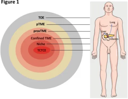
heterogeneous components or processes – from the role of the extracellular matrix to angiogenesis, immune infiltration, and to the microbiota – that are either located within the tumor or found more distantly. Drawing these lists of components, stemming from research done in different independent fields, was essential for the establishment of the TME field. Yet we propose that the scientific community working on the TME is now in need of a more precise and actionable view, namely a multi-layered view of cancer. We distinguish six layers of environmental interactions with the tumor and show that they are associated with specific location-related mechanisms, and ultimately with distinct therapeutic approaches. This leads to the recognition of different levels of the tumor environment organized as a “Russian doll” (Figure 1). Obviously, other hierarchical divisions of the TE than the six-layer approach we propose are possible; our main claim is not about the exact number of these layers, but rather about the crucial need to adopt a stratified approach to foster future developments in the TME field. This will improve our understanding of the TME and the TE in general and should be useful not only to scientists but also to clinicians involved in cancer research and treatment.
Tumor cell-to-tumor cell environment (TCTCE)
Tumor cells evolve in a microenvironment where important interactions occur between different cancer cells. Within the tumor, the environment greatly differs between the core and the edge of the tumor. For example, the core of the tumor tends to be characterized by hypoxia, while more oxygen is available at the periphery. A “metabolic symbiosis” has been described between hypoxic and oxygenated tumor cells, indicating that each population might create a particular environment for the other. For example, the hypoxic tumor cells create a gradient of lactic acid that fuels the oxidative metabolism of oxygenated tumor cells [13].
Other types of cooperative interactions between tumor cells take place at the level of individual cells, ex., lung cancer cells can create their own niche, by producing non- or slow-proliferating
cells that secrete Wnt [14] or Notch ligands [15] supporting the growth of the more proliferative cancer cells.
Moreover, feedback loops between cancer stem cells (CSCs) and more differentiated cancer cells can play a determinant role in tumor development. For example, in chronic myeloid leukemia (CML), mature cancer cells secrete interleukin-6 (IL-6), a pro-inflammatory cytokine that induces a bias of differentiation in CML progenitors/stem cells towards the granulomonocytic lineage, leading ultimately to a feedback loop promoting CML [16]. Interactions between individual cells may also involve the construction of cellular structures such as tunneling nanotubes (TNTs). TNTs are cellular extensions that connect tumor cells to each other and could play an important role in cell-to-cell communication by transferring information (via mRNAs or proteins) from one cell to another [17]. TNTs may be organized into networks in various tumor areas and may exhibit a heterogeneous distribution.
Lastly, accumulating data suggest that genetically different sub-clones can cooperate, and thus favor tumor growth [18]. Cooperation between tumor cells may not only occur at the primary or metastatic tumor site, but also in the circulation. Indeed, recent studies highlight the existence of circulating tumor cell (CTC) clusters [19], associated with adverse outcome when compared to single CTCs, suggesting that this cooperation between cancer cells might play an important role in metastasis.
Taken together, these data indicate that the complex interactions between tumor cells already create various local environments. The interactions described above rely on various sources of heterogeneity between cancer cells, which include the classical diversity in genetic and epigenetic alterations, but also differentiation states or metabolic states. The mechanisms involved are, obviously, very local and constrained to a confined space and involves close contact or short-range interactions. These include cell-to-cell interactions that are mediated by
various processes including metabolic exchange, low-range effects of cytokines, and local structural connections between cells such as nanotubes.
We suggest referring to this first layer of environment, where these interactions take place, as the “tumor-cell to tumor-cell environment” (TCTCE). The TCTCE differs from the TME in so far as it is composed of the tumor cells themselves exclusively. The TCTCE implies cooperative interactions between tumor cells which take place, as we have seen, between individual cells or between groups of cells. The cooperative interactions are most likely reciprocal where cells have a stake on each other. In the stakeholder model [20,21] derived from evolutionary biology, the benefit conditioned by the stake must be superior to the cost. The interaction may be symmetrical (both benefit) or asymmetrical (only one benefits). Whereas single-cell cooperativity involves signaling loops between individual cells, cooperativity between groups of cells relies on a common function of groups of cells where survival advantage for both is achieved. A good example is “metabolic symbiosis”, as described above [13].
The niche
The TME usually refers to the local environment in which the tumor grows. In this respect, the difference between the TME and the niche is not always clear. The notion of niche itself is used in a variety of contexts and refers to different entities.
The notion of niche is widely used in stem cell biology. Its origin can be traced back to Schofield, who introduced this notion in hematology to designate “the cellular environment which retains the stem cell”, “prevents it from maturing”, and “determines its behavior” [22][23]. Not only normal stem cells but also CSCs reside in particular niches. This is the case for hemato-oncology, where the evidence for cancer stem cells was first obtained [24], as well as for solid tumors [25]. The niche, therefore, describes the microenvironment of the CSCs specifically, as opposed to the whole tumor. It is often conceived as a “specialized
microenvironment” [26], a small unit or an anatomically distinct region within the microenvironment, in direct interaction with individual stem cells [25].
But the notion of niche is also used for cancer cells more widely. For example, in glioblastoma, a vascular tumor niche has been described and in lung metastasis the concept of metastatic and premetastatic niche has been proposed [27]. In this wider context, the notion of niche is sometimes used to refer to particular parts of the TME or to refer to the TME at a particular time point. The first case insists on the spatial aspect: the TME is composed of multiple niches. This has been proposed, for instance, in the case of glioblastoma. Hambarzumyan and Bergers [28] define niches as “communication centers in which tumor and host cell populations dynamically interact via direct cell contact or paracrine signaling cues”, and propose the presence of three types of niches in glioblastoma: a perivascular niche, a hypoxic niche, and an invasive niche. The second case insists on the temporal aspect: the niche represents the initiation site of tumor expansion. This initiation site can exist prior to tumor cell implantation, which would correspond to the concept of the premetastatic niche.
These three usages of the niche concept may overlap, but only to a certain degree. For instance, the perivascular niche is certainly found at a very early stage in tumor development whereas the hypoxic niche and invasion niche are assembled later in the life of the tumor, and these different niches can also apply to cancer stem cells. To sum up, despite this diversity in the uses of the concept of niche, they all appear as specifications of the broader notion of TME, either by focusing on the interactions of the TME with the CSCs, or by specifying various spatial or temporal small structures to which the notion of TME can broadly refer. To that extent, these various niches appear as more specific targets than the TME taken as a whole for scientific inquiry and for therapeutic interventions.
Confined TE, proximal TE, and peripheral TE
In the scientific and medical literature, the notion of TME refers to a plurality of elements [1– 3,7], and it is unclear whether a necessary condition for being part of the TME is to be confined to the tumor, or if elements located at more distant sites can also be part of the TME. Tumor lymphatics, for example, were not initially thought to be part of the TME in so far as they are localized at a certain distance from the tumor, and are only secondarily invaded by tumor cells. However, lymphatics were later detected inside tumors and for that reason regarded as an important part of the TME [29]. Similarly, tertiary lymphoid structures have been described in close vicinity of tumor mass, as well as within the tumor mass, in sites where priming of T cells can occur [30]. These ectopic lymphoid organs can exhibit a tumor-promoting or inhibiting role and may impact patient survival [31][32]. Cellular structures or matrix components that may impact tumor development stay outside the main tumor mass. This is the case for the tumor stroma around tumor masses, which may affect tumor development [8,33,34]. This is seen, for instance, in glioblastoma where a ring of reactive astrocytes around the tumor can be observed. Another example is pancreatic ductal adenocarcinoma (PDAC), where a very compact stroma surrounds tumor nodules, which hinders the ingrowth of vessels and the influx of immune cells into the tumor nodules [35]. Tumor-draining sentinel lymph nodes (TDNL), which have been proposed to represent an active part of the TME [36], are spatially found in a more distant location. Tumors can profoundly alter the functional characteristics of downstream lymph nodes (LNs) [37], such as priming LNs for metastatic colonization. LNs may also actively participate in tumor cell dissemination through the LN vasculature, which serves as an exit route for systemic dissemination of cancer cells [38].
Taken together, these examples illustrate the difficulty in delineating both conceptually and spatially the boundaries of the TME and the tumor environment (TE) in general. Yet the specific location and function of some elements of the TME/TE such as the stroma surrounding the
tumor or the lymph nodes located in the periphery suggest a distinction between three spatially different microenvironments, which could be referred to as the “proximal TE” (e.g., the surrounding stroma) and the “peripheral TE” (e.g., lymph nodes), in contrast to the “confined
TE” (corresponding to the classical notion of the TME and characterized by partially permeable
barriers, through which only some cells and molecules infiltrate).
Because of their specificities, these different parts of the TE might present very different challenges for tumor development. For example, from the tumor’s point of view, a key challenge in the confined TE is the local organization of the extracellular matrix, the intra-tumor vasculature, the infiltration of immune cells, and processes such as mechano-transduction [39]. The proximal TE has a double role in tumor development. On the one hand, it participates in the confinement of the tumor mass, which may contribute to tumor escape against immune responses and to therapy-resistance [40]; on the other, it maintains growth factor gradients and nutrient supply to tumor cells through vessel ingrowth or passive diffusion. For the peripheral TE, the relatively long-range communication with lymph nodes, via cellular and molecular messages, is crucial [41]. The fact that different layers are associated with different challenges and different mechanisms is of paramount importance because it delineates distinct therapeutic strategies. Indeed, approaches tailored to the reeducation of stromal cells [42] greatly differ in design and conception to those aimed at altering the mechanics or structure of the ECM [43,44], or to strategies aimed at fostering the homing at the tumor vicinity of new immune cells coming from lymph nodes [45]. Of course, these strategies are often complementary, but our point is that there exist location-dependent mechanisms in and around the tumor, and that these mechanisms can be targeted via distinct therapies.
Finally, one must consider the possible broader impact of the organism on tumor progression at a systemic level, suggesting the idea of an “extended microenvironment”, or “tumor macroenvironment” [46].
Perhaps the most obvious example of a long-distant effect is the priming of distant sites for secondary tumor development by soluble factors or exosomes produced by the primary tumor itself. For instance, VEGF-C derived from a primary tumor can prime lymphatic ganglions for homing of tumor cells [47]. Exosomes produced by breast cancer cells at the primary site prepare the premetastatic niche in lung tissue [48]. Along these lines, tumor-induced myeloid suppressor cells, a subpopulation of immune cells endowed with tumor-promoting activity, or tumor-primed neutrophils have been reported to promote the colonization of distant lungs by cancer cells [49,50].
Nevertheless, the role of distant parts of the organism is not restricted to metastasis; it can also concern the primary tumor. First, the hematopoietic and the immune system are connected to the tumor through the vasculature. Immune responses are induced at distance from primary tumors leading to the activation of effector immune cells, such as cytotoxic T cells, which circulate systemically through the bloodstream and migrate to the tumor sites where they exert cancer-inhibitory effects. Spitzer and colleagues have demonstrated that coordinated systemic and local immune responses are required for the success of immunotherapy [51], emphasizing thereby the importance of always seeing immune responses in the broader context of the whole organism. Second, the microbiota has been involved in the development of different tumor types (sometimes very remote from where the microbiota is localized) through its effect on the immune system. The microbiota impacts host response to various therapies, including chemotherapy and immunotherapies [12][52][53]. Such recently investigated examples further support the idea that distant elements can have a major influence on tumor development.
Moreover, cancer can have long-range effects that are well described but which feedback effect on the primary tumor or on metastases remains undetermined. For example, lung adenocarcinomas can deregulate the circadian clock of the liver and breast cancers and can impact distant tissues, in particular the hematopoietic system, by remodeling the bone marrow and increasing the immature hematopoietic compartment [54]. It has also been observed that malignant tumors can activate non-cell autonomous autophagy not only in the TME but also in distant tissues [55]. Another example is the fact that inflammatory factors induced by cancer-associated fibroblasts (CAFs) may have systemic effects and lead to decreased levels of blood
myeloid and plasmacytoid dendritic cells and to a partial activation with a semi-mature
phenotype and impaired immunostimulatory function [56]. Tumor cachexia can also contribute
to the general weakening of the organism which impacts indirectly on tumor development and host survival.
Taken together, these data suggest a greater involvement of the organism as a whole in cancer progression than previously thought, indicating that beyond the “micro-” environment, the tumor is also exposed to a wider “organismal” environment, that we suggest to call the “tumor organismal environment” (TOE). The TOE includes, but is not limited to, what McAllister and Weinberg call the “tumor-induced systemic environment” [57], as well as the recently suggested notion of “tumor immune microenvironment” [58]. The TOE represents the full set of all the components of the organism not located in the tumor or at its vicinity but which can affect cancer development.
The wider context of the TOE raises specific challenges, such as the long-distance communication between the microbiota and tumors; this communication can be mediated by specific metabolites, by the immune system, and by the endocrine system, among other possibilities. This also creates new opportunities for therapeutic interventions, either by determining and perhaps manipulating the specific microbes that influence anti-cancer
treatments [53,59], or by acting on the communication pathways between those distant sites, including cytokines and metabolites. Clearly, a key challenge for the future will be to thoroughly investigate the role of the TOE in cancer development, its specificity in term of mechanisms, and the new opportunities that it provides for therapeutic interventions.
Concluding Remarks
Over the last four decades, the study of the tumor microenvironment has been carried out by researchers from various disciplines [2], a fact that has contributed to explain the current loose definition and delineation of the TME in the scientific literature. We have shown herein that an undifferentiated approach to the TME, where many heterogeneous components are simply put together under a unique label, is no longer tenable. It is indeed crucial to develop a multi-layer view of the interactions between the tumor and its environments, because the challenges and mechanisms at each level are distinct, which implies that specific therapeutic strategies should be designed to target each of them. Indeed, not all tumors may harbor a complete set of layers and thus it would be important to determine which of these layers will impact tumor growth in a given tumor. For example, manipulating the microbiota through fecal transplant may not work for tumors that are immune-privileged such as PDAC. Thus, there may be tumors that command a more local intervention on the tumor environment while others require a more general approach. Of course, combinatorial strategies are important and it would be crucial for a given cancer to determine which one might work. Combination trials of anti-angiogenic therapy and immunotherapy are currently underway and we may expect that this will probably work for some tumors but not for others.
Even if we have proposed to distinguish six such layers of environmental interactions in the present paper, the exact number of layers matters much less than our more general defense of a multilevel perspective. Furthermore, the boundaries of these different levels are of course not
tight. For instance, factors that drive the TCTCE may also exhibit roles in the confined TE, such as FGFs or even VEGF which play a role in both cases [60].
One of the most pressing challenges for future research on the TME and the TE will be the dissection of the complex interactions between the different levels of tumor-environment interactions that we have distinguished (see: Outstanding Question Box). Not only is the “soil and seed” hypothesis constantly revisited by research that investigates how a local microenvironment can participate in the induction of distant metastatic sites [61], but it is becoming increasingly clear that overall characteristics of the host such as obesity can critically affect the local microenvironment, for example by altering the extracellular matrix [62,63]. In other words, the recognition of different levels in the TME does not mean a separation or isolation of each level, but, quite the contrary, an invitation to explore the complex and fascinating dialogue between those levels.
References
1 Maman, S. and Witz, I.P. (2018) A history of exploring cancer in context. Nat. Rev.
Cancer 18, 359–376
2 Witz, I.P. (2009) The Tumor Microenvironment: The Making of a Paradigm. Cancer
Microenviron. 2, 9–17
3 Li, H. et al. (2007) Tumor microenvironment: the role of the tumor stroma in cancer.
J. Cell. Biochem. 101, 805–815
4 Bissell, M.J. and Hines, W.C. (2011) Why don’t we get more cancer? A proposed role of the microenvironment in restraining cancer progression. Nat. Med. 17, 320–329
5 Hu, M. and Polyak, K. (2008) Microenvironmental regulation of cancer development.
Curr. Opin. Genet. Dev. 18, 27–34
6 Laconi, E. (2007) The evolving concept of tumor microenvironments. BioEssays News
Rev. Mol. Cell. Dev. Biol. 29, 738–744
7 Whiteside, T.L. (2008) The tumor microenvironment and its role in promoting tumor growth. Oncogene 27, 5904–5912
8 Bissell, M.J. and Radisky, D. (2001) Putting tumours in context. Nat. Rev. Cancer 1, 46–54
9 Fridman, W.H. et al. (2012) The immune contexture in human tumours: impact on clinical outcome. Nat. Rev. Cancer 12, 298–306
10 Schneider, G. et al. (2017) Tissue-specific tumorigenesis: context matters. Nat. Rev.
Cancer 17, 239–253
11 Hirata, E. and Sahai, E. (2017) Tumor Microenvironment and Differential Responses to Therapy. Cold Spring Harb. Perspect. Med. DOI: 10.1101/cshperspect.a026781
12 Iida, N. et al. (2013) Commensal bacteria control cancer response to therapy by modulating the tumor microenvironment. Science 342, 967–70
13 Sonveaux, P. et al. (2008) Targeting lactate-fueled respiration selectively kills hypoxic tumor cells in mice. J. Clin. Invest. 118, 3930–3942
14 Tammela, T. et al. (2017) A Wnt-producing niche drives proliferative potential and progression in lung adenocarcinoma. Nature 545, 355–359
15 Lim, J.S. et al. (2017) Intratumoural heterogeneity generated by Notch signalling promotes small-cell lung cancer. Nature 545, 360–364
16 Reynaud, D. et al. (2011) IL-6 Controls Leukemic Multipotent Progenitor Cell Fate and Contributes to Chronic Myelogenous Leukemia Development. Cancer Cell 20, 661–673 17 Winkler, F. and Wick, W. (2018) Harmful networks in the brain and beyond. Science 359, 1100–1101
18 Polyak, K. et al. Co-evolution of tumor cells and their microenvironment. , Trends in
Genetics, 25. (2009) , 30–38
19 Aceto, N. et al. (2014) Circulating tumor cell clusters are oligoclonal precursors of breast cancer metastasis. Cell 158, 1110–22
20 Roberts, G. (2005) Cooperation through interdependence. Anim. Behav. 70, 901–908 21 Aktipis, A. Principles of cooperation across systems: from human sharing to
multicellularity and cancer. Evol. Appl. 9, 17–36
22 Schofield, R. (1983) The stem cell system. Biomed. Pharmacother. Bioméd.
Pharmacothérapie 37, 375–80
23 Schofield, R. (1978) The relationship between the spleen colony-forming cell and the haemopoietic stem cell. Blood Cells 4, 7–25
24 Bonnet, D. and Dick, J.E. (1997) Human acute myeloid leukemia is organized as a hierarchy that originates from a primitive hematopoietic cell. Nat Med 3, 730–737
regulating stemness of tumor cells? Cell Stem Cell 16, 225–238
26 Baryawno, N. et al. Hematopoiesis: Reconciling Historic Controversies about the Niche. , Cell Stem Cell, 20. (2017) , 590–592
27 Kaplan, R.N. et al. Preparing the “soil”: The premetastatic niche. , Cancer Research, 66. (2006) , 11089–11093
28 Hambardzumyan, D. and Bergers, G. Glioblastoma: Defining Tumor Niches. , Trends
in Cancer, 1. (2015) , 252–265
29 Alitalo, A. and Detmar, M. Interaction of tumor cells and lymphatic vessels in cancer progression. , Oncogene, 31. (2012) , 4499–4508
30 Dieu-Nosjean, M.C. et al. Tertiary lymphoid structures in cancer and beyond. , Trends
in Immunology, 35. (2014) , 571–580
31 Hiraoka, N. et al. Tertiary lymphoid organs in cancer tissues. , Frontiers in
Immunology, 7. (2016)
32 Finkin, S. et al. (2015) Ectopic lymphoid structures function as microniches for tumor progenitor cells in hepatocellular carcinoma. Nat. Immunol. 16, 1235–1244
33 Ingber, D.E. (2002) Cancer as a disease of epithelial-mesenchymal interactions and extracellular matrix regulation. Differ. Res. Biol. Divers. 70, 547–560
34 Bissell, M.J. et al. (2002) The organizing principle: microenvironmental influences in the normal and malignant breast. Differ. Res. Biol. Divers. 70, 537–546
35 Joyce, J.A. and Fearon, D.T. (2015) T cell exclusion, immune privilege, and the tumor microenvironment. Science 348, 74–80
36 Swartz, M.A. et al. (2012) , Tumor microenvironment complexity: Emerging roles in cancer therapy. Cancer Research, 72, pp. 2473–2480
37 Munn D.H., Mellor A.L. (2006) The tumor-draining lymph node as an immune-privileged site. Immunol Rev. 213, 146-158
38 Brown, M. et al. (2018) Lymph node blood vessels provide exit routes for metastatic tumor cell dissemination in mice. Science 359, 1408–1411
39 Northcott, J.M. et al. (2018) Feeling Stress: The Mechanics of Cancer Progression and Aggression. Front. Cell Dev. Biol. 6:17,
40 Gajewski, T.F. et al. (2013) Innate and adaptive immune cells in the tumor microenvironment. Nat. Immunol. 14, 1014–1022
41 Chrisikos, T.T. et al. (2018) Molecular regulation of dendritic cell development and function in homeostasis, inflammation, and cancer. Mol. Immunol. DOI:
10.1016/j.molimm.2018.01.014
42 Quail, D.F. and Joyce, J.A. (2013) Microenvironmental regulation of tumor progression and metastasis. Nat. Med. 19, 1423–1437
43 Paszek, M.J. et al. (2005) Tensional homeostasis and the malignant phenotype.
Cancer Cell 8, 241–254
44 Ingber, D.E. (2008) Can cancer be reversed by engineering the tumor microenvironment? Semin. Cancer Biol. 18, 356–364
45 Tang, H. et al. (2016) Facilitating T Cell Infiltration in Tumor Microenvironment Overcomes Resistance to PD-L1 Blockade. Cancer Cell 29, 285–296
46 Al-Zoughbi, W. et al. (2014) Tumor macroenvironment and metabolism. Semin.
Oncol. 41, 281–295
47 Karaman, S. and Detmar, M. (2014) Review series Mechanisms of lymphatic metastasis. J. Clin. Invest. 124, 922–8
48 Becker, A. et al. Extracellular Vesicles in Cancer: Cell-to-Cell Mediators of Metastasis. , Cancer Cell, 30. (2016) , 836–848
49 Wculek, S.K. and Malanchi, I. (2015) Neutrophils support lung colonization of metastasis-initiating breast cancer cells. Nature 528, 413–417
50 Ouzounova, M. et al. (2017) Monocytic and granulocytic myeloid derived suppressor cells differentially regulate spatiotemporal tumour plasticity during metastatic cascade. Nat.
Commun. 8, 14979
51 Spitzer, M.H. et al. (2017) Systemic Immunity Is Required for Effective Cancer Immunotherapy. Cell 168, 487-502.e15
52 Dzutsev, A. et al. (2015) The role of the microbiota in inflammation, carcinogenesis, and cancer therapy. Eur. J. Immunol. 45, 17–31
53 Routy, B. et al. (2018) Gut microbiome influences efficacy of PD-1–based immunotherapy against epithelial tumors. Science 359, 91–97
54 Masri, S. et al. (2016) Lung Adenocarcinoma Distally Rewires Hepatic Circadian Homeostasis. Cell 165, 896–909
55 Katheder, N.S. et al. (2017) Microenvironmental autophagy promotes tumour growth.
Nature 541, 417–420
56 Tjomsland, V. et al. (2010) Semi mature blood dendritic cells exist in patients with ductal pancreatic adenocarcinoma owing to inflammatory factors released from the tumor.
PLoS ONE 5,
57 McAllister, S.S. and Weinberg, R.A. (2014) The tumour-induced systemic
environment as a critical regulator of cancer progression and metastasis. Nat Cell Biol 16, 717–727
58 Binnewies, M. et al. (2018) Understanding the tumor immune microenvironment (TIME) for effective therapy. Nat. Med. 24, 541–550
59 Matson, V. et al. (2018) The commensal microbiome is associated with anti–PD-1 efficacy in metastatic melanoma patients. Science 359, 104–108
60 Lu, K. V and Bergers, G. (2013) Mechanisms of evasive resistance to anti-VEGF therapy in glioblastoma. CNS Oncol. 2, 49–65
61 Friedl, P. and Alexander, S. (2011) Cancer Invasion and the Microenvironment: Plasticity and Reciprocity. Cell 147, 992–1009
62 Seo, B.R. et al. (2015) Obesity-dependent changes in interstitial ECM mechanics promote breast tumorigenesis. Sci. Transl. Med. 7, 301ra130-301ra130
63 Olson, O.C. et al. (2017) Obesity and the tumor microenvironment. Science 358, 1130–1131
64 Thiery-Vuillemin, A. et al. (2005) Molecularly targeted agents: Their promise as cancer chemopreventive interventions. Eur. J. Cancer 41, 2003–2015
65 Boesch, M. et al. (2016) Heterogeneity of Cancer Stem Cells: Rationale for Targeting the Stem Cell Niche. Biochim. Biophys. Acta 1866, 276–289
66 Su, S. et al. (2018) CD10+GPR77+ Cancer-Associated Fibroblasts Promote Cancer Formation and Chemoresistance by Sustaining Cancer Stemness. Cell 172, 841-856.e16 67 Moserle, L. et al. (2014) Antiangiogenic therapies: going beyond their limits. Cancer
Discov. 4, 31–41
68 June, C.H. and Sadelain, M. (2018) Chimeric Antigen Receptor Therapy. N. Engl. J.
Med. 379, 64–73
69 Steinman, R.M. and Banchereau, J. (2007) Taking dendritic cells into medicine.
Nature 449, 419–426
70 Zitvogel, L. et al. (2017) Nutrition, inflammation and cancer. Nat. Immunol. 18, 843– 850
Figure 1: The multiple layers of the tumor environment.
The figure on the left depicts the different layers of the tumor environment as a “Russian doll”. These different layers concern both the primary tumor and tumor metastasis.
The table on the right depicts possible therapeutic strategies related to the different TE layers. For each of the different layers, examples of its key constituents, as well as some therapeutic strategies associated with it, are listed. These therapeutic strategies are no more than possible examples, and some strategies might belong to two different layers. Yet the main message here is that distinguishing different layers in the tumor environment helps clarify the various anti-cancer treatments that can be used in the clinic. (TE, tumor environment; TCTCE, tumor-cell-to-tumor-cell environment; TOE, tumor organismal environment; TC, tumor cells; ECM, extracellular matrix; TNT, tunneling microtubes; CAFs, cancer-associated fibroblasts).