HAL Id: inserm-00483885
https://www.hal.inserm.fr/inserm-00483885
Submitted on 17 May 2010
HAL is a multi-disciplinary open access
archive for the deposit and dissemination of
sci-entific research documents, whether they are
pub-lished or not. The documents may come from
teaching and research institutions in France or
abroad, or from public or private research centers.
L’archive ouverte pluridisciplinaire HAL, est
destinée au dépôt et à la diffusion de documents
scientifiques de niveau recherche, publiés ou non,
émanant des établissements d’enseignement et de
recherche français ou étrangers, des laboratoires
publics ou privés.
Yehezkel Ben-Ari
To cite this version:
Yehezkel Ben-Ari. Basic developmental rules and their implications for epilepsy in the immature
brain.. Epileptic Disorders, John Libbey Eurotext, 2006, 8 (2), pp.91-102. �inserm-00483885�
Basic developmental rules
and their implications for
epilepsy in the immature brain
Yehezkel Ben-Ari
INMED/INSERM, Parc Scientifique de Luminy, Marseille, France
Plenary lecture given at the 26thInternational Epilepsy Congress – Paris 2005
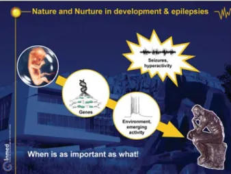
ABSTRACT–The construction of the human brain with its 1015 synapses follows a set of complex developmentally and environmentally regulated steps. A series of sequences have been described that are instrumental, in the sense that a failure of any one of them leads to dramatic, life-long consequences. Hence the importance of determining the sequential maturation of neurons, synapses and cortical maps. It is also important to determine how network-driven events become installed, as neuronal activity intervenes in all of these steps and modulates, for better or worse, the outcome. A fundamental conse-quence of these sequential events is that any disruption will have very different consequences depending on when it occurs, indeed, “when is as important as what”. An obvious aspect of these general features is related to seizures. In fact, the developing brain has both a higher incidence of seizures in human and animal models, and experiences seizures that can produce long-lasting conse-quences that are also stage-dependent.
This seminar and the series of slides presented are an introduction to these issues, summing up several studies made notably by INMED researchers during the last two decades (http://www.inmednet.com). It concentrates on four basic developmental rules: i) the generation by very immature neurons, of very large currents mediated by the activation of receptors in neurons that bear no synapses. This is due to the release of GABA that diffuses to distal sites and acts as a paracrine factor; ii) the excitatory/inhibitory shift of the actions of GABA during development because of a progressive reduction in the intracellular chloride concentration; iii) the sequential formation of GABAergic synapses and networks before glutamatergic ones, implying that, at an early stage, all the excitatory drive will be GABAergic; iv) the presence, at an early stage, of a unique, primitive pattern in all developing structures, this pattern disappears when most GABAergic synapses have shifted to their adult configuration-.Several consequences of these sequences are described including: i) a control of neuronal migration by GABA-acting drugs, and the possibility that migration disorders are also generated by environmental factors that include the effects of GABA-acting agents; ii) If GABA excites immature neurons and inhibits adult one, then GABA-acting agents will also produce different effects on the mother and the embryo; iii) early brain oscillations are generated by the periphery and propagate centrally – notably to the sensory-motor cortex, suggesting that peripherally-generated movements may provide an important signal for the formation of cortical maps, in keeping with the importance of embryonic movements; iv) “seizures beget seizures” in the developing brain. This has now been shown in a triple chamber with the two intact hippocampi that we developed, and with which it has been possible to show that only recurrent seizures that include high frequency oscillations can transform the naïve,
Correspondence: Y. Ben-Ari
Institut de Neurobiologie de la Méditerranée (INMED),
INSERM U29,
Parc Scientifique de Luminy, 13273 Marseille Cedex 09, France
contralateral hippocampus to an epileptic one that seizes spontaneously. Most interestingly, at an early developmental stage, when GABA excites many neurons and the density of glutamatergic synapses is not sufficiently high, purely glutamatergic seizures cannot lead to long-term consequences, the additional excitatory drive provided by GABAergic synapses is needed. In other words, at that stage, blocking GABA synapses generates seizures, as in adults, but these do not lead to long-term consequences. The mechanisms that underlie these differences is due to the need for high frequency oscillations (> 80 Hz or so), and these can only be generated when GABA synapses are operative in the developing brain: GABA receptor antagonists are ictogenic, but not epileptogenic.To facilitate teaching purposes the paper is published together with supplemental data (as a PowerPoint presentation included in the accompanying DVD), thus allowing an overview of important developmental steps and their implications.
[Published with supplemental data on CD]
Key words: neuronal migration, excitatory action of GABA, oscillation, seizure, antiepileptic drug, GABAergic interneuron
There is a paradox as far as epilepsy in the developing brain is concerned. The incidence of seizures is higher early in life and seizures lead to long- term deleterious sequel; yet immature neurons are less vulnerable than adult ones to a variety of insults including anoxo-ischemic episodes and seizures. In other words, seizures are more frequent - indicating that immature networks are endowed with a capacity for high excitability and synchronized events.
These observations lead to a number of conclusions. Firstly, seizures are more readily generated because one or more of the mechanisms that in adults prevent excessive activity, are not available. Secondly, seizures must pro-duce their consequences by altering fundamental proces-ses rather than “simply” causing neuronal lesions. This implies that understanding and treating neonatal and early seizures must incorporate a detailed analysis of the se-quences of expression of the principal constituent of cor-tical networks in order to determine where the missing mechanisms are that fail to prevent seizure generation, and which sequence is affected by hyperactivity leading to permanent alterations of the normal sequence of events. To further complicate the issue, it is essential to take into account that the developing brain is not a small adult brain, but a unique machine with channels, molecules and mechanisms that are entirely or largely absent in adults. Thus, it is not possible to rely on mechanisms operating in adults to unravel the causes of the paradox summarized above. The central issue is to determine when during development, neuronal activity can alter develop-mental processes (figure 1/DVD figure 2).
Studies performed mostly in Unit 29 of the French medical research council (INSERM) have unraveled several funda-mental rules of brain development and have shown that these rules most likely underlie the unique properties of the developing brain as far as epileptogenesis is concer-ned. This work summarizes the salient features of these observations and, relying on pictures from actual experi-ments, shows how they explain the high incidence of seizures and their long lasting consequences on the deve-loping brain. We shall divide the sequence into 3 steps:
migration, synapse formation, network construction
(fi-gure 2/DVD fi(fi-gure 3).
To evaluate the impact of activity on brain maturation, we shall examine successively the role of the principal trans-mitters, GABA and glutamate, on essential developmental
Figure 1. Nature and Nurture in development & epilepsies.
steps, and, in parallel, illustrate how this information impacts on neurological disorders. In the adult brain, glutamatergic neurons constitute the vast majority of neu-rons, and provide the main source of the excitatory drive. The dense networks of GABAergic interneurons that cons-titute 10-15% of the neurons on the other hand, provide the source of the inhibitory drive. In spite of this apparent imbalance, interneurons play a crucial role in the genera-tion of behaviorally relevant patterns (Freund and Buszaki, 1996). Various types of interneurons control the fine dis-charge of the principal cells, and the patterns that networks will generate. In the absence of functional GA-BAergic neurons and synapses, cortical networks seize and do not generate behaviorally relevant patterns. Let us now examine whether they are operative at early stages and whether they control essential developmental processes.
Neuronal migration
In a wide range of systems, receptors develop before synapses and the obvious question is whether they exert an influence. In other words, does modulation by environ-mental factors, including neuronal activity, start with syn-apse formation or does it occur at an earlier stage when synapses are not yet operative?
If transmitters are released and act on immature neurons endowed with receptors but not functional synapses, they should generate a current by activating post-synaptic re-ceptors. With this in mind, we therefore patch-clamp recorded immature neurons in which the conventional, fast synaptic responses cannot be generated and found that indeed, electrical stimuli evoked giant currents that are several order of magnitude larger than conventional synaptic currents (figure 3; DVD figure 4-6). These
cur-rents are mediated by the activation of GABA- and glutamate NMDA-type receptors – but not AMPA recep-tors that are responsible for the majority of fast acting, ionotropic synapses in the adult brain. We also found that there is a tonic release of GABA and glutamate at an early stage, since a GABA receptor antagonist generates a tonic current indicating an ongoing release of GABA (De-marque et al., 2002). The release mechanisms are unique in the sense that they do not depend on essential vesicular release proteins and factors including release proteins and machinery. They can even be generated in animals in which the release machinery has been knocked out. This paracrinic release of GABA acts on distal neurons to generate large currents prior to synapse formation. At a later stage, this system disappears and is replaced by the more conventional focal action of transmitters. Therefore, neurons communicate prior to synapse formation and there is a non-canonical release of transmitter that can act diffusely on distal sites (also see below).
Transmitter release and neuronal migration
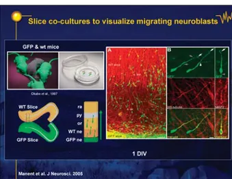
Is transmitter release functional and does it alter migra-tion? We tested the possible role of this paracrine release on migration in vitro, using an original preparation in which two hippocampal slices are cultivated next to each other: one of the slices was obtained from a GFP trans-genic mouse in which all neurons are green, allowing us to follow visually their course and migration on the other, dark slice (figure 4; DVD figure 7; Manent et al. 2005. We found (figure 5; DVD figure 8) that blocking GABA recep-tors retarded neuronal migration considerably indicating that migration – an activity-dependent process (DVDfig-ure 6; Komuro and Rakic) - is modulated by a non-focal,
paracrinic action of transmitters, most notably GABA. These and other observations are in line with the extensive
Figure 3. Receptors are formed before synapses. Demarque et al. Neuron 2004.
Figure 4. Slice co-cultures to visualize migrating neuroblasts. Manent et al. J Neurosci 2005.
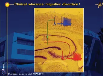
evidence that GABA is a morphogenic agent that, at an early stage, modulates neuronal migration in addition to synapse formation, and neuronal growth and differentia-tion. Thus, transmitters, notably GABA, can modulate early developmental processes by means of actions on receptors that are, in essence, extra -synaptic (also see Owens et al. 1999, 2002; Represa and Ben-Ari 2005). Therefore, agents acting on GABA receptors can be ex-pected to act on neurons that possess few or no synapses at all. This type of mechanism could be involved in the formation of heterotopic masses and other migration dis-orders such as those produced in pregnant rats leading to the formation of aberrant connections between the hip-pocampus and the neocortex (Chevassus-Au-Louis et al. 1998 and figure 5; DVD figure 8). At any rate, they draw our attention to the possible deleterious actions of drugs acting on GABA receptors, notably benzodiazepines or certain anti-epileptic agents. In keeping with this, prelimi-nary observations suggest that some AEDs indeed affect migration (Manent et al. in preparation). These actions are of the utmost importance in view of the part played by migration disorders in human epilepsies. A variety of animal models of these disorders exist, some of them such as the one produced by in utero injection of an anti-mitotic agent MAM (figure 5; DVD figure 8). Studies using this model show that the ectopic masses present in the hippoc-ampus are, in fact, neocortical neurons that were trapped in the hippocampus because of the delayed migration. Patch recordings from both the “normal” target of these neurons and their host structure shows that they have connections both with the neocortex and the hippocam-pus, creating thus an aberrant bridge connecting, via one synapse, two brain structures that are not connected nor-mally (figure 6; DVD figure 9; Chevassus-Au-Louis et al., 1998). Seizures will readily propagate from the hippocam-pus to the cortex.
Transmitter uptake systems
and early seizures
If transmitters act at this early stage, in a paracrinic man-ner, then the control of their concentration may play an important role. The concentration of GABA and glutamate in the extracellular space is controlled by a variety of transporters. We therefore tested the effects of blockers of the main transporters on neuronal activity at an early developmental stage E18 to P0 (DVD figure 10). We found that GABA transporters are not active at birth – blockers of this transport do not alter currents generated by GABA. This will allow GABA to diffuse and act on distal sites
(DVD figure 11). In contrast, glutamate transporters are
operative early on, indicating that the control of glutamate concentration is essential at this early stage to prevent possibly excitotoxic actions of glutamate (DVD figure 12). In keeping with this, glutamate transporter blockade gen-erates large oscillations in hippocampal and neocortical networks (DVD figure 13), and in vivo icv injections of a glutamate receptor antagonist in pups generates recurrent seizures with suppression bursts that are reminiscent of the encephalopathies (Demarque et al. 2004). Thus, deficien-cies in glutamate transport could mediate early, severe epilepsies.
Synapse formation
The formation of synapses (figure 7; DVD figure 14) is an important step as it signals a more organized, topical mode of communication and the beginning of network forma-tion with its generaforma-tion of behaviorally relevant patterns. The two principal transmitter systems that operate are GABA and glutamate, acting on receptors permeable to anions, chloride in particular for the former, and cations,
Figure 5. Blockade of GABA Rs retards migration. Manent et al. J Neurosci 2005.
Figure 6. Clinical relevance: migration disorders! Chevassus-Au-Louis et al. PNAS 2000.
sodium, potassium and calcium for the latter (figure 8;
DVD figure 15). Agents that augment the efficacy of
GABAergic synapses are often anti-epileptics and anxi-olytics; agents that increase the excitatory drive will in-duce hyperactivity and cell death (DVD figure 16). This raises the issue of how an equilibrium between these transmitters is reached throughout development (DVD
figure 17). This also raises the question of how to develop
cortical networks with functional activity at all stages – promote neuronal development and synapse formation yet preserve stability and prevent over excitation that will be excitotoxic, or dominant GABAergic inhibition that will retard developmental functions that are modulated by the increased intracellular calcium that results from the activation of glutamate synapses (DVD figure 17)? In 1989, three basic developmental rules (figure 9; DVD
figure 18) for the maturation of GABA and glutamate
systems were unraveled. These present a possible solution to this issue:
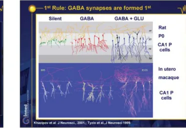
i) GABAergic synapses precede glutamate synapses (figure 10; DVD figure 19; Ben-Ari 2002; Tyzio et al. 1999). In the developing hippocampus, interneurons do not develop hand-in-hand with the pyramidal neurons: GABAergic interneurons and synapses mature first and are operative at a stage when the principal cells are silent (Ben-Ari et al. 2004). Interneurons divide prior to the glutamatergic neurons, extend their axons and dendrites prior to the principal cells, establish functional synapses before principal neurons and can generate patterns when principal neurons are essentially inert. We patch-clamp recorded a large sample of CA1 pyramidal neurons at birth and reconstructed them after identifying their PSCs. We found three populations of neurons, silent ones with no PSCs that have few or no dendrites, bigger neurons with
Figure 8. Inhibition and Excitation. Figure 7. Constructing a cortex.
C. Hammond, Molecular and Cellular Neurobiology, Elsevier.
Figure 9. Three rules preserved throughout evolution! Ben-Ari et al. J Physiol, 1989
Figure 10. First Rule: GABA synapses are formed first. Khazipov et al. J Neurosci 2001; Tyzio et al. J Neurosci 1999.
GABA but no glutamate synapses have a small, apical dendrite, neurons with both GABA and glutamate PSCs have a large, apical dendrite that reaches, for the first time, the distal lacunosum moleculare and a basal dendrite. Therefore, the first synapses are established on the apical dendrites and are GABAergic, neurons must reach the distal lacunosum moleculare to have functional glutamate synapses. Since the axons are there already, the critical factor is the degree of maturation of the post-synaptic target: GABAergic synapses are established when axons meet their post-synaptic target, but glutamatergic synapses will not be formed unless the target is more developed. This sequential expression of GABA and glutamate syn-apses has been confirmed in a wide range of species including primate neurons in utero (figure 10; DVD
fig-ure 19; Tyzio et al., see below,). We patch-clamped CA1
pyramidal neurons in slices obtained from macaque em-bryos delivered by caesarean section from mid-gestation to two weeks before full-term, and found an earlier forma-tion of GABAergic synapses on pyramidal neurons with a small, apical dendrite and both GABA and glutamate synapses on neurons with extended apical dendrites and basal dendrites. There were no neurons with functional glutamate but no glutamate synapses confirming the ear-lier formation of GABAergic synapses.
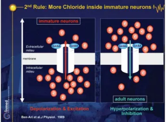
ii) GABA inhibits adult neurons but excites immature ones (figure 11; DVD figure 20)
This is due to a higher concentration of intracellular chloride in immature neurons, which leads to an efflux of chloride when GABA receptors are activated (Ben-Ari et
al. 1989, Tyzio et al. 2003). These gradients have been
observed in all species and brain structures studied, indi-cating that they have been retained throughout evolution (Gao and van den Pol 2001, Owens et al. 1999, 2002, Hubner et al. 2001). They are also observed in primate
neurons in utero, and are thus valid for human fetuses and preterm babies (Khazipov et al. 2001). The consequence is that when GABA receptors are activated there is an efflux of chloride instead of the usual influx, and a depolariza-tion that will generate acdepolariza-tion potentials, remove the voltage-dependent magnesium block from NMDA chan-nels leading to an influx of calcium (Ben-Ari et al. 1997). This early action of GABA reverses subsequently, with the delayed activation of a chloride co-transporter that exports chloride from the inside to the outside and install the adult concentration of chloride (figure 12; DVD figure 21; Rivera et al., 1999). We have suggested elsewhere that this sequence provides a solution to the problem of how to equilibrate, at all developmental stages, excitation and inhibition during brain maturation (DVD figure 22 and Ben-Ari 2002). Indeed, as the excitatory drive provided by GABAergic synapses is not toxic - as the E rev of GABA is always closer to the resting membrane potential than glutamate (E 50-60 mV as compared to 0 mV). This is also facilitated by the shunting actions of GABAergic synapses that inhibit overexcitation. Thus, GABA will provide a sufficient degree of excitatory drive to raise intracellular calcium via the activation of NMDA channels or voltage-dependent calcium channels, and thus to modulate devel-opmental processes without danger. Also, the longer ki-netics of GABAergic as compared to glutamate AMPA receptor-mediated PSCs enables an accumulation of syn-aptic current in neurons that possess but few synapses. Therefore, during early stages, when the glutamatergic drive is limited, GABA “does the job”, exciting neurons and raising intracellular calcium. When the density of glutamatergic synapses is sufficient and a strong inhibition is required to prevent seizure generation, GABA assumes its adult actions following the expression of a transporter that will reduce the intracellular concentration of chlo-ride. This model provides a rational solution to the issue
Figure 12. Higher [Cl-]
i: a question of pumps & transporters. Figure 11. Second Rule: More Chloride inside immature neurons!
raised above (DVD figure 22, Ben-Ari 2002). If GABAer-gic synapses are formed first, GABAerGABAer-gic interneurons should also develop at an early stage. In the developing hippocampus, interneurons do not develop hand-in-hand with the pyramidal neurons: GABAergic interneurons and synapses mature first and are operative at a stage when the principal cells are silent (Hennou et al. 2002, Ben-Ari et al. 2004). Interneurons divide prior to glutamatergic neurons, extend their axons and dendrites prior to the principal cells, establish functional synapses before principal neu-rons and can generate patterns when principal neuneu-rons are essentially inert. Most interestingly, the rules for establish-ing functional GABA or glutamate synapses between an axon and its dendritic target are not identical: GABA synapses are established once the contact is established, whereas glutamate synapses can only be formed when the target neuron has reached a certain degree of maturation. Therefore, in spite of a rather long journey – interneurons are not generated in the close vicinity of the principal neurons, but in the distal ganglionic eminence – interneu-rons must, for some reason, be at work at an early stage. In parallel studies, we found that already in utero, patterns generated exclusively by interneurons can be recorded (Ben-Ari et al., 2004).
iii) Developing networks speak only one language
(figures 13, 14; DVD figures 23-24)
We discovered some time ago that the developing hip-pocampal network has a single pattern – which we re-ferred to as giant depolarising potentials (GDPs) (Ben-Ari
et al. 1989; Ben-Ari 2001). This pattern is characterised by
recurrent bursts with large, polysynaptic currents medi-ated by GABA and glutamate receptors. Subsequent stud-ies revealed that a similar pattern is present in all develop-ing networks, in all species includdevelop-ing the developdevelop-ing primate hippocampus in utero. GDPs provide almost all the activity as long as GABAergic synapses are excitatory. The shift from excitatory to inhibitory GABA signals the
end of GDPs and the expression of more adult patterns; it is at that stage that a network is sufficiently complex that it can generate behaviorally relevant patterns. GDPs are generated by the combined action of excitatory GABA synapses and large NMDA receptor-mediated synaptic currents. The latter are of particularly long duration in the immature brain consequent to the presence of a subunit of NMDA receptors that confer to neurons very long kinetics. The synergistic action of GABA and NMDA receptors is the core of this pattern, the small depolarization produced by GABA is sufficient to remove the blockade from NMDA receptors and generates the long, poly-synaptic currents notably via the activation of an excitatory recurrent loop – the activation of interneurons leading, in return, to an enhancement of the excitation on principal cells and the generation of large synaptic currents. GDPs have now been recorded by a plethora of techniques – including cell-attached and whole cell recordings, as well as field and surface recordings – and preparations including slices and also cultures and in vivo. In the intact hippocampus in
vitro – a preparation developed in the laboratory that has
major advantages in comparison to slices – GDPs can be generated by a wide range of sites but they propagate along the developmental gradient – from rostral to caudal sites – indicating a developmental activity gradient (Kha-lilov et al. 1997, 2003). GDPs can be considered as a type of a primitive pattern with little information content that exists to signal activity in developing connections and to promote synapse formation and possibly provide early signals for the construction of operating functional units on the basis of neurons that fire together, wire together”. A general developmental curve depicting the maturation sequences in primate hippocampal neurons is shown in
figure 15 (DVD figure 25). This figure, that provides the
most complete curve available today, is a series of mea-sures (Boltzmann equations to depict the temporal
matu-Figure 13. Three rules preserved throughout evolution!
Figure 14. A primitive pattern in all developing structures! Ben-Ari et al. J Physiol 1989; Khazipov et al. J Physiol 1997.
ration of the various functions). Thus, proliferation (taken from the literature) is followed by axon (first) and dendritic extension (then, expressed in lm per day) and spine acquisition. Pyramidal neurons of CA1 have no spines at mid-gestation, but as many as 7000 glutamate synapses a few weeks before delivery. The earlier formation of GABA synapses is described quantitatively in the last curve. GDPs disappear when the density of glutamate synapses is sufficient to generate more complex, behaviorally relevant patterns and GABA assumes its adult actions. An obvious consequence of this early expression of functional GABAergic synapses is that GABA-mimetic agents and anti-epileptic drugs will exert a particularly strong action on the activity-dependent formation of the network (Represa and Ben-Ari 2005). The obvious implications of these gradients are that drugs targeted to a given molecular species will have complex. unexpected actions: they will exert no action on some neurons endowed with different molecular species and act only on that proportion of neurons that have shifted to adult types of molecular species. In addition, the gradient of heterogeneity will depend notably on the species: animals that have an extended period of brain development – such as monkeys and humans– will offer a picture of heterogeneity for an extended period of time thus allowing extrinsic factors to exert a stronger influence than animals in which this period is restricted to a few days.
Implications of these rules
There are several implications and consequences of these rules (figure 16; DVD figure 26).
Early activity may participate in the formation of cortical maps
Recordings in chronically implanted pups revealed the presence of sharp waves and tails that are reminiscent of
GDPs (figures 17, 18; DVD figures 27-28; Leinekugel et
al. 2002; Khazipov et al. 2004). These patterns are
gener-ated by GABA and glutamatergic synaptic currents as revealed by whole cell recordings in non-anaesthetized, restrained pups. Most importantly, when these events are recorded in both the periphery and the cortical centre – the sensori-motor cortex – the spindles in the periphery precedes those in the center. Lesions of the spinal cord reveal a large reduction of spontaneous spindles suggest-ing that the peripherally-generated spindles are the gen-erators of those in the cortical sites. These and other similar observations are in line with the concepts that developing circuits in the periphery and centres commu-nicate via a single pattern and that peripheral structures can provide a strong source of modulation of the centres. In a wider perspective, these studies raise the possibility that the programming of the construction of central
corti-Figure 15. Developmental sequences in Macaques. Khazipov et al. J Neuroscience 2002.
Figure 16. Implications of these rules.
Figure 17. Spindles in the developing cortex.
cal organization, including cortical maps and other func-tional entities, are controlled or at least modulated by peripheral activity. In view of this, a unified pattern of activity in developing structures is perfectly able to con-tribute to the wiring of distant structures. The alterations produced by aberrant activities such as seizures would produce their deleterious sequelae merely by interrupting these patterns rather than by more specific information-containing signals.
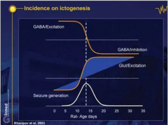
Impact on early seizures
Immature brains have a higher incidence of seizures early on. This is, at least in part, due to the late maturation of a potent inhibitory GABAergic system and the early excita-tory actions (figure 19; DVD figure 29; Holmes and Ben-Ari 1998). The peak seizure generation in the developing hippocampus (bottom curve) is attained when GABAergic excitation shifts to a dominant inhibition around P 10), glutamate excitatory drive steadily increases, and the
im-portance of GDPs declines, being replaced by more so-phisticated patterns that require a relatively elaborated network to function. In keeping with this, seizures are readily generated around the last trimester in primate hippocampal neurons in utero (DVD figure 30).
Seizures can lead to long lasting consequences
In vivo studies have documented extensively the possible
long-term actions of seizures at an early stage. As shown earlier, seizures such as those produced by administration of kainic acid during the first week of post-natal life are not associated with brain damage (Tremblay et al., 1984). Various convulsive agents lead to long-lasting conse-quences including persistent changes in neuronal excit-ability (DVD figure 31; Villeneuve et al. 2000). One way of mediating these long term actions could be a reduction in the seizure threshold after an episode of severe seizures; In other words, in this model, “seizures would beget seizures” as first suggested by Gower in the early nineties. This concept has however never received direct confirma-tion. We recently developed a unique preparation in which this property can be tested readily in vitro
(fig-ure 20; DVD fig(fig-ure 32; Khalilov et al. 1997, 2003).
The preparation consists of a triple chamber that can accommodate the two intact hippocampi and their con-necting commissure in three independent compartments. It is possible to apply a convulsive agent to one hippoc-ampus and allow a given number of seizures to propagate to the other hippocampus. We can then block the connec-tions reversibly and determine whether the hippocampus has been transformed by seizures. We found that a series of brief seizures generated by the kainate in one hippoc-ampus generated seizures that propagated to the other site leading to a transformation of the naive hippocampus to an epileptic one that generates spontaneous and evoked
Figure 18. Pattern oscillators: from spinal cord to centre.
Figure 19. Incidence on ictogenesis. Khazipov et al, 2003.
Figure 20. A novel preparation: interconnected intact hippocampi in vitro. Khalilov et al. Neuron 1997; Khazipov et al. J Neurophysiol 1999.
seizures (figure 21; DVD figures 33-35). A number of re-current seizures are required for this effect- five to 10. Most interestingly, the epileptogenic mirror focus is truly epilep-tic as even 48 hours later, it generates spontaneous ictal events, and when slices are prepared from the intact hippocampus, it generates high frequency ictal events: this is to the best of my knowledge, the only example of an ictal event generated in hippocampal slices. In contrast to interictal events that are readily generated in slices – including human slices obtained after resections from epileptic patients- ictal events are not generated.
We therefore used this preparation both to determine what the minimal requirements for the transformation to take place are - what the mechanisms of induction are - and what the mechanisms mediating the expression of the property are? Examining the pattern of oscillations in the propagated seizures, we observed that very high fre-quency events are present –80 to 120 Hz or more in the kainite-treated hippocampus (DVD figure 36). Initially, the seizures that propagate to the other side generate only low frequency events. After the propagation of several, high frequency events from the other side, the naïve hippocampus also becomes progressively capable of gen-erating high frequency events and this signals that the naïve hippocampus has become epileptogenic (DVD
figure 36). If high frequency oscillations are required for
“seizures to beget seizures”, then conditions that prevent the generation of these oscillations should also prevent the formation of a mirror focus. We found that blocking either GABA or NMDA receptors by applications of selective antagonists exclusively in the naïve side, the seizures still propagate from the other side, but they do not contain high frequency events (DVD figure 37). Applications of kainate to one hemisphere that generate high frequency events in the treated but not the naïve side, fail to transform the naïve side to an epileptic one. In other words, functional
GABAergic receptors are required for “seizures to beget seizures”. Without these high frequency events, seizures are not epileptogenic, and most likely will not induce long-term consequences on target structures. Interestingly, blocking GABA receptors in the kainite-treated side also led to a blockade of the long-term effect locally: the kainite-treated side had no high frequency oscillations and did not generate seizures after repeated applications; the other side that did not receive kainate but which was stimulated by low frequency recurrent seizures coming from the treated side, did generate high frequency oscilla-tions and became epileptic (Khalilov et al. 2005). Low frequency seizures can generate high frequency events in the contralateral side provided that GABA and NMDA receptors are operative there. This transformation of a naïve hippocampus to an epileptic one is thus analogous to a form of long-term plasticity – an alteration of synaptic plasticity, the activation by high frequency currents of GABA receptors leading to the activation of NMDA recep-tors, and this synergistic action leads to the increased synaptic efficacy.
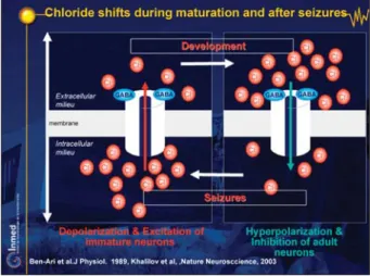
Interestingly, patching neurons from the epileptic hippoc-ampus, we found that GABA again excites neurons: the seizures induced a long-term transformation of the inhibi-tory actions of GABA to excitainhibi-tory, most likely because of a loss of the capacity to remove and regulate chloride via co-transporters (figure 22; DVD figure 38). To some ex-tent, epileptogenesis recapitulates ontogenesis (DVD
figure 39, Khalilov et al. 2005, Cohen et al. 2002), and
seizures re-induce a return to immature neurons with an excitatory GABA – except that now with the high density of glutamate synapses, seizures will be generated. There-fore, the expression of the epileptogenic properties is, in fact, due to a persistent accumulation of chloride, the seizures producing a loss in the efficacy of neurons to remove the excess chloride produced during the seizures.
Figure 21. Formation of an epileptogenic mirror focus. Figure 22. Chloride shifts during maturation and after seizures. Ben-Ari et al. J Physiol 1989 ; Khalilov et al., Nature Neurosccience, 2003.
This loss of efficacy of the transporters may be more readily triggered by high frequency oscillations.
In conclusion, these observations suggest (DVD figure 40)
that seizures are not necessarily epileptogenic – they can occur recurrently, without sequelae, if they do not include high frequency oscillations that require both operative GABA and NMDA receptors. The most likely explanation for this requirement is that the excitatory drive provided by GABA is required at this stage as the density of glutamate synapses may not suffice: a small but decisive activation of excitatory GABA, activates NMDA receptors by removing the voltage-dependent magnesium blockade leading to a large calcium influx, and a sequence of events that leads to a permanent consequence. This observation suggests that it may be possible to evaluate the epileptogenic capacity of seizures in children by evaluating their intrinsic, domi-nating, oscillatory frequencies.
GABA is both a pioneer and an essential actor in early development. It is operative first; it excites neurons at a time when neurons have few synapses, and it has a para-crine action on immature neurons, modulating neuronal migration. Then it acts in coordination with NMDA recep-tors stimulating developing networks intervening in the construction of cortical networks and exerting a wide range of trophic actions. Clearly, because of this plethora of actions and its unique properties, this transmitter is able to act very differently according to the conditions. It seems ideally suited to the job of regulating the complex func-tional sequences involved in brain development. And last but not least, GABA is greatly involved in early seizures, both in their induction and in their long-lasting conse-quences: seizures do not transform a naïve network to one that seizes unless GABA synapses are operative. Clearly, the effects of GABA-acting drugs on developing brains must be re-evaluated in the light of this information. M
References
Ben-Ari Y. Excitatory actions of GABA during development: the nature of the nurture. Nat Rev Neurosci 2002; 3: 728-39. Ben-Ari Y, Cherubini E, Corradetti R, Gaiarsa JL. Giant synaptic potentials in immature rat CA3 hippocampal neurones. J Physiol 1989; 416: 303-25.
Ben-Ari Y, Khalilov I, Represa A, et al. Interneurons set the tune of developing networks. Trends Neurosci 2004; 27: 422-7. Ben-Ari Y, Khazipov R, Caillard O, et al. GABAA, NMDA and AMPA receptors: a developmentally regulated “ménage à trois”.
Trends Neurosci 1997; 20: 523-9.
Ben-Ari Y. Developing networks play a similar melody. Trends
Neurosci 2001; 24: 353-60.
Chevassus-Au-Louis N, Congar P, Represa A, Ben-Ari Y,
Gaiarsa JL. Neuronal migration disorders: heterotopic neocorti-cal neurons in CA1 provide a bridge between the hippocampus and the neocortex. Proc Natl Acad Sci USA 1998; 95: 10263-8.
Cohen I, Navarro V, Clemenceau S, et al. On the origin of inter-ictal activity in human temporal lobe epilepsy in vitro. Science 2002; 298: 1418-21.
Demarque M, Represa A, Becq H, et al. Paracrine intercellular communication by a Ca2+- and SNARE-independent release of GABA and glutamate prior to synapse formation. Neuron 2002; 36: 1051-61.
Demarque M, Villeneuve N, Manent JB, Becq H, Represa A, Ben-Ari Y, Aniksztejn L, et al. Glutamate transporters prevent the generation of seizures in the developing rat neocortex. J Neurosci 2004; 24: 3289-94.
Freund T, Buszaki G. Interneurons of the hippocampus.
Hippoc-ampus 1996; 6: 347-470.
Gao XB, van den Pol AN. GABA, not glutamate, a primary trans-mitter driving action potentials in developing hypothalamic neu-rons. J Neurophysiol 2001; 85: 425-34.
Hennou S, Khalilov I, Diabira D, Ben-Ari Y, Gozlan H. Early se-quential formation of functional GABA(A) and glutamatergic synapses on CA1 interneurons of the rat foetal hippocampus. Eur
J Neurosci 2002; 16: 197-208.
Holmes GL, Ben-Ari Y. Seizures in the developing brain: perhaps not so benign after all. Neuron 1998; 21: 1231-4.
Hubner CA, Stein V, Hermans-Borgmeyer I, et al. Disruption of KCC2 reveals an essential role of K-CI transport already in early synaptic inhibition. Neuron 2001; 30: 515-24.
Khalilov I, Esclapez M, Medina I, et al. A novel in vitro prepara-tion: the intact hippocampal formation. Neuron 1997; 19: 743-9. Khalilov I, Holmes GL, Ben-Ari Y. In vitro formation of a second-ary epiletogenic mirror focus by interhippocampal propagation of seizures. Nat Neurosci 2003; 6: 1079-85.
Khalilov I, Le Van Quyen M, Gozlan H, Ben-Ari Y. Epileptogenic actions of GABA and fast oscillations in the developing hippoc-ampus. Neuron 2005; 48: 787-96.
Khazipov R, Esclapez M, Caillard O, et al. Early development of neuronal activity in the primate hippocampus in utero. J Neurosci 2001; 21: 9770-81.
Khazipov R, Sirota A, Leinekugel X, et al. Early motor activity drives spindle bursts in the developing somatosensory cortex.
Nature 2004; 432: 758-61.
Leinekugel X, Khazipov R, Cannon R, et al. Correlated bursts of activity in the neonatal hippocampus in vivo. Science 2002; 296: 2049-52.
Manent JB, Demarque M, Jorquera I, Pellegrino C, Ben-Ari Y, Aniksztejn L, Represa A. A noncanonical release of GABA and glutamate modulates neuronal migration. J Neurosci 2005; 25: 4755-65.
Owens DF, Kriegstein AR. Is there more to GABA than synaptic inhibition? Nat Rev Neurosci 2002; 3: 715-27.
Owens DF, Liu X, Kriegstein AR. Changing properties of GABA(A) receptor-mediated signaling during early neocortical development. J Neurophysiol 1999; 82: 570-83.
Represa A, Ben-Ari Y. Trophic actions of GABA in neuronal de-velopment. Trends Neurosci 2005; 28: 278-83.
Rivera C, Voipio J, Payne JA, et al. The k+/Cl- co-transporter KCC2 renders GABA hyperpolarizing during neuronal matura-tion. Nature 1999; 397: 251-5.
Tremblay E, Nitecka L, Berger ML, Ben-Ari Y. Maturation of kainic acid seizure-brain damage syndrome in the rat. I. Clinical, electrographic and metabolic observations. Neuroscience 1984; 13: 1051-72.
Tyzio R, Ivanov A, Bernard C, et al. Membrane potential of CA3 hippocampal pyramidal cells during postnatal development.
J Neurophysiol 2003; 90: 2964-72.
Tyzio R, Represa A, Jorquera I, et al. The establishment of GABAergic and glutamatergic synapses on CA1 pyramidal neu-rons is sequential and correlates with the development of the apical dendrite. J Neurosci 1999; 19: 10372-82.
Villeneuve N, Ben-Ari Y, Holmes GL, Gaiarsa JL. Neonatal sei-zures induced persistent changes in intrinsic properties of CA1 rat hippocampal cells. Ann Neurol 2000; 47: 729-38.