MASSACHUSETTS 1WTUTE
Use in
Optogenetics
Research
OF
TECHNOLOGY
by
OCT 332014
Patrick Erin Monahan
III
LIBRARIES
M.S., Cell Biology, Neurobiology and Anatomy, Loyola University Chicago, 2009
B.S., Biomedical Sciences, Marquette University, 2005
Submitted to the Program in Media Arts and Sciences, School of Architecture and
Planning, in partial fulfillment of the requirements for the degree of
Master of Science
atthe
Massachusetts Institute of Technology
September 2014
This work is licensed under a Creative Commons Attribution 3.0 Unported License
The author hereby grants MIT permission to reproduce and distribute publicly
paper and electronic copies of this thesis document in whole or in part.
Signature redacted
Author
Patrick Monahan
Program in Media Arts and
Signature
redacted
Sciences
August 8, 2014
Certifiedby4
Prof. Edward S. Boyden
Leader, Synthetic Neurobiology Group
Associate Professor, MIT Media Lab and McGovern Institute
Departments of Biological Engineering and
Use in Optogenetics Research
by
Patrick Erin Monahan III
Submitted to the Program in Media Arts and Sciences,
School of Architecture and Planning,
on August 8, 2014, in partial fulfillment of the
requirements for the degree of
Master of Science
Abstract
Optogenetic actuators such as Channelrhodopsin-2 (ChR2) are
seven-transmembrane proteins that function as light-gated ion channels. These naturally
occurring proteins are found in green algae and serve as sensory photoreceptors
controlling phototaxis.
Operationally, they contain the light-isomerizable
chromophore all-trans-retinal that, upon absorption of a photon at or around
473nm, a conformational change to 13-cis-retinal is induced. This change opens the
channel allowing cations to flow through. In the absence of light, the 13-cis-retinal
relaxes back to the resting all-trans-retinal conformation and the channel closes.
When an actuators packaged into a lox-containing Adeno-associated virus is used in
conjunction with a mouse that expresses the Cre recombinase enzyme in a specific
cell type, cell specific expression of the opsin is achieved. When used with LEDs,
lasers, or specifically fabricated light delivery tools, control of very specific neural
networks is realized. This thesis provides a review of optogenetics and details the
development and application of a novel wireless device to optically control neural
circuits and behavior.
Thesis Supervisor:
Prof. Edward S. Boyden
Leader, Synthetic Neurobiology Group
Associate Professor, MIT Media Lab and McGovern Institute
Departments of Biological Engineering and
I would like to thank Ed Boyden, Mitch Resnick and Linda Peterson for helping me
complete this thesis.
I would also like to thank the love of my life, my wife Sheena and my son Paddy, and
the rest of my family for putting up with (and sometimes enabling) my shenanigans.
Contents
Abstract...3
Acknow ledgem ents...5
Contents...6
Introduction...7
Light-Activated Ion Pumps and Channels for Temporally Precise Optical
Control of Activity in Genetically Targeted Neurons...
10
A wirelessly powered and controlled device for optical neural control of
freely-behaving anim als...
44
This thesis contains two previously published works in the field of
optogenetics. The first paper is a review discussing types of microbial opsins and
their functional characteristics as well as tools for controlling such opsins. The
second paper presents a novel wireless tool for neural circuit control and behavioral
modification using optogenetics.
Optogenetics gives neuroscientists the ability to turn on or off very specific
cell types and neural circuits in the brain in a temporally precise fashion. This
development has enabled the casual manipulation of neural activity to explore the
sufficiency and necessity of different neural patterns as they pertain to normally and
abnormally functioning neural circuits. When incorporated into in vivo systems,
optogenetics allows neuroscientists to dynamically control genetically specific
neurons by delivering light to deep brain nuclei to alter behavior, manipulate
individual components of cortical microcircuits to study local network dynamics
and target malfunctioning pathways to shed light on disease.
Beyond enabling our understanding of brain function, continuing
development of opsins and the related optical toolbox may spawn a new generation
of optical prosthetics for treating currently intractable brain disorders.
The second paper looks at the invention and application of a wireless
transmitter for controlling neural circuits and behavior with optogenetics. This is an
important advancement, as current optogenetic efforts require animals to be
commutator that can restrict natural motion and are impractical for long-term or
high-throughput experimentation.
My specific contributions to the first paper includes building the polyimide
cannula system to deliver light to specific deep structures of the brain, creating
schematics and aiding in the writing of the paper (for example, see Figure 8 and
text).
My specific contributions to the second paper includes performing all
surgical implantations of the wireless devices, monitoring post-surgery mice,
performing behavioral experiments, analyzing results and contributing to the
manuscript (detailed in Materials and Methods).
Neuromethods (2012) 67: 305-338
DOI 10.1007/7657-2011-10
0 Springer Science+Business Media, LLC 2011
Published online: 13 December 2011
Ught-Acivated Ion
Punps
and Channels
for TeMporaly
Precise Optical Control of Activity
In Geneicafly
TgPetsd
Neurons
1
Brian Y. Chow, Xue Han, Jacob G. Bernstein, Patrick E. Monahan,
and Edward S. Boyden
AbStrad
The ability to turn on and off specific cell types and neural pathways in the brain, in a temporally precise fashion, has begun to enable the ability to test the sufficiency and necessity of particular neural activity patterns, and particular neural circuits, in the generation of normal and abnormal neural computations and behaviors by the brain. Over the last 5 years, a number of naturally occurring
light-activated
ion pumps and light-activated ion-channels have been shown, upon genetic expression in specific neuron classes, to enable the voltage (and internal ionic composition) of those neurons to be controlled by light in a temporally precise fashion, without the need for chemical cofactors. In this chapter, we review three major classes of such genetically encoded "optogenetic" microbial opsins-light-gated ion channels such as channelrho-dopsins,light-driven
chloride pumps such as halorhodopsins, andlight-driven
proton pumps such as archaerhodopsins-that are in widespread use for mediating optical activation and silencing of neurons in species from Caeuorhabditu elegans to nonhuman primates. We discuss the properties of thesemolecules-including their membrane expression, conductances, photocycle properties, ion selectivity, and action
spectra-as well as genetic strategies for delivering these genes to neurons in different species, and hardware for performing
light
delivery in a diversity of settings. In the future, these molecules not only will continue to enable cutting-edge science but may also support a new generation of optical prosthetics for treating brain disorders.Key
words:
Channelrhodopsin, Optogenetics, Photosensitive proteins, Retinal, Halorhodopsin, Archaerhodopsin, Light-sensitive cation channel, Light-sensitive chloride pump, Light-sensitive pro-ton pump, Photocontrol of behavior1. IWtdu on
The
ability to turn on and off specific cell types and neural pathways in
the
brain, in a temporally precisefashion,
has begun to enable the ability to test the sufficiency and necessity of particular neural activity patterns, and particular neural circuits, in the generation of normal'This chapter is an updated version of reference (141).
305
and abnormal neural computations and behaviors by the brain. Most
electrophysiological
and imaging experiments
inneuroscience are
correlative-comparing a neural signal observed in the brain to a
behavior or pathology. In contrast, the power to manipulate specific
cells and circuits is opening up the ability to understand their causal
roles in brain functions. Over the last 5 years, a number of naturally
occurring
light-activated
ion pumps and light-activated ion channels
have been shown, upon genetic expression in specific neuron classes,
to enable the voltage (and internal ionic composition) of those
neu-rons to be controlled by
light
in a temporally precise
fashion.
These
molecules are microbial (type I) opsins, seven-transmembrane
pro-teins naturally found in archaea, algae, fungi, and other species, and
which possess
light-driven
electrogenic activity or contain
light-gated
ion pores. These molecules, when heterologously expressed in
neu-rons or other cells, translocate ions across cell membranes in response
to pulses oflight ofirradiances that are easily achievable with common
laboratory microscopes, LEDs, and lasers. These molecules have
begun to find widespread use in neuroscience due to their
ease
of
handling and use (each is a single gene, under 1 kb long, encoding for
a monolithic protein), their lack of need for chemical
supplementa-tion in many species (they utilize the naturally occurring
chromo-phore all-trans retinal, which appears to occur at sufficient quantities
in the mammalian nervous system), and their high speed of operation
(they can respond within tens of microseconds to milliseconds, upon
delivery of light, and shut off rapidly upon cessation of light, as
needed for neuroscience experiments).
Three major classes of such "optogenetic" microbial opsins
have been described to date. The first class, channelrhodopsins, is
exemplified by the light-gated inwardly rectifying nonspecific
cat-ion channel channelrhodopsin-2 (ChR2) from the green algae
Gblamydomonareinhardtii
(1), which, when expressed in neurons,
can mediate sizeable currents up to 1,000 pA in response to
milli-second-timescale pulses of blue light (2-5), thus enabling reliable
spike trains to be elicited in ChR2-expressing neurons by blue light
pulse trains (Fig.
1b).
Several additional channelrhodopsins useful
to biologists and bioengineers have been discovered or engineered,
with faster or slower kinetics, red-shifted activation, and cell-region
specific targeting, and explored in detail below (6-10). The
chan-nelrhodopsins
have been used to activate neurons in neural circuits
in animals from worms to monkeys, and have proven powerful and
easy to use. The second class of microbial opsins utilized for
biological control to date, halorhodopsins, is exemplified by the
light-driven
inwardly directed chloride pump halorhodopsin, from
the archacal species Natronomas pbaraonis (Halo/NpHR/pHR;
(11)), which, when expressed in neurons, can mediate modest
inhibi-tory currents on the order of 40-100 pA in response to yellow light
illumination (12, 13), enabling moderate silencing of neural activity
(Fig.
1c).
Halorhodopsins have some intrinsic kinetic limitations,
Uight-Activated Ion Pumps and Channels for Temporally...
307
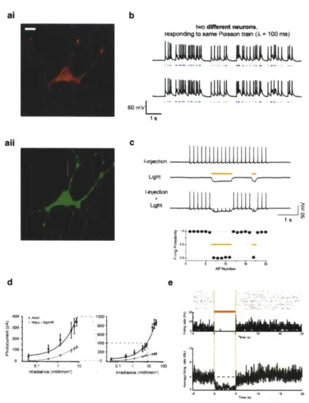
al
b
two dferen ruromn,
responding to SaM PoHseon train (1 100 Ms)
0 mVL
all
C
Ught 1.nowotion LightI
-s
d
7
7.
0' I 10 0Fig.
1. Three dm88.e
of
microbial
opeins
that
enable optical
neural activation
and silencing tools.
(a) Neuron
expressing
hChr2-mCherry
(at;
bar,
20
pmn)
and Halo-GFP
(aN).
(b)Polason tralns
of spikes elicited
by pulses
of
blue
light
(Mwt
dhes, intwo
different neurons. (C) Light-drivenspike
blockade, demonstrated (Top~fora
representative hippocampal neuron, and(Bottom) for
a
populationof
neurons (N =7). I-itkiid, neuronalfiring
induced
bypulsed somatic current injection
(300pA,
4ins).
Liht
hyperpolarizationinduced
by periodsof
yellowlight
(yeI~ow dashws. I-iiectki +Light,
yellowlight
drives Haloto
blockneuron spiking, leaving spikes elicited
during periodsof
darkness intact. Panelsa-c adapted
fromreferences
Boyden
at
al.
(3)
and Han and
Boyden, (12). (d) Photocurrents
of
Arch vs. Halo measured
as a
function
of
575
+25 nmight irradlance
(oreffective light
irradlance),in
patch-clampedcultured
neurons (N = 4-18neurons
foreach
point),for low (i) and
high (ii)light
powers. The lineis a single
Hillfit to the
data. (a) Top, NeuralactIvity in
arersnaieneuron
before, during,and after 5
sof
yellowlight
illumination,shown as
aspike
resterplot
(2.*, andas
ahistogram of Ins
urng rate flneaveraged across trials
(&uttwn; blnsize,
20me).
Bot~v, populationaverage of
instantaneous filing
rate before, duringand after
yellowlight illumination
(Nick line, mean; prayliws,
mean i SE;S= 13
unIte).
Panelsd-e adapted
fromreference Chow
atal.
(15).with some photocycles taking tens of minutes to complete (e.g., Fig. 4a, b; (12, 14)), and halorhodopsins also require improved trafficking for expression at high
levels
(15-17). A third class of microbial opsin, the bacteriorhodopsins, is exemplified by the light-driven outward proton pump archacrhodopsin-3 (Arch/aR3), fromthe archacal species Halorubrum sodomense. Arch can mediate strong
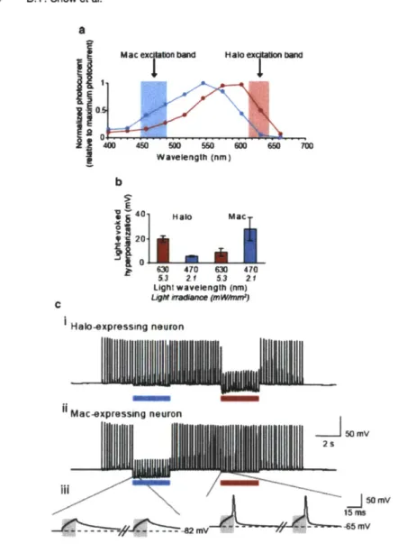
inhibitory currents of up to 900 pA in vitro (Fig.ld),
and in vivo is capable of mediating near-100% silencing of neurons in the awake behaving mouse or monkey brain in response to yellow-green light (Fig. Ic, (15, 18)). Protons are extremely effective as a charge carrier for mediating neural silencing, and proton pumps have greatly improved kinetics with respect to halorhodopsins (Fig. 4c, d), as well as a fast photocycle, and efficient trafficking to membranes. Furthermore, outward proton pumps, perhaps surprisingly, do not alter pH to a greater extent than do other opsins (such as ChR2) or than does normal neural activity. The broad and ecologically diverse class of outward proton pumps, which includes blue-green lightdrivable outward proton pumps such as the Leptosphaeria maculans
opsin Mac (Fig. 5), enables, alongside the yellow-red drivable Halo, multicolor silencing of independent populations of neurons (15). Also, because the neural silencers Halo and Arch are activated byyellow or yellow-green light, and the neural depolarizer ChR2 is
driven by blue light, expression of both a silencer and a depolarizer in the same cell (either by using two viruses, or by using the 2Alinker to combine two opsins into a single open reading frame (12, 19)) enables bidirectional control of neural activity in a set of cells, useful for testing necessity and sufficiency of a given set of neurons in the same animal, or disruption of neural synchrony and coordination through "informational lesioning" (19).In the sections below we describe the properties of these three opsin classes, as well as genetic (e.g., viral, transgenic) and hardware (e.g., lasers, LEDs) infrastructures for using these opsins to parse out the function of neural circuits in a wide variety of animal nervous systems. A theme of this field is that extremely rapid progress and adoption of these technologies has been driven by technology development curves in other fields such as gene therapy and optical imaging. Our hope is to convey a snapshot of this rapidly moving field as of 2011, summarizing the first half-decade of its existence, to teach both neuroengineers hoping to innovate by inventing new tools, as well as
neuroscientists
hoping to utilize these tools to answer new scientific questions. We will first survey the general properties of these opsins (Sect. 2), then go into thechannelrhodopsins (Sect. 3), followed by the neural silencing
pumps (halorhodopsins and bacteriorhodopsins, Sect. 4), themolecular strategies for delivering these genes to cells for
appropri-ate expression in vitro and in vivo (Sect. 5), and the hardware for illumination of these opsin-expressing neurons in vitro and in vivo (Sect. 6).Ught-Activated Ion Pumps and Channels for Temporally... 309
2. Properdas
of "Optogenetic"
Microbial (Type I)
Opsins
The three classes of molecule described to date are from organisms
such as unicellular algae, fungi, and archaea, whose native
environ-ments and membrane lipid composition are very different from
those of mammalian neurons. Thus, the performance of these
molecular tools in neurons can be difficult to predict based solely
upon their properties
in
other species, and must be assessed
empiri-cally for assurance of efficacy and safety. Nevertheless, there are
several molecular properties that contribute to efficacious,
tempo-rally precise optical control of neurons, which can be explored in a
unified and logical fashion:
"
Initial proteinexprezion
levels.
The
efficiency
of ribosomal
translation of a molecule is largely affected by codon
optimiza-tion. It is recommended that genes be used that are
codon-optimized for the target species.
* Membrane
insertion propertie, protein folding, and
interactions wilh local environment.
Increased membrane
local-ization will result in more functional molecules and thus
increased photocurrents. This property may also be inversely
associated with the potential property of
taicijy
since poorly
trafficked or fblded molecules may aggregate in the cytosol and
endoplasmic reticulum. On the other hand, if a molecule has
adverse intrinsic side effects, enhanced trafficking may
exagger-ate them. Furthermore, any given channel or pump will best
operate under defined conditions (e.g., chloride conductance,
pH,
lipid
environment, etc.), which may not exist in a given
target cell type.
* Innate conductance and permeability
Channels translocate
more ions per photocycde than pumps, since they open up a
pore in the membrane. On the other hand, pumps can move
ions against concentration gradients, unlike channels. Each
opsin furthermore passes a precise set of ions in a specific
cellular context, and not others.
-
Photocycle kinetics.
Both light-driven channels and pumps are
described by a photocycle, the list of states that a molecule goes
through after light exposure, including ion-translocating or ion
pore-forming steps. The faster the photocycle, the more
tem-porally precise the molecular function might be, and fur a
pump, the more ions will be translocated. (For a channel, a
faster photocycle may result in the channel entering the ion
pore-forming state more often, but may also reduce the time
spent in the open state.) If a molecule enters an inactive
photocyde state for an enduring period of time, it may be effcctively nonfunctional.
" Photositivity Molecules may require different amounts of
light to begin moving through their photocyce, based on the chromophore absorption efficiency. Furthermore, from a end-user standpoint, effective photosensitivity will appear to be a function of the overall photocycle; fur example, a pump that has a slow photocycle may appear to be light insensitive (because incident photons may have no effect on the molecule during the photocyce), whereas a channel that inactivates extremely slowly may appear to be light sensitive (because each photon will result in large charge transfers).
* Action spectrum. Different molecules are driven by different
colors of light. Multiple cell types can be orthogonally addressed with different colors of light, if they express opsins whose action spectra minimally overlap.* Ion seectivity Unlike traditional electrodes, microbial opsins
can generate ion specific currents, since they will pass specific ions such as chloride (CL) or calcium (Ca'2). This opens up novel kinds of experimental capability, such as the ability to test the sufficiency of a given ion, in a given location, for a given biological function.We will, in the fbllowing sections, frame current knowledge about cell-type specific optical control of neurons, in the context of decades of research in structure-function relationships of microbial (type I, or archaeal) opsins. In many ways, these molecules are similar in tertiary structure to mammalian (type II) rhodopsins (20), the pigments that confer photosensitivity to the rods and cones of the human retina. Both types are composed of seven transmembrane (7-TM) cx-helices, linked by six loop segments, and their photosensitivity is enabled by a retinal bound to a specific lysine residue near the C-terminus, forming a Schiff base that
undergoes a trans-cis or cir-grans isomerization upon illumination,
that then induces conformational changes in the protein. However, they are evolutionarily unrelated, and their differences have impor-tant implications for their use in perturbing neuronal activity. Mammalian rhodopsins (21) are very sensitive photon detectors, optimized for sensitivity rather than speed. They utilize 11-cisretinal as the primary chromophore, which isomerizes to all-trans
retinal upon absorbing a photon. The resultant structural change activates an associated G-protein, transducin, which then initiates a cascade of secondary messengers. The all-trans retinal dissociates from theopsin, is
converted back to its 11-cis form, and then reassociates with the apoprotein to reconstitute a functional mole-cule-a process that typically takes hundreds of milliseconds, tooslow to enable fast control ofneurons in the central nervous system.
Light-Activated Ion Pumps and Channels for Temporally... 311
On the other hand, a microbial opsin utilizes all-trans retinal as its
chromophore, which isomerizes to 13-cis retinal upon absorbing a
photon. The chromophore does not undergo a quasi-irreversible dissociationevent, but
rather thermally relaxes to its active all-trans form in the dark (although this process can be facilitated by light).The
trans-cis
isomerization sets off several coupled structural
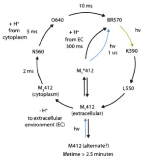
rear-rangements within the molecule that accommodate the passive conduction or active pumping of ions (22-24). Ultimately, this means that at the expense of light sensitivity, archaeal opsins can deflect the membrane potential of a cell on the scale hundreds of microseconds to a few milliseconds. However, it should be noted that genetically targetable, optical neural silencing has also been demonstrated using mammalian G-protein coupled receptors, which can couple to potassium channels (4), and genetically target-able optical neural activation has been demonstrated using mela-nopsins and invertebrate-style rhodopsins, at the price of temporal precision (25, 26).As an exemplar of a well-characterized microbial opsin reagent, with crystal structure and photocycle both well-described, Fig. 2 shows the crystal structure of the light-driven chloride-pump halor-hodopsin (Fig. 2a; (27)), as well as schematized structural rearran-gements that are hypothesized to occur as the molecule pumps a chloride ion across the membrane and into the cytoplasm (Fig. 2b; (24)). While it is convenient to consider the molecular tools dis-cussed here as toggle switches for turning neurons on and off, it is critical for use of these opsins to realize that the translocation of ions by microbial opsins is not as simple as a two-state toggle switch. The structural rearrangements constitute an active advance-ment through a complex photocycle with various intermediate states beyond the initial phototransition (Fig. 2c). There exists a very rich literature on type I microbial opsins from an evolutionary and protein structure-function perspective. The canonical mole-cules include the proton-pumping bacteriorhodopsin (BR), the
chloride-pumping
halorhodopsin
(HR/sHR/HsHR) from
Halo-bacterium salinarum (halobium),
and the halorhodopsin from N.
pharaonis (Halo/NpHR/pHR). These were some of the first
membrane proteins crystallized, and a myriad of structure-function studies have been performed on these molecules (11, 14, 20, 22-24, 27-45). These studies are not reviewed here, but it is important to point out their existence because much of what we know with respect to the photocyde and structure of opsins comes from these studies and from sequence homology of novel opsins to these canonical molecules. As we show later, for example, a deep understanding of the literature has enabled some researchers to derive powerful new variants of channelrhodopsin (5, 6, 8, 10), even though no crystal structure exists for this molecule.16
-b
Uptake and isomenzation
HR640 -20 m. HRlSa0 HROOO + C1.
" M
* o E C ( H hV
R 410 R640 H1 +HpS,0) HR520*-/ Release Switch HR520Fig. 2. Structure and function of halorhodopsin. (a) Crystal structure of halorhodopsin, which is composed of
7-transamembrane
ot-helices
(7-TM) and a retina chromophore that forms a Schiff base
to
a
lysine
near the C-terminus
(from Kolbe
et
al. (27)). (b) Schematic of the halorhodopsin structural rearrangements and their relation to pumping activity
at various points
inthe photocycle. Image
modifiedfrom Essen (24).
(C)The halorhodopsin photocycle at high-power
continuous illumination
on
the timescale of a typical photocycle (.e., condItions used for neural silencing, >few
milliseconds). The HR410 intermediate is the
origin
of the long-lived inactivation in neural silencing (e.g., Bamberg
et
al. (14); Han and Boyden (12)).. Optical
Neural
Stmulation:
Channel-rhodop-ins
Channelrhodopsins arc the primary photoreceptors in the eyespot of the unicellular algae that are responsible for phototactic and photophobic responses (46-48). Their name is derived from the fact that despite the sensory function, the 7-TM segment is in itself a light-activated ion channel. While the channel pore and properties remain poorly understood, it has recently been realized that chan-nelrhodopsins are likely proton pumps like many other microbial opsins, but with a leaky step in the photocycle during which the
opsin
lets
positive charge into cells (49). In C. reinhardiii, two
separate channelrhodopsins were originally identified (48), one with fast kinetics but poor light sensitivity, channelrhodopsin-I(ChRL) (50), and another with slower kinetics but improved sensi-tivity, channelrhodopsin-2 (ChR2) (1). Two more
channelrhodop-sins from Valvax carteri(VChR1, VChR2) have also been identified
(7, 51), and as we discuss later, many more ChRs are expected toaa
HR'500 HR580 C 14A 580 R_410 HR6a5 It Isomertzation HR565 HR60011. toewbwaace,
&Wd
Coniurt
32.
Basic
MWew
Of
kwtfts
aw
WaI&gfh
Se"ecUy
Uight-Activated
lon
Pumps and Channels for Temporally... 313exist. ChRL-style channelrhodopsins have red-shifted action
spec-tra (peak
Achj =500
nm,
Avchm =535
nm) relative to ChR2
(peak
chR2 =470 rnm), and thus in principle ChRi-stylc and
ChR2-style opsins could be used together to drive separate sets of
neurons with two different colors of
light,
if suitably spectrally
separatedopsin pairs could be found.
Channelrhodopsins (abbreviated as ChRs or chops) are
light-activated, inwardly rectifying cation channels that are, at neutral
pH, permeable to physiologically relevant cations such as H', Na',
K, and Ca2*, with permeabilties (relative to sodium) of 1 x 10⁶, 1,
0.5, and 0.1 respectively (1, 6, 46, 50). It is of particular note that
the proton conductance
(GH+)is 10
6-fold larger than the sodium
conductance
(GNa.),and thus near physiological pH, perhaps half
the photocurrent is carried by protons (46); thus, ChRs may rapidly
equilibrate the intracellular pH with its environment (10). Kinetic
selectivity analysis has shown that the mechanism ofion selectivity is
likely to be due to differential binding affinity of channelrhodopsin
channel residues for different ions, not differential ion transport
rates (46).
It was originally believed that ChRi was a selective proton
channel (50); however,
itwas later discovered that the poor
photo-currents at mammalian
pH were likely
attributable to
poor membrane localization (6), and the apparent lack of sodium
currents in the original report were due to the low pH used to
perform experiments in that study; the sodium conductance of
ChR1 lessens at low pH (6, 46), unlike that of ChR2 (6). This
highlights what is a recurrent theme throughout this chapter, that
effective
conductance in a heterologous system is determined not
only by the innate kinetic and transport properties of the molecule
but also by its trafficking and performance in the environment of
the heterologous system.
The single ion channel conductance of ChR2 has been
esti-mated at 50
fS
(1), which corresponds to approximately 3 x 10"
ions per second, or 300 ions per photocycle event, assuming a
10 ms turnover. This is considerably less than a typical voltage
dependent sodium channel that may have a conductance on the
order of -10 pS. It has been estimated from electrophysiological
data that
10
-106
membrane
embedded ChR2 molecules are
required to cause reliable spiking in cultured rodent hippocampal
neurons (52), with saturation blue
light
densities of several
milli-watts per square millimeter both in vitro (10) and in vivo (53).
Figure 3a shows a typical photocurrent trace from a
voltage-clamped neuron expressing ChR2 (top), and the spiking pattern
that would result in current-clamp mode (bottom). There is a large
transient peak with an opening time constant near 1 ms (1, 6, 10),
although photocurrent onset can be measured at <200 ps (1, 3,
18
a
" mpu 400PAK SWb
D470
hv
hv
P500
P480
C
-H-(EC)
P520
4AZ+H-(CP)
#M+leak
Fig. 3. Channelrhodopsin kinetic and photocycle properties, and impact on neural activity. (a) ChR2 currents elicited in a voltage-clamped hippocampal neuron expressing ChR2 and illuminated by blue light (b$*, and ChR2-drven spikes elicited in a current-clamped hippocampal neuron (three repetitions of the
same
blue light pulse in the same neuron) (bottom), under 1s
of blue light illumination. Adapted from Boydenet
al.
(3). (b) The photocycle of ChR2 determined by acombination of spectroscopy, site-directed mutagenesis, and electrophysiology, adapted from Feldbauer et
al.
(49). Theinner
circle summarizes the effective appearance of the photocycle, an approximation to the outer photocycle. (c) The interplay between photocycle, wavelength, and electrophysiological activity. ChR2 is excited with blue light for abrief period, and then a green light is turned on. The photocurrent initially
diminishes becausethe channel is
forcedto close, but then increases because the green light also pumps the
molecule
into its most highly efficient state.
Image modified from Bamann et
al.
(54).
54); this transient peak quickly decays to a stationary component that is typically <20-50% of the initial peak photocurrent (1, 3, 6, 10). Upon removing the light, ChR2 doses with a time constant of 10-20 ms (1, 6, 10). The transient photocurrent peak is highly dependent on the illumination intensity (51, 55) and history (1, 3,
10); the history dependence results from a desensitization of the
transient component that takes -5 s to recover from in the dark (3). The stationary component on the other hand, is less photosensitive and effectively history-independent (55). ChR2 absorbs maximally at 460 nm (1, 10), and the action spectra of both temporal components are nearly identical in ChR2.
The large and fast-onset peak enables ChR2-expressing neu-rons to spike with exquisite temporal precision on the millisecond timescale (Fig.
1b),
the timescale of an action potential. However, the large inactivation (or alternatively, the small stationary compo-nent) and its slow recovery in the dark, as well as the slow dosingrate of -10-15
ms,
ultimately limit the ability to drive reliable spike
rates >25 Hz (3, 10) because (1) the stationary photocurrent maybe too small to sufficiently depolarize a neuron to spike threshold,
and (2) the channel cannot physically close quickly enough to enable dc-inactivation of sodium channels. It should be noted, though, that many neurons, such as pyramidal cells, seldom fireC
40 .
100
pA
200 mUght-Activated Ion Pumps and Channels for Temporally... 315
action potentials at this rate on the individual neuron level (vs.
population synchrony or rhythmogenesis).
ChRi-style channelrhodopsins (VChRI, ChR1) (7, 50) on the
other hand demonstrate dramatically faster kinetics than ChR2-stylc
channelrhodopsins (VChR2, ChR2). The stationary photocurrents
of ChRis are >70% of the peak photocurrents, and the channels
open and close approximately two- to threefold faster than does
ChR2. Therefore, one would expect that given comparable
expres-sion, protein folding, membrane localization, and photosensitivity
(i.e., factors contributing to effective conductance), ChRis would
be capable of driving spike rates with greater fidelity than ChR2s.
However, poor membrane expression limits the performance of
natural ChRL -style channelrhodopsins (7,50). Chimeras composed
of the first five helices of ChR1 and last two helices of ChR2 have
been constructed (6, 10, 56), and these new variants exhibit the
small inactivation and action spectrum of ChRi, but the overall
effective
conductance of ChR2. These structure-function studies
are discussed in detail later in this chapter. A point mutant of this
chimera dubbed "ChIEF" (based on
itscomposition as a [Ch]
annelrhodopsin chimera with an [1]190 V substitution with
domains swapped between ChRI helix-[E] and ChR2 helix-[F]),
developed by Tsien and coworkers (10), appears to be a highly
improved tool for stimulating neurons. Its large stationary
photo-current and very fast channel closing, the latter conferred by the
1190 V mutation, contribute to far more reliable spiking (up to
100 Hz) than ChR2.
Based on the available characterization of the
channelrhodop-sins from V
carteri
(7, 51),
the general characteristics are similar to
those of the analogous molecules in C. reinuardhii. VChR2 and
ChR2 have nearly identical photocycles and action spectrum (51).
VChRI and ChR1 exhibit the similar reduced inactivation, and are
both red-shifted from their respective VChR2/ChR2 counterparts.
It has been proposed that VChR1 could be used for multicolor
optical stimulation in conjunction with ChR2, which is blue-shifted
by -70
nm,
but further improvements are likely required for
reli-able spiking because the VChR1 photocurrents are unfortunately
approximately four- to fivefold smaller (7), and also there is
signifi-cant spectral overlap between VChR1 and ChR2.
3.1 06taed
MWs
As previously mentioned,
itis critical to realize that the
transloca-of
ONNO
aw
tion of ions by opsins is not as simple as the operation of an on/off
Wamehwego SeWa~fsty
switch, but rather these opsins traverse a complex photocycle with
various intermediate states beyond the initial opening of the
chan-nel. Figure 3b shows the photocycle of ChRs based on
photophy-sical studies performed primarily by laser flash spectroscopy,
physiology, and site-directed mutagenesis (8, 49, 51, 54, 57).
Importantly, the intermediates of the photocyces themselves can
also undergo photoreactions, and thus they may be optically driven
or "short-circuited" (54) between photointermediates at much
faster rates (Fig. 3c). The ChR2 photocyde initially begins in its
closed dark-adapted state D470 (where the number in the state
name corresponds to the peak light absorption, in
nm,
of the
molecule in that state). The channel opens when D470 absorbs a
photon, after which the molecule will become a green absorbing
photoproduct or P-intermediate, P520, via thermal relaxation from
shorter lived photoproducts. This initial cascade of events takes
0.2-1.5 ins, depending on the transmembrane potential. The
open ChR2 can be closed by either optically pumping
P520
-+D470 with green
light,
or by decaying to P480 (via a yet
to be determined intermediate), a process that takes -6 s. The
inactivation toward the stationary photocurrent may be due to
molecules making the P520
-P480 transition, rather than the
optically induced P520
--D470 transition that would allow the
molecule to quickly open again. Assuming that ChRI and ChR2
photocycles arc topologically similar, e.g., the ChR2 D470 and
P480 equate to the ChRI peaks at 464 and 505
nm,
this
interpre-tation of the transient and sinterpre-tationary photocurrents is consistent
with the finding that, for ChRL, the stationary photocurrent is
red-shifted from the transient photocurrent (6).
The various wavelengths of absorption of rhodopsins and their
intermediates throughout the photocycle arises from the different
conformations of the chromophore and
itslocal environment,
which influences the chromophore charge distribution and the
protonation of the retinylidene Schiff base. Figure 3c demonstrates
this complex interplay in an experiment by Bamberg and coworkers
(54). After blue
light-excited
ChR2 has reached its steady state, a
green
light
costimulation is introduced. The photocurrent briefly
diminishes because the open channel is forced to close, but the
stationary photocurrent quickly improves because many molecules
have been pumped back into their highly
efficient,
peak producing
state. Thus, it is possible that slightly red-shifted or broadband
illumination of ChR2 may strike a balance between optimally
excit-ing the dark state (transient component) and reprintexcit-ing the dark
state (driving the red-shifted intermediate photoproduct). As we
discuss, optimal silencing with N. pharaonis halorhodopsin is
anal-ogously achieved by using both yellow light to hyperpolarizc the
neuron and blue light to drive the molecule out of
itsinactive state
(12, 14).
3A Mtants
As previously mentioned, even though no ChR crystal structure
aW
Valants
exists at the time of this writing, useful structure-function studies
have been performed based largely on sequence homology to
H. salinarum bacteriorhodopsin. The E90Q mutation (57) has
increased sodium selectivity (with respect to GH,) vs. wild-type
ChR2, and the H134Rmutant (5) demonstrates increased
conduc-tance by approximately twofold. Various mutations to C128 (8)
Light-Activated Ion Pumps and Channels for Temporally... 317
corresponding to bacteriorhodopsin T90, drastically slow down the rate of ChR2 closure from the open state, thus effectively creating a bistable open P520 state until illuminated with green light. By lengthening the time that ChR2 spends open on a per-photon basis, this mutation effectively decreases the amount of light needed to activate the channel, at the expense of temporal preci-sion. In contrast, the E123T mutant, combined with the H134R mutant, speeds channel closure and increases the precision of neural action potential firing at the expense of photocurrent and light sensitivity (58), resulting in a reagent nicknamed ChETA.
Chimeras of ChRI and ChR2 have been constructed by several researchers (6, 10, 56), one of which was that composed of ChRL helices A-E and ChR2 helices F-G (called abcdeFG, ChEF, or ChR1/2s/2 by various investigators). These chimeras displayed
the small inactivation of ChR1, but the large photocurrents of ChR2 on account of improved membrane localization and
light
sensitivity (based on quantitative confocal fluorescence microscopy, (6)). An 1190 V substitution to ChEF led to the molecule, "ChIEF," capable of driving more reliable fast spiking due to the much larger stationary current and faster channel closing kinetics after light offset (10). During these studies, it was also discovered that a single point mutation to wild-type ChR1, E87Q, eradicates its pH-dependent spectral shifts, and increases inactivation duringillumination (56).
The fact that the poor effective conductance of ChRi can be largely attributed to membrane localization rather than its photo-physical properties highlights the importance of considering and
improving the trafficking of heterologously expressed molecules. In
particular, ChRs are not localized to the outer membrane, but rather theeyespot,
in C. reinhardtii, and the membrane composi-tion of the organism is less than 20% phosphoglyceride (59), a primarylipid
type in mammalian neurons. As we later discuss in the context of halorhodopsins, the use of signaling peptides can improve outer membrane localization and reduce aggregation in the cytosol, endoplasmic reticulum, and Golgi apparatus.Along similar lines of using signal peptides to alter trafficking, the myosin binding domain (MBD) peptide promotes subcelular
localization of opsins to neuronal dendrites (9). This subcellular
localization strategy may prove to be helpful for enabling driving of electrical activity in specific neural compartments, or forhigh-reso-lution connectomic mapping in vivo. Two-photon excitation is a
powerful laser excitation technique that enables submicron resolu-tion in 3-D (60) relatively deep into the brain (-750 pm, or a significant fraction of the thickness of the mouse cortex), but its ability to induce action potentials in a neuronexpressing
ChR2 is limited by the interplay between molecule density and the extent of optical depolarization with respect to time (61, 62). The probability of inducing an action potential, at low powersthat are not destructive to tissue, is relatively low using a traditional
raster scan because the fraction of molecules excited at any point in time is small, and most photons that do hit the membrane are wasted (since the open time of ChR2 is long relative to a f&mtosec-ond laser photon delivery rate). Thus, with most conventional two-photon laser scanning methods, the aggregate contributions of the serially excited molecules never sufficiently depolarize the whole neuron to spike threshold. However, Rickgauer and Tank have demonstrated that neurons expressing ChR2 can be reliably excited by two-photon microscopy by optimizing the scan pattern to deliverlight
optimally to the cell membrane, in a fashion that reaches the maximum surface area while minimizing wastage of photons on already-light-driven channelrhodopsin molecules (62).Unlike microbial rhodopsins from archaea, significantly less is known about the photoelectrogenic molecules of unicellular algae, the only organisms known to date to have naturally occurring light-activated channels. Photoelectric responses have been measured in several green flagellates, as well as phylogenetically distant cryptophytes (47, 63-65). Interestingly, the two-compo-nent phototaxis strategy employed by C. reinhardtii, in which the response is mediated by a fast (ChRi) and slow (ChR2) rhodopsin, appears to be general (47), which begs the question whether
chi-meras
of their respective rhodopsins will also result in kinetic improvements and variants with interesting properties. Thus, as more phototaxis-mediating rhodopsins are isolated and sequenced, or as perhaps new depolarizing rhodopsin types are discovered, new molecular tools for controlling neurons will surely emerge.4. Opt|cal Neural
Silencing:
Halorho-dopsiks and
dRCI~riormo-dopsins
Whereas traditional electrodes can stimulate neurons with temporal
precision (albeit without cell type specificity), they are incapable of silencing neurons in order to assess their necessity for given neural computations, behaviors, and pathologies. Therefore, there is a largeneed for spatio-temporally precise methods for optical inhibition of
neurons. Inwardly rectifying chloride pumps and outwardly rectifyingproton pumps, halorhodopsins (HRs, hops) and bacteriorhodopsins
(BRs, bops), respectively, are electrogenic pumps that whenheterol-ogously expressed are capable ofsufficientlyhyperpolarizing a neuron
tosilence its activity (Fig. c-e;(12, 15,66)). They are thus far knowntoexistin everykingdom except for animals: archaca (22,23,67-69),
bacteria (70-76), fungi (77, 78) and algae (79). In addition to theiropposite electrophysiological
effect,
HRs and BRs differ primarily
4.1.
Iredpssnw:
ad
coitet
Ught-Activated Ion Pumps and Channels for Temporally...
319
from channelrhodopsins in that their physiological functions is chiefly due to their role as pumps as opposed to operating as passivechan-nels,
and thus can translocate ions against concentration gradients (but, typically only one ion per photocyce). Much is known about the photocydes and structure-function relationships of HRs and BRs because they have been crystallized (24, 27, 28, 80, 81) and heavily characterized via spectroscopy, mutagenesis, and physiology for dec-ades.This section focuses on two molecules in particular: N.
phar-aonis halorhodopsin (Halo/NpHR) and H. sodomens
bacteriorho-dopsin (Arch/AR-3), also known as an archaerhobacteriorho-dopsin (that is, abacteriorhodopsin from the halorubrum genus). Halorhodopsins
were shown in 2007 to be capable ofmediating modest optical neural
hyperpolarizations, and since have been improved in trafficking to boost their currents (12, 13, 16); bacteriorhodopsins were shown in 2009 to be able to mediate very powerful and kinetically versatilesilencing of multiple
neural
populations with different colors of light
(18). We discuss in the following sections "Conductance, permeabil-ity, and context" and "Kinetics and wavelength selectivity" of halor-hodopsins and bacteriorhalor-hodopsins for these two classes separately, followed by a joint discussion of the "Mutants and variants" and genomic "Diversity" in a unified section.N. pharaomis halorhodopsin (NpHR, Halo) is a highly selective,
inwardly rectifying, chloride pump, which can also conduct larger
monovalent anions (82). It has a reversal potential of approximately
-400 mV (82), and its chloride-dependence of pumping activity(full- and half-saturating chloride concentrations: [Cl-].,.
= 20
mM, [Cl~]1, 2 = 2.5 mM) (83) is appropriate for
operation
inmammalian cells, in contrast to H.
malmarum
halorhodopsin, which
is not capable of effective operation in mammalian neurons (15),
presumably
because of itslarge
chloride dependency: [Cl-]. .,. =5
M and[Cl~]
1 2 = 200mM
(83, 84). In the absenceof
anysignal peptide sequences to improve trafficking and membrane local-ization, Halo has been reported to generate 40-100 pA ofhyperpo-larizing current (12, 18, 66), with the differences in measured photocurrents between studies largely attributable to the power and wavelength of excitation used. This photocurrent is approximately 10-25-fold less than typical peak depolarizing currents generated by ChR2, highlighting one potential disadvantage inherent to a pump that translocates one ion per photocycle (e.g., Halo) vs. a channel that conducts 300 ions per photocycle (e.g., ChR2). To mediate these currents, there are an estimated ten million membrane-embedded Halo
molecules
per neuron (as assessed in hippocampal neuron cul-ture). Because the expressionlevels
are so high, Halois
known to form puncta or intraceilular blebs, aggregating in the endoplasmic reticulum (ER) and Golgi apparatus (16, 17). These issues are some-what addressed by attaching trafficking enhancement sequences to24
42. ffahw lepshsw:
bmatIcs
aW
VWM"
SAIWfVIRy
the molecule, e.g., sequences from the KiR2.1 protein (eNpHR,
eNpHR3.0),
which increases the effective conductance several fold by increasing membrane expression (16, 85).Recently, we have discovered that the crux-halorhodopsin (HR
from the haloarcula genus) from Haloarcula marismortui,
canoni-cally known as cHR-5 (69, 86), produces similar photocurrents to Halo with more uniform expression; even when highly over-expressed under high copy number transfcction conditions, no puncta or intracellular blebbing is observed (15). This molecule may better express than Halo in vivo, but it is unknown at this moment whether it will ultimately be more efficacious at altering mammalian behavior, given their statistically insignificantdiffer-ence
in photocurrent. However, the prolactin (Prl) ER-location sequence in conjunction with a signal sequence from a MHC class I antigen triples the Halo photocurrent (15, 18); we are now trying out multiple trafficking sequences in combination to see if they boost current further. However, it is important to note that if halorhodopsins have other side effects that are due to the protein's intrinsic properties-as an example, one paper quantitates the sig-nificantly altered neuronal capacitance that results from expressing halorhodopsin in neurons in vivo (87)-then boosting expression may only make such side effects worse.N. pbaraonis halorhodopsin is capable of silencing weakly firing
neurons on the millisecond timescale with its -100 pA-scale cur-rents, with rapid onset and offset (12), but during long periods of illumination, all halorhodopsins that we have tested so f&r and that have current (from N. pharamis, H. sodamense, Haoarcula
valismortis, H. marismortui, and Salinibacter ruber) inactivate b
approximately 30% every 15 s of illumination at 1-10 mW/mm
yellow (593 nm) light (Fig. 4a, b; (12, 18)). This slow inactivation stands in contrast to ChR2, which responds to light with a large transient peak that decays within seconds, followed by a stable stationary photocurrent. For all of the halorhodopsins named above, recovery in the dark from light-induced inactivation is slow, with a time constant of tens of minutes, as has been described for some halorhodopsins earlier (12, 14, 39) (Fig. 4a, b). This long-lasting inactivation property may hinder the use of halorhodopsins for silencing for prolonged periods, e.g., during repeated behavioral trials. Importantly, for all halorhodopsins investigated, the inactive photoproduct can be driven back into its active pumping state with a short (e.g., subsecond duration) pulse of blue or UV light (12, 14); thus, optimal use of Halo for neural silencing requires both yellow and blue light to be delivered to the same set of neurons, which is possible (88) but can complicate optics setups.The Halo photocyde is shown in schematic form in Fig. 2c. The time constants listed are the limiting ones, with all other transitions <100 ps. It should be noted that the names for
Qight-Activated Ion Pumps and Channels for Temporally... 321 C Halo 1 2 0% Tgp 15! so% I00% ' Arch 15s 30s 30s da da* dark 30s 30s dark dark 15S 18 Is 1 is 1 S
ight light light ht ht light
1OmV
""P'hUUJLIJL"S,
5s 0 I 0.8 90.7f
1
T%
06~
3060
1 4 0 3 W 9 1 1s Tirne trom end of in"ta 15 second iMumination (sec)
LL
Fg. 4. inetic comparisons between halorhodopsins and archasrhodopeins. (al) Time course of Halo-mediated hyperpolarizations
in
a representative current-dampedhippocampal
neuron during 15s
of continuous yelowlight
followed by four 1-s test pulses of yellow
light
(one every 30 s, starting 10 s afterthe
end of the first 15-s period of yelowlight).
(u1) Time course of Halo-mediated hyperpolarizaton for thesame
cellexdibited
in (a), but when Halo function is facilitated by a 400-ms pulse of blueight
in between the 15-s period of yellowight
and the first 1-s test pulse. (b) Population data for blue-ight facilitation of Halo recovery (N= 8 neurons). Plotted are the lypMrpolartzOnS elicited by the four 1-s test pulses of yellowlight,
normalized to the peak hyperpolarizatloninduced
by lt original 15-s yellowlight
pulse. Dots represent mean
SEM. ack
d*s represent experiments when no blue ight pulse was delivered (as inFig.
Sal.).
(Orm MW dols representexperiments
when 400 ms of blueight
was delivered to facilitate recovery (as in Fig.Sali.).
(c) Raw current trace of a neuron lentivirally infected with Arch, illuminated by a15 sight
pulse (575 25 nm, irradiance 7.8MW/mm
2), followed by 1 s test pulbes delivered starting 15,45, 75,105, and 135 s after the end of the 15 slight
pulse. (d) Population data of averaged Arch photocurrents (N = 11 neurons) sampled at thetimes
indicated by thevertcal dttd