THE JOURNAL OF INFECTIOUS DISEASES • VOL. 146, NO.4· OCTOBER 1982 ©1982byThe University of Chicago. All rights reserved. 0022-1899/82/4604-0007$01.02
Pathogenesis of Foreign Body Infection: Description and
Characteristics of an Animal Model
Werner Zimmerli, Francis A. Waldvogel, Pierre Vaudaux, and DrsE. Nydegger
From the Divisions of Infectious Disease and Hematology, Departments of Medicine and Microbiology, University Hospital, Geneva, Switzerland An animal model involving the subcutaneous implantation of tissue cages into guinea
pigs and subsequent infection with Staphylococcus aureus was used to study factors pertinent to foreign body infection. Whereas 108 colony-forming units (cfu) of
S. aureus strain Wood 46 did not produce any abscesses in the absence of foreign material, 102cfu was sufficient to infect95070 of the tissue cages despite the presence of polymorphonuclear leukocytes (PMNLs) in sterile tissue cage fluid. Opsonization of S. aureus by tissue cage fluid was adequate during the first hour of infection, but op-sonic coating of the organisms decreased at 20 hr after the induction of infection. PMNLs from sterile tissue cage fluid showed decreased phagocytic and bactericidal ac-tivities when compared with PMNLs from either blood or peritoneal exudate obtained after short- or long-term stimulation(P
<
0.001).The enhanced risk of bacterial infections in the vicinity of a foreign body - such as sutures and metallic or polymeric implants - has been re-peatedly documented in cardiovascular [1-3], or-thopedic [4-6], plastic reconstructive [7], and general [8] surgery. Staphylococcus aureus, Staphylococcus epidermidis, and Escherichia coli
are most frequently implicated as the etiologic agents [9]. Foreign body infection proceeds by two possible routes: early infection, due to local bacterial contamination during surgery [4], and late infection, after occasional seeding of micro-organisms by the hematogenous route [6]. Both types of infections, once established, rarely heal, and excision of the foreign body remains the only effective treatment.
Received for publication November 9, 1981, and in revised form April 26, 1982.
A preliminary abstract (no. 576) of this work was published in the Program and Abstracts of the 21st Interscience Con-ference on Antimicrobial Agents and Chemotherapy.
This work was supported by grant no. 3.836.0-79 from the Swiss National Research Foundation. Dr. Zimmerli is the reci-pient of fellowship no. 83.750.0-79 from the Swiss National Research Foundation.
We thank Dr. Kenneth Timmis for suggestions and help in revision of the manuscript; M. Bieri, E. Huggler, and A. Kahr for technical assistance; and F. Michaud for secretarial assistance.
Please address requests for reprints to Dr. Francis A. Waldvogel, Infectious Disease Division, Department of Medicine, University Hospital, 1211 Geneva 4, Switzerland.
487
The factors responsible for the enhanced risk of bacterial infection of foreign bodies are not well understood. Ineffectiveness of local host defense mechanisms against pyogenic organisms is sug-gested by the classical experiments of Elek and Conen [10], James and MacLeod [11], and Noble [12], which showed that infection could be pro-duced with as few as 100 cfu of S. aureus in the
vicinity of silk sutures. Although polymorpho-nuclear leukocytes (PMNLs) accumulating around a foreign body appear microscopically normal, their functional status as well as the opsonizing capacity of the surrounding fluid have never been explored because of the lack of an appropriate animal model. In the present communication we describe the development of an animal model that reproduces the clinical characteristics of a foreign body infection and thus allows the in vitro evalua-tion of the various phagocytic steps in its neigh-borhood [13-16].
Materials and Methods
Tissue cages, animals, and implantation of tissue cages. Rigid polymethacrylate and poly-tetrafluoroethylene tubes (internal and external diameters, 10 and 12 mm, respectively; length, 32 mm) were perforated by "-1250 regularly spaced holes (diameter, 1 mm) and sealed at each end with a cap of identical material.
Tricolor guinea pigs weighing 500-600 g each which were allowed free access to food and which
were housed individually were used throughout the experiments. After im induction of a neurolep-tanalgesia (0.1 ml/100 g of body weight; Hyp-norms; Duphar, Amsterdam), a 4-cm incision was made with an aseptic technique, and the sc space was dissected bluntly. One gas-sterilized tissue cage was implanted in each flank [17-20], and the skin was closed with metal clips, which were removed five days after surgery. The animals were used for experimentationtwp or more weeks after
implantation of the tissue cages, after full healing of the incision.
Cytologic, microbiologic, and protein deter-minations. Erythrocytes and leukocytes from freshly obtained tissue cage fluids were counted in a hemacytometer with trypan blue and Turk's stain, respectively. Cell viability was evaluated by exclusion of trypan blue. Quantitative bacterial cultures were performed on Mueller-Hinton agar, after lysis of PMNLs and appropriate serial dilu-tions in sterile water. Possible dumping of bacteria was checked for regularly and found to be minimal under these experimental conditions. The absence of spontaneous bacterial contamination of tissue cage fluids was confirmed by subcultur-ing undiluted tissue cage fluid on blood agar and incubating the plates for 48 hr. Protein was quan-titated by the Bio-Rad'" protein assay (Bio-Rad Laboratories, Richmond, Calif.).
Collection and storage of serum and tissue cage fluid. Blood was obtained by cardiac puncture and allowed to clot at 37 C for 30 min. Serum was separated thereafter by centrifugation at 1,500 g for 10 min at 4 C, stored at -70 C, and tested within three months. Tissue cage fluid was col-lected by percutaneous aspiration, added to ice-cold siliconized glass tubes, centrifuged, and stored under the same conditions as serum. Pools of either serum or tissue cage fluid were obtained by mixing such samples from at least five healthy guinea pigs.
Experimental infections. S. aureus strain Wood
46, stored in aliquots of the same initial culture at - 70 C, was used for all experiments. Overnight cultures in Mueller-Hinton broth were washed and resuspended in 0.9070 NaCI; 0.1 ml of an appro-priate dilution was injected into tissue cages, sc tissue, or the peritoneal cavity. The number of viable bacteria injected was checked by plating 0.1 ml of a 10-6 dilution of the washed overnight
culture, which contained 7-15 x 108cfu/ml.
In addition, the following infections were at-tempted under identical experimental conditions: (1)inoculation of sc cavities created by surgical ex-traction of tissue cages implanted four weeks previously; (2) inoculation of sc, longitudinally-halved tissue cages, thus allowing granulation tissue to grow on either side of the cage wall; or(3)
before inoculation S. aureus strain Wood 46 was
preopsonized by incubation with 10070 serum for 30 min at 37 C, followed by two washes with 0.9% NaCl. The efficacy of the preopsonization pro-cedure was assessed in an in vitro phagocytic assay without further addition of opsonins (serum or tissue cage fluid).
Preparation of leukocytes. Acute peritoneal exudates rich in PMNLs (>95%) were obtained from guinea pigs by ip injections of either 12% (wtlvol) sterile caseinate in 0.9% NaCI [21] or 0.1 % (wtlvol) sterile glycogen in 0.9070 NaCI [22]. Persistent peritonitis as a paradigm for a chronic exudate in the absence of a foreign body was pro-duced by repeated injections of glycogen over a period of two weeks. After such a sustained stimulus, these exudates showed the same leuko-cyte differential count as tissue cage fluid - 40% PMNLs and 60070 mononuclear leukocytes; histo-pathologic examination of sections confirmed the presence of a chronic inflammation. The exudates were collected by abdominal lavage with an 18-gauge needle and were filtered through four layers of gauze (pore size, 1 x 2 mm) into ice-cold siliconized glass tubes. The cell suspensions were washed three times in cold 0.9% NaCI by cen-trifugation at 150 g for 10 min and resuspended in 5 ml of a solution of phosphate-buffered saline (Dulbecco's Gibco-Bio-Cult Co., Glasgow, Scot-land). The total yield of PMNLs harvested by these techniques usually exceeded 108cells (>95 %
PMNLs) per animal with acute peritonitis and 6 x
107 cells(f\J40070 PMNLs) per animal with
persis-tent peritonitis. PMNLs from blood were ob-tained as a 60070-80070 pure suspension by the method described by Chenoweth et al. [23].
PMNLs from sterile tissue cage fluid were
aspirated percutaneously from tissue cages (two to five weeks after surgery) and washed as described above. When necessary, PMNLs were concen-trated by centrifugation or by pooling cells from two tissue cages from the same animal. PMNLs from infected tissue cages were aspirated with the
Pathogenesis of Foreign Body Infection
phosphate-buffered saline. This procedure as well as the dilution of the cell suspension to an ap-propriate concentration of PMNLs for the phago-cytic assay decreased the number of bacteria pres-ent in infected tissue cage fluid from 106 to 104
cfu/ml-that is, to 0.3070 of the number of in-dicator bacteria used in the assay.
Phagocytic bactericidal assay. The same phag-ocytic bactericidal assay previously described by our laboratory [13, 24], was used throughout the present study to measure opsonic activities of tissue cage fluid and serum, the opsonic coating of
S. aureusin the animal model, and the overall kill-ing by as well as the kill-ingestion rate of PMNLs.
Opsonic activity. An overnight culture of S. aureus strain Wood46was washed three times in distilled water by centrifugation at 3,000 g for 10min and finally resuspended in 1ml of 0.9% NaC1. Under such conditions, most bacteria ap-peared as single cocci on Gram stain, thus minimizing errors in cfu enumeration due to clumping, as we have shown previously [13, 24, 25].Guinea pig PMNLs, bacteria, and the opsonic source to be tested were suspended in phosphate-buffered saline in siliconized glass tubes in a final volume of 1 ml. Each 1 ml of incubation mixture contained 5 x 10⁶ PMNLs, 1.5-3 x 10⁶ cfu of
S.aureus,and0.1ml of a serially diluted source of opsonins. The incubation and plating conditions have been described [13, 24, 25]. The reciprocal value of the dilution of serum or tissue cage fluid that gave 50070 killing of S. aureuswas defined as the opsonic titer [13, 24]. Controls included (1) heat-inactivated (at56C for 30min) pooled serum or tissue cage fluid with PMNLs; (2) PMNLs without serum or tissue cage fluid; and (3) 10070 pooled serum without PMNLs. None of these con-trols exhibited any bactericidal activity against the test strain. The lack of bacterial killing with heat-inactivated serum as the opsonic source confirmed the previously defined opsonic requirements of S. aureusstrain Wood46 [13,24, 26].
Opsonic coating of bacteria. Experimentally infected tissue cage fluids were aspirated 1 and20 hr after inoculation of bacteria, and the opsonic coating of the bacteria was assessed as follows. The microorganisms were washed three times in 0.9% NaCI and resuspended in phosphate-buf-fered saline. For each assay0.5ml of this bacterial suspension was added to a suspension of0.5ml of peritoneal PMNLs (5 x 10⁶ ) in
phosphate-buf-489
fered saline and incubated without addition of any other opsonic source at 37 C in a shaking water bath. Samples of the test suspension were taken at zero-time and after 30 min of incubation, and bacterial survival was determined as described above. Simultaneous controls were obtained. by preopsonizing an aliquot of the same bacterial suspension with either 10% heat-inactivated (at 56C for 30min) or 10070 freshly obtained serum. Preopsonization of the bacteria with heat-in-activated serum did not increase the bacterial kill-ing measured in the phagocytic bactericidal assay.
Phagocytic bactericidal capacity of PMNLs.
The testing of the phagocytic bactericidal poten-tial of PMNLs obtained from tissue cage fluids or peritoneal exudates presupposed that the opsonic source used in the phagocytic assay was not rate-limiting. This requirement was met by using
~2.5% pooled serum or~lOOJo sterile tissue cage
fluid as the opsonic source [27].
Because PMNLs in tissue cages were unable to eliminate inocula as small as 102 cfu of S. aureus
(see below), we assessed the in vitro bactericidal performance of 2 x 10⁶ PMNLs/ml not only with
the conventional inoculum (4 X 10⁶cfu), but also
with a smaller inoculum of 8 x 10³cfu. After in-cubation of this suspension with0.2 ml of pooled, sterile tissue cage fluid at 37 C in a shaking water bath, samples were drawn at zero-time and 30, 120, and 240 min, diluted, mixed with an equal volume of0.2070TritonX-100 [25],and plated on Mueller-Hinton agar. Each test was accompanied by the following controls: (1) 20070 heat-in-activated tissue cage fluid, which did not promote any bacterial killing, and peritoneal PMNLs, and (2)20070 freshly obtained tissue cage fluid without PMNLs, which led to bacterial growth after 2 and 4 hr of incubation in all tubes. In this modified assay, control PMNLs exhibited killing capacity which was within the ranges of the standard assay. Control experiments with either 10070 serum or 400/0 tissue cage fluid as an opsonic source were also performed and showed identical killing rates in the presence of PMNLs. Moreover, preincuba-tion of 400/0 tissue cage fluid with PMNLs from blood, tissue cages, or exudates did not inhibit the cells' phagocytic bactericidal function. Finally, because for technical reasons PMNLs from tissue cages could not be separated from other leukocytes from tissue cages before the in vitro assays, an inhibitory effect of the latter cells on
PMNL function was excluded by appropriate mix-ing experiments with tissue cage cells and control PMNLs from peripheral blood.
Ingestion rate of PMNLs. Ingestion rates of PMNLs were tested in a similar phagocytic assay with use of lysostaphin [25, 28, 29]. In brief, this assay consisted of incubating 5 x 10⁶ PMNLs with 1x 10⁷ cfu of S. aureusstrain Wood 46 and
determining the overall killing in an aliquot after 30 min of incubation. Intracellular survival was assessed by incubation for another 30 min in the presence of 20 units of lysostaphin (Sigma Chemical Co., St. Louis), followed by treatment with 0.250/0 trypsin (Serva-Feinbiochemica, Heidel-berg, Federal Republic of Germany) and neutrali-zation of the proteolytic activity with 100/0 serum.
Complement studies. Total hemolytic comple-ment activity was measured in a manual assay [30] and expressed as the reciprocal dilution that gave 50% hemolysis of sensitized red blood cells (CHso). C3 levels were quantitated by single radial immu-nodiffusion with goat monospecific antibody to guinea pig C3 (Cappel Laboratories, Cochran-ville, Pa.) [31]. Values were expressed as percent-ages of the level in pooled normal guinea pig serum.
Statistical analysis. The significance of dif-ferences between mean values was determined by Student's r-test; the method of paired comparisons was used when appropriate (that is, comparison of opsonic coating at different conditions). For cor-relation analysis, the Spearman's corcor-relation coef-ficient was used. Results were expressed as means
± SEM.
Results
Composition of normal, sterile tissue cage fluid and histopathology of the inflammatory reaction.
After implantation of tissue cages, aspirated fluid showed the following characteristics. Erythrocyte counts decreased from 1.5 x 10S/ml (range, 0.3-3.6 x 10⁸) at two weeks after surgery to 1 x 10⁷/ml (range, 0.6-9 x .107 ) at 12 weeks after
surgery. Leukocyte counts decreased from 2.4 x 10⁶/ml (range, 0.3-8.4 X 10⁶ ) at two weeks after
surgery to 1 x 10s/ml (range, 0.5-8 X 10⁵) at 12 weeks after surgery. Differential cell counts per-formed two weeks after implantation of tissue cages showed a slight predominance of PMNLs (600/0 PMNLs and 40% mononuclear leukocytes),
as opposed to two months later (30070 PMNLs and 70% mononuclear leukocytes).
Histologic sections examined four weeks after implantation of tissue cages and stained with hematoxylin and eosin showed a richly vascu-larized granulation tissue containing lymphocytes, fibroblasts, and collagen fibers in close contact with the foreign body. There was heavy PMNL in-filtration in the vicinities of infected tissue cages. Biochemical determinations of levels of calcium, magnesium, phosphorus, and zinc, pH, P02, and pC02 confirmed the results obtained in other
animal species [19, 32]. The protein concentration in tissue cage fluid decreased from 33.0 ± 1.4 g/liter at two weeks to 22.8 ± 1.1 g/liter at 11
weeks. These values were significantly lower in tissue cage fluid than in serum (52.3 ± 0.9 g/liter) at any postoperative interval tested(P
<
0.001).Induction of infection in tissue cages and deter-mination of the minimal injecting dose. To de-termine whether implantation of tissue cages is a representative model of foreign body infection, it had to be shown that the minimal infecting dose was close to the values observed in other systems [10-12]. For this purpose, the minimal infecting dose - defined as the number of cfu of S. aureus
strain Wood 46 leading to a 95% infection rate-was determined for tissue cage fluid, as well as for sc tissue in the absence of a foreign body. Washed bacterial cultures were injected into tissue cage fluids or sc between weeks 2 and 13 after surgery. Inocula as small as 102 cfu (range, 46-210 cfu) produced infections in 44 (95.6%) of 46 of the tissue cages at 24 hr, as determined macroscopical-ly and by bacterial cultures, and inoculation of
~103cfu of S.aureusresulted in an infection in 40 of 40 tissue cages. The minimal infecting dose was identical whether polymethacrylate or poly tetra-fluoroethylene tissue cages were used. Autopsies of six animals showed the infection to be confined to the space within and around the tissue cage, without any evidence of spread to other organs; blood cultures were negative. In contrast, no fection could be produced in 30 animals by sc in-jection of 10L I 08cfu of S.aureusstrain Wood 46 in the absence of tissue cages. Ten animals were used as microbiologic controls for the sterility of sc tissue. For this purpose, the area of diffusion of the inoculated bacteria was determined in prelimi-nary experiments with Evan's blue (Hoffmann-La Roche, Basel, Switzerland). After injection of the
Pathogenesis ofForeign Body Infection 491
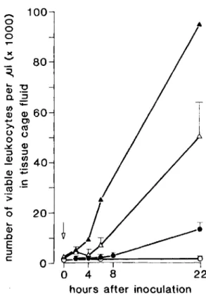
Figure 1. Viable leukocyte counts in tissue cage fluid during experimental infection of guinea pigs with
Staph-ylococcusaureusstrain Wood 46. At zero-time (arrow),
the indicated inocula were given: (0) 102 cfu (seven animals), (e) 104 cfu (four animals), (.6.)106 cfu (four
animals), and (A.) 108cfu (two animals). Data are mean
+SEM(bars) numbers of leukocytes x 1,OOO/jJIat inter-vals after infection.
infection. Bactericidal activity of PMNLs. Be-cause tissue cages could be infected with as few as 102 cfu of S. aureus despite the presence of 1 x
104-5 X 10⁶ PMNLs/ml, we concluded that the
presence of a tissue cage promoted infection even when the bacteria: PMNL ratio varied from 1 : 100 to 1: 50,000. We therefore examined PMNLs from sterile tissue cages as to their bactericidal function over various incubation times with various bacterial inocula and compared the data with the bactericidal activity of PMNLs from either peripheral blood or peritoneal exu-dates - that is, unstimulated or attracted PMNLs. Finally, to compare exudates of similar age, the function of PMNLs from tissue cages was com-pared for each experimental condition with that of peritoneal PMNLs obtained after two weeks of stimulation.
The bactericidal activity of tissue case-derived
~ "0 Q) a. :::J f/)
-Q) Q) 60-
0> >0- co 0 0 0 .:::t:. Q) :::J :::J Q) f/) 40 f/) Q) -.0 .S co >o
4 8 22hours after inoculation
o
80 20 100 >C -~ Q) .0 E :::J c:-
o
o
o
~-
ovarious bacterial inocula, the area corresponding to the previously determined diffusion zone was biopsied, homogenized, and checked for bacterial growth. All the biopsy specimens were sterile at 72 hr after injection of inocula as large as 108 cfu.
Evaluation of the role of tissue cage dead space.
Because the dead space present within tissue cages could create unfavorable conditions for surface phagocytosis, the following control experiments
were carried out. Tissue cages were halved
longitudinally and implanted as described. Histo-logic examination confirmed that granulation tissue had gained access to either side of the foreign body. Five of five of these modified tissue cages could still be infected with <103 organisms.
The negligible role of the dead space in promoting infection was further demonstrated by removing implanted, sterile tissue cages after four weeks and inoculating the healed sc pouches after removal of the clips with S.aureus strain Wood 46 suspended
in 4 ml of 0.9010 NaCl. In seven animals no infec-tion could be produced with 109 cfu, and in six
animals the peritoneal cavity (a natural dead space) could not be infected with 109 cfu.
Bacterial growth and leukocyte accumulation during experimental infections of tissue cages.
To assess the development of infection in the ex-perimental model, quantitative cultures ofS.aureus
strain Wood 46 were performed in tissue cages over seven days with various initial inocula. At 22 hr after inoculation of from 102 to 108 cfu,
bacteria reached concentrations ranging from 1.3 X 10⁵cfu/ml to 5.2
x
107 cfu/ml, respectively.Further observations over two to seven days showed that bacterial counts fluctuated between 105cfu/ml and 108cfu/ml, regardless of the initial
inoculum. These values were lower than those ob-tained under in vitro conditions in Mueller-Hinton broth (7-15 X 108cfu/ml after 24 hr).
Quantitation of viable (trypan blue-negative) leukocytes in tissue cage fluid at induction and during establishment of infection with various in-ocula is shown in figure 1. The observed increase in leukocyte counts depended markedly on the in-oculum size and on the time elapsed after inocula-tion, with a lag phase of 2-8 hr. At 22 hr, leukocyte counts had increased by factors of 5.1- and 40A-fold from initial inocula of 102and
108 bacteria, respectively.
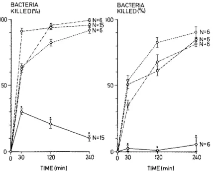
Phagocytic bactericidal activity of PMNLs from tissue cagefluid in the absence or presence of
BACTERIA
KILLED(%) Figure 2. Bactericidalcapacity0fpol
morphonuclear leukocytes (PMNL from sterile tissue cage fluid at 1 days after implantation in guinea pi!
(o~) compared with that ( peritoneal exudate PMNLs after sterile acute (0 - - 0) or protracte soluble (0-' -0) stimulation c PMNLs from peripheral blood(0- -0: The phagocytic bactericidal assa contained, in a final volume of 1 m of phosphate-buffered saline, 0.2 m of pooled tissue cage fluid, 2 x 10 PMNLs, and(left) 8 x 10³cfu (bac
teria : PMNL ratio, 1 : 250) or(right 4 x 10⁶cfu (bacteria: PMNL ratio,
2 : 1) of washed Staphylococcus
aureus.Data are means ± SEM(bars).
Asterisks indicate significant differ-ence(P
<
0.001) in comparison with each of the control PMNL prepara-tions. 240 120 TIME(min) BACTERIA KILLEO(%) o¥::::+=====~::::::::"_--ta
30 120 TIME(min) O~~---.---' a 30 240 50 100PMNLs was considerably lower than activities ob-served with PMNLs from acute and chronic peritoneal exudates and from peripheral blood (figure 2). This difference was significant with each bacterial inoculum and at each incubation in-terval tested, a result suggesting that the residen-tial PMNLs in tissue cages were unable to cope withS.aureus strain Wood46, even at minimal in-ocula. The viabilities of PMNLs from tissue cages, blood, and peritoneal exudate before and after in-cubation were comparable. Differential counts of leukocytes from tissue cages showed 40070-70%
PMNLs and 30070-60070 mononuclear leukocytes; these numbers were comparable to those obtained from persistent peritoneal exudates. In contrast to these results, in 10 assays the overall bactericidal activity of PMNLs accumulating in experimental-ly infected tissue cages at 20 hr after inoculation was 91.3% ± 1.9070 when tested by standard bactericidal procedure. Similar results were re-corded with control PMNLs from peritoneal exu-dates produced with glycogen or caseinate (bac-tericidal activity, 86.1 % ± 2.4% [16animals] and
79.6% ± 3.9% [10 animals], respectively).
Ingestion rates of PMNLs from sterile tissue cages. To define further the overall bactericidal defect observed with PMNLs from sterile tissue cage fluid, we measured their ingestion rate as well as their intracellular killing rate. In five animals,
PMNLs from tissue cages ingested49.6070 ± 2.3%
and killed 39.1 % ± 1.6% of the S. aureus,
whereas peritoneal PMNLs ingested 87.2070 ± 2.2% and killed83.1 % ± 2.5070 of the bacteria. After determination of the number of ingested bacteria, it could be calculated that 21.1 % ±
0.5070 of the ingested organisms remained viable in PMNLs from tissue cages, whereas only4.7% ±
0.5% of the bacteria ingested by control PMNLs survived intracellularly after30min of incubation.
Opsonic activity and complement levels of tissue cage fluid in the absence or presence of in-fection. After having identified a phagocytic and bactericidal defect in PMNLs from tissue cages, we analyzed another component of the phagocytic system - opsonization of the infecting organism. Figure 3 shows complement-mediated opsonic ac-tivity and total hemolytic complement levels in sterile tissue cage fluids, expressed as 50% killing andCHsounits, measured at various intervals after surgery. Both values in tissue cage fluid decreased progressively with time, the opsonic activity being
14.4% and the complement level being 14.8% of the serum values at 75 days after surgery. When expressed per gram of protein, the values of the tissue cage fluids corresponded to 33.1 % and
34.00/0of the serum values, respectively. The cor-relation in30animals between the total hemolytic complement level and the opsonic titer was highly
Pathogenesis of Foreign Body Infection 493
o
CH50 units K 50 units0
200
T 1200
tissue cage fluid,postop. interval
ID ltl N ID 600 o 7 9 guinea pig serum (poole d) 4 9 75 +' t" _ tD ~ N -# N
-DIm
\7 32 28 f.4 20 -25 26 -34. n= 8 10 day: 15a
100Figure 3. Opsonic activity and to-tal hemolytic complement level in tis-sue cage fluid from guinea pigs at in-tervals after implantation and in pooled normal guinea pig serum. Data are means ± SEM (bars) of the reciprocal dilutions that gave either 50070 killing of bacteria in a phago-cytic assay (K50) or 50% hemolysis in a hemolytic system (CH50). One as-terisk indicates
P<
0.01, and two as-terisks indicateP<
0.001 in compari-son with values in tissue cage fluid at 15 days after surgery.significant(r = 0.81; P
<
0.001). In 43 animals, the mean ± SEM C3 level in tissue cage fluid was18.60/0 ± 0.40/0 of the serum level.
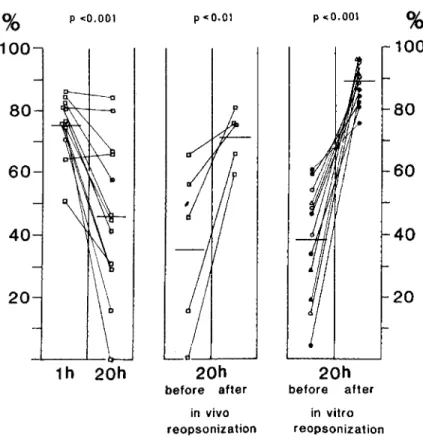
In view of these low levels of 500/0 killing,CHso, and C3 in tissue cage fluid, we considered the possibility that this fluid might be unable to pro-mote adequate opsonization, as has been de-scribed for empyema [13]. To test this hypothesis, S. aureus strain Wood 46 was preopsonized in vitro with pooled guinea pig serum before inocula-tion into tissue cages. Preopsonizainocula-tion of bacteria was confirmed by incubating an aliquot with peritoneal exudate PMNLs for 30 min in a phago-cytic assay which led to a killing rate of 80%. Despite optimal preopsonization, aliquots corre-sponding to 1.2 x 10² cfu injected into sterile tissue cages led to infection in four of four animals. Although the kinetics of bacterial growth and death were not formally tested in these experi-ments, low opsonic activity in tissue cage fluid could not be considered as a rate-limiting factor favoring the development of infection. During the development of infection, opsonic activity and the complement level were measured at 1 and 20 hr after inoculation of 107,cfu. The value of CHso in
six animals decreased moderately, from 267 ± 35.2 units to 172 ± 41.3 units (P
<
0.025). Op-sonic activity bound to S. aureus, however, showed a striking change during the course of the infection. In 20 animals, the opsonic coating of bacteria (figure 4, left) promoted 75.3% ± 2.9%killing at 1 hr after infection, whereas killing was decreased to 46.7% ± 7.40/0 at 20 hr (P
<
0.001). The opsonic coating of S. aureus could be im-proved (figure 4, middle) at 20 hr after infection by injection of 0.5 ml of fresh autologous serum (final concentration, 25%) into infected tissue cages, as demonstrated by aspirating the infecting bacteria 60 min after injection of fresh serum into tissue cages and testing the staphylococci in a phagocytic assay against peritoneal exudate PMNLs. In five assays, killing of these reop-sonized bacteria improved from 35.30/0 ± 12.4070to 71.6% ± 4.1 % (P<0.01). Finally, the opsonic coating of S. aureus at 20 hr was poor (figure 4, right) regardless of the size of the initial inoculum, which varied from 103 cfu to 108 cfu. In 12 assays,
killing of aspirated S. aureus could be improved
from 38.2% ± 5.3% to 88.8% ± 2.0% (P
<
0.001) by in vitro reopsonization with 100/0 pooled guinea pig serum. In summary, these experiments suggest that opsonic coating of S. aureus strain Wood 46 is incomplete at 20 hr after infection and that this defect can be corrected by addition of fresh guinea pig serum either into the tissue cage or to the phagocytic assay.
Discussion
The factors promoting foreign body infection are not well understood. In this report we have described an experimental guinea pig model that
%
10080
40
20 P <0.001 th 20h P<0.01 20h before after in vivo reopsonization P <0.001 20h before after in vitro reopsonization%
10080
60
40
20Figure 4. Opsonic activity bound to Staphylococcus aureus strain Wood 46 at intervals after experi-mental infection of tissue cages in guinea pigs:(left) 1 and 20 hr after in-fection with 107 cfu; (middle) 20 hr
after infection with 107 cfu before
and after in vivo supplementation of tissue cage fluid with 0.5 ml of fresh autologous serum (final concentra-tion, 25%); and(right) 20 hr after in-fection with(0) 103cfu, (e) 104cfu,
(~) 106 cfu, and (A.) 108 cfu before and after in vitro reopsonization with 10010pooled serum for a 30-min peri-od. Data are percentages of killing of S. aureus isolated from infected tis-sue cages after incubation for 30 min in a phagocytic assay with 5 x 10⁶
polymorphonuclear leukocytes from peritoneal exudate.
permits exploration of these factors and repro-duces the main characteristics observed in its clinical counterpart. After sc implantation of polymer tissue cages, injection of 102 cfu of an
organism of low virulence such asS. aureus strain
Wood 46 led uniformly to local infection in <20 hr. Infection did not spread to any other organ and healed after elimination of the foreign body. These observations, as well as the generally ac-cepted concept that the main host defense mecha-nisms against local infections with S. aureus in-clude opsonization and phagocytosis [13, 33], led us to focus our analysis of these host factors only over the first 20 hr of infection. S. aureus strain
Wood 46 was used in all experiments to avoid in-terference of staphylococcal protein A with IgG; this strain has been repeatedly shown to contain negligible amounts of protein A [26, 34] and to re-quire complement only for "optimal opsonization [13, 24, 26]. Although several biochemical deter-minations in tissue cage fluid differed from values in serum (data not shown), their variations were not sufficient to inhibit phagocytosis [35-39]. Such an effect, as well as a deleterious effect of
tissue cage fluid on PMNLs, was further ruled out by appropriate mixing experiments involving pre-incubation of tissue cage fluid with competent blood PMNLs.
Our observations have identified two host defense defects in the neighborhood of the foreign body that are manifested at distinct stages in an in-fection - namely, poor PMNL activity at the beginning and a steadily decreasing efficiency of opsonization during the course of the disease.
Initially, opsonization of strain Wood 46 was adequate and could therefore, not account for the high susceptibility to infection in our model. Moreover, in vitro preopsonization of the infect-ing strain did not prevent the development of in-fection after inoculation into tissue cages. Our data further suggest that at this early stage, the PMNLs residing in sterile tissue cage fluid were deficient in their phagocytic bactericidal function when compared with control PMNLs from either blood-the milieu from which they are originat-ing - or peritoneal exudates obtained by a soriginat-ingle soluble stimulus. This defect could be demon-strated under the standard conditions of a
conven-Pathogenesis ofForeign Body Infection
tional phagocytic assay, as well as at a bacterial PMNL ratio of 1 : 250, which was the condition encountered in tissue cage fluid. Finally, this phagocytic bactericidal deficit in PMNLs from tissue cages tested two weeks after implantation was also evident when these cells were compared with PMNLs from an exudate of the same age, produced by soluble stimuli administered over 14 days.
Several aspects of this phagocytic bactericidal defect in PMNLs from tissue cages merit com-ment. (1) The development of a control system reproducing the conditions of a chronic exudate in the absence of a foreign body was of importance to compare the phagocytic function of PMNLs of similar ages in the presence and the absence of a foreign body. The identical time elapsed between the onset of these protracted stimuli (tissue cages or glycogen), the cytology of the two exudates, and the results of histopathologic examination of the two surrounding tissues suggest the presence of cell populations of the same age. This issue, however, can be resolved unambiguously only by future experiments involving comparison of pulse-labeled PMNLs; for the time being, comparison of PMNLs from blood and exudate has been con-sidered as an adequate procedure in similar ex-periments [40]. (2) The observed phagocytic bactericidal defect in PMNLs from tissue cages is all the more striking because Van Epps and Garcia have demonstrated that PMNLs from peritoneal exudates of guinea pigs elicited by a soluble stimulus actually show increased, rather than de-creased, metabolic activity [40].
In keeping with our results obtained with PMNLs from tissue cages, Klock and Bainton [41] and Klock and Stossel [42] have demonstrated that intimate contact of PMNLs with glass or nylon wool will lead not only to activation of their ox-idative metabolism and to degranulation, but also to a decrease in the cells' phagocytic bactericidal function. Electron microscopic data obtained with our model have confirmed that PMNLs in close contact with tissue cages show decreased granule contents.' Degranulation and metabolic altera-tions have been described for other systems
in-1I. loris, W. Zimmerli, G. Majno, and F. A. Waldvogel,
"Foreign Body Reactions: Morphology of Inflammatory and Phagocytic Cells," manuscript in preparation.
495
volving PMNLs in contact with foreign, non-phagocytosable surfaces [43-45]. Taken together, these in vitro results suggest that intimate contact of PMNLs with foreign, nonphagocytosable sur-faces might be detrimental to these cells, as sug-gested by the results of our experimental model. Finally, the deficit observed in PMNLs from tissue cages affected ingestion as well as bacteri-cidal function, an observation suggesting that several subcellular mechanisms are altered. The mechanisms underlying these various alterations remain to be defined.
In contrast to these observations for the early stage of infection, opsonic coating of the infecting microorganism S. aureus strain Wood 46 - pre-sumably by functional C3 [46] - was markedly de-creased during the course of the infection. This observation is open to at least two interpretations. Either decreased opsonization of S. aureus might be a consequence of the imbalance between the rate of coating of bacteria with functionally active C3 and the logarithmic growth of the organisms, or cleavage of the opsonically active factor by fJ-I-H-globulin, C3b inactivator, and proteases [47-51] could have occurred during the develop-ment of a purulent infection. Loss of opsonic ac-tivity, as well as proteolytic cleavage of C3, has been recently demonstrated in several exudates rich in PMNLs [13, 15, 24]. Finally, our experi-ments showed that PMNLs attracted into the in-fected tissue cages displayed normal phagocytic bactericidal function, a finding which is apparent-ly in contradiction with our observations in sterile tissue cages. In the case of a sterile tissue cage, however, the foreign body itself is probably responsible for the attraction of PMNLs; in the case of an abscess, additional chemoattractants such as bacterial polypeptides [52] are liberated, thus allowing PMNLs to accumulate in the fluid phase without being in contact with the foreign body. Intimate contact of PMNLs with foreign surfaces has indeed been shown to be necessary to alter the cells' metabolism and phagocytic func-tions [41, 42].
In conclusion, the phagocytic bactericidal defect in PMNLs from sterile tissue cage fluid probably represents one of the several deter-minants of foreign body infection and allows other pathogenic factors to progress during the first hours of the disease. Some of these factors
may include organic substances coating the foreign body, which may protect bacteria from phagocytosis [53]; bacterial adhesion to foreign material [54]; and formation of a bacterial "gly-cocalyx" [55], which reduces bacterial accessibility to phagocytes and/or antibiotics. The animal model described here should permit the investiga-tion of these and other possible factors that predispose foreign bodies to infection.
References
1. Bernhard, V. M. Management of infected vascular pros-theses. Surg. Clin. North Am. 55:1411-1417, 1975. 2. Bhat, D. J., Tellis, V. A., Kohlberg, W. I., Driscoll, B.,
Veith, F. J. Management of sepsis involving expanded polytetrafluoroethylene grafts for hemodialysis access. Surgery 87:445-450, 1980.
3. Kloster, F. E. Complications of artificial heart valves. J.AM.A 241:2201-2203, 1979.
4. Charnley, J. Postoperative infection after total hip re-placement with special reference to air contamination in the operating room. Clin, Orthop. 87:167-187, 1972. 5. Infected hip prostheses [editorial]. Br. Med. J.
280:1241-1242, 1980.
6. Ahlberg,A.,Carlsson,A.S., Lindberg,L.Hematogenous infection in total joint replacement. Clin. Orthop. 137: 69-75, 1978.
7. Courtiss, E. H., Goldwyn, R. M., Anastasi, G. W. The fate of breast implants with infections around them. Plast. Reconstr. Surg. 63:812-816, 1979.
8. Georgiade, N. G., King, E. H., Harris, W. A, Tenery, J. H., Schlech, B. A. Effect of three proteinaceous foreign materials on infected and subinfected wound models. Surgery 77:569-576, 1975.
9. Hunter, G., Dandy, D. The natural history of the patient with an infected total hip replacement. J. Bone Joint Surg. [Br.] 59:293-297, 1977.
10. Elek, S. D., Conen, P. E. The virulence of Staphylo-coccus pyogenesfor man: a study of the problems of wound infection. Br. J. Exp. Pathol. 38:573-586, 1957. 11. James, R. C., MacLeod, C. J. Induction of staphylococ-cal infections in mice with small inocula introduced on sutures. Br. J. Exp. Pathol 42:266-277, 1961. 12. Noble, W. C. The production of subcutaneous
staphy-lococcal skin lesions in mice. Br. J. Exp. Pathol. 46:254-262, 1965.
13. Lew, P. D., Zubler R., Vaudaux, P., Farquet, J. J., Wald-vogel, F. A., Lambert.jPv-H. Decreased heat-labile op-sonic activity and complement levels associated with evidence of C3 breakdown products in infected pleural effusions. J. Clin. Invest. 63:326-334, 1979.
14. Lew, D. P., Despont, J.-P., Perrin, L. H., Aguado, M.-T., Lambert, P. H., Waldvogel, F. A. Demonstra-tion of a local exhausDemonstra-tion of complement components and of an enzymatic degradation of immunoglobulins in pleural empyema: a possible factor favouring the
per-sistence of local bacterial infections. Clin. Exp. Im-munol. 42:506-514, 1980.
15. Suter, S., Nydegger, U. E., Roux, L., Waldvogel, F. A. Cleavage of C3 by neutral proteases from granulocytes in pleural empyema. J. Infect. Dis. 144:499-508, 1981. 16. Stossel, T. P. Phagocytosis [parts 1-3J. N. Engl. J. Med.
290:717-723,774-780, 833-839, 1974.
17. Sveen, K., Hofstad, T. Use of preformed cavities in rab-bitsforthe quantitation of leukocyte chemotaxis caused by bacterial lipopolysaccharides. Acta Pathol. Micro-bioI. Scand. {B] 84:252-258, 1976.
18. Gerding, D. N., Moore, B. M., Russ, T. E., Peterson, L. R. Local cellular response to staphylococcal chal-lenge in an abdominal abscess model. In J. D. Nelson and C. Grassi [ed.]. Current chemotherapy and infec-tious diseases. American Society for Microbiology, Washington, D.C., 1980, p. 841-842.
19. Bergan, T. Pharmacokinetics of tissue penetration of anti-biotics. Rev. Infect. Dis. 3:45-66, 1981.
20. Gerding, D. N., Hall, W. H., Schierl, E. A., Manion, R.E.Cephalosporin and aminoglycoside concentrations in peritoneal capsular fluid in rabbits. Antimicrob. Agents Chern other. 10:902-911, 1976.
21. Stossel, T. P., Murad, F., Mason, R. J., Vaughan, M. Regulation of glycogen metabolism in polymorpho-nuclear leukocytes. J. BioI. Chern. 245:6228-6234, 1970.
22. Hisatsune, K., Kobayashi, K., Nozaki, S., Muramatsu,I. Phagocytosis-stimulating activity of tuftsin analogs. Microbiol. Immunol. 22:581-584, 1978.
23. Chenoweth, D. E., Lane, T. A., Rowe, J. G., Hugli, T. E. Quantitative comparisons of neutrophil chemotaxis in four animal species. Clin. Immunol, Immunopathol. 15: 525-535, 1980.
24. Zwahlen, A., Nydegger, U. E., Vaudaux, P., Lambert, P.-H., Waldvogel, F. A. Complement-mediated opsonic activity in normal and infected human cerebrospinal fluid: early response during bacterial meningitis.J. In-fect. Dis. 145:635-646, 1982.
25. Vaudaux, P., Waldvogel, F. A. Gentamicin antibacterial activity in the presence of human polymorphonuclear leukocytes. Antimicrob. Agents Chemother. 16:743-749, 1979.
26. Spika, J. S., Verbrugh, H. A., Verhoef, J. Protein A ef-fect on alternative pathway complement activation and opsonization ofStaphylococcus aureus.Infect. Immun. 34:455-460, 1981.
27. Leijh, P. C. J., van Den Barselaar, M. T., van Zwet, T. L., Dubbeldeman-Rempt,I., van Furth, R. Kinetics of phagocytosis of Staphylococcus aureus and
Escherichia coliby human granulocytes. Immunology 37:453-465, 1979.
28. Tan,J.S., Watanakunakorn,C.,Phair,J. P. A modified assay of neutrophil function: use of lysostaphin to dif-ferentiate defective phagocytosis from impaired in-tracellular killing.J.Lab. Clin. Moo. 78:316-322, 1971. 29. Mandell, G.L.Interaction of intraleukocytic bacteria and
antibiotics. J. Clin. Invest. 52:1673-1679, 1973. 30. Kabat, E. A., Mayer, M. M. Experimental
immunochem-istry. 2nd ed. Charles C Thomas, Springfield, Ill., 1961, p. 133-152.
Pathogenesis ofForeign Body Infection
31. Mancini, G., Carbonara, A. 0.,Heremans, J. F. Immu-nochemical quantitation of antigens by single radial im-munodiffusion. Immunochemistry 2:235-254, 1965. 32. Eickenberg, H.-U. What is interstitial fluid? Biochemical
and physiological analysis of fluid obtained from tissue cages. Scand. J. Infect. Dis. [Suppl.] 14:166-170, 1978. 33. Verbrugh, H. A. The phagocytic response in host resis-tance against staphylococcal infections. Spirit Offset, Rotterdam, 1979, p. 150.
34. Sjoquist, J., Stalenheim, G. Protein A from
Staphylococ-cus aureus. IX. Complement-fixing activity of protein A-IgG complexes. J. Immunol. 103:467-47:3, 1969. 35. Stossel, T. P., Alper, C A., Rosen, F. S.
Serum-depen-dent phagocytosis of paraffin oil emulsified with bac-terial lipopolysaccharide. J. Exp. Med. 137:690-705, 1973.
36. Craddock, P. R., Yawata, Y., VanSanten, L., Gilber-stadt, S., Silvis, S., Jacob, H. S. Acquired phagocyte dysfunction: a complication of the hypophosphatemia of parenteral hyperalimentation. N. Engl. J. Med. 290: 1403-1407, 1974.
37. Weston,W. L., Huff, J. C,Humbert, J. R., Hambidge, K. M., Neldner, K. H., Walravens, P. A. Zinc correc-tion of defective chemotaxis in acrodermatitis entero-pathica. Arch. Dermatol. 113:422-425, 1977.
38. Babior, B. M. Oxygen-dependent microbial killing by phagocytes [parts 1 and 2]. N. Engl. J. Med. 298:659-668,721-725,1978.
39. Mandell, G. L. Bactericidal activity of aerobic and an-aerobic polymorphonuclear neutrophils. Infect. Im-mun. 9:337-341, 1974.
40. Van Epps, D. E., Garcia, M. L. Enhancement of neutrophil function as a result of prior exposure to chemotactic fac-tor. J. Clin. Invest. 66:167-175,1980.
41. Klock, J. C., Bainton, D. F. Degranulation and abnormal bactericidal function of granulocytes procured by rever-sible adhesion to nylon wool. Blood 48:149-161, 1976. 42. Klock, J. C., Stossel, T. P. Detection, pathogenesis and prevention of damage to human granulocytes caused by interaction with nylon wool fiber: implications for fil-tration leukapheresis. J. Clin. Invest. 60: 1183-1190, 1977.
43. Henson, P. M. Interaction of cells with immune com-plexes: adherence, release of constituents, and tissue in-jury. J. Exp. Med. 134(Suppl.):114S-135S, 1971. 44. Wright, D. G., Gallin, J. I. Secretory reponses of human
497
neutrophils: exocytosis of specific (secondary) granules by human neutrophils during adherence in vitro and during exudation in vivo. J. Immunol. 123:285-294, 1979.
45. Yanai, M., Quie, P. G. Chemiluminescence by polymor-phonuclear leukocytes adhering to surfaces. Infect. Im-mun. 32:1181-1186, 1981.
46. Stossel, T. P., Field, R. J., Gitlin, J. D., Alper, C. A., Rosen, F. S. The opsonic fragment of the third com-ponent of human complement (C3). J. Exp. Med. 141: 1329-1347, 1975.
47. Law, S. K., Fearon, D. T., Levine, R. P. Action of the C3b-inactivator on cell-bound C3b. J. Immunol. 122: 759-765, 1979.
48. Law, S.K., Lichtenberg, N. A., Levine, R. P. Evidence for an ester linkage between the labile binding site of C3b and receptive surfaces. J. Immunol. 123:1388-1394, 1979.
49. Carlo, J. R., Ruddy, S., Studer, E. J., Conrad, D. H. Complement receptor binding of C3b-coated cells treated with C3b inactivator, f3-1-H globulin and tryp-sin. J. Immunol. 123:523-528, 1979.
50. Fearon, D. T., Austen, K. F. Activation of the alternative complement pathway with rabbit erythrocytes by cir-cumvention of the regulatory action of endogenous con-trol proteins. J. Exp. Med. 146:22-33, 1977.
51. Pangburn, M. K., Schreiber, R. D., Muller-Eberhard, H. J. Human complement C3b inactivator: isolation, characterization, and demonstration of an absolute re-quirement for the serum protein f3-1-H for cleavage of C3b and C4b in solution. J. Exp. Med. 146:257-270, 1977.
52. Russell, R. J., Wilkinson, P. C., Mclnroy, R. J., McKay, S., McCartney, A. C, Arbuthnott, J. P. Effects of staphylococcal products on locomotion and chemotaxis of human blood neutrophils and monocytes. J. Med. Microbiol. 9:433-449, 1976.
53. Peters, G., Locci, R., Pulverer, G. Microbial coloniza-tion 0f prosthetic devices. II. Scanning electron microscopy of naturally infected intravenous catheters. Zentralbl. Bakteriol. [B] 173:293-299, 1981.
54. Katz, S., Izhar, M., Mirelman, D. Bacterial adherence to surgical sutures: a possible factor in suture induced in-fection. Ann. Surg. 194:35-41, 1981.
55. Costerton, J. W., Geesey, G. G., Cheng, K.-J. How bac-teria stick. Sci. Am. 238(1):86-96, 1978.