DOI: 10.1093/jac/dkg277
Advance Access publication 29 May 2003
89
. . . .
Comparative efficacy of daptomycin and vancomycin in the therapy of
experimental foreign body infection due to Staphylococcus aureus
Pierre Vaudaux*, Patrice Francois, Carmelo Bisognano, Dongmei Li, Daniel P. Lew and
Jacques Schrenzel
Division of Infectious Diseases, Geneva University Hospital, CH-1211 Geneva 14, Switzerland
Received 8 January 203; returned 14 February 2003; revised 4 March 2003; accepted 6 April 2003
The therapeutic activity of daptomycin was compared with that of vancomycin in a rat model of subcutane-ously implanted tissue cages chronically infected with strain Rev1, a spontaneous methicillin-susceptible revertant of the methicillin-resistant Staphylococcus aureus strain MRGR3, showing equivalent virulence to its parent. The MIC and MBC of daptomycin (in Mueller–Hinton broth supplemented with 50 mg/L Ca2+) or
vancomycin for strain Rev1 were 1–2 and 2–4 or 1 and 2 mg/L, respectively. In vitro elimination of strain Rev1 in the presence of 50% tissue cage fluid was more rapid with daptomycin 4 mg/L compared with vancomycin. After 2 weeks of infection, viable counts of strain Rev1 averaged 6.49 log10 cfu/mL of tissue cage fluid (n = 87). Intraperitoneal administration of daptomycin 30 mg/kg once daily, or vancomycin 50 mg/kg twice daily, pro-duced antibiotic levels continuously above MBC. After 7 days of therapy with daptomycin or vancomycin, mean ± S.E.M. counts of Rev1 decreased (P < 0.05) by 1.11 ± 0.25 (n = 28) or 0.80 ± 0.31 (n = 35) log10 cfu/mL,
respectively, compared with cages of untreated animals, but were not significantly different from each other. In daptomycin-treated rats, three cages yielded subpopulations with reduced susceptibility to daptomycin. In conclusion, a low dose regimen of daptomycin was at least equivalent to vancomycin against chronic foreign body infections due to S. aureus. Drug dosage should be adapted to obtain inflammatory fluid levels of daptomycin minimizing emergence of resistant subpopulations.
Keywords: Gram-positive bacteria, chronic infections, antimicrobial agents
Introduction
Prosthetic device infections, in particular those resulting from Staphylococcus aureus, are notoriously difficult to treat, and micro-bial eradication frequently requires the removal of infected materials. Recent clinical studies using combinations of fluoroquinolones and rifampicin yielded promising results for the treatment of orthopaedic prosthetic S. aureus infections without prosthesis removal.1–4
Unfor-tunately, a vast majority of methicillin-resistant strains of S. aureus (MRSA) are also resistant to virtually all fluoroquinolones,5 which
severely limits the therapeutic armentarium for the treatment of foreign body infections. Thus, glycopeptide therapy with vanco-mycin or teicoplanin, alone or in combination with rifampicin,6–8
frequently remains the only available therapy for severe MRSA infections, but does not always prevent the emergence of rifampicin-resistant mutants.9 An additional concern is the recent discovery of
glycopeptide-intermediate or -resistant strains of S. aureus in several parts of the world.10–14 The increasing use of glycopeptides for
therapy of Gram-positive infections may further promote the emer-gence of more highly glycopeptide-resistant strains.15,16
Daptomycin is a lipopeptide antibiotic with potent in vitro bacteri-cidal activity against a wide range of Gram-positive pathogens, including antibiotic-resistant staphylococci.17–20 The mechanism of
action of daptomycin is not fully understood, but seems to be distinct from that of major cell wall-active agents, such as β-lactams and glycopeptides. Daptomycin binds in a calcium-dependent manner to Gram-positive cytoplasmic membranes,21,22 and disrupts membrane
function, dissipating membrane potential and inhibiting macro-molecular biosynthesis. Daptomycin is uniformly potent against S. aureus clinical isolates in large surveillance studies.23–27 The in vivo activity of daptomycin is currently being evaluated in both therapeutic trials and experimental models.19,28 Optimization of
dapto-mycin pharmacokinetics and pharmacodynamics against severe S. aureus infections17,28–32 requires maximum efficacy and safety, and
the maintenance of bactericidal levels in deep-seated compart-ments.17,19,33
. . . . *Corresponding author. Tel: +41-22-37-29-826. Fax: +41-22-37-29-830; E-mail: [email protected]
We previously showed the usefulness of a rat tissue cage model of S. aureus chronic foreign body infections for evaluating various categories of antimicrobial agents such as vancomycin,34
teico-planin,35 imipenem,36 and several fluoroquinolones including
fler-oxacin,34 sparfloxacin, temafloxacin, ciprofloxacin,37 levofloxacin
and trovafloxacin,38 alone or in combination with rifampicin.34,35,39
In this study, we evaluate the efficacy of a once-daily dosing of daptomycin compared with a twice-a-day regimen of vancomycin in the therapy of experimental chronic foreign body infections due to S. aureus.40
Materials and methods
Bacterial strainsStrain Rev1 is a spontaneous methicillin-susceptible revertant of MRSA strain MRGR3, a clinical isolate from a patient with catheter-related sepsis. Strain Rev1 was found to be as virulent in the rat model of chronic
S. aureus tissue cage infection as its MRSA parental strain.34,37,39 Except
for the loss of the methicillin resistance element, strain Rev1 exhibits an antibiotic resistance pattern identical to its MRSA parent, including resistance to penicillin, gentamicin, chloramphenicol, erythromycin, tetracycline and polymyxin B, but susceptibility to clindamycin, rifam-picin and all fluoroquinolones.34,37,39
Antimicrobial agents
For in vitro studies, daptomycin (Cubist Pharmaceutical, Inc., Lexing-ton, MA, USA) was solubilized in distilled water at a concentration of 1 mg/mL and further diluted in saline. For in vivo studies, daptomycin was solubilized in saline at a concentration of 7.5 mg/mL. Commercially available vancomycin hydrochloride (Lilly, Giessen, Germany) was suspended as recommended by the manufacturer.
In vitro studies
MICs of daptomycin and vancomycin for strain Rev1 were determined in cation-adjusted Mueller–Hinton broth (CAMHB) containing 20–25 mg/L Ca2+ and 10–12.5 mg/L Mg2+ by the standard broth macrodilution
method, with an average inoculum of 106 cfu/mL, as described by the
NCCLS.41 For daptomycin MIC, CAMHB was supplemented with
additional calcium to a physiological concentration of 50 mg/L Ca2+
(CSMHB).
To screen for the possible carryover effects of each antibiotic during the MBC determinations, 100 µL portions were taken from all tubes with no visible growth. These were subcultured, either undiluted or diluted 10-fold in saline, on Mueller–Hinton agar (MHA) for 36 h at 37°C. The MBC was defined as the lowest concentration that killed 99.9% of the original inoculum.
Killing kinetic studies
Portions of 100 µL containing 106 cfu of strain Rev1 (obtained from
exponential-phase cultures) were added to sterile plastic tubes of 1 mL of either CSMHB or CAMHB that included either daptomycin or vanco-mycin (4 mg/L), respectively, in a shaking waterbath at 37°C. The number of viable organisms was determined by subculturing 50 µL of 10-fold diluted portions on MHA after 0, 1, 3, 6 and 24 h of incubation. Colonies were enumerated with a laser colony counter (Spiral System) after 48 h of incubation at 37°C. The detection limit was 2 log10 cfu/mL. No significant carryover of antibiotics was observed by using these experimental conditions. To evaluate the impact of tissue cage fluid proteins on the bactericidal activity of daptomycin 4 mg/L, the rate of elimination of strain Rev1 from tubes containing 1 mL of a mixture of
CSMHB and sterile tissue cage fluid (pooled from 20 different cages of uninfected animals) in a 1:1 ratio was also recorded.
To evaluate the susceptibility to daptomycin or vancomycin of strain Rev1 recovered from infected tissue cage fluids, the bacteria were iso-lated from the tissue cage fluids by centrifugation, and were treated with 0.1% Triton X-100 and sonication to disrupt the host cells, as described previously.35 This procedure is used to reduce bacterial clumping, and
was shown repeatedly to be harmless for ex vivo bacteria regarding their ability to multiply and their susceptibility to antibiotics.35,42 Thereafter,
tissue cage bacteria were exposed to either daptomycin or vancomycin (4 mg/L each) in tubes containing 1 mL of either CSMHB or CAMHB, respectively, supplemented with 50% pooled tissue cage fluid, and their rate of elimination was compared with that of strain Rev1 grown in vitro. To make the comparison with ex vivo bacteria more relevant, bacteria grown in vitro were taken from saline-washed cultures of stationary phase organisms.35,38 The number of viable organisms after 0, 2, 4, 6 and
24 h of incubation was determined, as described above. For each group, the reductions in cfu counts from time zero to further times of incubation were expressed as ∆log10 cfu/mL. The means ± S.E.M. of the ∆log10 cfu/mL of three independent determinations were analysed for signifi-cant differences by unpaired t-tests. Data were considered signifisignifi-cant when P < 0.05 using two-tailed significance levels.
Treatment of chronic tissue cage infections
Experiments involving rats were approved by the Ethics Committee of the Faculty of Medicine, University of Geneva, and by the Veterinary Office of the State of Geneva. Four tissue cages were implanted sub-cutaneously, as described previously,34 in Wistar rats that had been
anaesthetized with an intraperitoneal injection of a mixture of ketamine (90 mg/kg) and xylazine (5 mg/kg). At 3 weeks post-implantation, tissue cage fluid was aspirated and checked for sterility. To establish a chronic
S. aureus infection, tissue cages were inoculated with 0.1 mL of saline containing 0.2 × 10⁶ to 2 × 10⁶ cfu of a log-phase culture of strain Rev1.
Two weeks later, all tissue cages containing more than 105 cfu/mL of
fluid were included in the therapeutic protocols.
Rats infected with strain Rev1 were randomized to receive (by the intraperitoneal route for 7 days) either a once-a-day regimen of dapto-mycin (30 mg/kg) or a twice-a-day regimen of vancodapto-mycin (50 mg/kg), or were left untreated.
At 12 h after the last injection of vancomycin or 24 h after the last injection of daptomycin, quantitative cultures of 10-fold serially diluted tissue cage fluids were performed on MHA. To optimize the yield of viable bacteria, tissue cage fluids were sonicated briefly (60 W, 1 min) to disrupt the biofilm and phagocytic cells before the serial dilutions and plating. Plates were incubated for 24–48 h at 37°C. The detection limit was 2 log10 cfu/mL of tissue cage fluid. The differences in cfu counts between days 1 and 8 were determined and expressed as ∆log10 cfu/mL. For each treatment group, results were expressed as means ± S.E.M. Com-parison of bacterial counts in the different groups was performed by one-way analysis of variance and t-tests corrected for multiple groups. Data were considered significant when P <0.05 using two-tailed significance levels.
Resistance to antimicrobial agents
In initial experiments, the bacteria recovered from cage fluids on day 8 of therapy were screened for the emergence of resistance to daptomycin on MHA. Samples (of 100 µL) of 10-fold-diluted cage fluid were plated onto MHA containing daptomycin 4 mg/L. Plates were incubated for 48 h at 37°C. The detection limit was 1 log10 cfu/mL of tissue cage fluid.
Further analysis of subpopulations that grew on the daptomycin-containing agar from some tissue cages was first performed on colonies
recovered from the primary antibiotic-containing plates. The identifica-tion of representative individual colonies as S. aureus was assessed by standard microbiological procedures, and their clonal relationship with strain Rev1 confirmed by pulse-field gel electrophoresis (PFGE). The overall antimicrobial susceptibility of 10 colonies isolated from each primary daptomycin-containing plate was compared with strain Rev1 by disc diffusion using MHA, according to NCCLS recommendations. The antibiotics tested were daptomycin (30 µg paper discs provided by Cubist) and 15 commercially available discs containing penicillin, oxacillin, gentamicin, norfloxacin, ciprofloxacin, trovafloxacin, clinda-mycin, erythroclinda-mycin, fusidic acid, co-trimoxazole, fosfoclinda-mycin, rifam-picin, vancomycin, teicoplanin or mupirocin. Three additional colonies from each daptomycin-containing plate were tested separately for increased daptomycin MICs, compared with strain Rev1, by a standard broth microdilution test, as recommended by NCCLS, using either CAMHB or CSMBH.41 Finally, the stability of the daptomycin MICs
was checked by agar dilution, broth microdilution and macrodilution assays performed on individual colonies that had been stored in skim milk for 6–12 months at –70°C.
Pharmacokinetics of antimicrobial agents
The pharmacokinetic properties of vancomycin in rat tissue cage fluid have been estimated previously.34 In rats treated with daptomycin, its
pharmacokinetic levels in tissue cage fluids were determined at various time intervals (0, 2, 4, 6, 12 and 24 h) on days 4 and 7 of therapy. Simi-larly, blood levels of rats treated with daptomycin were also determined at various time intervals (0.25, 0.5, 1, 2, 4, 6, 8, 12 and 24 h) after single-dose intraperitoneal administration of the antimicrobial agent. The blood was collected by cardiac puncture into heparinized tubes. Plasma was collected by centrifugation and samples were stored at –20°C until analy-sis.
Daptomycin analytical assay
Daptomycin was detected using an internal standard of ethylparaben, and was isolated by protein precipitation with methanol, followed by HPLC. The mobile phase consisted of 90% mobile phase A (acetonitrile: 0.5% NH4H2PO4 34:66, v/v) and 10% mobile phase B (acetonitrile: 0.5% NH4H2PO4 20:80, v/v) at a flow rate of 1.5 mL/min. Serum drug concen-trations were determined by reverse-phase HPLC using a Metachem Hypersil C8 analytical column and a Waters Xterra RP18 guard column (ANSYS Technologies, Inc., Lake Forest, CA, USA). At a flow rate of 1.5 mL/min, daptomycin shows a retention time of 14–16 min. Samples were analysed at 214 nm. The detection of daptomycin concentrations in rat plasma was linear across the range of 7.5–400 mg/L. This method has been validated for daptomycin over the concentration range of 3–500 mg/L, with a lower limit of quantification equal to the lowest calibration level of 3 mg/L.
The concentrations of daptomycin in rat tissue cage fluid were esti-mated by a previously described microbiological assay,43 except for the
use of Antibiotic medium 11 and Sarcina lutea as the test strain. To avoid a potential bias due to protein binding, all tissue cage fluids were diluted with one volume of PBS. Thus, tissue cage fluid protein concentrations of samples were equivalent to those of daptomycin standards prepared in PBS supplemented with 50% of sterile pooled tissue cage fluid. Under these experimental conditions, the limit of detection of the daptomycin assay was 1 mg/L.
The areas under the concentration–time curve (AUC) of daptomycin in either plasma or tissue cage fluid were estimated by the linear trapezoi-dal rule from 0–24 h (AUC0–24) on days 4 and 7 of administration of this antimicrobial agent.
Results
In vitro studiesThe MIC and MBC of daptomycin in CSMHB were 1–2 and 2–4 mg/L, respectively, for strain Rev1; the MIC and MBC of daptomycin in the presence of 50% tissue cage fluid added to CSMHB were 2 and 4 mg/L, respectively, for strain Rev1; the MIC and MBC of vanco-mycin in CAMHB were 1 and 2 mg/L, respectively, for strain Rev1.
Time–kill studies performed in CSMHB showed rapid elimin-ation of exponential-phase cultures of strain Rev1 by daptomycin 4 mg/L. The reduction in the viable counts of strain Rev1 by dapto-mycin exceeded 3 log10 cfu/mL after 3 h (data not shown). A similar reduction in the viable counts of strain Rev1 by daptomycin 4 mg/L was observed in CSMHB supplemented with 50% tissue cage fluid (data not shown).
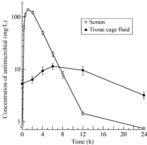
Pharmacokinetics of antimicrobial agents in tissue cage fluid At day 4 of therapy, the mean levels of daptomycin in tissue cage fluids (n = 6) of animals treated with daptomycin were 5.4 at time zero, 6.6 at 2 h, 9.8 at 4 h, 11.8 at 6 h, 10.0 at 12 h and 3.4 mg/L at 24 h, respectively, after administration of a 30 mg/kg once-daily regimen (Figure 1). Similar concentrations of daptomycin were recorded at day 7 of therapy (data not shown). Since residual levels of dapto-mycin were nearly equivalent to the MBC of this antimicrobial agent for strain Rev1, recorded in CSMHB containing 50% tissue cage fluid, the daptomycin once-daily regimen yielded bactericidal levels of daptomycin in infected tissue cage fluids throughout therapy. At day 4 of therapy, the tissue cage fluid AUC0–24 of daptomycin was 195.8 mg⋅h/L.
In comparison, blood levels of animals treated with a 30 mg/kg intraperitoneal regimen of daptomycin showed a Cmax of 141 mg/L at
1 h, and a concentration of 8.2 mg/L at 8 h. The trough level was 0, which differs from human pharmacokinetics in which daptomycin maintains trough levels of 1–9 mg/L. The AUC0–24 of daptomycin was 558 mg⋅h/L, which is only slightly higher than the clinical AUC0–24 of 468 mg⋅h/L.44
Figure 1. Pharmacokinetic levels of daptomycin in plasma, and tissue cage
The average peak and trough tissue cage fluid levels of vanco-mycin determined in a previous study34 were 12 and 2 mg/L at 4 and
12 h, respectively.
Treatment of chronic tissue cage infections
At the onset of therapy, average bacterial counts for cages infected with strain Rev1 were 6.87 ± 0.28 cfu/mL for controls (n = 24), 6.25 ± 0.17 log10 cfu/mL for animals receiving daptomycin once a day (n = 28), and 6.43 ± 0.13 log10 cfu/mL for animals receiving
vanco-mycin twice a day (n = 35). At the end of the 7 day treatment period, bacterial counts in the tissue cages of control animals showed a slight and non-significant increase of 0.24 ± 0.23 log10 cfu/mL (n = 24). In
contrast, both the daptomycin and vancomycin regimens (Figure 2) led to significant reductions in bacterial counts in tissue cage fluids of 1.11 ± 0.25 (n = 28; P = 0.001) and 0.80 ± 0.31 log10 cfu/mL (n = 35; P = 0.02), respectively, compared with tissue cage fluids of control animals. The higher average reduction in cfu counts of rats treated with daptomycin compared with that of vancomycin-treated animals did not reach statistical significance (P = 0.45).
Comparison of daptomycin activity on bacteria grown in vitro and in vivo
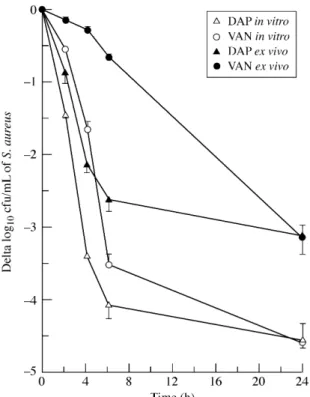
To evaluate the bactericidal activity of daptomycin compared with vancomycin against tissue cage fluid organisms of strain Rev1 in conditions relevant to therapy, we assayed, in parallel, the elimin-ation rates of tissue cage and in vitro grown bacteria by 4 mg/L of either antimicrobial agent in the presence of 50% sterile tissue cage fluid in CSMHB. Stationary-phase were preferred to log-phase organisms to make the comparison with ex vivo bacteria more relevant.35,38 Figure 3 shows a significantly (P < 0.001) higher
elimin-ation rate of stelimin-ationary-phase organisms by daptomycin 4 mg/L, compared with vancomycin, during the initial 4 h period of drug exposure, which led to >3 log10 reductions in viable counts at 4 versus 6 h, respectively. However, the most impressive differences in the bactericidal activities of daptomycin compared with vancomycin were seen with bacteria freshly removed from infected cages. During the initial 6 h period of drug exposure, the average reductions in viable counts of tissue cage bacteria by daptomycin 4 mg/L were 2.1 and 2.6 log10 cfu/mL at 4 and 6 h, respectively, compared with 0.3 and
0.6 log10 cfu/mL with vancomycin 4 mg/L (Figure 3). The presence
of 50% tissue cage fluid, yielding average protein concentrations of
10–15 mg per mL of CSMHB, did not significantly impair the bac-tericidal activity of daptomycin against bacteria grown in vitro or in vivo. Since tissue cage fluid was uniformly present in all assay con-ditions, this protein supplement was therefore not responsible for the markedly different elimination of tissue cage bacteria by daptomycin compared with vancomycin.
Screening of antibiotic resistance during daptomycin therapy After exclusion of six cages that contained undetectable numbers of bacteria (2 log10 cfu/mL), daptomycin resistance was screened
in 22 cage fluids by direct plating of post-therapy tissue cage bacteria on MHA containing daptomycin 4 mg/L. This low concentration of daptomycin, equivalent to its MBC for strain Rev1 in the presence of tissue cage fluid, was selective enough to prevent bacterial growth of post-therapy isolates in 19 of 22 cages of daptomycin-treated animals, whose average viable counts at day 8 were 5.05 ± 0.25 log10 cfu/mL. In contrast, three of the 22 cages whose average viable counts (6.86 ± 0.13 log10 cfu/mL) at day 8 were relatively high, yielded bacterial subpopulations on daptomycin-supplemented MHA with an average frequency of 2.2 × 10⁻⁴. The number of
colonies that grew on daptomycin-supplemented MHA from these three cages were 6, 23 and 31 cfu, respectively, yielding an average concentration of 3.21 ± 0.21 log10 cfu/mL. Phenotypical properties
(haemolysis, coagulase) and the PFGE pattern of the bacteria grown on daptomycin-supplemented MHA were identical to those of the parental strain Rev1. Further studies of daptomycin resistance phenotypes were performed with the two cages yielding the highest
Figure 2. Decrease in viable counts of S. aureus Rev1 in tissue cage fluids of rats
treated with the different regimens for 7 days. DAP, rats treated with daptomycin 30 mg/kg once a day; VAN, rats treated with vancomycin 50 mg/kg twice a day. n, number of evaluated cages in each group.
Figure 3. Comparison of daptomycin bactericidal activity on S. aureus Rev1,
either grown in MHB as stationary phase cultures (in vitro), or recovered from pooled, chronically infected tissue cages (ex vivo). Incubation with either dapto-mycin (DAP; 4 mg/L) or vancodapto-mycin (VAN; 4 mg/L) was carried out in a 1:1 mix-ture of MHB and sterile tissue cages fluids pooled from 20 different uninfected cages implanted in rats.
viable counts (>20 cfu per plate) of daptomycin-supplemented MHA.
The stability and homogeneity of the daptomycin resistance phenotypes were evaluated on 10 colonies randomly selected from each daptomycin-supplemented primary plate. We compared their overall antimicrobial susceptibility with that of strain Rev1, using disc diffusion on MHA. Each daptomycin-selected colony exhibited a consistent 3–4 mm reduction in zone sizes around the daptomycin discs (19–20 mm) compared with those recorded around the parental strain Rev1 (23 mm). In contrast, all other zone sizes around 15 additional antibiotic discs were identical for all daptomycin-selected colonies and Rev1, which further confirmed their respective clonal identity.
The resistance phenotypes of six daptomycin-selected colonies were found to be stable after 1 year of storage at –70°C, since the viable counts of all subclones on MHA supplemented with daptomycin 4 mg/L were nearly equivalent to those enumerated on antibiotic-free MHA. In contrast, the cfu counts of strain Rev1 plated on the same daptomycin-containing MHA medium represented only 10–8 of those
on daptomycin-free MHA, thus confirming the low spontaneous emergence of daptomycin resistance in the parental strain.
When tested by the broth microdilution or macrodilution methods in CAMHB adjusted to 50 mg/L Ca2+ or non-adjusted,
daptomycin-selected colonies showed average 4-fold and 8-fold increases in daptomycin MICs, respectively, compared with strain Rev1. Similar values were found after 1 year of storage at –70°C, or after repeated passages in daptomycin-free MHB (data not shown).
Discussion
Establishment of an optimal dosing of daptomycin for the safe and effective treatment of serious Gram-positive infections, in particular those resistant to multiple antibiotics, would represent an important therapeutic achievement. The rapid in vitro bactericidal activity of daptomycin, combined with its low potential for in vitro spontaneous acquisition of antibiotic resistance21 and absence of cross-resistance
with other antimicrobial agents, represent attractive properties for targeting microbial organisms involved in chronic or foreign body infections. Many of the early animal studies conducted in the 1980s and early 1990s indeed showed daptomycin’s efficacy in deep-seated infections, such as endocarditis,29,31,32 but did not define optimal
pharmacokinetic and pharmacodynamic properties for this antibiotic that might be of direct use for currently performed human trials. Another frequently mentioned problem associated with daptomycin in vitro testing is the requirement of well-characterized microbiologi-cal media, with well-adjusted contents of ionized microbiologi-calcium, to obtain reliable in vitro data on daptomycin susceptibility or resistance in staphylococci and enterococci.17–27 The lack of properly labelled
information on the cation content on a number of commercially avail-able liquid and solid media might be the source of significant errors in susceptibility testing of staphylococcal and enterococcal clinical isolates.
A useful property of subcutaneous tissue cage models of implant-associated infections due to S. aureus34–37,39 or Staphylococcus epidermidis 45,46 is the possibility of the direct assessment of the levels
of each antimicrobial agent in tissue cage fluids. This allows direct estimates to be made of the tissue cage concentration–time profile of each antimicrobial agent in tissue cage fluids. In recent years, pharmacodynamic modelling of the therapeutic efficacies of anti-microbial agents has been developed, and is a powerful tool that combines the pharmacokinetic properties of each agent with the
anti-microbial susceptibilities of their anti-microbial targets.47,48 These
pharmacodynamic concepts were applied recently to daptomycin in a murine thigh model of S. aureus infection, which indicated that the plasma AUC/MIC ratio was an important predictor of successful microbiological outcome.28 The plasma AUC
0–24 of daptomycin
recorded in our tissue cage rat model (which was slightly higher than the average AUC0–24 of human volunteers receiving a clinical dose of 4 mg/kg)44 falls within the AUC
0–24 values leading to bactericidal
activities in the murine thigh S. aureus infection model.28 Despite
being 65% lower than that recorded in rat plasma, the tissue cage fluid AUC0–24 of daptomycin was still sufficient to exert a significant bac-tericidal effect in the locally infected cage fluids, even against strain Rev1. The efficacy of daptomycin, compared with that of several other antimicrobial agents tested in the hard-to-treat rat model of chronic foreign body infection,34–39 was at least equivalent to a larger
daily regimen of vancomycin, and is an indication for its good bac-tericidal activity in vivo. This assumption was confirmed by in vitro testing demonstrating bactericidal activity of daptomycin at 4 mg/L, equivalent to its MBC against strain Rev1. Daptomycin is ∼90% bound by serum proteins.44 However, in vitro testing indicated that
the presence of tissue cage fluid components, which contain serum-derived proteins, did not significantly affect the bactericidal activity of daptomycin at a concentration of 4 mg/L against organisms, col-lected from either a stationary-phase culture or even freshly removed from infected cage fluids.
Tissue cage grown bacteria frequently express in vivo-induced tolerance to different antibiotics.34,35,42 This in vivo-induced
toler-ance, which is either not expressed or rapidly disappears under in vitro conditions, is referred to as phenotypical tolerance.49 In contrast
to the previously described high level of phenotypical tolerance expressed by tissue cage bacteria against teicoplanin,35 daptomycin
still demonstrates a high level of bactericidal activity against the potentially tolerant bacteria.
Since the relatively low concentrations of daptomycin reached in the chronically infected cage fluids, varying only from the MBC to three times the MBC levels, already showed therapeutic efficacy, these data suggest that optimization of the pharmacokinetic and pharmaco-dynamic parameters to this particular infection model potentially might lead to significantly improved therapeutic responses by reach-ing higher local levels of daptomycin, as suggested by a recent pre-liminary report.50
The emergence, in three infected cages, of subpopulations exhib-iting decreased susceptibility to daptomycin, compared with the parental strain Rev1, was an interesting microbiological finding whose real clinical significance is still uncertain. A single previous study mentioned the emergence of S. aureus subpopulations exhibit-ing diminished susceptibility durexhibit-ing daptomycin therapy of experi-mental endocarditis in rabbits.31 These rabbits were treated with
suboptimal dosages of daptomycin, which yielded isolates with an increase in MIC value in 13% of rabbits. Trough levels of dapto-mycin in tissue cage fluid were just equivalent to the MBC for strain Rev1. Animal-to-animal variability cannot exclude that daptomycin levels fell below the MBC in these three rats, allowing emergence of resistant subpopulations.
In conclusion, daptomycin showed an encouraging in vivo effi-cacy in the rat model of chronic foreign body infections due to S. aureus Rev1, and was equivalent to that of vancomycin. Dapto-mycin produced significant reductions in the bacterial burden of S. aureus. High tissue levels of daptomycin seem to be required to minimize emergence of subpopulations exhibiting decreased suscep-tibility to daptomycin. Prediction of the therapeutic efficacies of
var-ious antibiotics against foreign body infections may be difficult by relying exclusively on in vitro pharmacodynamic models derived from pharmacokinetic data in the plasma compartment. Our data further emphasize the value of performing experiments in animals for the primary evaluation of new therapeutic agents.
Acknowledgements
We thank Manuela Bento for technical assistance, and Dr Jeff Alder, Megan Robertson, Jared Silverman and Changfu Cheng for yielding the plasma pharmacokinetic data and providing help in manuscript preparation.
This work was supported in part by research grant from Cubist Pharmaceuticals, Inc., Lexington, Mass., and grants 4049–063250, 3200–63710.00 (to P.V.) and 632–57950.99 (to J.S.) from the Swiss National Science Foundation.
References
1. Widmer, A. F., Gaechter, A., Ochsner, P. E. et al. (1992). Anti-microbial treatment of orthopedic implant-related infections with rifampin combinations. Clinical Infectious Diseases 14, 1251–3.
2. Zimmerli, W., Widmer, A. F., Blatter, M. et al. (1998). Role of rifampin for treatment of orthopedic implant-related staphylococcal infec-tions—a randomized controlled trial. Journal of the American Medical Association 279, 1537–41.
3. Drancourt, M., Stein, A., Argenson, J. N. et al. (1993). Oral rifampin plus ofloxacin for treatment of Staphylococcus-infected ortho-pedic implants. Antimicrobial Agents and Chemotherapy 37, 1214–18.
4. Drancourt, M., Stein, A., Argenson, J. N. et al. (1997). Oral treat-ment of Staphylococcus spp. infected orthopaedic implants with fusidic acid or ofloxacin in combination with rifampicin. Journal of Antimicrobial Chemotherapy 39, 235–40.
5. Hooper, D. C. (2002). Fluoroquinolone resistance among
Gram-positive cocci. Lancet Infectious Diseases 2, 530–8.
6. Norden, C. W. & Shaffer, M. (1983). Treatment of experimental
chronic osteomyelitis due to Staphylococcus aureus with vancomycin and rifampin. Journal of Infectious Diseases 147, 352–7.
7. Faville, R. J., Jr, Zaske, D. E., Kaplan, E. L. et al. (1978). Staphylo-coccus aureus endocarditis. Combined therapy with vancomycin and rifampin. Journal of the American Medical Association 240, 1963–5.
8. Bayer, A. S. & Lam, K. (1985). Efficacy of vancomycin plus
rifampin in experimental aortic- valve endocarditis due to methicillin-resistant Staphylococcus aureus: in vitro–in vivo correlations. Journal of Infectious Diseases 151, 157–65.
9. Acar, J. F., Goldstein, F. W. & Duval, J. (1983). Use of rifampin for
the treatment of serious staphylococcal and gram-negative bacillary infections. Reviews of Infectious Diseases 5, Suppl. 3, S502–6.
10. Hiramatsu, K., Aritaka, N., Hanaki, H. et al. (1997). Dissemination in japanese hospitals of strains of Staphylococcus aureus heterogene-ously resistant to vancomycin. Lancet 350, 1670–3.
11. Tenover, F. C., Lancaster, M. V., Hill, B. C. et al. (1998). Charac-terization of staphylococci with reduced susceptibilities to vancomycin and other glycopeptides. Journal of Clinical Microbiology 36, 1020–7.
12. Hiramatsu, K. (1998). Vancomycin resistance in staphylococci. Drug Resistance Updates 1, 135–50.
13. Johnson, A. P. & Woodford, N. (2002). Glycopeptide-resistant Staphylococcus aureus. Journal of Antimicrobial Chemotherapy 50, 621–3.
14. Centers for Disease Control and Prevention. (2002).
Staphylo-coccus aureus resistant to vancomycin—United States, 2002. Morbidity Mortality Weekly Report 51, 565–7.
15. Hiramatsu, K. (2001). Vancomycin-resistant Staphylococcus aureus: a new model of antibiotic resistance. Lancet 1, 147–55.
16. Geisel, R., Schmitz, F. J., Fluit, A. C. et al. (2001). Emergence, mechanism, and clinical implications of reduced glycopeptide suscep-tibility in Staphylococcus aureus. European Journal of Clinical Micro-biology and Infectious Diseases 20, 685–97.
17. Tally, F. P. & DeBruin, M. F. (2000). Development of daptomycin
for Gram-positive infections. Journal of Antimicrobial Chemotherapy 46, 523–6.
18. Akins, R. L. & Rybak, M. J. (2001). Bactericidal activities of two
daptomycin regimens against clinical strains of glycopeptide intermediate-resistant Staphylococcus aureus, vancomycin-resistant Enterococcus faecium, and methicillin-resistant Staphylococcus aureus isolates in an in vitro pharmacodynamic model with simulated endocardial vegetations.
Antimicrobial Agents and Chemotherapy 45, 454–9.
19. Tally, F. P., Zeckel, M. L., Wasilewski, M. et al. (2001). Dapto-mycin: a novel agent for Gram-positive infections. Experimental Opinion on Investigational Drugs 8, 1223–38.
20. Fuchs, P. C., Barry, A. L. & Brown, S. D. (2002). In vitro bacteri-cidal activity of daptomycin against staphylococci. Journal of Antimicro-bial Chemotherapy 49, 467–70.
21. Silverman, J. A., Oliver, N., Andrew, T. et al. (2001). Resistance studies with daptomycin. Antimicrobial Agents and Chemotherapy 45, 1799–1802.
22. Fuchs, P. C., Barry, A. L. & Brown, S. D. (2000). Daptomycin
susceptibility tests: interpretive criteria, quality control, and effect of cal-cium on in vitro tests. Diagnostic Microbiology and Infectious Disease 38, 51–8.
23. King, A. & Phillips, I. (2001). The in vitro activity of daptomycin against 514 Gram-positive aerobic clinical isolates. Journal of Antimicro-bial Chemotherapy 48, 219–23.
24. Snydman, D. R., Jacobus, N. V., McDermott, L. A. et al. (2000). Comparative In vitro activities of daptomycin and vancomycin against resistant gram-positive pathogens. Antimicrobial Agents and Chemo-therapy 44, 3447–50.
25. Fuchs, P. C., Barry, A. L. & Brown, S. D. (2001). Evaluation of
daptomycin susceptibility testing by Etest and the effect of different batches of media. Journal of Antimicrobial Chemotherapy 48, 557–61.
26. Wise, R., Andrews, J. M. & Ashby, J. P. (2001). Activity of
dapto-mycin against Gram-positive pathogens: a comparison with other agents and the determination of a tentative breakpoint. Journal of Antimicrobial Chemotherapy 48, 563–7.
27. Barry, A. L., Fuchs, P. C. & Brown, S. D. (2001). In vitro activities of daptomycin against 2,789 clinical isolates from 11 North American medical centers. Antimicrobial Agents and Chemotherapy 45, 1919–22.
28. Louie, A., Kaw, P., Liu, W. et al. (2001). Pharmacodynamics of daptomycin in a murine thigh model of Staphylococcus aureus infection.
Antimicrobial Agents and Chemotherapy 45, 845–51.
29. Kennedy, S. & Chambers, H. F. (1989). Daptomycin (LY146032)
for prevention and treatment of experimental aortic valve endocarditis in rabbits. Antimicrobial Agents and Chemotherapy 33, 1522–5.
30. Cantoni, L., Glauser, M. P. & Bille, J. (1990). Comparative efficacy
of daptomycin, vancomycin, and cloxacillin for the treatment of Staphylo-coccus aureus endocarditis in rats and role of test conditions in this determination. Antimicrobial Agents and Chemotherapy 34, 2348–53.
31. Kaatz, G. W., Seo, S. M., Reddy, V. N. et al. (1990). Daptomycin compared with teicoplanin and vancomycin for therapy of experimental
Staphylococcus aureus endocarditis. Antimicrobial Agents and Chemo-therapy 34, 2081–5.
32. Voorn, G. P., Kuyvenhoven, J., Goessens, W. H. et al. (1994). Role of tolerance in treatment and prophylaxis of experimental Staphylo-coccus aureus endocarditis with vancomycin, teicoplanin, and dapto-mycin. Antimicrobial Agents and Chemotherapy 38, 487–93.
33. Oleson, F. B., Jr, Berman, C. L., Kirkpatrick, J. B. et al. (2000). Once-daily dosing in dogs optimizes daptomycin safety. Antimicrobial Agents and Chemotherapy 44, 2948–53.
34. Lucet, J. C., Herrmann, M., Rohner, P. et al. (1990). Treatment of experimental foreign body infection caused by methicillin-resistant
Staphylococcus aureus. Antimicrobial Agents and Chemotherapy 34, 2312–17.
35. Schaad, H. J., Chuard, C., Vaudaux, P. et al. (1994). Teicoplanin alone or combined with rifampin compared with vancomycin for prophy-laxis and treatment of experimental foreign body infection by methicillin-resistant Staphylococcus aureus. Antimicrobial Agents and Chemo-therapy 38, 1703–10.
36. Schaad, H. J., Chuard, C., Vaudaux, P. et al. (1994). Comparative efficacies of imipenem, oxacillin and vancomycin for therapy of chronic foreign body infection due to methicillin- susceptible and -resistant
Staphylococcus aureus. Journal of Antimicrobial Chemotherapy 33, 1191–1200.
37. Cagni, A., Chuard, C., Vaudaux, P. et al. (1995). Comparison of sparfloxacin, temafloxacin, and ciprofloxacin for prophylaxis and treat-ment of experitreat-mental foreign-body infection by methicilin-resistant
Staphylococcus aureus. Antimicrobial Agents and Chemotherapy 39, 1655–60.
38. Vaudaux, P., Francois, P., Bisognano, C. et al. (2002). Compari-son of levofloxacin, alatrofloxacin, and vancomycin for prophylaxis and treatment of experimental foreign-body-associated infection by methi-cillin-resistant Staphylococcus aureus. Antimicrobial Agents and Chemo-therapy 46, 1503–9.
39. Chuard, C., Herrmann, M., Vaudaux, P. et al. (1991). Successful therapy of experimental chronic foreign-body infection due to methicillin-resistant Staphylococcus aureus by antimicrobial combinations. Anti-microbial Agents and Chemotherapy 35, 2611–16.
40. Vaudaux, P., Bisognano, C., Francois, P. et al. (2001). Compara-tive efficacy of daptomycin and vancomycin in the therapy of experimen-tal foreign body infection due to Staphylococcus aureus. In Program and Abstracts of the Forty-first Interscience Conference on Antimicrobial Agents and Chemotherapy, Chicago, IL, 2001. Abstract 790. American Society for Microbiology, Washington, DC, USA.
41. National Committee for Clinical Laboratory Standards. (2000). Methods for Dilution Antimicrobial Susceptibility Tests for Bacteria that Grow Aerobically—Fifth Edition: Approved Standard M7-A5. NCCLS, Wayne, PA, USA.
42. Chuard, C., Lucet, J. C., Rohner, P. et al. (1991). Resistance of
Staphylococcus aureus recovered from infected foreign body in vivo to killing by antimicrobials. Journal of Infectious Diseases 163, 1369–73.
43. Bouchenaki, N., Vaudaux, P., Huggler, E. et al. (1990). Successful single-dose prophylaxis of Staphylococcus aureus foreign body infection in guinea pigs by fleroxacin. Antimicrobial Agents and Chemotherapy 34, 21–4.
44. Wise, R., Gee, T., Andrews, J. M. et al. (2002). Pharmacokinetics and inflammatory fluid penetration of intravenous daptomycin in volun-teers. Antimicrobial Agents and Chemotherapy 46, 31–3.
45. Widmer, A. F., Frei, R., Rajacic, Z. et al. (1990). Correlation between in vivo and in vitro efficacy of antimicrobial agents against foreign body infections. Journal of Infectious Diseases 162, 96–102.
46. Schwank, S., Rajacic, Z., Zimmerli, W. et al. (1998). Impact of bacterial biofilm formation on in vitro and in vivo activities of antibiotics.
Antimicrobial Agents and Chemotherapy 42, 895–8.
47. Karabalut, N. & Drusano, G. L. (1993). Pharmacokinetics of the
quinolone antimicrobial agents. In Quinolone Antimicrobial Agents
(Hooper, D. C. & Wolfson, J. S., Eds), pp. 195–223. American Society for Microbiology, Washington, DC, USA.
48. Turnidge, J. D. (1990). Prediction of antibiotic dosing intervals
from in vitro susceptibility, pharmacokinetics and post-antibiotic effect: theoretical considerations. Scandinavian Journal of Infectious Diseases. Supplementum 74, 137–41.
49. Vaudaux, P. (1998). Phenotypic antibiotic tolerance of Staphylo-coccus aureus in implant-related infections: relationship with in vitro
colonization of artificial surfaces. Drug Resistance Updates 1, 352–7.
50. Vaudaux, P., Schaad, H., Francois, P. et al. (2003). Efficacy of a high dose regimen of daptomycin compared with oxacillin and vanco-mycin in the therapy of experimental foreign body infection due to methi-cillin-susceptible Staphylococcus aureus. In Program and Abstracts of theForty-second Interscience Conference on Antimicrobial Agents and Chemotherapy, San Diego, CA. Abstract B-274, p. 32. American Society for Microbiology, Washington, DC, USA.