Genetic advances in dermatophytes
Maria Grumbt1, Michel Monod2& Peter Staib11Leibniz Institute for Natural Product Research and Infection Biology – Hans Knoell Institute, Junior Research Group Fundamental Molecular Biology of
Pathogenic Fungi, Jena, Germany; and2Department of Dermatology, Centre Hospitalier Universitaire Vaudois, Lausanne, Switzerland
Correspondence: Peter Staib, Leibniz Institute for Natural Product Research and Infection Biology – Hans Knoell Institute, Junior Research Group Fundamental Molecular Biology of Pathogenic Fungi, Beutenbergstr. 11a, D-07745 Jena, Germany. Tel.: 149 3641 532 1600; fax: 149 3641 532 0809; e-mail: [email protected]
Received 8 March 2011; accepted 30 March 2011.
Final version published online 9 May 2011.
DOI:10.1111/j.1574-6968.2011.02276.x
Editor: Derek Sullivan
Keywords
transformation; selection marker; gene targeting;Arthroderma; Trichophyton; filamentous fungi.
Abstract
Millions of superficial fungal infections are annually observed in humans and animals. The majority of these mycoses are caused by dermatophytes, a specialized group of filamentous fungi that exclusively infect keratinized host structures. Despite the high prevalence of the disease, dermatophytosis, little is known about the pathogenicity mechanisms of these microorganisms. This drawback may be related to the fact that dermatophytes have been investigated poorly at the molecular level. In contrast to many other pathogenic fungi, they grow comparatively slowly under in vitro conditions, and in the last decades, only a limited number of molecular tools have been established for their manipulation. In recent years, however, major promising approaches were undertaken to improve genetic analyses in dermato-phytes. These strategies include efficient systems for targeted gene inactivation and gene silencing, and broad transcriptional profiling techniques, which have even been applied in sophisticated infection models. As a fundamental prerequisite for future genetic analyses, full genome sequences of seven different dermatophyte species have become available recently. Therefore, it appeared timely to review the available molecular tools and methodologies in dermatophyte research, which may provide future insights into the virulence of these clinically important pathogens.
Introduction
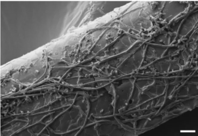
Genetic approaches have allowed fundamental insights into almost all areas of microbial pathogenesis research. Yet, today, such methodologies have only rarely been established in dermatophytes, in contrast to other clinically important fungal pathogens, for example Candida albicans, Aspergillus fumigatus or Cryptococcus neoformans. Consequently, little is known about the pathogenicity of dermatophytes at the molecular level. Dermatophytes constitute a group of highly specialized fila-mentous fungi that share the peculiar ability to digest and grow on keratinized host structures such as skin stratum corneum, hair and nails (Fig. 1) (Ajello, 1974). Keratin utilization by these microorganisms as the sole carbon and nitrogen source has been linked to extracellular proteolysis, and a large number of secreted proteases were identified in different dermatophyte species (reviewed in Monod, 2008). Despite these major efforts, however, the role of individual proteases during infection remains almost elusive. Moreover, dermatophyte pathogenicity likely tends to be more complex and involves fungal mechan-isms that still have to be identified. At the same time, it appears to be of particular note that the adaptation of dermatophytes to
specific host niches is associated with variable clinical signs, i.e. chronic vs. inflammatory disease, suggesting distinct, almost unknown pathophysiological reactions. Therefore, studies on dermatophyte pathogenicity focus not only on fungal attributes but also on host immune response mechanisms (Vermout et al., 2008; Brasch, 2009).
Comprehensive up-to-date review articles covering der-matophyte epidemiology and clinical importance as well as genetic approaches in taxonomy and diagnosis are already available (Binstock, 2007; Abdel-Rahman, 2008; Gr¨aser et al., 2008; Kanbe, 2008; Seebacher et al., 2008; Ameen, 2010). These topics will not be a part of the present overview. Nevertheless, some basic information on species diversity and medical impact will be provided in order to better convey the recent achievements in molecular genetic research in this fascinating group of microorganisms.
Dermatophytes: clinical importance
and taxonomy
Dermatophytoses belong to the most common infectious diseases in humans, affecting 10–20% of the population
MICR
worldwide. These infections are usually not life threatening, but occur even in immunocompetent hosts, and in many cases, are long lasting, recurrent and difficult to cure (Borgers et al., 2005). Depending on their predominant natural reser-voir, dermatophyte species are classified into three groups: anthropophilic, zoophilic and geophilic (Weitzman & Sum-merbell, 1995). The natural hosts of anthropophilic and zoophilic species are humans and animals, respectively, whereas geophilic dermatophytes are soil saprophytes. Symp-toms of dermatophytosis can vary from chronic to highly inflammatory, depending on the causative agent and the body location affected. The given disease is described with the word ‘tinea,’ followed by a term referring to the infected body site, for example tinea pedis (feet), tinea capitis (scalp or head), tinea corporis (body or trunk) and tinea unguium (nails, also called onychomycosis) (Degreef, 2008). Major prominent anthropophilic species, for example, Trichophyton rubrum, Trichophyton interdigitale and Trichophyton tonsurans, are mostly associated with more chronic, less inflammatory infections. In contrast, zoophilic species, for example, Micro-sporum canis, Arthroderma benhamiae, Arthroderma vanbreu-seghemii, Trichophyton erinacei and Trichophyton verrucosum as well as geophilic dermatophytes such as Microsporum gypseum often induce highly inflamed lesions in humans.
Dermatophytes are ascomycete fungi. The anamorphs (asexual forms) are classified into three genera: Trichophy-ton, Microsporum and Epidermophyton. Teleomorphs (sexual forms) belong to the Arthroderma genus in the Ascomyco-tina subphylum. Dermatophytes are heterothallic (mating types are designated as either ‘1’ or ‘ ’); however, in many zoophilic and anthropophilic species, sexual reproduction has not been observed. Recent progress in molecular taxon-omy and insights into mating revealed that Trichophyton mentagrophytes was a complex of anthropophilic and zoo-philic species that produce different teleomorphs, leading to
a current confusion in species denomination. For example, A. benhamiae is the teleomorph obtained by mating isolates from rodents (Ajello & Cheng, 1967), whereas A. vanbreuse-ghemii is the teleomorph from strains isolated from humans and certain rodents (Takashio, 1979). Both zoophilic species A. benhamiae and A. vanbreuseghemii cause highly inflam-matory tinea capitis, tinea corporis and tinea faciei. They are designated T. mentagrophytes and T. mentagrophytes var. asteroides in many textbooks and publications. The anthro-pophilic strains of the T. mentagrophytes species complex produce noninflammatory tinea pedis and tinea unguium. Sexual reproduction has not been observed and the fungus is still called by the anamorph name T. interdigitale (or T. mentagrophytes var. interdigitale) (Symoens et al., 2011). Therefore, the formerly widely used species description, T. mentagrophytes, should nowadays only be used for isolates referring to the reference strain designated as a neotype (Gr¨aser et al., 1999). This hint appears to be noteworthy, because many of the genetic studies in dermatophytes were performed using species of the T. mentagrophytes complex, i.e. A. benhamiae and A. vanbreuseghemii. How-ever, in the case of the latter species, the name T. menta-grophytes was used (e.g. Yamada et al., 2005, 2008, 2009a, b; Alshahni et al., 2011).
Broad-scale gene discovery in
dermatophytes
Transcriptional profiling
Broad-scale gene discovery by differential cDNA analysis, expressed sequence tag (EST) sequencing and cDNA-based microarrays allows global insights into cellular adaptation at the level of gene expression. In dermatophytes, such techniques were recently established and revealed the transcriptional response of these fungi under different biologically interesting and also pathogenicity-related con-ditions. A comprehensive T. rubrum Expression Database was launched by Wang et al. (2004, 2006), offering a platform for ESTs and cDNA microarray-based transcrip-tional profiles (http://www.mgc.ac.cn/TrED/). Documented in a number of publications, this approach resulted in the identification of T. rubrum genes, whose expression is linked to distinct developmental growth phases or the presence of selected drugs (Liu et al., 2007; Yang et al., 2007; Yu et al., 2007; Zhang et al., 2007, 2009). Broad transcriptional analyses were also performed in our work on T. rubrum and A. benhamiae, with a focus on genes putatively impli-cated in extracellular proteolysis. Herein, ESTs from T. rubrum grown on protein as the sole carbon and nitro-gen source were analysed and used for the construction of a cDNA microarray containing at least 23 protease genes (Zaugg et al., 2009). Major dermatophyte-secreted
Fig. 1. Scanning electron micrograph of the dermatophyte Arthroder-ma benhamiae colonizing huArthroder-man hair. Sterilized hair was infected with A. benhamiae microconidia and incubated for 14 days at 30 1C. Scale bar = 10 mm.
keratinases have been known before and were correlated with the degradation of hard compact keratin (for a review, see Monod, 2008). Notably, dermatophytes were shown to secrete multiple serine proteases of the subtilisin family (Sub) as well as metalloproteases of the fungalysin family (Mep) [S8 and M36 family, respectively, in the MEROPS proteolytic enzyme database (http://merops.sanger.ac.uk)]. Microarray analysis during the growth of T. rubrum or A. benhamiae on soy and keratin protein confirmed the activation of particular SUB and MEP genes as well as genes encoding secreted exoproteases such as leucine aminopepti-dases and dipeptidylpeptiaminopepti-dases. In addition, other specifi-cally induced factors playing a potential role in protein utilization were identified, including heat shock proteins, various transporters, metabolic enzymes, transcription fac-tors and hypothetical proteins with unknown functions (Zaugg et al., 2009; Staib et al., 2010). Similar approaches were also supported by the analysis of suppression subtractive hybridization libraries, applied for the identification of novel dermatophyte genes specifically expressed by T. rubrum cells upon contact with keratin, in response to varying pH or to other environmental stimuli (Kaufman et al., 2005; Baeza et al., 2007; Maranhao et al., 2007, 2009; Peres et al., 2010; Silveira et al., 2010). A comparative transcriptional analysis in the two closely related species T. tonsurans and Trichophyton equinum detected differential, species-specific expression levels of selected genes encoding secreted proteases upon growth on keratin (Preuett et al., 2010).
Gene expression profiling inA. benhamiae
during infection
In order to unravel pathogenicity-related adaptation me-chanisms of dermatophytes during infection, we explored the transcriptional response of the fungal cells in an animal model. For this approach, the zoophilic dermatophyte A. benhamiae was selected as an appropriate species for several reasons (Fig. 2). Arthroderma benhamiae is zoophilic and causes inflammatory cutaneous infections not only in hu-mans but also in guinea-pigs, allowing the establishment of an animal model (Staib et al., 2010). Under laboratory conditions, A. benhamiae grows relatively fast and produces abundant microconidia, single-nucleated round-oval cells that are useful for transformation. Cleistothecia formation further facilitates genetic analyses and allows to shed light on the basis of sexual development in dermatophytes. As a major additional prerequisite, the genome of our A. benha-miae strain, which had been isolated from a patient with highly inflammatory tinea faciei (Fumeaux et al., 2004), has recently been decoded and annotated (Burmester et al., 2011) (Fig. 2). Transcriptional analysis in A. benhamiae cells isolated during experimental cutaneous infection of guinea-pigs uncovered a distinct protease gene expression profile, which is essentially different from the pattern displayed during in vitro growth on keratin. Most notably, a differ-ential expression of genes coding for members of the Sub and Mep protease families was detected. Instead of the
Guinea-pig infection model available Hyphae Microconidia Macroconidia Human infection Molecular tools Cleistothecia sexual reproduction Haploid genome sequenced Fig. 2. Summary of the basic characteristics that
make Arthroderma benhamiae a useful model for molecular research in dermatophytes. The centre shows a typical colony of A. benhamiae on Sabouraud glucose agar after 5 days of growth at 30 1C. Detailed explanations are given in the text.
major keratinase genes expressed in vitro, others were activated specifically during infection, suggesting functions that are not necessarily associated with the degradation of keratin. Future studies will address the strong in vivo activation of the gene encoding the serine protease Sub6, a known major allergen in the related dermatophyte T. rubrum. The broad A. benhamiae in vivo gene expression profile further revealed other putatively pathogenicity-related factors, whose role has to be studied by straightfor-ward functional analysis. Other interesting, putatively pathogenicity-related dermatophyte genes have been identi-fied recently in a broad transcriptome approach in A. benhamiae during the interaction with human keratino-cytes (Burmester et al., 2011).
Transformation and gene targeting in
dermatophytes
Transformation and selection markers
In comparison with many other fungi, dermatophytes have been shown to be less amenable to genetic manipulation. As a result, site-directed mutagenesis in dermatophyte species has been evidenced only in a very small number of cases. This drawback is assumed to be a result of both low transformation frequency and inefficient homologous inte-gration, processes that are indispensable for targeted genetic manipulations. The first successful transformation of a dermatophyte has been described in 1989 by Gonzalez et al. (1989) in T. mentagrophytes (Table 1). The transformation protocol applied was based on a standard protoplast/poly-ethylene glycol (PEG)-mediated procedure that has been established widely in filamentous fungi, for example
Asper-gillus nidulans, Neurospora crassa and others (for a review, see Fincham, 1989; Weld et al., 2006). As a marker for the selection of T. mentagrophytes transformants, the system used the bacterial hygromycin B phosphotransferase gene hph. Plasmid DNA was stably integrated into the fungal genome with varying integration sites and numbers of insertions in the resulting transformants. Thereafter, no further attempts on dermatophyte transformation have been reported until 2004, when Kaufman et al. (2004) described PEG-mediated protoplast transformation and restriction-enzyme-mediated integration in T. mentagro-phytes, using the hph gene as a selectable marker and the gene encoding the enhanced green fluorescent protein (eGFP) as a reporter. PEG-mediated transformation and transformant selection via hygromycin resistance was further demonstrated in M. canis (Yamada et al., 2005, 2006; Vermout et al., 2007) and T. rubrum (Fachin et al., 2006; Ferreira-Nozawa et al., 2006). Different other drugs/ dominant markers have meanwhile been proven successful for the selection of transformants in T. mentagrophytes, i.e. two other aminoglycoside antibiotics/resistance genes, nourseothricin/Streptomyces noursei nourseothricin acetyl-transferase gene nat1 (Alshahni et al., 2010) and geneticin (G-418)/Escherichia coli neomycin phosphotransferase gene neo (Yamada et al., 2008). The latter marker as well as hph were also used successfully in A. benhamiae (Grumbt et al., 2011). Besides PEG-mediated protoplast transformation, other techniques facilitating gene transfer were also mean-while adopted in dermatophytes. A promising Agrobacter-ium tumefaciens-mediated transformation (ATMT) system was established recently for T. mentagrophytes (Yamada et al., 2009b). ATMT has already strongly advanced func-tional genomics in various filamentous fungi before (for a
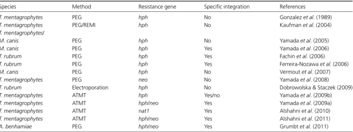
Table 1. Chronological overview of successful genetic transformation experiments in dermatophytes
Species Method Resistance gene Specific integration References
T. mentagrophytes PEG hph No Gonzalez et al. (1989) T. mentagrophytes PEG/REMI hph No Kaufman et al. (2004) T. mentagrophytes/
M. canis PEG hph No Yamada et al. (2005)
M. canis PEG hph Yes Yamada et al. (2006)
T. rubrum PEG hph Yes Fachin et al. (2006)
T. rubrum PEG hph Yes Ferreira-Nozawa et al. (2006)
M. canis PEG hph No Vermout et al. (2007)
T. mentagrophytes PEG neo No Yamada et al. (2008) T. rubrum Electroporation hph No Dobrowolska & Staczek (2009) T. mentagrophytes ATMT hph Yes/no Yamada et al. (2009b) T. mentagrophytes ATMT hph/neo Yes Yamada et al. (2009a) T. mentagrophytes ATMT nat1 Yes Alshahni et al. (2010) T. mentagrophytes ATMT hph/neo Yes Alshahni et al. (2011) A. benhamiae PEG hph/neo Yes Grumbt et al. (2011)
REMI, restriction-enzyme-mediated integration; hph, hygromycin phosphotransferase gene; neo, neomycin phosphotransferase gene; nat1, nourseothricin acetyltransferase gene.
review, see Michielse et al., 2005). Notably, Yamada and colleagues used the system for both random integration of T- (transferred) DNA and targeted insertion, for example disruption of the areA/nit-2 gene. As another alternative transformation technique, electroporation of germinated conidia was applied in T. rubrum, allowing the random integration of hph and eGFP (Dobrowolska & Staczek, 2009). Although not many comparative data on transforma-tion efficiency are available – some species have not even been addressed at all – different dermatophyte species appear to be more or less amenable to DNA uptake and/or stable integration. Therefore, transformation protocols es-tablished for a selected species are not necessarily transfer-able to another, but require precise modifications. From our own work, we know for example that our standard PEG-protocol for the efficient transformation of A. benhamiae was not directly applicable for T. rubrum or M. canis. The reasons for this observation are likely multifactorial, includ-ing differential protoplast stability, cell wall composition, microconidia production, etc.
Targeted gene inactivation
Filamentous fungi are known to only poorly support site-directed insertion of linear DNA cassettes in the genome by homologous recombination, in contrast to yeasts such as Saccharomyces cerevisiae or the opportunistic pathogen C. albicans. Therefore, in filamentous fungi, identification of transformants with a desired genetic alteration has proven laborious in many cases. In order to circumvent this obstacle, parental strains were generated in diverse species that lack the nonhomologous end joining (NHEJ) recombi-nation pathway, for example in N. crassa (Ninomiya et al., 2004), Aspergillus spp. (da Silva Ferreira et al., 2006; Krappmann et al., 2006; Nayak et al., 2006), and since recently, also in T. mentagrophytes (Yamada et al., 2009a) and A. benhamiae (Grumbt et al., 2011) (Table 1). Mutants deficient in NHEJ processes allow a strongly increased frequency of targeted insertions; however, an altered risk of unforeseen genetic variations cannot be excluded. In derma-tophyte species, only a small number of genes have so far been analysed by targeted inactivation, for example pacC and MDR2 in T. rubrum (Fachin et al., 2006; Ferreira-Nozawa et al., 2006), Ku80, areA and Trim4 in T. mentagrophytes (Yamada et al., 2009a, b), areA in M. canis (Yamada et al., 2006) and Ku70 and AcuE in A. benhamiae (Grumbt et al., 2011). Interestingly, A. benhamiae has been shown in our work to allow efficient targeted gene deletion not only in a ku70 mutant background but also in the wild-type strain. This has been demonstrated by the construction of mutants in malate synthase AcuE, KU70 and other candidates (Grumbt et al., 2011; M. Grumbt and P. Staib, unpublished data). The use of two different dominant
selection markers, hph and neo, even allowed for the first time the site-directed complementation of knockout mutant strains. Because the deletion of KU70 had no adverse effect on the virulence of A. benhamiae in a guinea-pig infection model, both the wild type and the ku70 mutant appear to be suitable parental strains for future pathogenicity research. In general, isogenic strain construction is assumably facilitated in species such as A. benhamiae and T. mentagrophytes by the fact that they easily allow the production of abundant single nucleated cells in the form of microconidia as a starting material.
RNA silencing
RNA interference, originally described in the nematode Caenorhabditis elegans, is based on a cellular process by which an introduced double-stranded RNA induces the degradation of specific mRNAs of interest (Fire et al., 1998). RNA silencing was widely applied as an efficient tool to address gene function in multiple research areas, espe-cially when conventional site-directed gene inactivation is difficult or, due to knockout lethality, impossible. As an-other advantage, the technique offers the possibility to inhibit several genes at the same time, a characteristic that might be useful for the functional analysis of homologous genes within large families, for example those encoding secreted endoproteases in dermatophytes. Here, the system was first established by Vermout et al. (2007) by the construction of M. canis transformants in which the expres-sion of genes encoding secreted proteases Sub3 and dipepti-dyl peptidase IV, respectively, was suppressed. Using the SUB3 RNA-silenced strain, the authors revealed a contribu-tion of this protease in the adherence of M. canis to feline epidermis, whereas a function in epidermal invasion and virulence of the fungus during cutaneous guinea-pig infec-tion was not assigned (Baldo et al., 2010).
Genome sequencing projects
Given the fact that powerful tools have meanwhile become available for the genetic manipulation of dermatophytes, the advent of dermatophyte genome sequencing projects now offers a fundamental basis for future research. Annotated genome sequences of seven different dermatophyte species have become available recently (http://www.broadinstitute. org/annotation/genome/dermatophyte_comparative/Multi Home.html), provided by projects headed by the Broad Institute (Cambridge) and the Hans Knoell Institute (Jena, Germany), respectively. The latter institution has recently published the first report on dermatophyte genomes, pre-senting a comparative study on the two closely related zoophilic, human pathogenic species A. benhamiae (major reservoir are guinea-pigs) and T. verrucosum (major
reservoir are cattle) (Burmester et al., 2011). The genome sequences identified were compared not only with each other but also with those of other species of the Onygenales, i.e., Coccidioides posadasii and Coccidioides immitis, and with the mould A. fumigatus. The 22–23 Mb genomes of A. benhamiae and T. verrucosum, containing 7980 and 8024 predicted protein-encoding genes, respectively, were found to be smaller than those of Aspergillus (e.g. 28 and 37.3 Mb for Aspergillus clavatus and Aspergillus niger, respectively), Coccidioides spp. (27–29 Mb) or Histoplasma capsulatum (30–39 Mb). Special attention was paid not only to the analysis of genes that are putatively associated with host adaptation, for example genes encoding secreted proteases. Genes involved in the biosynthesis of secondary metabolites and mating were also found to be of future interest (Burmester et al., 2011). Additional insights are expected from the envisaged genome comparison including the other five sequenced human pathogenic dermatophyte species. The species selection was based on different biological parameters and pathogenicity-related hypotheses (White et al., 2008), and the basic traits of the selected strains such as growth rate and resistance to diverse antibiotics were already monitored (Achterman et al., 2011). Because these species encompass anthropophilic (T. rubrum, the most common inducer of dermatophytosis in humans worldwide; T. tonsurans, often associated with tinea capitis in America), zoophilic (T. equinum, associated with horses; M. canis, associated with cats and dogs) and geophilic (M. gypseum) dermatophytes, a comparative genome analysis will, among other topics, address factors that are potentially involved in host preference, adaptation during chronic vs. inflammatory infection and saprophytic growth.
Conclusion
An increasing, lively interest in the molecular biology of dermatophytes combined with the establishment of fundamental genetic approaches has strongly advanced the research in these filamentous fungi. Basic prerequisites have been launched, such as genome sequencing projects, expression profile data sets and efficient targeted gene inactivation techniques. Nevertheless, molecular research is still preliminary in these genetically less amenable micro-organisms. Therefore, further efforts have to be undertaken for the improvement of existing and the establishment of additional genetic tools and methodologies. Such efforts will be worthwhile, given the fact that dermatophytoses are widespread and of particular clinical interest. Using the available techniques, now fundamental questions can be addressed in dermatophytes, related to the patho-genicity as well as general host and environmental adaptation mechanisms, sexual development, basic biology and evolution.
Acknowledgements
We are sorry that space limitations did not allow us to cite all important papers. We thank Axel A. Brakhage, Christoph Heddergott and the electron microscopy centre at the University Hospital Jena for providing the scanning electron micrograph in Fig. 1, and Bernard Mignon for the photo-graph visualizing the guinea-pig animal model in Fig. 2. Work in our laboratory is supported by the Deutsche Forschungsgemeinschaft and the Hans Knoell Institute.
References
Abdel-Rahman SM (2008) Strain differentiation of dermatophytes. Mycopathologia 166: 319–333. Achterman RR, Smith AR, Oliver BG & White TC (2011)
Sequenced dermatophyte strains: growth rate, conidiation, drug susceptibilities, and virulence in an invertebrate model. Fungal Genet Biol 48: 335–341.
Ajello L (1974) Natural history of the dermatophytes and related fungi. Mycopath Mycol Appl 53: 93–110.
Ajello L & Cheng SL (1967) The perfect state of Trichophyton mentagrophytes. Sabouraud 5: 230–234.
Alshahni MM, Makimura K, Yamada T, Takatori K & Sawada T (2010) Nourseothricin acetyltransferase: a new dominant selectable marker for the dermatophyte Trichophyton mentagrophytes. Med Mycol 48: 665–668.
Alshahni MM, Yamada T, Takatori K, Sawada T & Makimura K (2011) Insights into a nonhomologous integration pathway in the dermatophyte Trichophyton mentagrophytes: efficient targeted gene disruption by use of mutants lacking ligase IV. Microbiol Immunol 55: 34–43.
Ameen M (2010) Epidemiology of superficial fungal infections. Clin Dermatol 28: 197–201.
Baeza LC, Bailao AM, Borges CL, Pereira M, Soares CM & Mendes Giannini MJ (2007) cDNA representational difference analysis used in the identification of genes expressed by Trichophyton rubrum during contact with keratin. Microbes Infect 9: 1415–1421.
Baldo A, Mathy A, Tabart J et al. (2010) Secreted subtilisin Sub3 from Microsporum canis is required for adherence to but not for invasion of the epidermis. Brit J Dermatol 162: 990–997. Binstock JM (2007) Molecular biology techniques for identifying
dermatophytes and their possible use in diagnosing onychomycosis in human toenail: a review. J Am Podiat Med Assn 97: 134–144.
Borgers M, Degreef H & Cauwenbergh G (2005) Fungal infections of the skin: infection process and antimycotic therapy. Curr Drug Targets 6: 849–862.
Brasch J (2009) Current knowledge of host response in human tinea. Mycoses 52: 304–312.
Burmester A, Shelest E, Glockner G et al. (2011) Comparative and functional genomics provide insights into the pathogenicity of dermatophytic fungi. Genome Biol 12: R7.
da Silva Ferreira ME, Kress MR, Savoldi M et al. (2006) The akuB(KU80) mutant deficient for nonhomologous end joining is a powerful tool for analyzing pathogenicity in Aspergillus fumigatus. Eukaryot Cell 5: 207–211.
Degreef H (2008) Clinical forms of dermatophytosis (ringworm infection). Mycopathologia 166: 257–265.
Dobrowolska A & Staczek P (2009) Development of transformation system for Trichophyton rubrum by electroporation of germinated conidia. Curr Genet 55: 537–542.
Fachin AL, Ferreira-Nozawa MS, Maccheroni W Jr & Martinez-Rossi NM (2006) Role of the ABC transporter TruMDR2 in terbinafine, 4-nitroquinoline N-oxide and ethidium bromide susceptibility in Trichophyton rubrum. J Med Microbiol 55: 1093–1099.
Ferreira-Nozawa MS, Silveira HC, Ono CJ, Fachin AL, Rossi A & Martinez-Rossi NM (2006) The pH signaling transcription factor PacC mediates the growth of Trichophyton rubrum on human nail in vitro. Med Mycol 44: 641–645.
Fincham JR (1989) Transformation in fungi. Microbiol Rev 53: 148–170.
Fire A, Xu S, Montgomery MK, Kostas SA, Driver SE & Mello CC (1998) Potent and specific genetic interference by double-stranded RNA in Caenorhabditis elegans. Nature 391: 806–811. Fumeaux J, Mock M, Ninet B et al. (2004) First report of
Arthroderma benhamiae in Switzerland. Dermatology 208: 244–250.
Gonzalez R, Ferrer S, Buesa J & Ramon D (1989) Transformation of the dermatophyte Trichophyton mentagrophytes to
hygromycin B resistance. Infect Immun 57: 2923–2925. Gr¨aser Y, El Fari M, Vilgalys R, Kuijpers AF, De Hoog GS, Presber
W & Tietz H (1999) Phylogeny and taxonomy of the family Arthrodermataceae (dermatophytes) using sequence analysis of the ribosomal ITS region. Med Mycol 37: 105–114.
Gr¨aser Y, Scott J & Summerbell R (2008) The new species concept in dermatophytes – a polyphasic approach. Mycopathologia 166: 239–256.
Grumbt M, Defaweux V, Mignon B, Monod M, Burmester A, W¨ostemeyer J & Staib P (2011) Targeted gene deletion and in vivo analysis of putative virulence gene function in the pathogenic dermatophyte Arthroderma benhamiae. Eukaryot Cell, DOI: 10.1128/EC.00273-10.
Kanbe T (2008) Molecular approaches in the diagnosis of dermatophytosis. Mycopathologia 166: 307–317.
Kaufman G, Horwitz BA, Hadar R, Ullmann Y & Berdicevsky I (2004) Green fluorescent protein (GFP) as a vital marker for pathogenic development of the dermatophyte Trichophyton mentagrophytes. Microbiology 150: 2785–2790.
Kaufman G, Berdicevsky I, Woodfolk JA & Horwitz BA (2005) Markers for host-induced gene expression in Trichophyton dermatophytosis. Infect Immun 73: 6584–6590.
Krappmann S, Sasse C & Braus GH (2006) Gene targeting in Aspergillus fumigatus by homologous recombination is facilitated in a nonhomologous end-joining-deficient genetic background. Eukaryot Cell 5: 212–215.
Liu T, Zhang Q, Wang L et al. (2007) The use of global transcriptional analysis to reveal the biological and cellular events involved in distinct development phases of Trichophyton rubrum conidial germination. BMC Genomics 8: 100. Maranhao FC, Paiao FG & Martinez-Rossi NM (2007) Isolation
of transcripts over-expressed in human pathogen Trichophyton rubrum during growth in keratin. Microb Pathogenesis 43: 166–172.
Maranhao FC, Paiao FG, Fachin AL & Martinez-Rossi NM (2009) Membrane transporter proteins are involved in Trichophyton rubrum pathogenesis. J Med Microbiol 58: 163–168. Michielse CB, Hooykaas PJ, van den Hondel CA & Ram AF
(2005) Agrobacterium-mediated transformation as a tool for functional genomics in fungi. Curr Genet 48: 1–17. Monod M (2008) Secreted proteases from dermatophytes.
Mycopathologia 166: 285–294.
Nayak T, Szewczyk E, Oakley CE et al. (2006) A versatile and efficient gene-targeting system for Aspergillus nidulans. Genetics 172: 1557–1566.
Ninomiya Y, Suzuki K, Ishii C & Inoue H (2004) Highly efficient gene replacements in Neurospora strains deficient for nonhomologous end-joining. P Natl Acad Sci USA 101: 12248–12253.
Peres NT, Sanches PR, Falcao JP et al. (2010) Transcriptional profiling reveals the expression of novel genes in response to various stimuli in the human dermatophyte Trichophyton rubrum. BMC Microbiol 10: 39.
Preuett BL, Schuenemann E, Brown JT, Kovac ME, Krishnan SK & Abdel-Rahman SM (2010) Comparative analysis of secreted enzymes between the anthropophilic–zoophilic sister species Trichophyton tonsurans and Trichophyton equinum. Fungal Biol 114: 429–437.
Seebacher C, Bouchara JP & Mignon B (2008) Updates on the epidemiology of dermatophyte infections. Mycopathologia 166: 335–352.
Silveira HC, Gras DE, Cazzaniga RA, Sanches PR, Rossi A & Martinez-Rossi NM (2010) Transcriptional profiling reveals genes in the human pathogen Trichophyton rubrum that are expressed in response to pH signaling. Microb Pathogenesis 48: 91–96.
Staib P, Zaugg C, Mignon B et al. (2010) Differential gene expression in the pathogenic dermatophyte Arthroderma benhamiae in vitro versus during infection. Microbiology 156: 884–895.
Symoens F, Jousson O, Planard C, Fratti M, Staib P, Mignon B & Monod M (2011) Molecular analysis and mating behaviour of the Trichophyton mentagrophytes species complex. Int J Med Microbiol 301: 260–266.
Takashio M (1979) Taxonomy of dermatophytes based on their sexual states. Mycologia 71: 968–976.
Vermout S, Tabart J, Baldo A, Monod M, Losson B & Mignon B (2007) RNA silencing in the dermatophyte Microsporum canis. FEMS Microbiol Lett 275: 38–45.
Vermout S, Tabart J, Baldo A, Mathy A, Losson B & Mignon B (2008) Pathogenesis of dermatophytosis. Mycopathologia 166: 267–275.
Wang L, Ma L, Leng W et al. (2004) Analysis of part of the Trichophyton rubrum ESTs. Sci China Ser C 47: 389–395. Wang L, Ma L, Leng W et al. (2006) Analysis of the dermatophyte
Trichophyton rubrum expressed sequence tags. BMC Genomics 7: 255.
Weitzman I & Summerbell RC (1995) The dermatophytes. Clin Microbiol Rev 8: 240–259.
Weld RJ, Plummer KM, Carpenter MA & Ridgway HJ (2006) Approaches to functional genomics in filamentous fungi. Cell Res 16: 31–44.
White TC, Oliver BG, Gr¨aser Y & Henn MR (2008) Generating and testing molecular hypotheses in the dermatophytes. Eukaryot Cell 7: 1238–1245.
Yamada T, Makimura K, Uchida K & Yamaguchi H (2005) Reproducible genetic transformation system for two dermatophytes, Microsporum canis and Trichophyton mentagrophytes. Med Mycol 43: 533–544.
Yamada T, Makimura K & Abe S (2006) Isolation,
characterization, and disruption of dnr1, the areA/nit-2-like nitrogen regulatory gene of the zoophilic dermatophyte, Microsporum canis. Med Mycol 44: 243–252.
Yamada T, Makimura K, Hisajima T, Ito M, Umeda Y & Abe S (2008) Genetic transformation of the dermatophyte, Trichophyton mentagrophytes, based on the use of G418
resistance as a dominant selectable marker. J Dermatol Sci 49: 53–61.
Yamada T, Makimura K, Hisajima T, Ishihara Y, Umeda Y & Abe S (2009a) Enhanced gene replacements in Ku80 disruption mutants of the dermatophyte, Trichophyton mentagrophytes. FEMS Microbiol Lett 298: 208–217. Yamada T, Makimura K, Satoh K, Umeda Y, Ishihara Y & Abe S
(2009b) Agrobacterium tumefaciens-mediated transformation of the dermatophyte, Trichophyton mentagrophytes: an efficient tool for gene transfer. Med Mycol 47: 485–494.
Yang L, Wang L, Peng J et al. (2007) Comparison between gene expression of conidia and germinating phase in Trichophyton rubrum. Sci China Ser C 50: 377–384.
Yu L, Zhang W, Wang L et al. (2007) Transcriptional profiles of the response to ketoconazole and amphotericin B in Trichophyton rubrum. Antimicrob Agents Ch 51: 144–153. Zaugg C, Monod M, Weber J et al. (2009) Gene expression
profiling in the human pathogenic dermatophyte
Trichophyton rubrum during growth on proteins. Eukaryot Cell 8: 241–250.
Zhang W, Yu L, Leng W et al. (2007) cDNA microarray analysis of the expression profiles of Trichophyton rubrum in response to novel synthetic fatty acid synthase inhibitor PHS11A. Fungal Genet Biol 44: 1252–1261.
Zhang W, Yu L, Yang J, Wang L, Peng J & Jin Q (2009) Transcriptional profiles of response to terbinafine in Trichophyton rubrum. Appl Microbiol Biot 82: 1123–1130.