Case report
Takotsubo cardiomyopathy associated with opiate
withdrawal
A. SARCON
1, J.-R. GHADRI
2, G. WONG
1, T.F. LU
¨ SCHER
2, C. TEMPLIN
2* and
E. AMSTERDAM
1*
From the
1Division of Cardiovascular Medicine, University of California (Davis) School of Medicine
and Medical Center, Sacramento, CA, USA and
2University Heart Center, Department of Cardiology,
University Hospital Zurich, Zurich, Switzerland
Address correspondence to Jelena-Rima Ghadri, MD, University Heart Center Zurich, Department of Cardiology, 8091 Zurich, Switzerland. email: [email protected]
*These authors contributed equally to this work.
Learning Point for Clinicians
Classically, takotsubo cardiomyopathy (TTC) or ‘broken-heart-syndrome’, describes the associ-ation of a trigger factor such as ‘death of a loved one’ with induction of a transient cardio-myopathy. However, the trigger factor can be any one of a diverse group of physical or emotional stressors. It has recently been reported that drug withdrawl, particularly from opiates, can also initiate TTC. Thus, prompt recognition of with-drawal-associated TTC is essential for appropriate management and optimal clinical outcome.
Case report
A 60-year-old woman with hepatitis C, major de-pression and chronic pain syndrome presented to the emergency department complaining of abdom-inal discomfort, chills, diaphoresis, nausea and emesis for 72 hr. Several days prior to presentation, she had run out of MS Contin 100 mg (qid), and because of her symptoms she had discontinued all her medications for 48 hr. Her cardiovascular risk factors were cigarette smoking and hypertension.
On admission, the patient was in distress, reiter-ating her need for morphine. Vital signs were blood pressure 141/89 mm Hg, pulse 117/min and
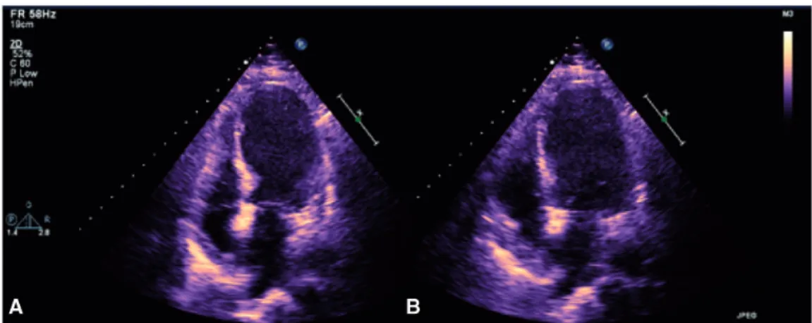
respiratory rate 24/min. Her chemistry panel and venous blood gases revealed a mixed acid base dis-order and liver function tests were normal. Shortly after she complained of sudden onset of chest pain and her electrocardiogram showed ST-segment vation in the precordial leads. Troponin I was ele-vated (21.12 ng/ml, ref <0.04). Echocardiography revealed left ventricular (LV) ejection fraction of 35% with apical akinesis and hypokinesis of the mid-anterior and mid-inferior segments (Figure 1). The patient received urgent cardiac catheterization, which revealed no significant coronary artery dis-ease. Left ventriculography demonstrated LV apical akinesis, basal hypercontractility, and LV ejection fraction of 40%, and LV end diastolic pressure of 29 mm Hg. These findings were consistent with takotsubo cardiomyopathy (TTC) and the patient was transferred to the cardiac care unit where a nitroprusside infusion was initiated and morphine was administered. The patient’s hospital course was complicated by pulmonary edema, which required intensive therapy including diuresis, vaso-dilators and brief ventilatory support. Over the next several days, therapy with an angiotensin converting enzyme inhibitor and beta-blocker was started. Morphine was progressively decreased and discon-tinued after <1 week. The patient’s condition stabi-lized over the next 3 weeks. By hospital day 27, she ! The Author 2013. Published by Oxford University Press on behalf of the Association of Physicians.
All rights reserved. For Permissions, please email: [email protected]
Q J Med 2014;107:301–302
was fully ambulatory, her echocardiogram had nor-malized, and her electrocardiogram had only re-sidual anterior T-wave inversion.
Discussion
TTC is characterized by transient, regional systolic dysfunction of the left ventricle without obstructive epicardial coronary artery disease. It predominantly affects postmenopausal women and is frequently triggered by an emotional or a physical stressor.1 TTC related to medication withdrawal has been re-ported in a diverse group of patients. Interestingly, Americans account for approximately 80% of the global consumption of prescription analgesics.2
The rising consumption of analgesic medications worldwide suggests a potential increase in the fre-quency of withdrawal-associated TTC.
The pathophysiology of TTC is not well under-stood, but excessive catecholamine and adrenergic stimulation have been implicated.3Drug withdrawal can be associated with a hyperadrenergic state and central nervous system irritability,4which may pro-vide a mechanism for withdrawal-associated TTC. We have identified seven additional cases of drug withdrawal-associated TTC in our International Takotsubo Registry at the leading hospital Zurich, Switzerland (www.takotsubo-registry.com). We sug-gest that withdrawal-associated TTC is under-diag-nosed due to limited awareness. The majority of
these patients was women with ST-segment eleva-tion and all demonstrated the LV apical ballooning pattern, with reduced ejection fraction of 36%. In-hospital complications occurred among our patients including one death. Our patient’s complex presen-tation involving opiate withdrawal and TTC was manifested by acute pulmonary edema and meta-bolic alkalosis, which prolonged her hospitalization. Recognition of the potential for patients to develope TTC during drug withdrawal to develop TTC is es-sential for appropriate management and favorable outcome.
Conflict of interest: None declared.
References
1. Merchant EE, Johnson SW, Nguyen P, Kang C, Mallon WK. Takotsubo cardiomyopathy: a case series and review of the literature. West J Emerg Med 2008;9:104–411.
2. Manchikanti L, Fellows B, Ailinani H, Pampati V. Therapeutic use, abuse, and nonmedical use of opioids: a ten-year perspective. Pain Physician 2010;13:401–35. 3. Wittstein IS, Thiemann DR, Lima JA, Baughman KL,
Schulman SP, Gerstenblith G, et al. Neurohumoral features of myocardial stunning due to sudden emotional stress. N Engl J Med352:539–48.
4. McDonald T, Hoffman WE, Berkowitz R, Cunningham F, Cooke B. Heart rate variability and plasma catecholamines in patients during opioid detoxification. J Neurosurg Anesthesiol 1999;11:195–9.
Figure 1. Echocardiography reveals left ventricular ejection fraction of 35% with apical ballooning pattern (A in systole and B in diastole).