Surgery of the dilated aortic root and ascending aorta in pediatric patients:
techniques and results
Thierry Carrel
a,*, Pascal Berdat
a, Mladen Pavlovic
b, Sergei Sukhanov
c,
Lars Englberger
a, Jean-Pierre Pfammatter
aaClinic for Cardiovascular Surgery, University Hospital Berne, CH-3010 Berne, Switzerland b
Division of Pediatric Cardiology, University Hospital Berne, CH-3010 Berne, Switzerland
c
The Perm Heart Institute, Perm, Russia
Received 9 February 2003; received in revised form 25 April 2003; accepted 26 April 2003
Abstract
Objective: Dilatation of the aortic root is a well-known cardiovascular manifestation in children and adult patients with connective tissue disease (e.g. Marfan syndrome). Dilatation of the ascending aorta is extremely rare and may be associated with bicuspid aortic valve. This report evaluates the incidence of dilatative aortic root and ascending aortic pathology in patients younger than 18 years and analyzes the results obtained after repair and replacement strategies. Methods: Between 1/1995 and 12/2002, a total of 752 operations on the thoracic aorta were performed in adult and pediatric patients. We present our experience with a group of 26 patients , 18 years of age, who required isolated surgery of the aortic root and/or ascending aorta because of a dilatative lesion. Fifteen patients had isolated aortic root dilatation (13 of them suffered from Marfan syndrome), eight patients presented with an idiopathic dilatation of the ascending aorta and three patients had dilatation in association with a bicuspid aortic valve. Mean age was 10 ^ 4.8 years (4 – 18 years). Repair of the aortic root with preservation of the aortic valve (Yacoub, David or selective sinus repair) was performed in nine patients, replacement using a homograft was performed in five patients, composite graft with mechanical prosthesis in two patients, with biological prosthesis in one patient and Ross operation was performed in one case. Isolated supracoronary graft replacement was performed in eight patients. Results: Two patients died during hospitalization: a 10-year old girl developed respiratory failure on the 2nd postoperative day and autopsy revealed Ehlers – Danlos syndrome with a massive intrapulmonary emphysema. A 14-year-old Marfan patient with severely depressed preoperative LV function died from low cardiac output following composite-graft, mitral and tricuspid valve repair. One patient required aortic valve replacement 7 days after an aortic valve sparing root repair. There was no additional perioperative morbidity. In the long-term, two patients died from rupture of the thoracic aorta, both following minor non-cardiovascular surgical procedures. Both had normal sized descending and abdominal aorta. Conclusion: Repair of the aortic root and/or ascending aorta in children and adolescent patients can be performed with acceptable early and late results. While the presence of severe comorbidity may adversely affect early outcome, long-term survival was mainly determined by rupture of the descending aorta.
q2003 Elsevier Science B.V. All rights reserved.
Keywords: Aortic root; Ascending aorta; Dilatation; Marfan syndrome; Surgical technique; Results
1. Introduction
The most frequent surgical procedures on the thoracic aorta in neonates, infants and children are performed to repair aortic coarctation and hypoplastic or interrupted aortic arch. Isolated dilatation of the aortic root and/or ascending aorta is a rare but well-known cardiovascular
manifestation, which is usually encountered in patients with underlying connective tissue diseases (e.g. Marfan syn-drome, Ehlers – Danlos and Turner syndrome) [1 – 4]. The dilatation of the aortic root may lead to secondary aortic valve regurgitation and exposes the patients to the risk of acute aortic dissection or rupture. Pediatric patients rarely present with primary aortic valve regurgitation; in this case, the main mechanism of valve regurgitation is dilatation of the aortic sinuses, of the aortic anulus or spreading of the commissures at the sino-tubular level [5,6]. Another small group of patients may suffer from dilatation of the
1010-7940/03/$ - see front matter q 2003 Elsevier Science B.V. All rights reserved. doi:10.1016/S1010-7940(03)00302-6
www.elsevier.com/locate/ejcts
* Corresponding author. Tel.: þ 41-31-632-23-75; fax: þ 41-31-632-44-43.
supracoronary ascending aorta, with or without the presence of a bicuspid aortic valve.
If morphology of the aortic valve leaflets is normal, aortic root repair includes complete excision of the pathological aortic tissue with preservation of the normal aortic valve, either by reimplantation into a prosthetic graft or by remodeling the aortic root[7 – 9]. Preservation of the native valve may have several advantages: excellent hemo-dynamic characteristics, avoidance of oral anticoagulation and some growth potential in younger patients. However, controversy still exists regarding the durability of aortic valve-sparing procedures especially in the context of Marfan syndrome, in which the fibrillin defect involves not only the aortic wall but also the aortic valve leaflets as well [10]. We present our experience with a group of 26 patients , 18 years of age, who required isolated surgery of the aortic root and/or ascending aorta because of a dilatative lesion.
2. Methods
Between 1/1995 and 12/2002, a total of 752 operations on the thoracic aorta were performed in adult and pediatric patients because of aneurysm or dissection. Procedures performed because of isolated coarctation, stenosis of the ascending aorta (e.g. Williams – Beuren syndrome), and hypoplastic or interrupted aortic arch were excluded. Twenty-six patients , 18 years of age required isolated surgery of the aortic root and/or ascending aorta because of a dilatative lesion during the same time period. Sixteen patients were male and 10 were female. Mean age was 10 ^ 4.8 years, ranging from 4 to 18 years.
Fifteen patients (13 of them had proven Marfan syndrome according to the Gent nosology[11]) had isolated aortic root dilatation. Eight patients presented with an idiopathic dilatation of the ascending aorta; three of them had had previous cardiovascular surgery: PDA closure in one, coarctation repair in one and repair of complete atrioventricular canal in one patient. Finally, three patients had a dilatation in association with bicuspid aortic valve, one of them had severe valvular aortic stenosis and the other two had a normal valve function (Table 1).
Surgical procedures are summarized in Table 2. Repair of the aortic root with preservation of the native valve was performed in nine patients (Yacoub repair [n ¼ 6], David repair [n ¼ 2] or selective one-sinus repair [n ¼ 1]). Replacement using a homograft was performed in five patients, composite graft with mechanical prosthesis in two patients (in one of them in association with mitral and tricuspid valve repair), composite graft with biological prosthesis in one patient. Pulmonary autograft transfer (Ross procedure) was performed in one young girl with severe valvular stenosis and poststenotic dilatationTable 2. Supracoronary graft replacement was performed in eight patients. In an 18-year-old girl who presented with
aneurysm of the ascending aorta, hypoplastic aortic arch and coarctation, a supracoronary graft repair was combined to an ascending-to-descending bypass to treat the complete pathology in one step (Fig. 1).
The majority of patients was completely asymptomatic from their disease. Children and adolescent patients with a proven Marfan syndrome and aortic root dilatation were followed with repeated echocardiographic examination aimed to assess the size and the evolution of the diameter of the aortic root and at different levels (mid-ascending, arch, proximal descending, thoraco-abdominal hiatus, abdominal). Five out of eight patients with idiopathic dilatation had their aortic disease discovered incidentally, mainly by conventional chest X-ray performed because of respiratory infection or minor thoracic trauma. The three others had had prior cardiovascular surgery and were discovered to have ascending aortic aneurysm during follow-up examination. Since there are no absolute values
Table 1
Patients characteristics
Mean age 10 ^ 4.8 years (4 – 18 years)
Gender (m/f) 16:10
Diagnosis
Marfan syndrome 13 Idiopathic dilatation (aortic root) 2 Idiopathic dilatation (ascending aorta) 8 Bicuspid aortic valve þ 3 supracoronary dilation
Mean aortic diameter (at the site of the lesion)
41 ^ 13 mm (34 – 71 mm)
Previous surgery 3
PDA closure 1
Coarctation repair 1 Repair complete av canal 1 Two patients were operated in Perm, Russia
Table 2
Surgical procedures
Mean cardiopulmonary bypass time
Aortic root repair or replacement (n ¼ 18) 105 ^ 22 min Supracoronary graft repair (n ¼ 8) 46 ^ 26 min Mean aortic cross-clamping time
Aortic root repair or replacement (n ¼ 18) 73 ^ 18 min Supracoronary graft repair (n ¼ 8) 29 ^ 14 min Deep hypothermic circulatory arrest time 8, 10, 11 min
(three cases)
Yacoub procedure of aortic root repair 6 mitral valve repair in 1
David procedure of aortic root repair 2 One sinus aortic root repair 1
Homograft 5
Mitral valve repair in 1
Composite graft (mechanical prosthesis) 2 Mitral and tricuspid valve repair in 1
Composite graft (biological prosthesis) 1 Pulmonary autograft (Ross) 1 Supracoronary aortic replacement 8
of aortic diameter in children and adolescents, we found repair or replacement of the aortic root or ascending aorta to be justified when the diameter of the dilated segment was more than twice the size of a ‘reference‘ diameter, obtained by the addition of the diameter at the level of the aortic arch and the diaphragmatic hiatus and divided by 2. Indication was also found justified when an increase in size of more than 5 – 8 mm during 6 – 12 months was observed.
3. Operative technique
Selection of the operative procedure was based on the location and extension of the disease and the mechanism and severity of aortic regurgitation if present. Patients with isolated dilatation of the ascending aorta and normal aortic anulus, aortic valve leaflets and aortic sinuses underwent isolated supracoronary graft replacement, the proximal. anastomosis being performed at the level of the sino-tubular junction (Fig. 2). In three out of eight patients, the distal anastomosis was performed in the open arch technique using a short period of deep hypothermic circulatory arrest to allow complete exclusion of the most cranial part of the dilated ascending aorta. Eight patients with dilated aortic root and Marfan syndrome received an aortic valve sparing operation. In one of them, dilatation was not limited to the aortic root but extended in the ascending aorta as well (Fig. 3a). In these patients the sinuses of Valsalva are excised, leaving a rim of approximately 3 mm of sinus wall adjacent to the aortic anulus. The size of the Dacron vascular graft is chosen to fit the diameter of the aorto – ventricular junction. The anulus is reinforced with interrupted sutures and a strip of pericardin. The proximal end of the graft is cut so that the edges conformed to the insertion line of the aortic valve leaflets, according to the technique described by Yacoub
[12]. Three running sutures are performed following closely the insertion of the leaflets in the anulus.Fig. 3b shows a re-implanted aortic valve before coronary anastomoses to the ascending aortic graft. The coronary artery ostia are reimplanted into their respective neoaortic sinus using a 6.0 polypropylene running suture.
In cases of isolated dilatation of the supracoronary aorta, the ascending aorta is transected at the level of the com-missures of the aortic valve and distally at an appropriate level to allow complete exclusion of the dilated segment.
Root replacement using a homograft was performed in five patients, a composite graft with mechanical prosthesis in two patients (the parents wanted to have the most defi-nitive repair with the lowest probability of redo-surgery), with biological prosthesis in one patient and a Ross opera-tion was performed in a 10-year-old girl with severe valvular aortic stenosis and poststenotic dilatation. Two patients underwent concomitant repair of the mitral valve (anuloplasty) and one had mitral and tricuspid valve repair. In all patients intraoperative transesophageal echocardiography was performed to assess the function of the preserved aortic valve and the quality of the mitral repair. In those patients who underwent aortic root replacement with homograft or as a Ross procedure, echocardiography was performed to demonstrate normal motion of the homograft leaflets and absence of significant regurgitation.
4. Results
Cardiopulmonary bypass time and aortic cross-clamping time were significantly longer in patients (n ¼ 18) who underwent repair or replacement of the aortic root (105 ^ 22 min, resp. 73 ^ 18 min) than in those (n ¼ 8)
with isolated supracoronary graft replacement
(46 ^ 26 min, resp. 29 ^ 14 min). Duration of deep hypothermic circulatory arrest at 16 – 18 8C was 8, 10 and 11 min, respectively.
Hospital mortality occurred in two cases: a 10-year-old girl died from respiratory failure on the 2nd post-operative day and post-mortem revealed a massive intra-pulmonary emphysema with rarified interalveolar septa and several of the typical characteristics of Ehler – Danlos syn-drome. A 14-year-old Marfan patient died after being extubated on postoperative day 2 from low cardiac output following composite-graft, mitral and tricuspid valve repair. He had severely reduced left ventricular function preopera-tively with an ejection fraction calculated at 30% in the presence of severe mitral regurgitation.
One patient required mechanical valve replacement 1 week after a valve-sparing procedure because of severe aortic valve regurgitation. Echocardiography showed a large eccentric aortic regurgitation jet and at redo-surgery, regurgitation was found to be caused by a tear in an aortic leaflet close to the commissure between the right
Fig. 1. Angio-CT scan of an 18-year-old girl with severe hypoplastic aortic arch, aortic coarctation and aneurysm of the ascending aorta (arterial pressure was normal due the large collaterals).
and non-coronary sinus. Except this major adverse event, there was no additional significant perioperative morbidity. In the long-term two patients died, most probably from distal aortic rupture; both acute events occurred in the early postoperative course following minor surgical procedures. One patient died after inguinal hernia repair and the second died several days after laser therapy of a large cervico-facial naevus flammeus. The first patient was under cumadines, whereas the second did not receive any anticoagulants.
Both had a normal sized descending and abdominal aorta. Unfortunately, autopsy was not allowed by the relatives but aortic rupture was suspected on the basis of the clinical presentation and thought to be related, at least in one case, to hypertensive episodes due to postoperative pains.
Following aortic surgery, continued meticulous echo-cardiographic and/or CT-scan follow-up were performed in all patients to evaluate the size of non-operated aortic segments, the function of the preserved aortic valve or of the homograft valve as well as beginning signs of deterioration. Aortic valve replacement was necessary in one patient who presented with severe calcification and retraction of the aortic cusps of a homograft 6 years postoperatively.
5. Comment
It is inappropriate to think of children and adolescent patients with the Marfan syndrome and those young patients with idiopathic dilatation of the aortic root and/or the
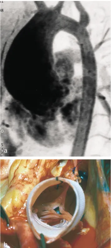
Fig. 2. (a) Severe supracoronary dilatation of the ascending aorta (7 cm) in a 12-year-old patient without aortic valve dysfunction. (b) Intraoperative situs following supracoronary graft replacement of the ascending aorta.
Fig. 3. Seventeen-year-old Marfan patient with atypical dilatation of the aortic root and the ascending aorta, and aortic regurgitation grade I. Intraoperative view following aortic root repair with reimplantation of the native aortic valve in a 22-mm vascular prosthesis.
ascending aorta as just smaller people with the same disease. There are special accommodations related to growth issues that have to be made in terms of diagnosis and management. Pediatric and adolescent patients presenting with aortic root dilatation with or without aortic valve regurgitation or isolated supracoronary dilatation of the ascending aorta usually demonstrate a progression of their disease [5,6]. Beneficial effects of beta-adrenergic blockers have been demonstrated in some subsets of patients [13]. Repair or replacement of the aortic root has been recognized as an established prophylactic procedure, which prevents aortic dissection and rupture as well as the development of severe aortic valve regurgitation [14,15]. Historically, standard surgical treatment has been replacement of the entire aortic root and the aortic valve with a composite valve graft including replantation of the coronary ostia [16]. The mechanical valve inserted in a composite graft is known for long-term durability but these young patients are exposed to a long-term risk of thromboembolism and oral anticoagulation. However, the majority of them will tolerate anticoagulation very well and they and their parents do not wish to accept the risk of another operation. Over the past 20 years, replacement of the aortic root with a composite graft prosthesis has proven to be a low risk operation in specialized centers, and indeed a very durable one[17,18]. The results of 20 years of composite graft with a mechanical valve are excellent and constitute the gold standard for patients with and without Marfan syndrome [1,15,17,18]. However, some patients have medical contraindications making indefinite anticoagulation inadvisable, others are not medically compliant enough for anticoagulation to be safe, some younger individuals have lifestyles making anticoagulation hazardous and some have an aversion to anticoagulation. In these circumstances, the aortic root and valve may be replaced with a cryopreserved homograft or with a xenograft aortic root. However, long-term durability of these substitutes is limited and may considerably differ inter-individually. Furthermore, the risk of reoperation is not as low as it may be at the initial operation because the homograft or xenograft may be densely calcified. Sternal reentry may be difficult, the coronary ostia may need to be reimplanted again and the aortic anulus may be excessively scarred. In our series, one patient had severe calcifications and retraction of the aortic leaflets and a mechanical valve was implanted within the homograft to simplify the redo-procedure. Another option for those patients who want to avoid anticoagulation is aortic root repair with preservation of the native aortic valve. The native aortic valve has been demonstrated to be a good alternative because the hemodynamics cannot be surpassed by those of any mechanical or biological prosthesis and the procedure does not require permanent anticoagulation [19]. Valve sparing aortic root replacement became popular in the 1990s as many patients with a normally functioning aortic valve wanted to avoid indefinite anticoagulation [7 – 9,20,21]. Unfortunately, the majority of published series do not
describe enough patients remaining at risk beyond 10 years to answer the question: how many years will the valve last before a second operation becomes necessary? Even in the personal experience of leading surgeons a substantial proportion of patients have required reoperation for aortic regurgitation while others have substantial regurgitation at follow-up, which will require reoperation. In the series of Vouhe´’s group, two patients required mechanical valve replacement 3 years after a Yacoub procedure [2]. In the present series, one patient had early acute failure of the aortic valve after a sparing procedure. The aortic valve was replaced during the same hospitalization.
Some cases with deteriorating valve function are probably due to ongoing dilatation of the aortic anulus or progressive disease of the aortic valve leaflets. In that sense, the reimplantation technique seems to produce less recurrence of aortic regurgitation in the long-term [20,21]. With this technique, the aortic graft is anchored at the ventriculo – aortic junction, but the valve is surrounded by the tubular graft without sinuses. Many variations of the David procedure have been designed to create aortic pseudo-sinuses in the graft, which have been described to be essential to prevent early valve failure. Anular reinforce-ment should be performed in every Marfan case. However, the role of ongoing pathology in the aortic leaflets of Marfan patients remains to be determined. In our experience, the adverse outcome and the problems encountered during the follow-up were not primarily determined by the pathology of the aortic root but mainly by a significant co-morbidity and by events in the more distal aortic segments.
The timing of surgery is crucial in these patients. While difficult to determine in some patients, it is related to the aortic diameter, the rate of progression of the dilatation and the function of the aortic valve.
In one of the patients in the present series, aneurysm of the ascending aorta was associated to a tubular hypoplastia of the aortic arch and a coarctation. The patient did not favor two operations with a different approach and did not accept the risk of deep hypothermic circulatory arrest. Angio-graphy showed a long extent of the hypoplastic arch and descending aortic and numerous large collateral vessels. For this reason, we decided to proceed with a single straight-forward intervention via sternotomy and performed a supracoronary graft replacement and an extra-anatomic ascending-to-descending aortic bypass[22,23].
6. Conclusion
In pediatric and adolescent patients suffering from a dilatation of the aortic root and/or ascending aorta, a surgical technique that does not require long-term couma-dine treatment (aortic valve sparing operation, selective sinus repair and supracoronary graft replacement) can be offered in the majority of cases. However, composite graft repair is still a good option for those who want to minimize
the probability of a second operation. Despite root repair or replacement, the long-term outcome may be complicated by unexpected events in the more distal aortic segments.
References
[1] Gott VL, Greene PS, Alejo DE, Cameron DE, Naftel DC, Miller DC. Replacement of the aortic root in patients with Marfan’s syndrome. N Engl J Med 1999;340:1307 – 13.
[2] Abdel-Massih T, Vouhe´ P, Mauriat P, Mousseaux E, Sidi D, Bonnet D. Replacement of the ascending aorta in children: a series of fourteen patients. J Thorac Cardiovasc Surg 2002;124:411– 3.
[3] Pfammatter JP, Pavlovic M, Berdat P, Carrel T. Dilatation of the ascending aorta in childhood: 4 cases without obvious predisposing disease. Cardiol Young 2001;11:169 – 72.
[4] Lin AE, Lippe B, Rosenfeld RG. Further delineation of aortic dilatation, dissection and rupture in patients with Turner syndrome. Pediatrics 1998;102:e12.
[5] Roman MJ, Devereux RB, Niles NW. Aortic root dilatation as a cause of isolated severe aortic regurgitation: prevalence, clinical and echocardiographic patterns and relation to left ventricular hypertrophy and function. Ann Intern Med 1987;106:800– 7.
[6] Bonderman D, Gharebhaghi-Schnell E, Wollenek G, Maurer G, Baumgartner H, Lang IM. Mechanisms underlying aortic dilatation in congenital aortic valve malformation. Circulation 1999;99:2138 – 43. [7] David TE, Feindel CM, Bos J. Repair of the aortic valve in patients with aortic insufficiency and aortic root aneurysms. J Thorac Cardiovasc Surg 1995;109:345 – 52.
[8] Scha¨fers HJ, Fries R, Langer F, Nikoloudakis N, Graeter T, Grundmann U. Valve preserving replacement of the ascending aorta—remodeling versus reimplantation. J Thorac Cardiovasc Surg 1998;116:990– 6.
[9] Birks EJ, Webb C, Child A, Radley-Smith R, Yacoub MH. Earlyx and long-term results of a valve-sparing operation for Marfan syndrome. Circulation 1999;100(Suppl. II):29 – 35.
[10] Gott VL, Laschinger JC, Cameron DE, Dietz HC, Greene PS, Gillinov AM, Pyeritz RE, Alejo DE, Fleischer KJ, Anhalt GJ, Stone CD,
McKusick VA. The Marfan syndrome and the cardiovascular surgeon. Eur J Cardiothorac Surg 1996;10:149 – 58.
[11] De Paepe A, Devereux RB, Dietz HC, Hennekam RC, Pyeritz RE. Revised diagnostic criteria for the Marfan syndrome. Am J Med Genet 1996;62:417– 26.
[12] Yacoub MH, Gehle P, Chandrasekaran V, Birks E, Child A, Radely-Smith R. Late results of a valve-sparing operation in patients with aneurysms of the ascending aorta and root. J Thorac Cardiovasc Surg 1998;115:1080 – 90.
[13] Shores J, Berger KR, Murphy EA, Pyeritz RE. Progression of aortic dilatation and the benefit of long-term (-adrenergic blockade in Marfan’s patients. N Engl J Med 1994;330:1335 – 41.
[14] Coady MA, Rizzo JA, Hammond GL. What is the appropriate size criterion for resection of thoracic aortic aneurysms? J Thorac Cardiovasc Surg 1997;113:476– 91.
[15] Kouchoukos NT, Dougenis D. Surgery of the thoracic aorta. N Engl J Med 1997;336:1876 – 88.
[16] Bentall H, de Bono A. A technique for complete replacement of the ascending aorta. Thorax 1968;23:338 – 9.
[17] Tambeur L, David TE, Unger M, Amstrong S, Ivanovf J, Wee G. Results of surgery for aortic root aneurysms in patients with the Marfan syndrome. Eur J Cardiothorac Surg 2000;17:415 – 9. [18] Miller DC. Valve-sparing aortic root replacement in patients with
Marfan syndrome. J Thorac Cardiovasc Surg 2003;125:773– 8. [19] de Oliveira NC, David TE, Ivanov J, Armstrong S, Eriksson MJ,
Rakouski H, Webb G. Results of surgery for aortic root aneurysm in patients with Marfan syndrome. J Thorac Cardiovasc Surg 2003;125: 789 – 96.
[20] Harringer W, Pethig J, Hagl C, Meyer GP, Haverich A. Ascending aortic replacement with aortic valve reimplantation. Circulation 1999; 100(Suppl. II):24– 9.
[21] David TE, Amstrong S, Ivanov J, Feindel CM, Omran A, Webb G. Results of aortic valve sparing operations. J Thorac Cardiovasc Surg 2001;122:39– 46.
[22] Berdat PA, Go¨ber V, Carrel T. Extra-anatomic aortic bypass for complex coarctation and hypoplastic aortic arch in adolescents and adults. Interactive Cardiothorac Surg (2003) in press.
[23] Carrel TP, Berdat PA. Ascending aortic aneurysim coarctation of the aortic isthmus and hypoplestic aortic arch: simultaneous treatment through stermotomy. Ann Thorac Surg 2003, in press.