. . . .
. . . .
BASIC SCIENCE
Targeting prolyl-isomerase Pin1 prevents
mitochondrial oxidative stress and vascular
dysfunction: insights in patients with diabetes
Francesco Paneni
1,2,3†, Sarah Costantino
1,2†, Lorenzo Castello
4, Rodolfo Battista
5,
Giuliana Capretti
4, Sergio Chiandotto
4, Domenico D’Amario
6, Giuseppe Scavone
7,
Angelo Villano
6, Alessandra Rustighi
8, Filippo Crea
6, Dario Pitocco
7, Gaetano Lanza
6,
Massimo Volpe
3,4, Giannino Del Sal
8, Thomas F. Lu¨scher
1, and Francesco Cosentino
1,2*
1
Cardiology and Cardiovascular Research, Institute of Physiology and University Hospital, Zu¨rich, Switzerland;2
Cardiology Unit, Department of Medicine, Karolinska University Hospital, Solna, 171 76 Stockholm, Sweden;3
IRCCS Neuromed, Pozzilli, Italy;4
Cardiology, Department of Clinical and Molecular Medicine, University of Rome ‘Sapienza’, Rome, Italy;5 Internal Medicine Unit, Civil Hospital, Sora, Italy;6
Department of Cardiovascular Medicine, Catholic University, Rome, Italy;7
Diabetes Care Unit, Internal Medicine, Catholic University, Rome, Italy; and8
Laboratorio Nazionale CIB, AREA Science Park and Department of Life Sciences, University of Trieste, Trieste, Italy Received 14 January 2014; revised 12 March 2014; accepted 31 March 2014; online publish-ahead-of-print 6 May 2014
This paper was handled by Stephanie Dimmeler (Prof. Johan-Wolfgang Goethe Universita¨t, [email protected]).
Aim Diabetes is a major driver of cardiovascular disease, but the underlying mechanisms remain elusive. Prolyl-isomerase Pin1
recognizes specific peptide bonds and modulates function of proteins altering cellular homoeostasis. The present study investigates Pin1 role in diabetes-induced vascular disease.
Methods and results
In human aortic endothelial cells (HAECs) exposed to high glucose, up-regulation of Pin1-induced mitochondrial
trans-location of pro-oxidant adaptor p66Shcand subsequent organelle disruption. In this setting, Pin1 recognizes Ser-116
inhibitory phosphorylation of endothelial nitric oxide synthase (eNOS) leading to eNOS – caveolin-1 interaction and reduced NO availability. Pin1 also mediates hyperglycaemia-induced nuclear translocation of NF-kB p65,
trigger-ing VCAM-1, ICAM-1, and MCP-1 expression. Indeed, gene silenctrigger-ing of Pin1 in HAECs suppressed p66Shc
-dependent ROS production, restored NO release and blunted NF-kB p65 nuclear translocation. Consistently,
diabetic Pin12/ 2mice were protected against mitochondrial oxidative stress, endothelial dysfunction, and vascular
in-flammation. Increased expression and activity of Pin1 were also found in peripheral blood monocytes isolated from diabetic patients when compared with age-matched healthy controls. Interestingly, enough, Pin1 up-regulation was
associated with impaired flow-mediated dilation, increased urinary 8-iso-prostaglandin F2aand plasma levels of
adhe-sion molecules.
Conclusions Pin1 drives diabetic vascular disease by causing mitochondrial oxidative stress, eNOS dysregulation as well as NF-kB-induced inflammation. These findings provide molecular insights for novel mechanism-based therapeutic strategies in patients with diabetes.
-Keywords Oxidative stress † Endothelial function † Inflammation † Diabetes mellitus
†F.P. and S.C. contributed equally to this work.
*Corresponding author. Tel:+46 851775398, Fax: +46 8344964, Email:[email protected]
Published on behalf of the European Society of Cardiology. All rights reserved.&The Author 2014. For permissions please email: [email protected].
European Heart Journal (2015) 36, 817–828 doi:10.1093/eurheartj/ehu179
Translational Perspective
The present study demonstrates that Pin1 is a common activator of key pathways involved in diabetic vascular disease in different experimental settings including primary human endothelial cells, knockout mice, and diabetic patients. Gene silencing and genetic disruption of Pin1 prevent hyperglycaemia-induced mitochondrial oxidative stress, endothelial dysfunction, and vascular inflammation. Moreover, we have translated our findings to diabetic patients. In line with our experimental observations, Pin1 up-regulation is associated with impaired flow-mediated dilation, increased oxidative stress, and plasma levels of adhesion molecules. In perspective, these findings may provide the rationale for mechanism-based therapeutic strategies in patients with diabetes.
Introduction
Risk of cardiovascular complications is extremely high in patients with
diabetes mellitus.1In this setting, hyperglycaemia is a key player in the
development of atherosclerotic disease. Accumulation of reactive oxygen species and inflammation are major features of diabetic
vas-cular phenotype.2However, the underlying mechanisms remain to
be elucidated. A better understanding of the pathways involved in hyperglycaemia-induced vascular damage may provide the basis for novel therapeutic strategies to reduce the burden of cardiovascular disease in patients with diabetes.
Phosphorylation of proteins on serine or threonine residues pre-ceding proline is emerging as a key signalling mechanism in several
physiological and pathological processes.3The peptidyl-prolyl cis-trans
isomerase Pin1 recognizes specific phosphorylated Ser/Thr-Pro-peptide bonds and regulates their conformational changes with high
ef-ficiency.3Pin1-catalysed isomerization modulates function of proteins
involved in cellular homoeostasis. On the other hand, impaired expres-sion and activity of Pin1 is implicated in the pathogenesis of cancer and
Alzheimer’s disease.4Indeed, Pin1 is overexpressed in most human
cancers and correlates with prognosis.5–7This isomerase activates
nu-merous oncogenes/growth enhancers and inhibits crucial tumour
sup-pressors.4 Moreover, Pin1 is required for the activation of NF-kB
signalling in cancer cells8–10and participates to mitochondrial
localiza-tion of oxidant proteins p66Shcand p53.11–14
Recent work has suggested that Pin1 may also play a role in the
vascular endothelium.15In bovine endothelial cells, Pin1 negatively
modulates endothelial nitric oxide synthase (eNOS) activity via
iso-merization of the phosphorylated Ser-116 residue.15In
hypergly-caemic conditions, p66Shc, eNOS, and NF-kB are key players
triggering vascular complications.2,16–18The mitochondrial adaptor
p66Shc is involved in the generation of reactive oxygen species
leading to cellular apoptosis and vascular damage. Although the
role of p66Shcas a regulator of lifespan in mammals has been
chal-lenged by recent controversial findings,19 its importance is
well-established in the setting of diabetic vascular complications.
Genetic deletion of p66Shcprotects against ROS-dependent
endo-thelial dysfunction in diabetic mice.16Notably, p66Shcexpression is
increased in peripheral monocytes of patients with type 2 diabetes
(T2DM) and correlates with oxidative stress.20Nitric oxide
bioavail-ability is a key marker of vascular health and preservation of eNOS function warrants endothelial homoeostasis and prevents
athero-thrombosis.2,21,22 In addition, nuclear translocation of NF-kB p65
leads to endothelial up-regulation of adhesion molecules critically
involved in the diabetic atherosclerotic phenotype.23–25Hence,
Pin1-dependent isomerization may alter function of proteins involved in the
diabetic vascular phenotype.18,26
In experimental and human diabetes, we show that Pin1 is up-regulated by hyperglycaemia and triggers detrimental pathways leading to vascular complications. Targeting Pin1 protects against hyperglycaemia-induced mitochondrial oxidative stress, endothelial dysfunction, and vascular inflammation and may provide novel therapeutic insights in patients with diabetes.
Methods
A detailed description of the methods is provided in Supplementary material online.
Cell culture
Human aortic endothelial cells (HAECs, passages 5 – 7) were exposed for 3 days either to normal glucose (5 mmol/L) or high glucose concentra-tions (25 mmol/L). Mannitol (25 mmol/L) was used as an osmotic control.
Animals
Four- to six-month-old male C57BL/6 mice, Pin1 WT mice and Pin12/2 mice were used in all the experiments. Animal experiments were con-ducted in accordance with the guidelines approved by the Institutional Animal Care Committee of the University of Zu¨rich, Switzerland, Univer-sity of Rome ‘Sapienza’ and UniverUniver-sity of Trieste, Italy.
Study population
Thirty-seven patients with T2DM and 20 age-matched healthy subjects were consecutively recruited at the Cardiology Units of Sant’Andrea Hospital, University ‘Sapienza’ and Catholic University (Rome, Italy). The study protocol was approved by Local Ethics Committee and, in ac-cordance with institutional Guidelines, all the participants were aware of the investigational nature of the study and gave written consent for their participation.
Statistical analysis
All data are presented as means + SEM. Statistical comparison were made by using Student’s t-test for unpaired data and one-way ANOVA, followed by Bonferroni’s post hoc test, when appropriate. The between-variable correlations were measured by Spearman’s analysis. Probability values ,0.05 were considered statistically significant. All analyses were performed with GraphPad Prism (version 5.0) and SPSS (version 20) softwares.
F. Paneni et al.
Results
High glucose increases Pin1 expression
and activity in human endothelial cells
To investigate the effects of hyperglycaemia on Pin1 expression, HAECs were exposed to high (HG, 25 mmol/L) and normal (NG, 5 mmol/L) glucose concentrations for 72 h. High glucose levels caused a significant up-regulation of Pin1 both at the mRNA and
protein level (Figure1A). Moreover, HG increased enzyme activity
(Figure1B). Interestingly, we found that Pin1 up-regulation is
modu-lated by DNA-remodu-lated epigenetic changes. Indeed, Pin1 promoter methylation, an important repressor of gene transcription, was significantly reduced in HAECs exposed to high glucose when
com-pared with normal glucose (Figure1C). Mannitol, used as an osmotic
control, did not exert any effect on Pin1 expression or activity (data not shown).
Pin1 mediates p66
Shc-dependent
mitochondrial ROS production
The molecular link between the isomerase Pin1 and mitochondrial
adaptor p66Shc was investigated by pull-down experiments
per-formed in HAECs exposed to HG and NG conditions. We observed
that Pin1 recognizes Ser-36 phosphorylation of p66Shcinduced by
high glucose (Figure1D and E). Interestingly, Pin1-dependent
isomer-ization is able to induce p66Shcmitochondrial translocation and
sub-sequent O22generation (Figure1F and G). Indeed, gene silencing of
Pin1 blunted mitochondrial translocation of p66Shc, preventing
hyperglycaemia-induced ROS generation (Figure1F and G).
Knockdown of Pin1 prevents
mitochondrial network derangement
and cytochrome c release
Loss of mitochondrial integrity is emerging as a determinant of
endothelial dysfunction in diabetes.27In this regard, we investigated
whether targeting Pin1 protects against hyperglycaemia-induced mitochondrial network disruption. Under control conditions, HAECs showed a complex network of thread-like mitochondria while exposure to high glucose induced a marked loss of mitochon-drial networks characterized by smaller punctuate mitochondria, as
shown by a confocal microscopy (Figure 2A). Interestingly, Pin1
knockdown prevented mitochondrial rupture and DNA
fragmenta-tion (Figure2A and B). Moreover, isolated mitochondria were
chal-lenged with calcium overload and the rate of swelling determined by light scattering. Organelles from HAECs exposed to normal glucose showed stable absorbance throughout the 20-min time-course. In contrast, HG-treated cells displayed mitochondrial
Figure 1 Hyperglycaemia-induced Pin1 up-regulation mediates mitochondrial oxidative stress in human aortic endothelial cells. (A and B) Pin1 gene (n ¼ 5) and protein (n ¼ 6) expression, as well as Pin1 activity (n ¼ 7) in human aortic endothelial cells exposed to normal and high glucose concentrations. (C ) Quantitative analysis of Pin1 promoter methylation in human aortic endothelial cells (n ¼ 6). UM, unmethylated; IM, intermediately methylated; HM, hypermethylated CpG dinucleotides. (D) Western blot and densitometric quantification of p66Shcactivating Ser-36 phosphorylation in human aortic endothelial cells exposed to normal and high glucose concentrations (n ¼ 4). (E) Western blot showing the interaction of Pin1 with the mitochondrial adaptor p66Shcupon high glucose exposure. IB, immunoblotting; IP, immunoprecipitation. (F ) Western blot showing p66Shcmitochondrial translocation in cells exposed to normal and high glucose in the presence or in the absence of Pin1
siRNA. Scrambled siRNA was used as a control. GAPDH and COX4 indicate loading controls for cytosolic and mitochondrial fractions, respectively. (G) ESR spectroscopy analysis of mitochondrial O22generation (n ¼ 7). Results are presented as means + SEM. NG, normal glucose; HG, high
glucose, O22, superoxide anion.
swelling which was prevented by Pin1 down-regulation (Figure2C). Silencing of Pin1 also protected against glucose-induced cytochrome
c release, as assessed by double staining and western blot (Figure2D
and E).
Pin1 inhibits endothelial nitric oxide
synthase activity
On the basis of previous studies showing that Pin1 regulates eNOS activity, we investigated whether Pin1 may affect eNOS in the pres-ence of HG concentration. We found that endothelial NO release was impaired after exposure to HG, whereas concomitant silencing
of Pin1 restored NO availability (Figure2F). Pull-down experiments
demonstrated that Pin1 recognizes high glucose-induced Ser-116 eNOS inhibitory phosphorylation, favouring association of eNOS
with caveolin-1, an important repressor of eNOS catalytic activity28
(Figure 2G – I ). Accordingly, Pin1 knockdown prevented eNOS –
caveolin-1 interaction, contributing to the preservation of NO
availability in our setting (Figure2I).
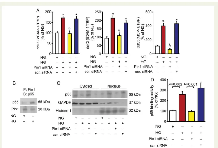
Pin1 mediates hyperglycaemia-induced
up-regulation of adhesion molecules
Since Pin1 is a critical mediator of inflammation in cancer, we inves-tigated whether this isomerase contributes to endothelial inflamma-tion in hyperglycaemic condiinflamma-tions. Exposure to HG caused a
Figure 2 Targeting Pin1 rescues hyperglycaemia-induced mitochondrial disruption and endothelial nitric oxide synthase dysregulation. (A) Con-focal microscopy images of mitoTracker-based mitochondrial staining showing alteration of an organelle network in human aortic endothelial cells exposed to high glucose in the presence or in the absence of Pin1 siRNA. Under normal glucose concentrations, human aortic endothelial cells show thread-like mitochondria (Figure1A and B) while exposure to high glucose leads to smaller punctuate mitochondria (Figure2A and B). Pin1 knockdown significantly attenuated mitochondrial disruption (Figure3A and B). Scrambled siRNA was used as a control (Figure4A and B). (B) Mitochondrial DNA integrity by real-time PCR (n ¼ 6). *P , 0.01 vs. NG§; P , 0.01 vs. HG. (C ) Mitochondrial swelling assay. Line graphs represent time-course of light absorbance decrease with (blue line) and without (red line) calcium overload (n ¼ 6). *P , 0.05 vs. untreated. (D) Confocal microscopy images showing cytochrome c (green) release (arrows) from mitochondria stained with organelle marker MitoTracker (red). (E) Representative western blot (WB) showing cytochrome c release from mitochondria. (F ) ESR spectroscopy analysis of nitric oxide release across the experimental conditions (n ¼ 6). (G) Western blot and densitometric quantification of endothelial nitric oxide synthase inhibitory Ser-116 phosphorylation in human aortic endothelial cells exposed to normal and high glucose concentrations (n ¼ 4). (H ) Immunoprecipitation showing endothelial nitric oxide synthase – Pin1 association in cells exposed to normal or high glucose concentrations. (I ) Endothelial nitric oxide synthase – caveolin-1 inter-action in high glucose-treated human aortic endothelial cells with or without Pin1 siRNA. IB, immunoblotting; IP, immunoprecipitation. Results are presented as means + SEM. NG, normal glucose, HG, high glucose.
F. Paneni et al.
significant up-regulation of the adhesion molecules vascular adhesion cell molecule-1 (VCAM-1), intercellular cell adhesion molecule -1
(ICAM-1), and MCP-1 in HAECs (Figure3A). In this setting, Pin1
recognizes NF-kB p65 favouring its nuclear translocation and
increased binding activity (Figure3B – D). Interestingly, Pin1
down-regulation by siRNA prevented NF-kB p65 activation and subsequent
overexpression of adhesion molecules (Figure3A, C and D).
Diabetic Pin1
2/2mice are protected
against endothelial dysfunction
Diabetes induced a significant increase of Pin1 expression in mouse
aorta (Figure4A). WT and Pin12/2mice were studied to investigate
whether Pin1 contributes to hyperglycaemia-induced endothelial dysfunction in vivo. Diabetic animals did not differ for body weight, glucose levels, blood pressure, and lipids (Supplementary material online, Table S1). Endothelial function was normal in control WT
and Pin12/2 mice (Figure4B). Interestingly, enough,
endothelium-dependent relaxation to acetylcholine (1029– 1026mol/L) was
impaired in WT diabetic mice but not in Pin12/2mice (Figure4B).
In contrast, endothelium-independent relaxation to sodium
nitro-prusside (10210– 1025mol/L) did not differ across the experimental
groups (data not shown).
Genetic deletion of Pin1 prevents
mitochondrial oxidative stress
In line with our in vitro findings, mitochondrial ROS generation
was abolished in Pin12/2diabetic vessels (Figure4C). Accordingly,
genetic disruption of the isomerase prevented diabetes-related mitochondrial DNA fragmentation and swelling, two important
hall-marks of mitochondrial dysfunction (Figure4D and E). To further
in-vestigate the link between Pin1 and vascular dysfunction in vivo, knockdown of Pin1 was achieved by using siRNA technology in WT diabetic mice. Interestingly, Pin1 silencing rescued endothelial dysfunction when compared with scrambled-treated WT diabetic
mice (Figure4F).
Figure 3 Pin1 triggers up-regulation of inflammatory adhesion molecules via nuclear factor kappa-B signalling. (A) Gene expression of inflamma-tory adhesion molecules vascular adhesion cell molecule-1, intercellular cell adhesion molecule-1, and MCP-1 in human aortic endothelial cells exposed to normal and high glucose concentrations in the presence or in the absence of Pin1 siRNA (n ¼ 6). *P , 0.01 vs. NG§; P , 0.01 vs. HG. (B) Immunoprecipitation showing the interaction between Pin1 and NF-kB p65 in cells treated with high or normal glucose. IB, immunoblotting; IP, immunoprecipitation. (C ) Representative western blot showing NF-kB p65 nuclear translocation. GAPDH and Histone 1 indicate loading con-trols for cytosolic and nuclear fractions, respectively. (D) NF-kB p65 binding activity (n ¼ 5). Results are presented as means + SEM. NG, normal glucose, HG, high glucose. VCAM-1, vascular adhesion cell molecule-1; ICAM-1, intercellular cell adhesion molecule-1; MCP-1, monocyte chemo-attractant protein-1, NF-kB, nuclear factor kappa-B.
Suppression of diabetes-induced vascular
inflammation in Pin1
2/2mice
To investigate the effects of Pin1 on vascular inflammation, the ex-pression of VCAM-1 and ICAM-1 was assessed in the aorta of WT
and Pin12/2diabetic mice. Aortas isolated from WT diabetic mice
displayed a significant up-regulation of these adhesion molecules,
as shown by immunofluorescence and real-time PCR (Figure 5A
and B). Such an effect was explained by increased NF-kB p65
nuclear translocation and binding activity (Figure5C and D).
Interest-ingly, we found that diabetic mice lacking Pin1 were protected against
vascular inflammation (Figure5A – D).
Pin1 expression and activity in patients
with diabetes
Pin1 gene expression and activity were assessed in peripheral blood monocytes of 37 patients with T2DM and 20 age-matched healthy controls. Type 2 diabetes mellitus subjects had higher BMI, waist cir-cumference, blood pressure, and lower HDL levels (Supplementary
material online, Table S2). Expression and activity of Pin1 were
increased in T2DM and correlated with HbA1c as well as fasting
plasma glucose (FPG, Figure6A – C ).
Importantly, FPG (b ¼ 0.39, P ¼ 0.021) and HbA1c (b ¼ 0.31,
P ¼ 0.034) were independently associated with Pin1 gene expres-sion, as shown by linear regression analysis adjusted for age, gender, waist circumference, blood pressure, HDL, and medications.
Pin1 up-regulation, endothelial
dysfunction, and oxidative stress
Endothelial function, assessed by flow-mediated dilation (FMD), was significantly impaired in T2DM subjects when compared with controls (Supplementary material online, Table S3). Nitroglycerine-mediated dilatation was comparable in the two groups (Supplemen-tary material online, Table S3) and no differences were observed in arterial diameter as well as resting or hyperaemic flow (data not shown). In agreement with an impairment of endothelial function,
diabetic patients showed higher urinary levels of 8-isoPGF2a, in vivo
Figure 4 Genetic deletion of Pin1 protects against diabetes-related endothelial dysfunction and mitochondrial oxidative stress. (A) Representative western blot and densitometric quantification of Pin1 expression in the aorta of WT and Pin12/2mice, with or without diabetes (n ¼ 5). ND, not
detectable. (B) Isometric tension studies in aortic rings isolated from the four experimental groups (WT control ¼ 10, WT diabetic ¼ 12, Pin12/2 control ¼ 8, Pin12/2diabetic ¼ 7) per group. *P , 0.05 and **P , 0.01 vs. WT diabetic. NE, norepinephrine. (C ) ESR spectroscopy analysis of
mitochondrial superoxide anion (O22) generation (n ¼ 6). (D) Mitochondrial DNA integrity assessed by real-time PCR in the four experimental
groups (n ¼ 7). *P , 0.01 vs. WT control§; P , 0.01 vs. WT diabetic. (E) Mitochondrial swelling assay (n ¼ 6). *P , 0.05 vs. untreated. (F ) Endothelium-dependent relaxation in diabetic mice treated with scramble or Pin1 siRNA. *P , 0.05 and **P , 0.01 vs. scrambled siRNA. Results are presented as means + SEM.
F. Paneni et al.
marker of oxidative stress (Supplementary material online, Table S3). Interestingly enough, Pin1 gene expression and activity significantly correlated with endothelial dysfunction and oxidative stress
(Figure7A and B). This finding was confirmed by linear regression
ana-lysis adjusted for confounding factors (Supplementary material online, Table S4).
Pin1 correlates with plasma adhesion
molecules
We also investigated the correlation between Pin1 and plasma adhe-sion molecules. Vascular adheadhe-sion cell molecule-1, ICAM-1, and MCP-1 were significantly higher in T2DM when compared with con-trols (Supplementary material online, Table S4). Of interest, a signifi-cant association was found between Pin1 expression/activity and adhesion molecules, as shown by correlation and linear regression
analysis (Figure8A, Supplementary material online, Table S4).
Collect-ively, these findings show that Pin1 may be critically involved in the
vascular disease phenotype observed in diabetic patients (Figure8B).
Discussion
Here, we show that Pin1 is up-regulated by hyperglycaemia and orchestrates pivotal molecular events triggering diabetic vascular disease. Several lines of evidence support our conclusions. Pin1 ex-pression and activity are significantly increased in human endothelial cells exposed to high glucose and in aortas of diabetic mice.
Activatory Ser-36 phosphorylation of adaptor protein p66Shcis
spe-cifically recognized by Pin1 and this leads to p66Shcmitochondrial
translocation, ROS production, and organelle disruption. Pin1 also recognizes Ser-116 eNOS inhibitory phosphorylation contributing to impaired NO availability. Furthermore, this isomerase is required for hyperglycaemia-induced nuclear translocation of NF-kB p65, leading to up-regulation of inflammatory adhesion molecules. Im-portantly, genetic deletion of Pin1 prevented vascular oxidative stress, endothelial dysfunction, and NF-kB-driven inflammation in diabetic mice. In addition, the expression and activity of Pin1 are increased in peripheral blood monocytes of T2DM patients and correlate with brachial artery FMD, urinary levels of the oxidative
marker 8-isoPGF2a, and plasma adhesion molecules.
Pin1 is emerging as a novel regulator of cellular function via modifications of protein structure upon recognition of a specific
phosphorylation motif (Ser/Thr-Pro).29Pin1-dependent
isomeriza-tion leads to stabilizaisomeriza-tion of proteins in active configuraisomeriza-tion and enhances their degradation or accessibility for further
modifica-tions by other enzymes.3The biological relevance of such a
signal-ling mechanism is supported by the notion that Pin1 dysregulation may contribute to diverse pathological conditions such as cancer
and Alzheimer’s disease.3,4Indeed, this isomerase is overexpressed
in 38 out of 60 different human cancers and predicts prognosis.5–7
In cancer cells, Pin1 recognizes a phosphorylated Thr-254-Pro motif of NF-kB p65 and inhibits its binding to IkBa, resulting in
increased nuclear translocation.8 Moreover, Pin1 controls
mito-chondrial trafficking of pro-oxidant proteins.11,13,30 In murine
Figure 5 Loss of Pin1 suppresses vascular inflammation in diabetic mice. (A) Fluorescence microscopy images showing the expression of adhesion molecules vascular adhesion cell molecule-1 and intercellular cell adhesion molecule-1 (green) in aortic cross sections from WT and Pin12/2mice with or without diabetes. Nuclei stained blue with DAPI. Higher magnification panels (bottom) show vascular adhesion cell molecule-1 and inter-cellular cell adhesion molecule-1 signals only in WT diabetic mice (arrows). (B) Gene expression of vascular adhesion cell molecule-1 and intercel-lular cell adhesion molecule-1 by real-time PCR (n ¼ 8). *P , 0.01 vs. WT control§; P , 0.01 vs. WT diabetic. (C ) Representative western blot of NF-kB p65 nuclear translocation in aortas isolated from the different experimental groups. (D) NF-kB p65 binding activity (n ¼ 5). Results are pre-sented as means + SEM.
fibroblasts, Pin1 blockade prevents translocation of p66Shcto the
mitochondria and oxidative stress.11Although Pin1 is an emerging
trigger of proliferative, inflammatory, and pro-apoptotic signalling in cancer, its role in the pathogenesis of vascular disease remains to be elucidated. A recent work in bovine endothelial cells showed that Pin1 recognizes Ser-116 eNOS inhibitory
phosphor-ylation and blunts NO release.15Indeed, pharmacological blockade
of Pin1 induced a 30% increase in NO production.15Since
mito-chondrial oxidative stress, reduced NO availability, and vascular
in-flammation are major hallmarks of diabetic vascular disease,18we
were prompted to investigate whether Pin1 is affected by hyper-glycaemia and mediates vascular damage in this setting. Our find-ings show that Pin1 is up-regulated by high glucose levels and is
responsible for mitochondrial translocation of p66Shcin the
vascu-lar endothelium. We and others have demonstrated that p66Shcis
a key mediator of ROS generation and, hence, a major contributor
of diabetic vascular complications.16,17,31,32Of note, p66Shcgene
expression is increased in mononuclear cells obtained from
patients with T2DM and correlates with oxidative stress.20 The
present study demonstrates that Pin1 is the upstream regulator
Figure 6 Pin1 expression and activity are increased in diabetic patients and correlate with glycaemic markers. (A) Scatter plots show Pin1 mRNA (controls ¼ 20, T2DM ¼ 37) and activity (controls ¼ 12, T2DM ¼ 37) in diabetic subjects and healthy controls. Results are presented as means + SEM. (B and C ) Correlation of Pin1 expression and activity with HbA1cand FPG. r ¼ Spearman’s correlation coefficient. HbA1cindicates glycosylated
haemoglobin; FPG, fasting plasma glucose.
F. Paneni et al.
of p66Shcand its inhibition prevents oxidative stress and mitochon-drial disruption in human endothelial cells and in mice under hyperglycaemic conditions. The relevance of our results is strengthened by recent work suggesting that loss of mitochondrial
integrity may affect endothelial function in diabetic patients.27,33
We also found that in hyperglycaemic conditions Pin1 impairs eNOS activity via isomerization of its inhibitory Ser-116 residue.
Proline-directed phosphorylation, requiring a proline at the P+ 1
position, is a pre-requisite for Pin1 interaction and subsequent iso-merization of its substrates. Of the five known serine/threonine phosphorylation sites in human eNOS, only Ser-116 conforms to
the P+ 1 proline requirement.34 In our study, Pin1 isomerizes
Ser-116 favouring eNOS interaction with caveolin-1, an important
repressor of eNOS catalytic activity in the endothelium.28,35
Accord-ingly, Pin1 deletion suppressed eNOS trafficking to the plasmalem-mal caveolae and restored NO availability. This finding is supported by another study showing that transient Pin1 overexpression blunts
NO release in unstimulated endothelial cells.15Furthermore,
silen-cing of Pin1 prevents hyperglycaemia-induced oxidative stress in
renal tubular cells12and neointima formation in mice by affecting
pro-liferation of vascular smooth muscle cells.12,36,37These latter studies
provided insights on the role of Pin1 in the vessel wall. However, our work demonstrates for the first time that Pin1 activation is a key driver of vascular damage in diabetes. Indeed, this study clearly shows that hyperglycaemia-induced Pin1 up-regulation leads to a deleterious vascular phenotype which can be prevented by targeting
Pin1. Accordingly, Pin12/2 diabetic mice are protected against
hyperglycaemia-induced endothelial dysfunction and mitochondrial oxidative stress.
We also provide strong evidence that Pin1 drives vascular inflam-mation in the setting of diabetes. Although it was recently shown that
Pin1 promotes NF-kB p65 signalling in cancer cells,8no previous
work investigated whether this isomerase mediates inflammation in the cardiovascular system. In our study Pin1-induced NF-kB p65 nuclear translocation and up-regulation of adhesion molecules in vitro and in vivo. We show here that deletion of Pin1 suppresses NF-kB p65 and expression of VCAM-1, ICAM-1, and MCP-1. These findings deserve attention since NF-kB activation has been
reported in the endothelium of humans with T2DM.23
In contrast with our results, a previous study showed that
Pin12/2 mice exhibit a significant impairment of
endothelium-dependent relaxation and high blood pressure values.38Here, we
Figure 7 Pin1 correlates with endothelial function and oxidative stress. (A and B) Correlation of Pin1 expression and activity with flow-mediated vasodilation and urinary 8-isoPGF2alevels. Red and blue circles indicate healthy controls (n ¼ 12 – 20) and T2DM patients (n ¼ 37), respectively.
FMD, flow-mediated dilation; 8-isoPGF2a, 8-iso-prostaglandin F2a. r ¼ Spearman’s correlation coefficient.
do not observe any endothelial dysfunction or hypertension in mice lacking Pin1. Different age of animals and experimental conditions may contribute to explain such discrepancy. In line with our find-ings, it was recently reported that blood pressure values and
cardiac phenotype are comparable in WT and Pin12/2mice.39
Add-itional experimental observations suggest that silencing of Pin1 pre-vents angiotensin II-dependent oxidative stress, indicating that Pin1
blockade may rather be protective against arterial hypertension.12
Another study also confirmed our results by showing that Pin1 overexpression impairs endothelium-dependent
vasorelaxa-tion in mice.15
In the present study, we have also translated our experimental findings to diabetic patients. Indeed, we found a significant up-regulation of Pin1 in subjects with T2DM when compared with age-matched healthy controls. Moreover, Pin1 correlated with
bra-chial artery FMD, urinary 8-isoPGF2a, and plasma levels of adhesion
molecules. Linear regression analyses showed that Pin1 was asso-ciated with oxidative stress, endothelial dysfunction, and vascular
in-flammation regardless of confounding factors. FPG and HbA1cwere
also independent predictors of Pin1 up-regulation. Although the assessment of Pin1 expression in peripheral monocytes cannot be directly related to endothelial dysfunction, these findings are in
Figure 8 Pin1 correlates with inflammatory adhesion molecules in patients with diabetes. (A) Correlation of Pin1 expression and activity with plasma adhesion molecules vascular adhesion cell molecule-1, intercellular cell adhesion molecule-1, and monocyte chemoattractant protein-1. Red and blue circles indicate healthy controls (n ¼ 12 – 20) and diabetic patients (n ¼ 37), respectively. r ¼ Spearman’s correlation coefficient. VCAM-1, vascular adhesion cell molecule-1; ICAM-1, intercellular cell adhesion molecule-1; MCP-1, monocyte chemoattractant protein-1. (B) Schematic representation of the Pin1 role in diabetic vascular disease. Diabetes causes up-regulation of Pin1 favouring its interaction with phos-phorylated serine and threonine residues of adaptor protein p66Shc, endothelial nitric oxide synthase, and NF-kB p65. Pin1-p66Shcinteraction is required for translocation of p66Shc, mitochondrial ROS generation, and disruption. Phosphorylated endothelial nitric oxide synthase at Ser-116 is recognized by Pin1 leading to endothelial nitric oxide synthase – caveolin-1 interaction, blunted endothelial nitric oxide synthase activity and reduced NO release. Pin1 also triggers diabetes-induced nuclear translocation of NF-kB p65 and subsequent up-regulation of inflammatory adhe-sion molecules. All together these findings indicate that Pin1-induced conformational changes are critically involved in the diabetic vascular disease phenotype.
F. Paneni et al.
accordance with our experiments in HAECs demonstrating a key role of the isomerase in the human endothelium. The strength of our work is the observation of Pin1 as a key mediator of hyperglycaemia-related vascular damage across different experimen-tal settings including knockout mice, primary human endothelial cells, and T2DM patients. Another novel finding is that hyperglycaemia reduces methylation of the Pin1 promoter. Since methylation is an
important repressor of gene transcription,40,41our findings suggest
that this epigenetic signature may contribute to Pin1 up-regulation in this setting. Undoubtedly, further studies are needed to better characterize epigenetic-driven Pin1 transcription in disease states.
In conclusion, we have shown that Pin1-dependent isomerization modulates key proteins involved in diabetic vascular disease
(Figure8B). Targeting Pin1 may restore vascular health by preventing
such deleterious events. These findings have important implications for future mechanism-based therapeutic strategies in patients with diabetes.
Supplementary material
Supplementary material is available at European Heart Journal online.
Funding
This study was supported by grants from the Swiss Heart Foundation, Italian Ministry of Education, University and Research, PRIN 2010-2011 (to F.C.), and the Swiss National Research Foundation to T.F.L (3100-06811802/1). AIRC Special Program Molecular Clinical Oncology ‘5 per mille’ and Italian Ministry of University and Research (RBAP10XKNC_003 and PRIN 2009- 2009YP9AE5) to G.D.S. F.P was the recipient of a PhD programme in Experimental Medicine at the University of Rome ‘Sapienza’.
Conflicts of interest: none declared.
References
1. Beckman JA, Paneni F, Cosentino F, Creager MA. Diabetes and vascular disease: pathophysiology, clinical consequences, and medical therapy: part II. Eur Heart J 2013;34:2444 – 2452.
2. Paneni F, Beckman JA, Creager MA, Cosentino F. Diabetes and vascular disease: pathophysiology, clinical consequences, and medical therapy: part I. Eur Heart J 2013;34:2436 – 2443.
3. Lu KP, Zhou XZ. The prolyl isomerase PIN1: a pivotal new twist in phosphorylation signalling and disease. Nat Rev Mol Cell Biol 2007;8:904 – 916.
4. Lee TH, Pastorino L, Lu KP. Peptidyl-prolyl cis-trans isomerase Pin1 in ageing, cancer and Alzheimer disease. Expert Rev Mol Med 2011;13:e21.
5. Girardini JE, Napoli M, Piazza S, Rustighi A, Marotta C, Radaelli E, Capaci V, Jordan L, Quinlan P, Thompson A, Mano M, Rosato A, Crook T, Scanziani E, Means AR, Lozano G, Schneider C, Del Sal G. A Pin1/mutant p53 axis promotes aggressiveness in breast cancer. Cancer Cell 2011;20:79 – 91.
6. Tan X, Zhou F, Wan J, Hang J, Chen Z, Li B, Zhang C, Shao K, Jiang P, Shi S, Feng X, Lv N, Wang Z, Ling Y, Zhao X, Ding D, Sun J, Xiong M, He J. Pin1 expression contri-butes to lung cancer: prognosis and carcinogenesis. Cancer Biol Ther 2010;9: 111 – 119.
7. Fukuchi M, Fukai Y, Kimura H, Sohda M, Miyazaki T, Nakajima M, Masuda N, Tsukada K, Kato H, Kuwano H. Prolyl isomerase Pin1 expression predicts prognosis in patients with esophageal squamous cell carcinoma and correlates with cyclinD1 expression. Int J Oncol 2006;29:329 – 334.
8. Ryo A, Suizu F, Yoshida Y, Perrem K, Liou YC, Wulf G, Rottapel R, Yamaoka S, Lu KP. Regulation of NF-kappaB signaling by Pin1-dependent prolyl isomerization and ubiquitin-mediated proteolysis of p65/RelA. Mol Cell 2003;12:1413 – 1426. 9. Atkinson GP, Nozell SE, Harrison DK, Stonecypher MS, Chen D, Benveniste EN. The
prolyl isomerase Pin1 regulates the NF-kappaB signaling pathway and interleukin-8 expression in glioblastoma. Oncogene 2009;28:3735 – 3745.
10. Wang J, Ray PS, Sim MS, Zhou XZ, Lu KP, Lee AV, Lin X, Bagaria SP, Giuliano AE, Cui X. FOXC1 regulates the functions of human basal-like breast cancer cells by ac-tivating NF-kappaB signaling. Oncogene 2012;31:4798 – 4802.
11. Pinton P, Rimessi A, Marchi S, Orsini F, Migliaccio E, Giorgio M, Contursi C, Minucci S, Mantovani F, Wieckowski MR, Del Sal G, Pelicci PG, Rizzuto R. Protein kinase C beta and prolyl isomerase 1 regulate mitochondrial effects of the life-span determinant p66Shc. Science 2007;315:659 – 663.
12. Sun L, Xiao L, Nie J, Liu FY, Ling GH, Zhu XJ, Tang WB, Chen WC, Xia YC, Zhan M, Ma MM, Peng YM, Liu H, Liu YH, Kanwar YS. p66Shc mediates high-glucose and angiotensin II-induced oxidative stress renal tubular injury via mitochondrial-dependent apoptotic pathway. Am J Physiol Renal Physiol 2010;299:F1014 – F1025. 13. Sorrentino G, Mioni M, Giorgi C, Ruggeri N, Pinton P, Moll U, Mantovani F, Del Sal G.
The prolyl-isomerase Pin1 activates the mitochondrial death program of p53. Cell Death Differ 2013;20:198 – 208.
14. Grison A, Mantovani F, Comel A, Agostoni E, Gustincich S, Persichetti F, Del Sal G. Ser46 phosphorylation and prolyl-isomerase Pin1-mediated isomerization of p53 are key events in p53-dependent apoptosis induced by mutant huntingtin. Proc Natl Acad Sci USA 2011;108:17979 – 17984.
15. Ruan L, Torres CM, Qian J, Chen F, Mintz JD, Stepp DW, Fulton D, Venema RC. Pin1 prolyl isomerase regulates endothelial nitric oxide synthase. Arterioscler Thromb Vasc Biol 2011;31:392 – 398.
16. Camici GG, Schiavoni M, Francia P, Bachschmid M, Martin-Padura I, Hersberger M, Tanner FC, Pelicci P, Volpe M, Anversa P, Luscher TF, Cosentino F. Genetic deletion of p66(Shc) adaptor protein prevents hyperglycemia-induced endothelial dysfunc-tion and oxidative stress. Proc Natl Acad Sci USA 2007;104:5217 – 5222. 17. Paneni F, Mocharla P, Akhmedov A, Costantino S, Osto E, Volpe M, Luscher TF,
Cosentino F. Gene silencing of the mitochondrial adaptor p66(Shc) suppresses vas-cular hyperglycemic memory in diabetes. Circ Res 2012;111:278 – 289.
18. Giacco F, Brownlee M. Oxidative stress and diabetic complications. Circ Res 2010; 107:1058 – 1070.
19. Ramsey JJ, Tran D, Giorgio M, Griffey SM, Koehne A, Laing ST, Taylor SL, Kim K, Cortopassi GA, Lloyd KC, Hagopian K, Tomilov AA, Migliaccio E, Pelicci PG, McDonald RB. The Influence of Shc Proteins on Life Span in Mice. J Gerontol A Biol Sci Med Sci 2013. doi:10.1093/gerona/glt198.
20. Pagnin E, Fadini G, de Toni R, Tiengo A, Calo L, Avogaro A. Diabetes induces p66shc gene expression in human peripheral blood mononuclear cells: relationship to oxi-dative stress. J Clin Endocrinol Metab 2005;90:1130 – 1136.
21. Forstermann U, Munzel T. Endothelial nitric oxide synthase in vascular disease: from marvel to menace. Circulation 2006;113:1708 – 1714.
22. Tabit CE, Chung WB, Hamburg NM, Vita JA. Endothelial dysfunction in diabetes mel-litus: molecular mechanisms and clinical implications. Rev Endocr Metab Disord 2010; 11:61 – 74.
23. Tabit CE, Shenouda SM, Holbrook M, Fetterman JL, Kiani S, Frame AA, Kluge MA, Held A, Dohadwala MM, Gokce N, Farb MG, Rosenzweig J, Ruderman N, Vita JA, Hamburg NM. Protein kinase C-beta contributes to impaired endothelial insulin sig-naling in humans with diabetes mellitus. Circulation 2013;127:86 – 95.
24. Kouroedov A, Eto M, Joch H, Volpe M, Luscher TF, Cosentino F. Selective inhibition of protein kinase Cbeta2 prevents acute effects of high glucose on vascular cell adhesion molecule-1 expression in human endothelial cells. Circulation 2004;110:91– 96. 25. Baker RG, Hayden MS, Ghosh S. NF-kappaB, inflammation, and metabolic disease.
Cell Metab 2011;13:11 – 22.
26. Paneni F, Volpe M, Luscher TF, Cosentino F. SIRT1, p66Shc, and Set7/9 in vascular hyper-glycemic memory: bringing all the strands together. Diabetes 2013;62:1800–1807. 27. Shenouda SM, Widlansky ME, Chen K, Xu G, Holbrook M, Tabit CE, Hamburg NM,
Frame AA, Caiano TL, Kluge MA, Duess MA, Levit A, Kim B, Hartman ML, Joseph L, Shirihai OS, Vita JA. Altered mitochondrial dynamics contributes to endothelial dys-function in diabetes mellitus. Circulation 2011;124:444 – 453.
28. Li C, Ruan L, Sood SG, Papapetropoulos A, Fulton D, Venema RC. Role of eNOS phosphorylation at Ser-116 in regulation of eNOS activity in endothelial cells. Vascul Pharmacol 2007;47:257 – 264.
29. Lu KP. Phosphorylation-dependent prolyl isomerization: a novel cell cycle regulatory mechanism. Prog Cell Cycle Res 2000;4:83 – 96.
30. Napoli M, Girardini JE, Piazza S, Del Sal G. Wiring the oncogenic circuitry: Pin1 unleashes mutant p53. Oncotarget 2011;2:654 – 656.
31. Fadini GP, Albiero M, Menegazzo L, Boscaro E, Pagnin E, Iori E, Cosma C, Lapolla A, Pengo V, Stendardo M, Agostini C, Pelicci PG, Giorgio M, Avogaro A. The redox enzyme p66Shc contributes to diabetes and ischemia-induced delay in cutaneous wound healing. Diabetes 2010;59:2306 – 2314.
32. Menini S, Amadio L, Oddi G, Ricci C, Pesce C, Pugliese F, Giorgio M, Migliaccio E, Pelicci P, Iacobini C, Pugliese G. Deletion of p66Shc longevity gene protects against experimental diabetic glomerulopathy by preventing diabetes-induced oxi-dative stress. Diabetes 2006;55:1642 – 1650.
33. Kluge MA, Fetterman JL, Vita JA. Mitochondria and endothelial function. Circ Res 2013;112:1171 – 1188.
34. Mount PF, Kemp BE, Power DA. Regulation of endothelial and myocardial NO syn-thesis by multi-site eNOS phosphorylation. J Mol Cell Cardiol 2007;42:271 – 279. 35. Frank PG, Woodman SE, Park DS, Lisanti MP. Caveolin, caveolae, and endothelial cell
function. Arterioscler Thromb Vasc Biol 2003;23:1161 – 1168.
36. Ki SH, Lee JW, Lim SC, Hien TT, Im JH, Oh WK, Lee MY, Ji YH, Kim YG, Kang KW. Protective effect of nectandrin B, a potent AMPK activator on neointima formation: inhibition of Pin1 expression through AMPK activation. Br J Pharmacol 2013;168: 932 – 945.
37. Lv L, Zhang J, Zhang L, Xue G, Wang P, Meng Q, Liang W. Essential role of Pin1 via STAT3 signalling and mitochondria-dependent pathways in restenosis in type 2 dia-betes. J Cell Mol Med 2013;17:989 – 1005.
38. Chiasson VL, Munshi N, Chatterjee P, Young KJ, Mitchell BM. Pin1 deficiency causes endothelial dysfunction and hypertension. Hypertension 2011;58:431 – 438. 39. Toko H, Konstandin MH, Doroudgar S, Ormachea L, Joyo E, Joyo AY, Din S,
Gude NA, Collins B, Volkers M, Thuerauf DJ, Glembotski CC, Chen CH, Lu KP, Muller OJ, Uchida T, Sussman MA. Regulation of cardiac hypertrophic signaling by prolyl isomerase Pin1. Circ Res 2013;112:1244 – 1252.
40. Paneni F, Costantino S, Volpe M, Luscher TF, Cosentino F. Epigenetic signatures and vascular risk in type 2 diabetes: a clinical perspective. Atherosclerosis 2013;230: 191 – 197.
41. Handy DE, Castro R, Loscalzo J. Epigenetic modifications: basic mechanisms and role in cardiovascular disease. Circulation 2011;123:2145 – 2156.
CARDIOVASCULAR FLASHLIGHT
. . . . doi:10.1093/eurheartj/ehu468
Online publish-ahead-of-print 17 December 2014
Extensive xanthomas with severe coronary artery disease in a young patient
with familial hypercholesterolemia
Vikas Agrawal* and Bhupendra Kumar Sihag
Department of Cardiology, Institute of Medical Sciences, Banaras Hindu University, Varanasi 221005, India
*Corresponding author. Tel:+91 9453300628, Email:[email protected]
A 20-year-old female presented to us with recent onset unstable angina. She had a strong family his-tory of premature coronary artery disease (two elder sisters dying at a young age due to CAD). She had large tendinous xanthomas over extensor tendons of metacarpophalangeal joints and patella (Panels A – C). She also had xanthelesma, gaint tuber-ous xanthomas over extensor aspects of limbs and buttocks and Plane xanthoma involving the neck (Panels D – H). The ECG showed deep ST depres-sion in all leads with ST elevation in avR (left main pattern, Panel I). Laboratory tests showed total cholesterol of 810 mg/dL (20.9 mmol/L), low-density lipoprotein cholesterol (LDL) of 733 mg/ dL (18.9 mol/L), triglycerides (TG) of 195 mg/dL (2.2 mmol/L), and high-density lipoprotein choles-terol of 39 mg/dL (1.0 mmol/L). Her coronary
angi-ography revealed severe left main disease involving ostium and mid shaft and also ostial RCA disease (Panels I, J, and K). Genetic study showed homozygotic mutation in LDL receptor gene. The patient is being planned for urgent PCI with intensive medical therapy.
Panel A: Large tendinous xanthomas over extensor tendons of metacarpophalangeal joints. Panel B: Large tendinous xanthomas over extensor tendons of metatarsophalangeal joints. Panel C: Large tendinous xanthomas over patella.
Panel D: Xanthelesma.
Panel E: Gaint tuberous xanthomas over extensor aspects of elbow. Panel F: Gaint tuberous xanthomas buttocks.
Panel G: Plane xanthoma involving the neck.
Panel H: Xanthoma involving extensor aspect of forearm.
Panel I: ECG showing deep ST depression in all leads with ST elevation in avR (left main pattern). Panel J: Coronary angiography showing severe left main disease involving ostium and mid shaft. Panel K: Coronary angiography showing severe ostial RCA disease.
Published on behalf of the European Society of Cardiology. All rights reserved.&The Author 2014. For permissions please email: [email protected].
F. Paneni et al.