Mutations that disable the DNA repair gene XPG in a
xeroderma pigmentosum group G patient
Thierry Nouspikel and Stuart G.CIarkson*
Department of Genetics and Microbiology, Centre Medical Universitaire (CMU), 9 avenue de Champel, 1211 Geneva 4, Switzerland
Received March 3, 1994; Revised and Accepted March 30, 1994 EMBL accession nos X69978 and X78329
The human XPG (ERCC5) gene encodes a large acidic
protein that corrects the ultraviolet light sensitivity of
cells from both xeroderma pigmentosum
complemen-tation group G and rodent ERCC group 5. Here we
characterize five XPG sequence alterations and a minor
splicing defect in XP-G patient XP125LO. Three of these
changes are polymorphic variants whereas the
remain-ing two, one in each XPG allele, inactivate
complementation in vivo. These single point mutations
provide formal proof that defects in XPG give rise to
the group G form of xeroderma pigmentosum, and their
locations suggest ways in which this may occur.
INTRODUCTION
The rare autosomal recessive disease xeroderma pigmentosum
(XP) is characterized by sunlight-induced skin and eye lesions,
skin cancers, and in some cases neurological abnormalities (1).
Most patients fall into seven complementation groups (A—G) that
are thought to be due to defects in an early step of nucleotide
excision repair (2). This pathway, which removes short patches
of DNA containing a wide variety of lesions, has been extensively
characterized in Escherichia coli (3) but much less is known of
how it functions in eukaryotes. By now, genes that complement
five of the seven classical XP groups ( A - D and G) have been
cloned (4—8). Some of these correspond to human ERCC genes
that are able to correct the ultraviolet light (UV) sensitivity of
certain rodent cell lines. Thus the XPB and ERCC3 genes are
identical, as are XPD and ERCC2. All five cloned XP genes also
show significant similarity to DNA repair genes from
Saccharomyces cerevisiae (2,9), attesting to the importance of
this repair pathway.
The one most recently characterized, XPG, is identical to
ERCC5 (10,11). It generates a mRNA of ~ 4 kb and encodes
an acidic protein of 1186 amino acids that shares two regions
of extensive homology with the yeast DNA repair protein RAD2.
The XPG protein restores normal levels of UV resistance and
unscheduled DNA synthesis when expressed in vivo in a
lymphoblastoid cell line from an XP-G patient (8). The most
straightforward interpretation of this finding is that defective XPG
alleles are responsible for the group G form of XP. It is formally
possible, however, that the defects reside in another gene and
that overexpressed XPG protein somehow is able to bypass the
normal repair pathway. To distinguish between these possibilities
and to try to define the molecular basis for this disorder, we have
analysed XPG cDNAs from an XP-G patient for the presence
of mutations and for their effects on DNA repair in vivo.
RESULTS
Patient XP125LO expresses both XPG alleles
The — 4 kb XPG mRNA is present in normal amounts (8) in
XPG83, a lymphoblastoid cell line (10) derived from XP-G
patient XP125LO (12). We made two cDNA libraries from this
cell line and screened them with XPG specific oligonucleotides.
Seventeen clones were recovered, four of which were fully
sequenced. These analyses revealed a total of six differences from
the original XPG cDNA sequence (EMBL X69978).
The first was a T—C transition at position 335. It represents
a silent polymorphism at His46 and it destroys a Styl restriction
site. To confirm this point mutation and to determine its parental
origin, we performed reverse transcription-polymerase chain
reaction (RT-PCR) on total RNA from cell lines from the
proband and from her obligate heterozygote mother (XPG83 and
XPG81, respectively; 10). Figure la shows that the expected 151
bp product containing the putative polymorphic site is amplified
from both sources, that it is digested by Styl to yield 130 bp and
21 bp bands in the case of the mother, but that a substantial
fraction of the amplified DNA from the patient remains uncut.
The incomplete digestion is not due to poor DNA quality because
the RT-PCR product from the mother is fully cut by £coRI,
and by a mixture of Styl and EcoRI, whereas part of the patient
DNA is again refractory to Styl under conditions in which £coRI
cuts to completion (Fig. la). Analyses of genomic PCR products
gave identical results (data not shown).
We conclude that patient XP125LO is heterozygous for this
silent polymorphism, that she inherited the T at position 335 from
her mother, and that both alleles are expressed and yield similar
levels of XPG mRNA.
Patient XP125LO is homozygous for two more
poly-morphisms
The second XPG sequence alteration was a C—G transversion
at position 3507. This generates a His—Asp substitution at amino
acid 1104. It also creates a new MboU site which permitted the
RT-PCR analysis shown in Fig. lb. The simultaneous presence
of 136 bp, 107 bp and 29 bp fragments in the sample from the
mother indicates that she is heterozygous for the transversion,
whereas her daughter is homozygous. Despite the nature of this
substitution, it is unlikely to have a causative role in XP-G because
it has been found in a functional ERCC5 cDNA sequence (11).
The third change was in the 3' untranslated region of XPG
at position 3842. This G—A transition, 84 bp beyond the TAA
stop codon, is present in both alleles of the patient (data not
shown) and also in ERCC5 cDNA (11). Hence, it too is unlikely
to be implicated in the XPG clinical phenorype.
HiS46CAT-»CAC HIS1104—> Asp
81 83
- S E ES - S E ES
21 ^ ^
60 48 25 85 29 107
Figure 1. Styl and MboU restriction analyses of RT-PCR products from lymphoblastoid cells from patient XP125LO (83) and her mother (81). —: Undigested XPG products. L: HaeM digest of pGEM3 DNA, with fragment lengths in bp. (a) S: Styl digestion. The inner open arrow indicates the undigested 151 bp R T - P C R product that lacks a Styl site (C/CWWGG) due to the T - C change at position 335 (X). (The small amount of this band in the S lane from the mother is due to partial digestion.) Inner filled arrows point at two Styl fragments of 130 bp and 21 bp derived from the second allele. E: control digestion with EcoRI. ES: double digestion with EcoRI and Styl. The outer open arrow indicates the 91 bp £coRI fragment specific for the transition-carrying allele, and outer filled arrows point at 70 bp and 21 bp fragments resulting from Sfyl digestion, (b) M: MboU digestion. The C —G transversion at position 3507 (X) creates a new MboU site (GAAGA[8/7], boxed), generating two fragments of 107 bp and 29 bp (open arrows). The filled arrow points at the intact 138 bp fragment derived from the second allele.
A minor fraction of XPG pre-mRNA is abnormally spliced
One cDNA derived from the paternal allele was found to be
missing the 55 bp from positions 1078-1132. This introduces
a frameshift that results, nine amino acids later, in a TGA stop
codon. Such a truncated protein of 302 amino acids would
certainly be expected to be non-functional. However, PCR
amplification of this region reveals that a shorter product is
present in only a minor fraction of the cDNA from both the
proband and her mother (Fig. 2). It is absent, however, from
their genomic DNA, suggesting that it is due to a splicing defect.
Sequence analysis of the genomic PCR products indeed reveals
the presence of a 315 bp intron located at the 5' end of the deleted
region (EMBL X78329). We speculate that the short mRNA
results from aberrant splicing of this intron to a downstream
cryptic acceptor site. Consistent with this idea, the 3' end of the
deleted region, (Y)nCAG/C (Fig. 2), shows an excellent fit with
the 3' splice site consensus sequence; only the presence of a C
instead of a purine in the first position of the 'exon' is somewhat
unusual, but it is found in 13% of primate splice acceptor sites
(14). To determine if this aberrant splicing is specific for this
disorder, we performed R T - P C R on nine unrelated non-XP
individuals. All nine generated the shorter product, in quite
variable amounts (data not shown). We conclude that this aberrant
splicing of XPG pre-mRNA is irrelevant to the XP phenotype.
RT Genomic 81 83 L _8J_ _83_ - H - H - H - H 605 297 61 Hinfl Hinfl 61 | 105 | 291 Hinfl II 142 605 Normal splice 61 I 81 It 142 290
I I
Abnormal splice 6 1 1 V 7 4 tetcttccttcttccagpAA 235 605Figure 2. Hinfl restriction analyses of RT-PCR and genomic PCR products from lymphoblastoid cells from patient XP125LO (83) and her mother (81). —: Undigested XPG products. H: Hinfl digestion. L: HaelU digest of pGEM3 DNA. Genomic PCR generates a 605 bp band due to the presence of a 315 bp intron located between positions 1077 and 1078 of the cDNA sequence. Splicing of this intron at the normal donor and acceptor sites results in a 290 bp RT - PCR product which is cut by Hinfl to yield 142 bp, 81 bp and 61 bp fragments; additional bands at 87 bp and 148 bp are presumed to be due to incomplete digestion at the two Hinfl sites 6 bp apart. Both cell lines also generate a minor 235 bp RT—PCR product and a minor 174 bp fragment after Hinfl digestion. These products can be explained by an occasional aberrant splicing event at the indicated cryptic acceptor site, thereby generating an XPG mRNA lacking positions
1078-1132.
However, the presence of some shorter PCR product in every
individual examined suggests that this 'leaky' splicing may
contribute to the low abundance of functional XPG mRNA (8).
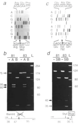
Candidate nonsense and missense mutations
The last two XPG sequence alterations found in patient XP125LO
provided candidate mutations for her acute sun sensitivity. One
is a G—T transversion at position 3075 (Fig. 3a) which
transforms the Glu960 codon into a TAA stop codon and, in so
doing, destroys a site for Apol. This enzyme cuts the RT-PCR
products from the mother to completion but only incompletely
digests those of the proband (Fig. 3b); control incubations show
that both DNAs can be digested to completion with BamYW. We
conclude that this nonsense mutation would generate a truncated
XPG protein of 959 amino acids and that it is carried by the
paternal allele.
The candidate mutation in the maternal allele is a C—T
transition at position 2572 (Fig. 3c) which causes an Ala—Val
Arg Glu Phe AGAGAATTT -~, f A H N c G M I T I fill I A Mil c G I I
I I III I
^> \ ^
AGATAATTT Arg Stop 960 81 83 L - A B - A B/-. Glu Ala Gin GAGGCGCAG A Ij 1/ c I M I I G f III f! A II I I c I I II G | Illllli GAGGTGCAG Glu Val Gin
792
81 83 L - S B - S B
175
35 5
Figure 3. Paternal (left) and maternal (right) XPG mutations in patient XP125LO.
(a) and (c). Nucleotide sequence of wild-type (top) and mutant (bottom) cDNAs showing (a) the G — T transversion at position 3075 that generates a premature stop codon, and (c) the C—T transition at position 2572 that leads to an amino acid substitution, (b) and (d) Restriction analyses of RT-PCR products from lymphoblastoid cells from the patient (83) and her mother (81). —: Undigested products. L: HaelR digest of pGEM3 DNA, with fragment lengths in bp. (b) A: Apol digestion. The open arrow points at a 175 bp fragment that remains intact due to the destruction of the Apol site (R/AATTY) by the mutation (X). Filled arrows show the 95 bp and 80 bp Apol digestion products specific to the wild-type allele. B: control digestion with BamW. (d) SB: double digestion with
Styl and Bsgl. The open arrow points at a 35 bp fragment due to the new Bsgl
site (GTGCAG[16/14], boxed) created by the mutation (X). The filled arrow points at the 40 bp Styl fragment specific to the wild-type allele.
substitution at amino acid 792. This transition occurs within a
CpG, suggesting that it arose by deamination of 5-methylcytosine.
Once again, the mutation creates a new restriction site, in this
case for Bsgl. Double digests of the RT-PCR products generate
35 bp Styl-Bsgl and 40 bp Styl fragments (Fig. 3d), thereby
showing that both the proband and her mother are heterozygous
for this missense mutation.
The candidate mutations inactivate XPG complementation
activity
For functional tests, we cloned full length XPG cDNAs containing
each candidate mutation into EBO-pLPP, an Epstein-Barr
virus-based episomal expression vector (15), and transfected them into
XPG83 cells. After hygromycin selection, stable transfectants
were challenged with UV, and their survival after 48 h was
assayed with BCECF-AM, a carboxyfluorescein derivative that
is hydrolysed to a fluorescent form by esterases within living cells
(16).
Transfectants containing the EBO-pLPP vector alone are as
highly sensitive to UV as untransfected cells, whereas the
100JT
10..
O C§ 100
3 5=I
CD on 10---x— Untransfected -o— Vector -.— wt XPG 1 1 1 h _T_ A-A/-XPG - A - Stop-XPG0 \ V
4 k A
UV dose (J/m2)Figure 4. UV sensitivity of transfected lymphoblastoid XPG83 cells from patient
XP125LO. (a) Untransfected cells (crosses), transfectants with EBO-pLPP vector alone (open circles), and transfectants with wild-type XPG cDNA in EBO-pLPP (filled circles), (b) EBO-pLPP-XPG transfectants carrying the paternal nonsense mutation (filled triangles), and EBO-pLPP-APG transfectants carrying the maternal missense mutation (filled inverted triangles). Results are expressed as per cent of the BCECF fluorescence of non-irradiated cells. Error bars show the standard deviations of groups of four measurements.
presence of wild type XPG cDNA restores UV resistance to
normal levels (Fig. 4a, and ref 8). Transfectants carrying the
nonsense mutation at codon 960 appear to be slightly resistant
at low UV doses but at higher doses are extremely UV sensitive
(Fig. 4b). The seemingly innocuous Ala—Val substitution at
codon 792 also has a severe effect; transfectants carrying this
missense mutation show a similar UV sensitivity to those
containing just the vector (Fig. 4b). We conclude that both
mutations disable XPG and render it incapable of correcting the
DNA repair defect in XPG83 cells.
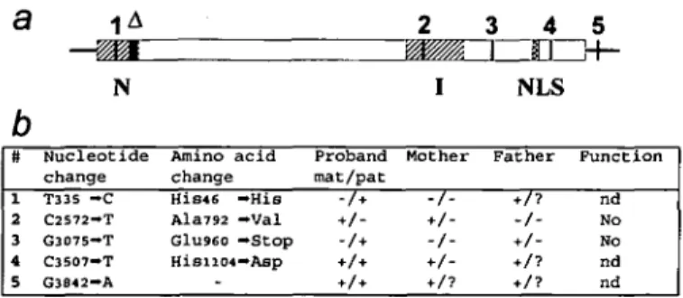
DISCUSSION
Figure 5 summarizes our major findings: patient XP125LO
inherited from her mother a missense Ala—Val mutation (#2)
located in the conserved I region of XPG. She is homozygous
for a His—Asp polymorphism (#4) in the C-terminal part of
a
b
#
i 2 3 4 51
A— ^ H
N
Nucleotide change T335 - C C2572-T G3075—T C3507-T G3842-A Amino change acid His«6 - H i s Ala792 —Val G1U960 —Stop HisiiO4— Asp2
I
Proband Mother mat/pat -A*-','-3
4
nrr
NLS
Father + /-5
3f-F u n c t i o n nd No No nd nd
Figure S. Summary of the XPG alleles of patient XP125LO. (a) The XPG open
reading frame is boxed and is flanked by 5' and 3 ' untranslated regions. The conserved N and I regions are hatched and the putative bipartite nuclear localization signal (NLS) is cross-hatched. A locates the 55 nucleotides missing in a minor fraction of XPG mRNA.Vertical bars and numbers locate the single nucleotide changes listed in the table, (b) Nucleotides are numbered from the XPG cDNA sequence (EMBL X69978). + and - indicate the presence or absence of a change in either allele of the proband (mat: maternal; pat: paternal) or her parents. In the case of the father, these are deductions (?: no deduction possible). Function indicates if transfection of a cDNA carrying that change corrects the UV sensitivity of XPG83 cells (nd: not determined).
the protein and for a G—A transition (# 5) in the 3' untranslated
region. Her paternal allele carries a silent T—C transition (# 1)
in the conserved N region, and a nonsense mutation (# 3) that
would generate a truncated XPG protein of 959 amino acids.
Although the latter might represent a new germline mutation,
paternal inheritance is highly likely because the proband has an
affected brother, XP124LO, who has also been assigned to XP
group G (12).
Two of these point mutations, the paternal nonsense and the
maternal Ala—Val missense, lead to functional inactivation. They
thus provide formal proof that XPG is indeed the causative gene
for XP group G. They also provide the initial information needed
for a genetic screening test for this disorder. Moreover, as
outlined below, their locations have potential implications for
XPG function.
The truncated protein of 959 amino acids resulting from the
paternal nonsense mutation would contain both the conserved N
and I regions but not the putative bipartite nuclear localization
signal or basic C-terminus (8). Conceivably, this shortened form
of XPG might be active if it could bypass the requirement for
nuclear entry. Alternatively, the loss of just the C-terminus may
be the critical event because such a deletion inactivates
S.cerevisiae RAD2 (17). Use of an in vitro DNA repair assay
(18,19) should help to distinguish between these possibilities.
At first sight, it may seem surprising that the maternal
Ala—Val substitution disables XPG complementing activity.
However, Ala792 is located in a highly conserved 27 amino acid
core within the I region of the RAD2 protein family, whose
members include human XPG and its Xenopus laevis counterpart,
S.cerevisiae RAD2 and YKL510, and Schizosaccharomyces
pombe radl3 and rad2 (8,11,20). In particular, this invariant Ala
is flanked by glutamate residues that have been found in the active
site of some DNA nucleases (8). That this core may indeed have
functional significance is suggested by the recent finding of a
DNA endonuclease activity intrinsic to S.cerevisae RAD2 (21).
Clearly, a similar activity is anticipated for XPG, and we
speculate that it is the endonuclease activity that is disabled by
the maternal missense mutation in patient XP125LO.
With only seven known patients world-wide, XP group G is
one of the rarest forms of this disorder. It is also one of the most
clinically heterogeneous. Patient XP125LO and her affected
brother have only mild cutaneous changes and no neurological
abnormalities (12), whereas the first two patients belonging to
this group exhibited severe XP pigmentation and mental
retardation (22-24). Recently, two very severely affected
patients with neurological abnormalities characteristic of
Cockayne's syndrome (CS) were assigned to XP group G
(25,26). This degree of clinical heterogeneity is surprising and
not yet understood. Perhaps mildly affected patients possess some
residual DNA repair activity despite their deficiencies in cellular
repair assays. It is also possible that severely affected patients
have mutations that inactivate a second function. In any event,
we expect the two XP-G/CS patients to possess different
mutations from those characterized here and we hope that their
locations will also yield clues to functionally important domains
within the XPG protein.
MATERIALS AND METHODS
Cell lines
The Epstein-Barr virus immortalized cell lines XPG83 from XPG patient XP125LO (12) and XPG81 from her mother were kindly provided by R.D.Wood. Cells were grown in suspension at 37°C in RPMI 1640 medium supplemented with 2 mM glutamine and 10% fetal calf serum under 5% CO2.
cDNAs
Twenty-five fig (dT)15 or 2 pg of a specific primer complementary to XPG
positions 3815-3829 were phosphorylated with T4 polynucleotide kinase (Boehringer) and were separately annealed with 20 /»g poly(A)+ RNA. The RNA was a kind gift from D.Scherly and was prepared by lysing XPG83 cells into guanidinium isothiocyanate followed by cesium chloride step-gradient centrifugation and isolation on an oligo-dT cellulose column. cDNA synthesis was performed as described in the Superscript-Plus instructions (Gibco BRL) with minor modifications, using E.coli DNA Pol I and T4 DNA polymerase (New England Biolabs) and RNase H and E.coli DNA ligase (Boehringer). Two oligonucleotides (pTGGCCGTCGACTAC and pGTAGTCGACGGCCAGTG) were annealed together and 6.3 #ig were added to the cDNA and ligated overnight at 16°C in a total volume of 100 p] using 32 U/pl T4 DNA ligase and 5% polyethylene glycol. The cDNA was fractionated on a 5 -20% sucrose gradient by centrifugation at 335 000 g for 5 h at 4°C. Aliquots were analysed on a 1 % agarose gel, and fractions >2.5 kb were pooled.
Libraries
The vector Bluescript KS+ with a Sfil-CAT cassette (27) inserted in the EcoRV
site was kindly provided by V.Steimle. The CAT sniffer was removed by digestion with 5/iI and separation on a 5-20% sucrose gradient, generating non-compatible termini. 680 ng vector was ligated to the cDNA with 440 U//tl T4 DNA ligase at 20°C overnight then transformed into electro-competent bacteria (DH5 for the specific primer library, Sure cells (Stratagene) for the (dT),5 library) with a
Gene-Pulser (Bio-Rad) set at 2500 V, 25 pF, 200 Ohm. The libraries were titred and plated at 40 000 colonies/dish on HATF filters (Millipore). 5 x 10⁵ clones were
plated from the specific library, and 106 clones from the (dT)15 library. Two
replicas were made on BA85 filters (Schleicher & Schuell) for hybridization screening.
Hybridization
Pairs of [ T3 2P ] - A T P labelled oligonucleotides (positions 1 - 2 6 and 1337-1312
for the specific library, and positions 966-985 and 2472-2492 for the (dT)13
library) were incubated with the filters in 5XSSC, lOxDenhardt's, 20 mM sodium phosphate (pH 7.0), 7% SDS and 100 ftg/ml herring sperm DNA, at 5°C below Tm for 5 h. After washing in 6xSSC at 10°C below Tm for 15 min, positive
clones were revealed by autoradiography, then recovered from the original HATF filters and submitted to a second round of purification. Purified clones were prepared by CsCl centrifugation and sequenced with a deaza-T7 sequencing kit (Pharmacia).
PCR restriction analysis
First strand cDNA was synthesized from 5 pg total RNA with Superscript-Plus (Gibco-BRL) as specified by the manufacturer. Genomic DNA was prepared by
digesting cells with 100 /jg/ml proteinase K in 100 mM NaCl, 10 mM Tris-HCl (pH 8.0), 25 mM EDTA and 0.5% SDS at 50°C overnight. After phenol extraction and ethanol precipitation, samples were digested with 100 /ig/ml RNase A for 1 h at 37°C, then again extracted with phenol and precipitated with ethanol. PCR reactions (50 id) contained 1/8 of the cDNA preparation or 250 ng genomic DNA, 100 ng of each primer, 50 nM of each dNTP, 1 - 2 mM MgCl2 and 4 U
Ampli-Taq (Perkin-Elmer). Samples were first subjected to 10 touchdown cycles of: 30 s at 94°C, 30 s at 10°C above Ta, decreasing by 1°C at each cycle, 30 s
at 72°C, and then to 20 standard cycles at calculated Ta. The following primer
pairs were used: 312-333 and 462-438, 966-985 and 1255-1236, 2472-2492 and 2597-2578, 2995-3015 and 3170-3149, 3330-3349 and 3624-3607. PCR products were phenol extracted, ethanol precipitated, digested with restriction enzymes, fractionated on 6% or 8% polyacrylamide gels then stained for 15 min with 100 /ig/1 ethidium bromide. The genomic PCR 291 bp and 105 bp Hinfi products (Fig. 2) were made flush-ended then cloned separately into the Smal site of Bluescript KS+. Three clones of each were sequenced from T3 and T7
primers, yielding the sequence of a 315 bp intron (EMBL X78329) that disrupts XPG at the codon for Gly294.
Transfection and UV survival assay
Candidate mutations were introduced separately into a functional
EBO-pLPP-XPG recombinant (8) by substituting appropriate Stol-BgKI or Saul-Stul
restriction fragments. XPG83 cells (107/ml) were transfected by electroporation
with a Bio-Rad Gene Pulser at 250 V, 960 pF in RPMI 1640 medium containing 20 /jg/ml EBO-pLPP vector or recombinant DNA and 400 ^g/ml E.coli tRNA. Selection was achieved with increasing doses of Hygromycin B (Calbiochem) up to 200-250 /ig/ml. Selected cells were UV-irradiated at 254 nm in Hank's buffer at a flux of 12 ^W/cm2 for increasing times. Duplicate 200 id samples
were cultured with 1.4 ml RPMI medium containing 10% inactivated fetal calf serum in 24-well plates. Forty-eight and 72 h later, 500 id aliquots were removed and incubated for 30 min with 1 /jg/ml 2',7'-bis-(2-carboxyethyl)-5-(and-6)-carboxyfluorescein, acetoxymethyl ester (BCECF-AM, Molecular Probes). Cells were centriftiged for 5 min at 2000 g and resuspended in 250 id Hank's buffer plus 1 % Triton X-100 at 4°C. The fluorescence of duplicate 100 /d aliquots was measured in 96-well plates in a Cytofluor (Millipore) with excitation/emission filters of 485/530 nm (16).
13. Saiki, R.K., Scharf, S., Faloona, F., Mullis, K.B., Horn, G.T., Erlich, H.A. and Arnheim, N. (1985) Science 230, 1350-1354.
14. Shapiro, M.B. and Senepathy, P. (1987) Nucleic Acids Res. 15, 7155-7174. 15. Spickofsky, N., Canfield, V. and Margolskee, R.F. (1990) DNA Prot. Eng.
Techniq. 2, 14-18.
16. Leeder, J.S., Dosch, H-M., Harper, P.A., Lam, P. and Spielberg, S.P. (1989)
Anal. Biochem. 177, 364-372.
17. Madura, K. and Prakash, S. (1986) J. Baaeriol. 166, 914-923. 18. Wood, R.D., Robins, P. and Lindahl, T. (1988) Cell 53, 97-106. 19. Sibghat-UUah, Husain, I., Carlton, W. and Sancar, A. (1989) Nucleic Acids
Res. 17, 4471^484.
20. Carr, A.M., Sheldrick, K.S., Murray, J.M., Al-Harithy, R., Watts, F.Z. and Lehmann, A.R. (1993) Nucleic Acids Res. 21, 1345-1349. 21. Habraken, Y., Sung, P., Prakash, L. and Prakash, S. (1993) Nature 366,
365-368.
22. Cheesbrough, M.J. and Kinmont, P.D.C. (1978) Br. J. Dermatol. 99, Suppl. 16, 61.
23. Keijzer, W., Jaspers, N.G.J., Abrahams, P.J., Taylor, A.M.R., Arlett, C.F., Zelle, B., Takebe, H., Kinmont, P.D.C. and Bootsma, D. (1979) Mutat.
Res. 62, 183-190.
24. Arlett, C.F., Harcourt, S.A., Lehmann, A.R., Stevens, S., Ferguson-Smith, M.A. and Morley, W.N. (1980) Carcinogenesis 1, 745-751.
25. Jaeken, J., Klocker, H., Schwaiger, H., Bellmann, R., Hirsch-Kauffmann, M. and Schweiger, M. (1989) Hum. Genet. 83, 339-346.
26. Vermeulen, W., Jaeken, J., Jaspers, N.G.J., Bootsma, D. and Hoeijmakers, J.H.J. (1993) Am. J. Hum. Genet. 53, 185-192.
27. Steimle, V., Otten, L.A., Zufferey, M. and Mach, B. (1993) Cell 75, 135-146.
ACKNOWLEDGEMENTS
We are most grateful to D.Scherly, V.Steimle and R.D.Wood for materials, advice and critical comments on the manuscript, Janine Corlet for help with sequencing, and Glaxo 1MB (Geneva) for access to their multiwell fluorimeter. This work was supported by grant 31-36481.92 from the Swiss National Science Foundation.
ABBREVIATIONS
CS, Cockayne's syndrome; ERCC, excision repair cross complementing; PCR, polymerase chain reaction; RT-PCR, reverse transcription—polymerase chain reaction; UV, ultraviolet light; XP, xeroderma pigmentosum.
REFERENCES
1. Cleaver, J.E. and Kraemer, K.H. (1989) In Scriver, C.R., Beaudet, A.L., Sly,.W.S. and Valle, D. (eds), The Metabolic Basis of Inherited Disease., McGraw-Hill, New-York, 6th edn, pp. 2949-2971.
2. Tanaka, K. and Wood, R.D. (1994) Trends Biochem. Sci. 19, 83-86. 3. Selby, C.P. and Sancar, A. (1993) Science 260, 5 3 - 5 8 .
4. Tanaka, K., Satokata, I., Ogita, Z., Uchida, T. and Okada. Y. (1989) Proc.
Nail. Acad. Sci. USA 86, 5512-5516.
5. Weeda, G., van Ham, R.C.A., Masurel, R., Westerveld, A., Odijk, H., de Wit, J., Bootsma, D., van der Eb, A. and Hoeijmakers, J.H.J. (1990)
Mol. Cell. Biol. 10, 2570-2581.
6. Legerski, R.J. and Peterson, C.A. (1992) Nature 359, 7 0 - 7 3 . 7. Weber, C.A., Salazar, E.P., Stewart, S.A. and Thompson, L.H. (1988)
Mol. Cell. Biol. 8, 1137-1146.
8. Scherly, D., Nouspikel, T., Corlet, J., Ucla, J., Bairoch, A. and Clarkson, S.G. (1993) Nature 363, 182-185.
9. Prakash, S., Sung, P. and Prakash, L. (1993) Anna. Rev. Genet. 27, 33-70. 10. O'Donovan, A. and Wood, R.D. (1993) Nature 363, 185-187. 11. Maclnnes, M.A., Dickson, J.A., Hernandez, R.R., Learmonth, D., Lin,
G.Y., Mudgett, J.S., Park, M.S., Schauer, S., Reynolds, R.J., Strniste, G.F. and Yu, J.Y. (1993) Mol. Cell. Biol. 13, 6393-6402.
12. Norris, P.G., Hawk, J.L. M., Avery, J.A. and Giannelli, F. (1987) Br.