1981 by the Bishop Museum
FINE STRUCTURE OF SPERMIOGENESIS IN THE TICK
AMBLYOMMA HEBRAEUM (ACARI: IXODIDAE): LATE STAGES OF
DIFFERENTIATION AND STRUCTURE OF THE
MATURE SPERMATOZOON
1Ashraf El Said,2*3 Zdzislaw Swiderski,2 Andre Aeschlimann4 and Peter A. Diehl4
Abstract. Spermatids of Amblyomma hebraeum were examined by light and electron microscopy. These large, elongate, tubular cells are pointed at one end and truncate at the other. Each is composed of an outer sheath and an inner cord. The outer sheath lining and the inner cord surface are composed of cel-lular processes joined at the truncate end of the sperm cell. The nucleus is in the outer sheath. The semispherical apex of the inner cord is covered by numerous bulbous expansions. The final stage of spermiogenesis is the simultaneous evagi-nation of the inner cord at the pointed end and invagievagi-nation of the outer sheath at the truncate end of the cell. The mature spermatozoon, ca. 200 /xm long and consisting of the inner cord added to the former outer sheath, is clavate anteriorly and cau-date posteriorly, where the nucleus is located. The mature sper-matozoon contains /3-glycogen particles and large peripheral granules. A prominent acrosomal complex associated with the nucleus is observed in the aging spermatozoon. The cellular processes and/or the fibrillar granular cytoplasm may have a role in sperm movement.
Tick spermatogenesis has been described by Bonnet (1907), Samson (1909), Casteel (1917), Warren (1933), Oppermann (1935), Tuzet & Mil-lot (1937), Sharma (1944), Wagner-Jevseenko (1958), Till (1961) and Khalil (1969, 1970). Other investigations using electron microscopy were con-ducted by Rothschild (1961), Reger (1961, 1962, 1963, 1974), Breucker & Horstmann (1968, 1972), Feldman-Muhsam & Filshie (1974, 1976, 1979), Feldman-Muhsam etal. (1975), Wuestetal. (1978), and El Said & Swiderski (1980a, b, c).
The present study was undertaken to determine the ultrastructure of the differentiating spermatids and mature spermatozoa in Amblyomma hebraeum Koch (Ixodidae) and to compare our results with those reported for other tick species.
Spermato-1
This investigation was supported in part by Grant 3.916.72 from the Swiss National Science Foundation. Request reprints from Dr. Z. Swiderski at the University of Geneva.
2
Department of Animal Biology, Laboratory of Comparative Anatomy and Physiology, University of Geneva, CH-1211 Ge-neva 4, Switzerland.
3
Present address: Embassy of the Arab Republic of Egypt, The Hague, The Netherlands.
4
Institute of Zoology, University of Neuchatel, CH-2000 Neuchatel, Switzerland.
genesis and sperm maturation in A. hebraeum have not previously been studied by light or electron microscopy.
MATERIALS AND METHODS
Unfed adult A. hebraeum. (kindly supplied by Les Barges Research Unit, Ciba-Geigy, Vouvry, Swit-zerland) were placed for feeding in metal capsules fixed to the backs of laboratory rabbits.
Copulation was sometimes observed in the lab-oratory when engorged males and partly engorged females were placed together in a petri dish shield-ed from light by a black cloth and warmshield-ed by a lamp (El Said et al., data in prep.). As soon as the ticks assumed the mating position, they were ob-served for the rest of the process under a binocular microscope.
To study the spermatid and the elongation pro-cess, the mating process was interrupted after for-mation of the sperm-containing spermatophore, which was removed from the male genital orifice before reaching the female.
To study mature spermatozoa, females that had mated and engorged were dissected in physiolog-ical saline for light microscopy or in 0.1 M caco-dylate buffer (pH 7.4) for electron microscopy.
Endospermatophores containing spermatozoa were dissected free from the female seminal re-ceptacles. Both ecto- and endospermatophores were immediately pierced with a fine pin to facil-itate penetration of fixatives and embedding me-dia.
Mitochondrial distribution was studied in fresh tissue stained with 0.2% Janus green B. For in vivo observations, phase contrast microscopy was some-times used. For electron microscopy, freshly dis-sected tissue was prefixed in 4% glutaraldehyde in 0.1 M cacodylate buffer at pH 7.4 for at least 24 h. The material was then washed in 2 or 3 changes of the same buffer for 24 h. Postfixation was car-ried out in cold 2% OsO4 for 1 h; material was
1981 El Said et al.: Spermiogenesis in Amblyomma hebraeum seminal receptacle ampullae 465 tube 1 2 4 hour old endospermatophore newly evaginated endospermatophore empty endospermatophore
differentiating spermatid mature spermatozoon
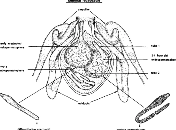
FIG. 1. Schematic diagram of the seminal receptacle of mated 9 containing endospermatophores of different ages. Note the differentiating spermatid in the newly evaginated endospermatophore and the mature sperma-tozoon in the 24-h-old endospermatophore.
and, after 2 changes of propylene oxide, embed-ded in Spurr or Durcupan ACM Fluka. Fine and semifine sections were cut with glass knives on a Reichert ultra microtome. Fine sections were stained with uranyl acetate (Watson) and lead cit-rate (Reynolds) and examined with a Hitachi HS 7S electron microscope.
To demonstrate glycogen, the periodic acid-thiocarbohydrazide silver proteinate method of Thiery (1967) was used.
RESULTS
During copulation, an ectospermatophore eject-ed from the male genital orifice contains an en-dospermatophore into which seminal fluid and sperm cells are injected. This endospermatophore evaginates in the female genital tract where the contents of the ectospermatophore are released.
One to 5 endospermatophores may be found in the seminal receptacle of mated females.
Sperm cells in the newly evaginated endosper-matophore are still immature. These spermatids elongate and develop into mature spermatozoa in 1-2 days (Fig. 1). The mature spermatozoa leave the endospermatophore about 2 days later, mi-grate up the oviducts and accumulate in great numbers in the ampulla region.
Fine structure of the spermatid
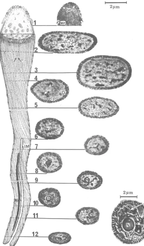
The main body of the spermatid (Fig. 2-15) is composed of 2 long tubes: the outer sheath and the inner cord, the former surrounding the latter. Both tubes are connected at the posterior truncate end of the spermatid (Fig. 2, 6, lla). The anterior end (Fig. lla) is slightly broader, with its apex acutely tapering. The outer sheath and inner cord
are separated by the cisternal cavity (Fig. 2) con-taining a granular material.
The rod-shaped, electron-dense nucleus extends for about V2 the anterior length of the spermatid (Fig. lla). When observed in vivo, the nucleus is easily seen lying over the layer of cellular processes beneath the membrane of the outer sheath and distending the sperm surface (Fig. 2, l i b , c).
A semispherical structure of unknown function, entirely covered by numerous regular rows of bul-bous expansions, lies at the anterior tip of the in-ner cord (Fig. 2-4). In the midregion of this semi-sphere, the last row of bulbous expansions extend as a fringe of motile processes (Fig. 2, point 5). At the anterior end of the spermatid, the acrosome underlies the cellular membrane.
In the anterior part of the spermatid, the mod-erately dense granular cisternal cavity contains sev-eral small vesicles (Fig. 2, point 1). The cytoplasm of the middle and posterior parts is fibrillar-gran-ular (Fig. 5, 6). The granules may represent fibers in cross section. Both fibrillar and granular ele-ments measure about 100-150 A in diam.
The fibrillar-granular cytoplasm in the middle and distal part of the cell accumulates as dense clusters bordering the inner surface of the inner tube (Fig. 2, points 7-9). In this cortical region, there are also numerous dense fibers, appearing in longitudinal sections to extend the length of the spermatid.
The cellular processes extend from the foot of the semispherical structure for the length of the spermatid and cover the entire surface of the inner cord to the posterior apex of the cell. At the point of fusion of inner cord and outer sheath, these processes fold externally upon themselves and ex-tend to the pointed end of the spermatid (Fig. 2, 6, 11). Externally, the cellular processes curve around the sperm surface (Fig. 12); they are equi-distant and essentially parallel (Fig. 8, 9) and most begin immediately following the termination of another (Fig. 9, arrows). Where certain terminat-ing processes do not follow another, flankterminat-ing pro-cesses draw closer and are ultimately parallel (Fig. 12, arrows). Each cellular process is attached to the cell membrane by a short stalk (Fig. 15).
Cellular processes seen in cross section are ar-ranged in 2 concentric circles inside the cisternal cavity (Fig. 7, 10), 1 in the outer sheath and 1 in the inner cord (Fig. lla). Each process is pyriform in cross section and composed of 3 subunits of varying electron density (Fig. 10). The 1st (a) is homogeneous and of relatively low density. The
2nd (b) is crescentic and darkly staining; it overlaps type a at the broad end of the pyriform process. The small, filamentous 3rd form (c) usually rep-resents a terminating end of the process.
Elongation
Final development of the spermatozoon is com-pleted in the endospermatophores within the sem-inal receptacle of mated females and requires at least 24 h (Fig. 1).
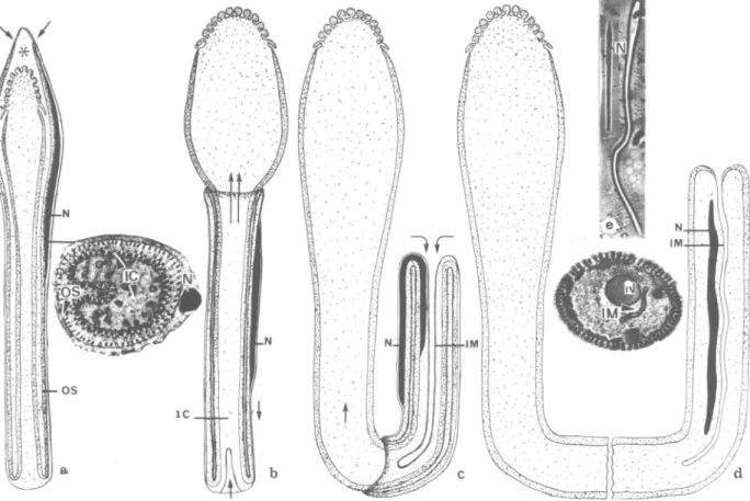
An operculum (Borut 8c Feldman-Muhsam 1976, Wiiest et al. 1978) detaches from the pointed tip of the spermatid (Fig. 1 la) during the forward movement of the conical apex of the inner cord. The freed conical apex bulges (Fig. lib) and the outer sheath carrying the nucleus slips posteriorly to invaginate at the truncate end of the elongating spermatid (Fig. l i b , c). A fold in the freed bulge of the inner cord folds out and swells, forming the eventual clavate anterior part of the mature sper-matozoon (Fig. l i b , c).
The anterior bulge results from the outward ex-pansion of the longitudinal fold seen in cross sec-tions of the immature spermatid (Fig. 7, 10). Evagination and invagination occur simultaneously and continue until the outer sheath and the inner cord merge. Thus, a single clavate structure, dou-ble the length of the original spermatid, is formed.
The mature spermatozoon
The mature spermatozoon remains in the en-dospermatophore within the seminal receptacle for 3-4 days, then migrates up the oviducts to the ampulla and very rarely to the ovaries.
The mature spermatozoon is composed mainly of the anterior region of the former inner cord added to the former outer sheath that has turned inside out (Fig. 12).
The internal structure of the spermatozoon is similar to the inner cord of the spermatid, thus only differences observed between both stages will be mentioned.
The final external features of the mature sper-matozoon are characterized by a thick clavate an-terior end carrying a semispherical apical structure covered by numerous small expansions. A short fringe of motile processes encircles the apical structure just posterior to the bulbous expansion (Fig. 12).
Upon completing spermiogenesis, the clavate anterior end flattens and the posterior part forms a long, thin "caudal" region.
1981 El Said et al.: Spermiogenesis in Amblyomma hebraeum 467
M
Fie. 2. Three-dimensional reconstruction of the spermatid with micrographs of the corresponding cross and longitudinal sections through different regions. Note the cellular processes (cp) of the outer sheath (OS) encircling the bulbous expansions (be) at the tip of the inner cord, followed by motile processes (nip) and cellular processes (cp) inside the cisternal cavity (cc). The fibrillar-granular region (fg) at the anterior part is denser than in the posterior nucleate region (N). The cytoplasm contains numerous mitochondria (m), and dense fibers (D). Several vesicles (v) are observed in the cisternal cavity (cc).
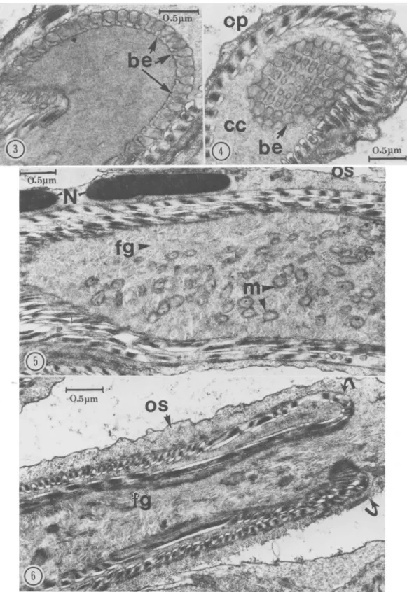
FIG. 3-6. 3, Longitudinal section through the bulbous expansions (be) connected by short stalks to the tip of the inner cord; 4, Cross section through the conical tip apex showing linear arrangement of bulbous expansions (be) inside the cisternal cavity (cc); 5, Longitudinal section through the nucleate part of the spermatid. Note the electron-dense nucleus (N) in the outer sheath, mitochondria (m), and fibrillar-granular cytoplasm of inner cord; 6, Posterior region of the spermatid representing point of attachment (arrows) of inner cord and outer sheath (OS).
1981 El Said et al.: Spermiogenesis in Amblyomma hebraeum 469
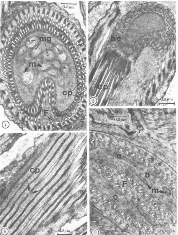
FIG. 7-10. 7, Cross section of spermatid showing accumulation of mitochondria (m) containing large electron-dense mitochondrial granules (mg). Note the fold (F) in the inner cord and the 2 concentric circles of cellular processes (cp) inside the asternal cavity (*); 8, Longitudinal section of the anterior part of spermatid showing (he origin of the cellular processes (cp) at the base of the bulbous expansions (be); 9, Tangential section of cellular processes (cp), showing arrangement, internal structure, and terminal points (arrows); 10, Cross section through anterior region of the spermatid showing the deep fold containing numerous elongated mitochondria (m). Note 3 following components of the cellular processes: (a) a homogeneous moderately dense part, (b) an electron-dense crescentic part, and (c) a dwarfed terminal part.
FIG. 11. Schematic diagram of the elongation process of the spermatid: a, Arrows show the point of detach-ment of the outer sheath (OS). Nucleus (N) is present in outer sheath (OS) at anterior end of spermatid. The inner cord (IC) is internal. Cisternal cavity (*); b, The evagination of the inner cord tip (arrows) and the evagi-nation of the outer sheath (OS) at the truncate end; c, The evagievagi-nation-invagievagi-nation process. Note the migration of the nucleus (N) within the outer sheath (OS) into the sperm body; d, The mature spermatozoon after com-pleting elongation process. Note the bulbous head of the sperm and the inner and posterior position of the nucleus; e, Micrograph of spermatid before the elongation process and the mature spermatozoon.
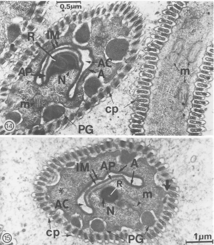
the acrosome and nucleus are finally located in the "caudal" end of the sperm closely adjacent to the invaginated cell membrane (=imagination mem-brane), which lines the acrosomal canal (Fig. 11, 12). In young spermatozoa, the acrosome is rep-resented by a thin, flattened vesicle, which is lim-ited by a membrane; only a very narrow cytoplas-mic zone separates the cell membrane from the acrosomal membrane. As the spermatozoon ages, this structure develops and its association with the nucleus is easily observed. The schematic diagram (Fig. 13) illustrates the acrosomal region and the nucleo-acrosomal association. An acrosomal plate (Fig. 14) becomes evident and is characterized as a moderately dense thickening of the acrosome. The plate extends for a short distance below and above its point of attachment with the nucleus; it is continuous with the acrosome (Fig. 13, 15). The base of the nucleus joins the nucleus to the acro-somal plate. A long, thin, moderately dense rod
embedded in the nucleus and extending into it for a short distance apparently functions to strengthen the nucleo-acrosomal liaison by further fixing the acrosomal plate to the base of the nucleus. The nuclear membrane is always present.
The cell membrane (=invagination membrane) and the acrosome line a canal (=acrosomal canal) created by the invagination of the outer sheath during the elongation process (Fig. 11-15). Nu-merous tubular vesicles are seen accumulating within the acrosomal canal (Fig. 13).
The external surface of the mature spermato-zoon appears ridged owing to cellular processes winding slightly in more or less parallel rows over the entire surface of the mature sperm.
The dense fibers observed in the inner cord of the spermatid appear now in the cortical region of the cytoplasm, usually underlying the spermato-zoon plasma membrane.
extracel-1981 El Said et al.: Spermiogenesis in Amblyomma hebraeum 471
FIG. 12. Three-dimensional reconstruction of the spermatozoon with corresponding micrographs of different, regions. Note the glycogen granules (G) and mitochondria (m) at anterior end, as well as the cell membrane (cm) and underlying acrosome, nucleus (N), glycogen (G), and peripheral granules (PG) at the posterior end. Inset: Cross section through nuclear part of spermatozoon demonstrating /3-glycogen particles stained by the periodic acid-thiosemicarbazide-proteinate silver technique of Thiery (1967).
NUCLEO-ACROSOMAL REGION
OF SPERMATOZOON
>' /, , • , , JJ
m
FIG. 13. Nucleo-acrosomal region of the spermatozoon (arrows indicate localization within the mature sperm). The acrosomal plate (AP) and the nucleus (N) are connected at the nuclear base (N base) by a long rod (R) centrally placed in the nucleus. The cell membrane (IM) and the acrosome (A) line the acrosomal canal (AC) in which numerous tubular vesicles (tv) are situated, cp: cellular processes.
1981 El Said et al.: Spermiogenesis in Amblyomma hebraeum 473
cp
FIG. 14-15. 14, PG: peripheral granules. Site of acrosomal plate (AP) attachment with nucleus (N) and acrosomal canal (AC). A: acrosome. The moderately electron-dense rod (R) connects the acrosomal plate (AP) to the nucleus (N) base. Peripheral granules (PG) become prominent at this stage and the electron lucent space separating them from the sperm cell membrane becomes more evident; 15, The cell membrane ( = invagination membrane IiVl), acrosomal plate (AP) and acrosome (A) at the "caudal" end of spermatozoon. Prominent pe-ripheral granules (PG), fibrillar-granular cytoplasm (fg), and a single layer of cellular processes (cp) are shown. A short stalk connects a cellular process to the sperm cell membrane ( *).
lularly between the cellular processes and the plas-ma membrane, which separates them from the cor-tical cytoplasm. In older spermatozoa found in the oviducts and ampulla, these granules become more dense and appear to have a granular matrix (Fig. 14). Glycogen, represented by individual /3-glyco-gen particles, is located in the central region of the spermatozoon (Fig. 12 inset).
Motility
Sperm movement was examined in fresh squash preparations under phase contrast. The sperm was seen to move with the clavate mitochondrial region followed by the thin nucleate "caudal" part. The fascinating sperm movement is a glide, usually in a straight line or occasionally in a rotating move-ment. When the sperm meets an obstacle, it sur-mounts it or turns away from large objects. The bulbous expansions on the conical apex and the surrounding circlet of motile processes are seen on close observation to be in continuous motion. A peculiar rolling-up and folding-out motion of the tip of the flattened anterior region was observed.
DISCUSSION
The results show that the fine structures of sper-matids and spermatozoa of Amblyomma hebraeum are similar in most respects to those of Amblyomma
dissimili Koch, 1844 and Dermacentor andersoni
Stiles, 1908 (Reger 1961, 1974) but that certain differences exist between these species.
The elongation process by which the A. hebraeum spermatid develops into a mature spermatozoon is like that described in argasid and ixodid ticks by Brinton et al. (1974), Borut 8c Feldman-Muhsam (1976), and Wiiest et al. (1978).
The groove observed by Reger (1961) in the A.
dissimili spermatid is interpreted here as an
ante-rior fold that expands, giving volume to this region once the evaginating anterior part of the sperma-tozoon is freed from the outer sheath. Suleiman & Brown (1978) stated that the "groovelike depres-sion" is absent in the anterior end of the spermatid of Dermacentor variabilis (Say); however, their fig.
11 of the nucleus and the groove shows this part to be in the anterior end.
Suleiman & Brown (1978) mentioned a unique pluglike structure at the tip of the nucleate part of the spermatozoon. No pluglike structure was ob-served by us or in scanning views of this region by Feldman-Muhsam & Filshie (1976) and Wuest et al. (1978).
We have demonstrated that the motile processes
encircling the tip of the spermatozoon, observed by Casteel (1917) but contradicted by Oppermann (1935), are present in the A. hebraeum spermatid but concealed by the outer sheath. This view is in accord with the findings of Reger (1961) in A.
dis-simili.
Christophers (1906), Bonnet (1907), and Nor-denskiold (1920) misinterpreted the spermatid in the male vas deferens as the mature sperm. They did not envisage further developmental stages in the female genital tract. In Hyalomma "aegyptium (Linnaeus) 1758," Bonnet (1907) observed 2 forms of sperm, 1 rod shaped, the other filiform. These were obviously the spermatid before evagination and the mature spermatozoon, respectively.
Opinions differ regarding localization of mito-chondria within the sperm body. In A. hebraeum, they are abundant in the anterior or clavate region, but few and randomly dispersed elsewhere and absent in the conical tip. According to Casteel (1917), who studied Argas miniatus Koch, 1844, and Sokolov (1958), who studied Ornithodoros
tho-lozani (Laboulbene & Megnin) 1882 (=O. papillipes),
mitochondria are arranged in rows or lines. Cas-teel (1917) observed that "the mitochondria are arranged in straight lines so that the front end of the tube appears longitudinally striated." The lin-ear arrangement of mitochondria observed at the light microscopic level was recently confirmed on the ultrastructural level in O. moubata (Murray) 1877 (Breucker & Horstmann 1968). Our results confirm that mitochondria accumulate in the an-terior part; however, their specific arrangement in lines or rows was not observed in A. hebraeum. This is also concluded from the results of Oppermann (1935) and Reger (1961). The different results of Casteel (1917), Sokolov (1958), Breucker & Horst-mann (1968), and ourselves concerning mitochon-drial distribution may be due to structural differ-ences in individual species.
During the elongation process leading to the mature spermatozoon, an acrosomal canal forms by invagination of the cell membrane (=invagina-tion membrane) and the underlying acrosome. The acrosomal canal was termed a "flagellum" by Oppermann (1935), Tuzet 8c Millot (1937), Shar-ma (1944), Wagner-Jevseenko (1958), and Khalil (1969). These authors described an evagination process by which this "flagellum" protrudes out-ward from the nucleate end of the spermatozoon and the nucleus migrates into the "flagellum" base. This process occurs when the sperm leaves the seminal receptacle or in the oviducts (Khalil 1969).
1981 El Said et al.: Spermiogenesis in Amblyomma hebraeum 475
The seminal receptacle, oviducts, and ovaries of female A. hebraeum were examined before, during, and after ovulation, but this process was not ob-served. At no time were the sperm of this tick "fla-gellate." Evaginated "flagella" were said by Casteel (1917) to be nonfunctional and nonmotile and no axoneme was demonstrated. Thus, we refrain from calling this structure a "flagellum" and con-clude that tick sperm are aflagellate cells.
The role of the acrosome in egg penetration has not been demonstrated. Therefore, we are uncer-tain of the true identity of this structure. The ac-rosome described here is similar to the acac-rosome identified by Breucker & Horstmann (1968) in 0.
moubata and by Reger (1963) in A. dissimili. Motility
All observers are in general agreement regard-ing the unusual type of tick sperm movement. The clavate mitochondrial part is anucleate but repre-sents the active end, by which the spermatozoon moves forward, and is followed by the nucleate end.
Casteel (1917) observed that the anterior thick part, of the sperm flattens at the terminal phase of maturation. He described movement at the tip of the anterior region as contracting and expanding. Recent observations on living tick sperm by Reger (1974) corroborate those of Christophers (1906), who described vermicular contortions of the an-terior part of the sperm. Our observations on movement of this part showed that the tip moves in a peculiar rolling-up and folding-out manner and that the bulbous expansions are in continuous motion.
The general type of motility of tick spermatozoa was described by Christophers (1906) and Reger (1974) as a steady gliding movement and Sharma (1944) described it as rotary. Our observations on living sperm confirm the gliding movement and some rotary movement. A contractile system is probably involved in these movements, as suggest-ed by Rothschild (1961), a view also acceptsuggest-ed by Feldman-Muhsam & Filshie (1976), who proposed that certain spermiophore movements may be ex-plained by contraction of the cytoplasmic fila-ments. So-called "contractile elements" (Casteel 1917), "elastic strands" (Sokolov 1958), "filament bundles" (Breucker & Horstmann 1968), and "sub-surface filaments" (Reger 1963) have been ob-served in spermatozoa of different ticks. These ele-ments, also observed in A. hebraeum, are termed "dense fibers." Microtubules are lacking in tick
sperm cells but their function may be compensated for by the presence of dense fibers extending the entire length of these cells. The diameters of these fibers (100-150 A) correspond to the diameter of actin and myosin fibers, thus they may represent the above-mentioned contractile proteins fre-quently found in nonmuscle cells and thought to be involved in generating various "movements."
Apart from the dense-fibrillar elements, cellular processes or the fibrillar-granular region, or both, are probably involved in motion, as suggested by Reger (1974). The fringe of motile processes around the tip of the sperm head, which are in constant movement (Casteel 1917), may also con-tribute to motile activity.
The large accumulation of mitochondria and high concentration of /3-glycogen particles among them are certainly related to energy production, which is essential for motility. The /3-glycogen par-ticles are considered to be an easily assimilable form of glucose storage. Glycogen presumably serves as an endogenous source of energy under aerobic conditions within the mitochondria.
Acknowledgments. We express our deepest gratitude to Dr H.
Hoogstraal for critically reading the manuscript. We also wish to thank Professor H. Huggel, Head of the Laboratory of Com-parative Anatomy and Physiology, for providing us with a working place in his laboratory and for help and encourage-ment. Thanks are also extended to Mr Bouvard of Les Barges Research Unit, Ciba-Geigy, for kindly supplying tick materials for this study. Sincere thanks are due to Madame Seif for tech-nical assistance.
LITERATURE CITED
Bonnet, A. 1907. Sur les organes genitaux males et la
sper-matogenese chez les Ixodes. C. R. Assoc. Fr. Av. Sci. 35: 544-49.
Borut, S. & B. Feldman-Muhsam. 1976. A new observation
on spermateleosis in ticks./. Parasitol. 62: 318-20.
Breucker, H. 8c E. Horstmann. 1968. Die Spermatozoen der
Zecke Ornithodorus moubata (Murr.). Z. Zellforsch. Mikrosk.
Anal. 88: 1-22.
1972. Die Spermatogenese der Zecke Ornithodoros moubata (Murr.). Z. Zellforsch. Mikrosk. Anal. 123: 18-46.
Brinton, L. P., W. Burgdorfer & J. H. Oliver, Jr. 1974.
His-tology and fine structure of spermatozoa and egg passing in the female tract of Dermacentor andersoni Stiles (Acari: Ixodidae). Tissue &f Cell 6: 109-25.
Casteel, D. B. 1917. Cytoplasmic inclusions in male germ cells
of the fowl tick. J. Morphol. 28: 643-93.
Christophers, S. R. 1906. The anatomy and histology of ticks.
Sci. Mem. Off. Med. Sanit. Dep. Gov. India, n.s. No. 23. 55 p.
El Said, A. & Z. Swiderski. 1980a. Regional specialization of the sperm membrane in the tick Amblyomma hebraeum Koch (Acari: Ixodidae). Cell Tissue Res. 208: 35-45.
1980b. A freeze etching study on the origin of cellular pro-cesses in spermatids of the tick Amblyomma hebraeum Koch (Acari: Ixodidae). Proc. 7th Europ. Congr. Electr. Microsc.
1980c. Comparative freeze-etch replica and thin section studies on the phagocytotic process in the sperm of the tick Amblyomma hebraeum (Acari: Ixodidae). Proc. 7 th Europ. Congr. Electr. Microsc. The Hague, 1980. 2: 62-63.
Feldman-Muhsam, B. & B. K. Filshie. 1974. The ultrastruc-ture of the connecting links between tick spermiophores and their symbionts. Abstr. Proc. 3rd. Int. Congr. Parasitol., Munich, 1974. 2: 965-66.
1976. Scanning and transmission electron microscopy of the spermiophores of Omithodoros ticks: an attempt to explain their modlity. Tissue & Cell 8: 411-19.
1979. A SEM study of spermiophores of Omithodoros ticks, p. 681-85. In: Proc. 4th. Int. Congr. Acarol. Saalfelden, 1974.
Feldman-Muhsam, B., B. K. Filshie & S. Borut. 1975. EM study on tick prospermium relevant to onset of sperma-teleosis. Abstr. 50th Annu. Meet. Am. Soc. Parasitol., Louisi-ana, p. 49.
Khalil, G. M. 1969. Biochemical and physiological studies of certain ticks (Ixodoidea). Gonad development and game-togenesis in Argas (Persicargas) arboreus, Kaiser, Hoogstraal and Kohls (Argasidae)./. Parasitol. 55: 1278-97.
1970. Biochemical and physiological studies of certain ticks (Ixodoidea). Gonad development and gametogenesis in Hyalomma (H.) anatolicum excavatum Koch (Ixodidae). J. Parasitol. 56: 596-610.
Nordenskiold, N. A. E. 1920. Spermatogenesis in Ixodes ricinus L. Parasitology 12: 159-65.
Oppermann, E. 1935. Die Entstehung des Riesenspermien von
Argas columbarum (Shaw) {reflexus F.). Z. Mikrosk. Anat. Forsch. 37: 538-60.
Reger, J. F. 1961. The fine structure of spermatids from the tick Amblyomma dissimili. J. Ultrastruct. Res. 5: 584-99. 1962. A fine-structure study on spermiogenesis in the tick
Amblyomma dissimili with special reference to the develop-ment of motile processes. J. Ultrastruct. Res. 7: 550-65.
1963. Spermiogenesis in the tick Amblyomma dissimili as re-vealed by electron microscopy. J. Ultrastruct. Res. 8: 607-21.
1974. The origin and fine structure of cellular processes in spermatozoa of the tick Dermacentor andersoni. J. Ultrastruct. Res. 48: 420-34.
Rothschild, Lord. 1961. Structure and movement of tick sper-matozoa (Arachnida, Acari). Q. J. Microsc. Sci. 102: 239-47.
Samson, K. 1909. Zur Spenniohistiogenese der Zecken.
Sit-zungsber. Ges. Naturf. Fr. Berlin No. 8: 486-99.
Sharma, G. P. 1944. Studies on spermatogenesis in ticks. Proc.
Natl. Inst. Sci. India 10: 305-16.
Sokolov, I. I. 1958. Cytological studies of the development of male germ cells in Omithodoros papillipes Bir. (Acari, Ixo-doidea). Entomol. Obozr. 37: 260-81. (In Russian.) Suleiman, A. S. & G. G. Brown. 1978. Spermiogenesis in the
dog tick Dermacentor variabilis (Say). Iowa State J. Res. 53: 93-108.
Thiery, J. P. 1967. Mise en evidence des polysaccarides sur coupes fines au microscope electronique. J. Microsc. Pans 6: 987-1118.
Till, W. M. 1961. A contribution to the anatomy and histology of the brown ear-tick Rhipicephalus appendiculatus New-mann. Mem. Entomol. Soc. South. Afr. 6: 5-124.
Tuzet, O. & J. Millot. 1937. Recherches sur la spermiogenese des Ixodes. Bull. Biol. Fr. Belg. 71: 190-205.
Wagner-Jevseenko, O. 1958. Fortpflanzung bei Ornithodorus
moubata und genitale Ubertragung von Borrelia duttoni. Acta Trop. 15: 118-68.
Warren, E. 1933. On atypical modes of sperm development in certain arachnids. Ann. Natal Mus. 7: 151-94.
Wiiest, J., A. El Said, Z. Swiderski & A. Aeschlimann. 1978. Morphology of the spermatid and spermatozoon of Am-blyomma hebraeum Koch (Acarina: Ixodidae). Z. Parasitenkd. 55: 91-99.
NOTICE
FORUM ON PACIFIC SYSTEMATICS COLLECTIONS AND
BIOGEOGRAPHY SYMPOSIUM
At the annual meetings of the Association of Systematics Collections, to be held 23-26 May 1982, at the Bishop Museum, Honolulu, two symposia will be held. The first symposium will be a one-day forum on the status, care and use of collections of Pacific area biota. This forum will relate primarily to collections housed on Pacific islands, but will also involve Pacific collections in museums elsewhere. Matters of location and extent of collections, their curation, availability, safe-keeping and future will be discussed. Speakers will address present situations in certain island nations or nearby countries and will speak on curatorial, legal, service and other aspects of collections. A second, one-day symposium will deal with Pacific island biogeography relating to insects, other animals and plants, mostly terrestrial but also marine. Emphasis will be on tropical Pacific island groups.
Everyone is welcome to come to these symposia and to participate in the discussions, as well as the regular meetings of the Association. For further information, please write: Forum, % Dr J. L. Gressitt or Dr F. J. Radovsky, Bishop Museum, P.O. Box 19000-A, Honolulu, Hawaii 96819.