RESEARCH OUTPUTS / RÉSULTATS DE RECHERCHE
Author(s) - Auteur(s) :
Publication date - Date de publication :
Permanent link - Permalien :
Rights / License - Licence de droit d’auteur :
Institutional Repository - Research Portal
Dépôt Institutionnel - Portail de la Recherche
researchportal.unamur.be
University of Namur
Effect of ABCB1 genetic polymorphisms on the transport of rivaroxaban in HEK293
recombinant cell lines
Sennesael, Anne Laure; Panin, Nadtha; Vancraeynest, Christelle; Pochet, Lionel; Spinewine,
Anne; Haufroid, Vincent; Elens, Laure
Published in:
Scientific Reports
DOI:
10.1038/s41598-018-28622-4
Publication date:
2018
Document Version
Publisher's PDF, also known as Version of record
Link to publication
Citation for pulished version (HARVARD):
Sennesael, AL, Panin, N, Vancraeynest, C, Pochet, L, Spinewine, A, Haufroid, V & Elens, L 2018, 'Effect of
ABCB1 genetic polymorphisms on the transport of rivaroxaban in HEK293 recombinant cell lines', Scientific
Reports, vol. 8, no. 1, 10514, pp. 10514. https://doi.org/10.1038/s41598-018-28622-4
General rights
Copyright and moral rights for the publications made accessible in the public portal are retained by the authors and/or other copyright owners and it is a condition of accessing publications that users recognise and abide by the legal requirements associated with these rights. • Users may download and print one copy of any publication from the public portal for the purpose of private study or research. • You may not further distribute the material or use it for any profit-making activity or commercial gain
• You may freely distribute the URL identifying the publication in the public portal ? Take down policy
If you believe that this document breaches copyright please contact us providing details, and we will remove access to the work immediately and investigate your claim.
Effect of ABCB1 genetic
polymorphisms on the transport
of rivaroxaban in HEK293
recombinant cell lines
Anne-Laure Sennesael
1,2, Nadtha Panin
3, Christelle Vancraeynest
2, Lionel Pochet
2,
Anne Spinewine
1,4, Vincent Haufroid
3,5& Laure Elens
3,6Direct oral anticoagulants (DOAC) are substrates for the ABCB1 transporter (also called P-glycoprotein), an active efflux pump. ABCB1 polymorphisms have been previously reported to influence the
pharmacokinetics of several drugs such as immunosuppressants and tyrosine kinase inhibitors. Recently, in vivo studies have suggested that genetic variants might contribute to the inter-individual variability in DOAC plasma concentrations. Therefore, we evaluated the in vitro effect of the most common coding ABCB1 single nucleotide polymorphisms (SNP), 1236 C > T-2677G > T-3435C > T, and the coding ABCB1 1199 G > A SNP on the transport activity towards rivaroxaban. HEK293 cells were transfected to overexpress the ABCB1 wild-type (1236C-2677G-3435C, 1199 G) or variant proteins (1236C-2677G-3435T, 1236T-2677T-3435T or 1199 A). ABCB1 expression decreased the intracellular accumulation of rivaroxaban, when compared to control cells. This confirms the involvement of ABCB1 in the active transport of rivaroxaban. However, the ABCB1 1236 C > T-2677G > T-3435C > T and 1199 G > A SNPs had no significant influence on the intracellular accumulation of rivaroxaban when compared to the wild-type protein. These results suggest that the ABCB1 coding SNPs investigated in the present study are unlikely to contribute to the inter-individual variability in rivaroxaban plasma concentrations.
The landscape of oral anticoagulant therapy has significantly changed in the last years. Direct oral anticoagulants (DOAC) have been approved for stroke prevention in non-valvular atrial fibrillation and the treatment as well as the secondary prevention of venous thromboembolism. They are increasingly used in clinical practice, partly due to their higher convenience for clinicians and patients (fixed-dose regimen, fewer interactions with drugs and food) compared with vitamin K antagonists (VKA)1. Four DOACs are currently available: three direct factor Xa
inhibitors (apixaban, edoxaban, rivaroxaban) and one direct thrombin inhibitor (dabigatran etexilate). In 2016, guidelines issued by the European Society of Cardiology or the American College of Chest Physicians have rec-ommended DOACs over VKAs in eligible patients2,3.
All DOACs are substrates for the ABCB1 transporter, also called P-glycoprotein (P-gp) or formerly designated as the multidrug resistance 1 (MDR1) protein4. This active efflux pump belongs to the ATP-binding cassette
transporter superfamily and is involved in the disposition of multiple drugs from diverse classes such as antican-cer agents, immunosuppressants or antibiotics5. ABCB1 expression at the apical membrane of enterocytes limits
absorption, while its localization on the luminal membrane of hepatocytes and renal tubular cells enhances biliary and renal excretion respectively. In patients taking rivaroxaban, the ABCB1 protein expression in renal tubular cells is particularly important given that more than one third of the dose is eliminated unchanged in the urine6.
1Clinical Pharmacy Research Group, Louvain Drug Research Institute, Université catholique de Louvain (UCL),
Brussels, Belgium. 2Department of Pharmacy, Namur Research Institute for LIfe Sciences, Namur Thrombosis and
Hemostasis Center (NTHC), University of Namur, Namur, Belgium. 3Louvain Centre for Toxicology and Applied
Pharmacology, Institut de Recherche Expérimentale et Clinique, UCL, Brussels, Belgium. 4Department of Pharmacy,
NTHC, CHU UCL Namur, Yvoir, Belgium. 5Department of Clinical Chemistry, Cliniques Universitaires Saint-Luc,
UCL, Brussels, Belgium. 6Integrated PharmacoMetrics, PharmacoGenomics and PharmacoKinetics, Louvain Drug
Research Institute, UCL, Brussels, Belgium. Correspondence and requests for materials should be addressed to A.-L.S. (email: [email protected])
Received: 15 February 2018 Accepted: 18 June 2018 Published: xx xx xxxx
www.nature.com/scientificreports/
Many medications that are commonly prescribed in patients with atrial fibrillation (AF) can inhibit or induce ABCB1 activity and thereby influence DOAC pharmacokinetics7. For instance, it has been demonstrated that the
concurrent use of moderate inhibitors of ABCB1 such as amiodarone or verapamil led to a nearly 40% increase in rivaroxaban exposure8,9. Combining this with renal impairment or older age, two common characteristics in AF
patients, produced even stronger effect on rivaroxaban pharmacokinetics.
Genetic polymorphisms have also been reported to influence ABCB1 activity and/or expression5. The ABCB1
gene, located on chromosome 7, is composed of 29 exons in a 251.3-kb genomic region. More than 60 coding single nucleotide polymorphisms (SNP) have been described for ABCB110. The three most common SNPs in
the coding region are rs1128503 (1236 C > T, Gly412Gly), rs2032582 (2677 G > T, Ala893Ser) and rs1045642 (3435 C > T, Ile1145Ile). They are in strong linkage disequilibrium and present a minor allelic frequency around 50% in the Caucasian population. They have been widely investigated, with inconsistent effects on drug dispo-sition, drug response and toxicity5. Another SNP of interest is the 1199 G > A non-synonymous coding SNP
(rs2229109), with an allelic frequency around 6% in Caucasians. The amino acid change (Ser400Asn) caused by this SNP is located in a cytoplasmic loop involved in substrate recognition. In previous in vitro studies, it has been shown that 1199 G > A affects the transport of tacrolimus, vinblastine or tyrosine kinase inhibitors but that cyclosporine A, Rh123 and doxorubicin are transported in a similar extent by the wild-type and variant ABCB1 proteins11,12.
Despite the fixed-dose regimen of DOACs, significant inter-individual variability in peak and trough plasma concentrations has been described13,14. In a prospective cohort study, 40% of patients with atrial fibrillation had
rivaroxaban plasma measurements outside the 5th–95th percentile interval observed in phase 3 trials15. Moreover,
in an analysis of the ROCKET-AF trial, rivaroxaban exposure predicted the risk of major bleeding16. Inversely, it
was recently highlighted that DOAC patients experiencing thromboembolic events had lower plasma levels in the first month of treatment17. Genetic polymorphisms have been proposed to explain why patients taking the same
DOAC dose present highly variable plasma concentrations18. Therefore, this study aimed to evaluate the in vitro
effect of the ABCB1 1236 C > T-2677G > T-3435C > T and 1199 G > A SNPs on the transport activity towards rivaroxaban.
Results
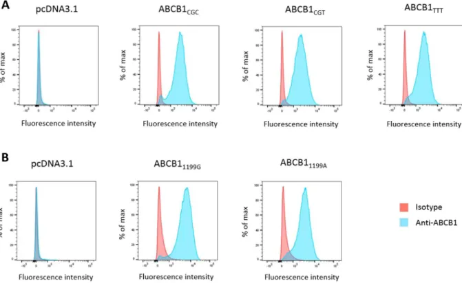
Generation of ABCB1 recombinant cell lines.
HEK293 cells overexpressing ABCB1 wild-type and vari-ant proteins have been previously generated and characterized11,19. For ABCB1 1236 C > T-2677G > T-3435C > T,the recombinant models used consisted of stably transfected cell lines with the pcDNA3.1 empty vector, HEKpcDNA3.1, the wild-type vector, HEK1236C-2677G-3435C or two different combinations of the variant cDNA,
HEK1236C-2677G-3435T and HEK1236T-2677T-3435T, hereafter referred to as HEKcontrol, HEKCGC, HEKCGT and HEKTTT
respectively. For ABCB1 1199 G > A, three recombinant cell lines were used: HEKcontrol, HEK1199G (wild-type
pro-tein) and HEK1199A (variant protein). To ensure similar ABCB1 surface expression among the different cell lines,
cells were sorted using fluorescence activated cell sorting (FACS) with fluorescence parameters gated on the same level of intensity. As shown in Fig. 1A, similar ABCB1 surface expression was observed by analytical flow cytom-etry among recombinant cell lines, both for the ABCB1 1236 C > T-2677G > T-3435C > T and 1199 G > A SNPs. In contrast, there was no fluorescence detected in HEKcontrol ensuring no or at least very low basal expression in
our negative controls.
Impact of ABCB1 1236 C > T-2677G > T-3435C > T polymorphisms on the intracellular
accumu-lation of rivaroxaban.
First, we assessed the impact of ABCB1 1236 C > T-2677G > T-3435C > T on the ABCB1 transport activity towards rivaroxaban. HEKcontrol, HEKCGC, HEKCGT and HEKTTT were incubated withdifferent concentrations of rivaroxaban ranging from 50 to 1000 ng/ml. These concentrations were chosen to cover rivaroxaban plasma levels encountered in clinical practice (concentrations up to 600 ng/ml were described in real-life patients with AF)14,15. As shown in Fig. 2, the intracellular accumulation of rivaroxaban was
signifi-cantly decreased in recombinant cell lines overexpressing ABCB1 when compared to control cells (Fig. 2, [50, 250 and 500 ng/ml], p < 0.01; [100 and 1000 ng/ml], p < 0.001). However, we observed that the intracellular accumu-lation of rivaroxaban was similar between HEKCGC, HEKCGT and HEKTTT (Fig. 2, p > 0.05 at all concentrations).
Impact of ABCB1 1199 G > A polymorphism on the intracellular accumulation of rivaroxaban.
Same experiments were conducted to investigate the impact of ABCB1 1199 G > A on the ABCB1 activity towards rivaroxaban. Again, the intracellular accumulation of rivaroxaban was lower in cell lines overexpressing ABCB1, when compared with control cells whatever their genotype (Fig. 3, [from 50 to 1000 ng/ml], p < 0.001). As for the three other variants, we did not find any statistical difference in the intracellular accumulation of rivaroxaban between cells overexpressing the ABCB1 1199 G and 1199 A proteins (Fig. 3, p > 0.05 at all concentrations).Discussion
In this study, we investigated the in vitro impact of ABCB1 genetic polymorphisms on the transport activity towards rivaroxaban. Previously validated HEK293 recombinant cell models were used11,19. We confirmed
the involvement of ABCB1 in the active transport of rivaroxaban6. Indeed, we found that ABCB1 expression
decreases the intracellular accumulation of the drug. However, we failed to show a significant effect of the dif-ferent SNPs investigated. The ABCB1 1236 C > T-2677G > T-3435C > T and 1199 G > A SNPs had no significant influence on the intracellular accumulation of rivaroxaban when compared to the wild-type protein.
To our knowledge, this is the first in vitro study to explore the role of ABCB1 genetic determinants in the dis-position of DOACs. Few in vivo studies have been published so far. Recently, a case has been described reporting a rivaroxaban-treated patient admitted at the hospital for severe anemia related to gastrointestinal bleeding20. This
unexpected delayed clearance. The authors suggested that the homozygous presence of ABCB1 variant alleles 2677–3435TT might have contributed to the altered drug clearance. This observation seems in contradiction with our results showing no influence of those SNPs using in vitro models. However, it must be stressed that this observation relates to an isolated clinical case and, given the relatively high frequency of the homozygous gen-otype (up to 25% of the population of Caucasian origin is expected to be 2677–3435TT), it might simply reflect a spurious association that is only due to chance. Furthermore, this patient received concomitant simvastatin (40 mg q.d) which is known to inhibit CYP3A4 enzyme but also the ABCB1 and ABCG2 transporters and might have explained the findings. Finally, the patient presented a moderate renal impairment that could have worsened the renal elimination of the drug.
Figure 1. ABCB1 cell surface expression. Flow cytometry histograms of HEK293 cells transfected with (A) the empty pcDNA3.1 vector, ABCB1CGC, ABCB1CGT or ABCB1TTT and (B) the empty pcDNA3.1 vector, ABCB11199G,
ABCB11199A. Cells were incubated with a FITC anti-ABCB1 antibody (blue) or a matched isotypic control (red).
Figure 2. Impact of ABCB1 1236 C > T-2677G > T-3435C > T on the intracellular accumulation of rivaroxaban. Intracellular accumulation of rivaroxaban after 120 min of incubation (N = 3) at different concentrations in HEKcontrol, HEKCGC, HEKCGT and HEKTTT. The absolute amount of rivaroxaban (in ng)
was divided by the total amount of proteins in cell extracts (in mg). *Compared to control cells: *p < 0.05, **p < 0.01, ***p < 0.001.
www.nature.com/scientificreports/
More recently, a randomized, two-center, crossover study was performed in healthy volunteers to study the influence of ABCB1 polymorphisms on the pharmacokinetics of dabigatran and rivaroxaban. Peak plasma concentration and area under the curve (AUC) were compared across three different ABCB1 genotypes i.e. 2677GG-3435CC, 2677GT-3435CT and 2677TT-3435TT and the effect of clarithromycin co-administration on the pharmacokinetics was also assessed. In agreement with the fact that rivaroxaban and dabigatran etexilate are both substrates for ABCB1, clarithromycin co-administration led to a two-fold increase in both drugs’ AUC, irre-spective of ABCB1 genotype. Their conclusion meets the observations made in the present investigation as they inferred that ABCB1 genotype is not a significant determinant of inter-individual variability in dabigatran and rivaroxaban pharmacokinetics but that co-administration of a P-gp/CYP3A4 inhibitor with dabigatran etexilate or rivaroxaban may warrant caution.
ABCB1 is involved in the transport of many endogenous, dietary and drug compounds that do not share obvious common structural features. The ABCB1 1236 C > T-2677G > T-3435C > T SNPs have been inconstantly described to influence the clinical pharmacokinetics of immunosuppressants, anticancer agents, antiepileptic drugs or antidepressants5. A trend towards higher concentrations in patients carrying the variant haplotype seems
to emerge, but therapy adjustments are currently not required or even recommended. The ABCB1 1199 G > A SNP has previously shown a substrate-dependent impact on transport activity. For instance, the variant allele (1199 G) decreased ABCB1 activity towards tacrolimus in HEK293 and K562 recombinant cell lines, but did not affect the transport of cyclosporine11. Similarly, the in vitro effect of 1199 G > A was lower for nilotinib compared
to imatinib, two tyrosine kinase inhibitors12. Structural flexibility and multiple binding sites were suggested as
explanations for the polyspecificity of ABCB121. In that way the 1199 G > A SNP, which is located in a cytoplasmic
loop involved in substrate recognition, may affect the binding of some ABCB1 substrates while the transport of the others remains unchanged as this seems to be the case for rivaroxaban11.
Another explanation for our negative findings may rely in the fact that rivaroxaban is described as a weak to moderate substrate for ABCB14,22. In Caco-2 cells for instance, ABCB1 inhibition reduced rivaroxaban efflux
by only 23% whereas the efflux of dabigatran was inhibited by 87%4. Moreover, rivaroxaban is also substrate for
the ABCG2 transporter, known as breast cancer resistance protein (BCRP), and the cytochrome P450 (CYP) 3A4/3A5 enzyme. The active renal secretion by ABCB1 and ABCG2 and hepatic transformation by CYP3A4/3A5 account for 30% and 18% of total drug elimination respectively6. Although combined ABCB1, ABCG2 and
CYP3A4 inhibitors were shown to increase rivaroxaban plasma concentrations by up to 150%, the impact of ABCB1 inhibition alone seems marginal23,24. Gong and colleagues demonstrated that rivaroxaban clearance was
significantly reduced in mice lacking both ABCB1 and ABCG2, but did not differ for individual transporter knockout25. This suggests a compensatory role of one transporter when the other is inhibited. It is important to
note that, in HEK293 cells, the endogenous expression of ABCB1 and ABCG2 is reported to be very low26.
Other genetic determinants could contribute to the inter-individual variability in DOAC plasma concentra-tions. Future studies are needed to assess the impact of ABCG2 polymorphisms on the transport of rivaroxaban. The ABCG2 rs2231142 (421 C > A, Gln141Lys) SNP, with an allelic frequency around 5–10% in Caucasians and 30–60% in East Asians, is of particular interest as it significantly affected plasma trough concentrations in patients treated with apixaban27,28. In the same study the CYP3A5*3 allele, which is highly prevalent in Caucasians, also
contributed to higher apixaban levels. Finally, it could be relevant to investigate the CYP3A4*22 allele, which was
associated with a lower CYP3A4 expression and/or activity29.
In summary, the ABCB1 1236 C > T-2677G > T-3435C > T and 1199 G > A SNPs did not affect the efflux of rivaroxaban in HEK293 recombinant cell lines. Therefore, we believe that these SNPs are unlikely to contribute to the inter-individual variability in rivaroxaban plasma concentrations. Future studies should focus on the role of
ABCG2, CYP3A4 and CYP3A5 genetic polymorphisms in the transport of rivaroxaban.
Figure 3. Impact of ABCB1 1199 G > A on the intracellular accumulation of rivaroxaban. Intracellular accumulation of rivaroxaban after 120 min of incubation (N = 3) at different concentrations in HEKcontrol,
HEK1199G and HEK1199A. The absolute amount of rivaroxaban (in ng) was divided by the total amount of proteins
Materials and Methods
Materials.
Rivaroxaban and [13C6]-rivaroxaban-d5 were purchased from Alsachim (Strasbourg, France).G418 was purchased from Roche Applied Science (Vilvoorde, Belgium).
Cell culture.
Human Embryonic Kidney (HEK293) cells were grown in Dulbecco’s Modified Eagle medium (DMEM) Glutamax 4.5 g/l glucose (Gibco, Invitrogen) supplemented with 10% (v/v) of Fetal Bovin Serum (Gibco, Invitrogen) and 1% (v/v) of Penicillin-Streptomycin solution (Gibco, Invitrogen) at a temperature of 37 °C in the presence of 5% of CO2.Generation of stable recombinant cell lines.
The generation of stable recombinant cell lines has been described in previous studies11,19. Briefly, the expression vectors pcDNA3.1 with cDNA encoding ABCB1(ABCB11199G and ABCB11236T-2677T-3435T) were used. Mutated plasmids ABCB11199A, ABCB11236C-2677G-3435C and
ABCB11236C-2677G-3435T were obtained by site-directed mutagenesis. Plasmids were sequenced to confirm the
pres-ence of mutations and HEK293 cells were then transfected with pcDNA3.1 vectors. G418 (concentration 1 mg/ml) was added to select stable recombinant cell lines, which were characterized by flow cytometry, western-blot and immunofluorescence.
Characterization of ABCB1 cell surface expression.
After one week of culture in the presence of G418, ABCB1 cell surface protein expression was characterized by flow cytometry as previously described with minor changes11. For each recombinant cell line, 106 cells were harvested by centrifugation and washed twice withice-cold buffer solution (phosphate buffer saline (PBS), FBS 1%, EDTA 1 mM). Cells were then incubated 45 min on ice in the dark in buffer solution containing FITC Mouse anti-P-glycoprotein antibody diluted 1:10 (clone17F9 557002, BD Pharmingen) or its isotypic control diluted 1:10 (FITC Mouse IgG 2bk, clone27-35 555742, BD Pharmingen). Cells were finally washed with ice-cold buffer solution, centrifuged and resuspended in buffer solution. Samples were analyzed using Fluorescence-activated cell sorting (FACS) Canto II (BD).
Rivaroxaban accumulation.
Rivaroxaban accumulation experiments were performed as previously described for tacrolimus or cyclosporin A11. One day before the experiment, 350.000 cells were seeded inpoly-L-lysine-coated 24-well plates in 500 µl of complete medium. Rivaroxaban was added at 5 different concen-trations: 50, 100, 250, 500 and 1000 ng/ml. Recombinant cells were incubated for 120 min at 37 °C, 5% of CO222.
Cells were then washed twice with ice-cold PBS. Cold conditions were applied to block enzymatic activity and to avoid active transport out of the cells, thereby preventing any drug loss during washing steps. After centrifugation and removal of the supernatant, cells were detached with ice-cold PBS containing 0.2% EDTA. Cell pellets were conserved at −80 °C until quantification by LC-MS/MS analysis.
Rivaroxaban quantification.
The amount of rivaroxaban extracted from the cell pellet was quantified by LC-MS/MS analysis according to a previously described method, with minor adaptive changes in the sample preparation30. The chromatography system was made up of an UPLC Acquity H-Class system (Waters) coupledwith a tandem-quadrupole mass spectrometer Xevo TQ-S (Waters). Separation of the analytes was achieved on a Waters Cortecs UPLC C18 column (2.1 × 100 mm, 1.6 µm). Rivaroxaban was extracted with 100 µl of metha-nol and water (volume ratio 7:3) containing the internal standard ([13C6]-rivaroxaban-d5) at 2 ng/ml. Blank cell
pellets were prepared similarly for the calibration curve, with extraction solutions containing rivaroxaban at concentrations ranging from 0.125 ng/ml to 50 ng/ml. Samples were vortexed and then sonicated for 10 minutes in an ultrasound bath. After centrifugation at 20,000 g for 10 minutes, the supernatant was transferred to a HPLC vial (Macheray-Nagel, Düren, Germany). An aliquot (2 µL) of the final extract was injected into the LC-MS/MS system. For each experiment, the absolute amount of rivaroxaban recovered from the cell extracts was normalized to the amount of proteins measured with the BCA kit (Thermoscientific, Ghent, Belgium).
Statistical analysis.
All experiments were repeated twice. Statistical analyses were performed using GraphPad Prism 7.0 for Windows (San Diego California, USA). Analyses of variance were used under the null hypothesis that the means of the different groups were equal. When differences among means were significant, post-hoc Student-Newman-Keuls tests were carried out. In all cases, a p-value of less than 0.05 was considered statistically significant.Data availability.
All data generated or analyzed during the current study are available from the correspond-ing author on reasonable request.References
1. Czuprynska, J., Patel, J. P. & Arya, R. Current challenges and future prospects in oral anticoagulant therapy. Br. J. Haematol., https:// doi.org/10.1111/bjh.14714 (2017).
2. Kirchhof, P. et al. 2016 ESC Guidelines for the management of atrial fibrillation developed in collaboration with EACTS. Europace
18, 1609–1678, https://doi.org/10.1093/europace/euw295 (2016).
3. Jain, A. & Cifu, A. S. Antithrombotic therapy for venous thromboembolic disease. JAMA 317, 2008–2009, https://doi.org/10.1001/ jama.2017.1928 (2017).
4. Hodin, S. et al. In Vitro Comparison of the Role of P-Glycoprotein and Breast Cancer Resistance Protein on Direct Oral Anticoagulants Disposition. Eur. J. Drug Metab. Pharmacokinet., https://doi.org/10.1007/s13318-017-0434-x (2017).
5. Wolking, S., Schaeffeler, E., Lerche, H., Schwab, M. & Nies, A. T. Impact of Genetic Polymorphisms of ABCB1 (MDR1, P-Glycoprotein) on Drug Disposition and Potential Clinical Implications: Update of the Literature. Clin. Pharmacokinet. 54, 709–735, https://doi.org/10.1007/s40262-015-0267-1 (2015).
6. Mueck, W., Stampfuss, J., Kubitza, D. & Becka, M. Clinical pharmacokinetic and pharmacodynamic profile of rivaroxaban. Clin.
www.nature.com/scientificreports/
7. Jungbauer, L., Dobias, C., Stollberger, C. & Weidinger, F. The frequency of prescription of P-glycoprotein-affecting drugs in atrial fibrillation. J. Thromb. Haemost. 8, 2069–2070, https://doi.org/10.1111/j.1538-7836.2010.03943.x (2010).
8. Greenblatt, D. J. et al. Impaired Rivaroxaban Clearance in Mild Renal Insufficiency With Verapamil Coadministration: Potential Implications for Bleeding Risk and Dose Selection. J. Clin. Pharmacol. 58, 533–540, https://doi.org/10.1002/jcph.1040 (2018). 9. Cheong, E. J. Y., Goh, J. J. N., Hong, Y., Kojodjojo, P. & Chan, E. C. Y. Rivaroxaban With and Without Amiodarone in Renal
Impairment. J. Am. Coll. Cardiol. 71, 1395–1397, https://doi.org/10.1016/j.jacc.2018.01.044 (2018).
10. Hodges, L. M. et al. Very important pharmacogene summary: ABCB1 (MDR1, P-glycoprotein). Pharmacogenet. Genomics 21, 152–161, https://doi.org/10.1097/FPC.0b013e3283385a1c (2011).
11. Dessilly, G. et al. ABCB1 1199G > A genetic polymorphism (Rs2229109) influences the intracellular accumulation of tacrolimus in HEK293 and K562 recombinant cell lines. PLoS One 9, e91555, https://doi.org/10.1371/journal.pone.0091555 (2014).
12. Dessilly, G. et al. ABCB1 1199G > A polymorphism (rs2229109) affects the transport of imatinib, nilotinib and dasatinib.
Pharmacogenomics 17, 883–890, https://doi.org/10.2217/pgs-2016-0012 (2016).
13. Chaussade, E. et al. Real-life peak and trough dabigatran plasma measurements over time in hospitalized geriatric patients with atrial fibrillation. J Nutr Helath Aging, https://doi.org/10.1007/s12603-017-0982-4 (2017).
14. Testa, S. et al. Plasma levels of direct oral anticoagulants in real life patients with atrial fibrillation: Results observed in four anticoagulation clinics. Thromb. Res. 137, 178–183, https://doi.org/10.1016/j.thromres.2015.12.001 (2016).
15. Gulilat, M. et al. Interpatient Variation in Rivaroxaban and Apixaban Plasma Concentrations in Routine Care. Can. J. Cardiol. 33, 1036–1043, https://doi.org/10.1016/j.cjca.2017.04.008 (2017).
16. Eikelboom, J. W., Quinlan, D. J., Hirsh, J., Connolly, S. J. & Weitz, J. I. Laboratory Monitoring of Non-Vitamin K Antagonist Oral Anticoagulant Use in Patients With Atrial Fibrillation: A Review. JAMA cardiology 2, 566–574, https://doi.org/10.1001/ jamacardio.2017.0364 (2017).
17. Testa, S. et al. Low drug levels and thrombotic complications in high risk atrial fibrillation patients treated with direct oral anticoagulants. J. Thromb. Haemost., https://doi.org/10.1111/jth.14001 (2018).
18. Pare, G. et al. Genetic determinants of dabigatran plasma levels and their relation to bleeding. Circulation 127, 1404–1412, https:// doi.org/10.1161/circulationaha.112.001233 (2013).
19. Dessilly, G., Panin, N., Elens, L., Haufroid, V. & Demoulin, J. B. Impact of ABCB1 1236C > T-2677G > T-3435C > T polymorphisms on the anti-proliferative activity of imatinib, nilotinib, dasatinib and ponatinib. Sci. Rep. 6, 29559, https://doi.org/10.1038/srep29559 (2016). 20. Ing Lorenzini, K., Daali, Y., Fontana, P., Desmeules, J. & Samer, C. Rivaroxaban-Induced Hemorrhage Associated with ABCB1
Genetic Defect. Front. Pharmacol. 7, 494, https://doi.org/10.3389/fphar.2016.00494 (2016).
21. Chufan, E. E., Sim, H. M. & Ambudkar, S. V. Molecular basis of the polyspecificity of P-glycoprotein (ABCB1): recent biochemical and structural studies. Adv. Cancer Res. 125, 71–96, https://doi.org/10.1016/bs.acr.2014.10.003 (2015).
22. Gnoth, M. J., Buetehorn, U., Muenster, U., Schwarz, T. & Sandmann, S. In vitro and in vivo P-glycoprotein transport characteristics of rivaroxaban. J. Pharmacol. Exp. Ther. 338, 372–380, https://doi.org/10.1124/jpet.111.180240 (2011).
23. Mueck, W., Kubitza, D. & Becka, M. Co-administration of rivaroxaban with drugs that share its elimination pathways: pharmacokinetic effects in healthy subjects. Br. J. Clin. Pharmacol. 76, 455–466, https://doi.org/10.1111/bcp.12075 (2013). 24. Hellwig, T. & Gulseth, M. Pharmacokinetic and pharmacodynamic drug interactions with new oral anticoagulants: what do they
mean for patients with atrial fibrillation? Ann. Pharmacother. 47, 1478–1487, https://doi.org/10.1177/1060028013504741 (2013). 25. Gong, I. Y., Mansell, S. E. & Kim, R. B. Absence of both MDR1 (ABCB1) and breast cancer resistance protein (ABCG2) transporters
significantly alters rivaroxaban disposition and central nervous system entry. Basic Clin. Pharmacol. Toxicol. 112, 164–170, https:// doi.org/10.1111/bcpt.12005 (2013).
26. Ahlin, G. et al. Endogenous gene and protein expression of drug-transporting proteins in cell lines routinely used in drug discovery programs. Drug Metab. Dispos. 37, 2275–2283, https://doi.org/10.1124/dmd.109.028654 (2009).
27. Ueshima, S. et al. Impact of ABCB1, ABCG2, and CYP3A5 polymorphisms on plasma trough concentrations of apixaban in Japanese patients with atrial fibrillation. Pharmacogenet. Genomics 27, 329–336, https://doi.org/10.1097/fpc.0000000000000294 (2017). 28. Hira, D. & Terada, T. BCRP/ABCG2 and high-alert medications: Biochemical, pharmacokinetic, pharmacogenetic, and clinical
implications. Biochem. Pharmacol., https://doi.org/10.1016/j.bcp.2017.10.004 (2017).
29. Elens, L., van Gelder, T., Hesselink, D. A., Haufroid, V. & van Schaik, R. H. CYP3A4*22: promising newly identified CYP3A4 variant allele for personalizing pharmacotherapy. Pharmacogenomics 14, 47–62, https://doi.org/10.2217/pgs.12.187 (2013).
30. Sennesael, A. L. et al. An optimized dRVVT-based assay to estimate the intensity of anticoagulation in patients treated with direct oral anticoagulants. Thromb. Res. 157, 29–37, https://doi.org/10.1016/j.thromres.2017.06.034 (2017).
Acknowledgements
The authors would like to thank S. Ibouraadaten and F. Uwambayinema for their assistance in analytical flow cytometry, Dr N. Dauguet for his help in cell sorting and R. Siriez for his contribution to LC-MS/MS analysis. AL Sennesael is a Research Fellow of the Fonds National de la Recherche Scientifique (FNRS).
Author Contributions
A.L.S., L.E., V.H., A.S. designed the research study. A.L.S. and N.P. performed the experiments and analysed the results. C.V. and L.P. supervised the LC-MS/MS analysis. A.L.S. wrote the first draft of the manuscript and the final version. All the co-authors were responsible for the review of the manuscript.
Additional Information
Competing Interests: The authors declare no competing interests.
Publisher's note: Springer Nature remains neutral with regard to jurisdictional claims in published maps and institutional affiliations.
Open Access This article is licensed under a Creative Commons Attribution 4.0 International License, which permits use, sharing, adaptation, distribution and reproduction in any medium or format, as long as you give appropriate credit to the original author(s) and the source, provide a link to the Cre-ative Commons license, and indicate if changes were made. The images or other third party material in this article are included in the article’s Creative Commons license, unless indicated otherwise in a credit line to the material. If material is not included in the article’s Creative Commons license and your intended use is not per-mitted by statutory regulation or exceeds the perper-mitted use, you will need to obtain permission directly from the copyright holder. To view a copy of this license, visit http://creativecommons.org/licenses/by/4.0/.