HAL Id: tel-03139837
https://tel.archives-ouvertes.fr/tel-03139837
Submitted on 12 Feb 2021
HAL is a multi-disciplinary open access archive for the deposit and dissemination of sci-entific research documents, whether they are pub-lished or not. The documents may come from teaching and research institutions in France or abroad, or from public or private research centers.
L’archive ouverte pluridisciplinaire HAL, est destinée au dépôt et à la diffusion de documents scientifiques de niveau recherche, publiés ou non, émanant des établissements d’enseignement et de recherche français ou étrangers, des laboratoires publics ou privés.
Regulation of gametophyte-to-sporophyte transitions
during the file cycle of Ectocarpus
Haiqin Yao
To cite this version:
Haiqin Yao. Regulation of gametophyte-to-sporophyte transitions during the file cycle of Ectocarpus. Development Biology. Sorbonne Université, 2019. English. �NNT : 2019SORUS424�. �tel-03139837�
Sorbonne Université
Sorbonne Université
Ecol
Ecole Doctorale 515 Complexité du Vivant
e Doctorale 515 Complexité du Vivant
UMRUMR 82278227 CNRSCNRS –– SorbonneSorbonne UniversitéUniversité Laboratoire
Laboratoire dede BioloBiologiegie IntégrativeIntégrative desdes ModèlesModèles MarinsMarins Equipe
Equipe GénétiquGénétiquee desdes AlguesAlgues
Régulation
Régulation des
des transitions
transitions de
de gamétophyte
gamétophyte àà
sporophyte
sporophyte pendant
pendant le
le cycle
cycle de
de vie
vie d'Ectocarpus
d'Ectocarpus
Regulation
Regulation of
of gametophyte-to-sporophyte
gametophyte-to-sporophyte transitions
transitions
during
during the
the life
life cycle
cycle of
of Ectocarpus
Ectocarpus
Par
Par Haiqin
Haiqin YAO
YAO
Thèse de doctorat de Biologie du développement
Thèse de doctorat de Biologie du développement
Dirigée par J.
Dirigée par J. Mark C
Mark Cock et Susana M. Coelho
ock et Susana M. Coelho
Prés
Présentée entée et soutenueet soutenue publpubliquement liquement le e 99 DécemberDécember 201 20199 CChercheur Indépendanthercheur Indépendant
Université de Rouen Normandie Université de Rouen Normandie Sorbonne Univer
Sorbonne Université ‐ Csité ‐ CNRSNRS Ghent University, Bel
Ghent University, Beligumigum Sorbonne Univer
Sorbonne Université –sité – CCNRSNRS Devant un jury composé de :
Dr Akira F. Peters, Rapporteur Dr Azeddine Driouich, Rapportrice Dr Bernard Kloareg, Président du Jury Dr Kenny Bogaert, Examinateur
Acknowledgements
I am grateful to everyone who helped me complete this work. I am grateful to my supervisors, Dr. Mark Cock and Dr. Susana Coelho, for providing me with the chance to do a PhD in their laboratory, Biologie Integrative des Modèles Marins, and helping me embark on a memorable journey of science in the most romantic country France. They have been the best mentors and have brought constructive suggestions, deep trust, constant encouragement and great patience to me throughout all three years of my PhD. I owe my sincere respect and gratitude to them.
I am also thankful to my committee thesis members Dr. Philippe Potin, Dr. Cécile Hervé and Dr. Gorge Pohnert for their critical input and advice which helped me to carry out and improve my research during this thesis. I also thank the director of our unit (UMR 8227) Stéphane Egee and the director of Station Biologique de Roscoff Catherine Boyen.
I am extremely thankful to Dr. Murielle Jam, Dr. Delphine Scornet, Dr. Sébastien Colin, Dr. Diane Jouanneau and Qikun Xing. They helped me a lot and gave me guidance in experiment techniques. Without their help I would not have been able to complete my experiments on time. Also, a very special word of thanks goes out to Dr. Akira Peters whose scientific spirit and kindness encouraged me a lot, together with his cake made of algae. Moreover, I have to thank Lydie Correia for her help with peptide sequencing, Sylvie Rousvoal for organising training courses, Elodie Rolland, Laurence Dartevelle and other colleagues, also those wonderful teachers or trainers who have taught me and generously supported me during these three years.
I was very fortunate that during my academic life because I received guidance and support from Professor Feijiu Wang, who was my master supervisor. I am very grateful to him for bringing me to the feild of algal research. I would also like to acknowledge here the constant encouragement and motivation given to me by Dr. Fuli Liu. Without his recommendation, I would never have the chance to meet my supervisor Mark Cock, I appreciate him very much.
iii I would like to thank all the team members: present and past, Aga Lipinska, Yacine Badis, Olivier Godfroy, Zofia Nehr, Laurent Peres, Marie-Mathilde Perrineau, Svenja Heesch, Céline Caillard, Nick Toda, Komlan Avia and as well as the doctors, Simon Bourdareau, Laure Mignerot, Josselin Guéno.
I would like to express my thanks to all people with whom I made friends in this friendly country. Especially thanks to Maria Matard-Man, Eugénie Grigorian and my Landlords Mrs Marie Helene Pors and Mr Louis Pors. Goulou, they provided a lot of help during my life in France.
I would like to acknowledge the scholarship provided by the China Scholarship Council (CSC). Also, I would like to thank the CNRS for providing me with funding during the final phase of my PhD.
Finally, I want to thank my parents, my sisters and brothers, they have been with me forever despite the distance. A special thanks to Zaihui Zhou, my fiance. Without their understanding, encouragement and support, it would have been impossible for me to stay alone in France. Last but not least, I am thanks to the peaceful and picturesque town, Roscoff, where make me fall in love with nature and the country of France for giving me the experience of a lifetime. Merci beaucoup!
Contents
Acknowledgements ii
Chapter I
1
General Introduction 1
1. Eukaryotic life cycles 2
1.1 Eukaryotic life cycle structure 2
1.2 Life cycle diversity in eukaryotic lineages 5
1.3 The relationship between life cycle structure and multicellularity 8
1.4 Life cycle diversity in the brown algae 9
1.5 Model systems to study life cycle regulation 13
2. Alternation between gametophyte and sporophyte generations during the life cycle 15
2.1 The gametophyte and sporophyte generations during life cycle progression 15 2.2 Gene expression during the gametophyte and sporophyte generations of
haploid-diploid life cycles 16
2.3 Evolution of gametophyte and sporophyte developmental programs 18
2.4 Evolution of patterns of gametophyte and sporophyte gene expression 19
3. Genetic control of transitions between the gametophyte and the sporophyte
generation 24
3.1 Genetic regulators of life cycle transitions 24
3.2 Non-cell autonomous regulation of life cycle generation identity 27
3.3 Composition of brown algal cell walls 31
v
Chapter Ⅱ
36
Protocol for the production and bioassay of a diffusible factor that induces gametophyte-to-sporophyte developmental reprogramming in Ectocarpus 36
Introduction 37
Production of sporophyte-conditioned medium 39
Culture of Ectocarpus tissues 39
SCM production 39
Collection and storage of the SCM 39
Bioassay to detect the diffusible sporophyte-inducing factor 40
Production of meio-spores 40
Bioassay of the diffusible factor 40
References 42
Figures 44
Chapter Ⅲ
48
Characterisation of a diffusible factor that induces gametophyte-to-sporophyte developmental reprogramming in Ectocarpus 48
Abstract 49
Introduction 50
Results 52
Rapidly released meio-spores are more responsive to the sporophyte-inducing diffusible
factor 52
The sporophyte-inducing diffusible factor is stable when stored at 4°C or at -20°C 52 Effect of sporophyte culture conditions on the production of the sporophyte-inducing
The diffusible factor is a large molecule 53
Mass spectrometry analysis of proteins in concentated SCM 54
The sporophyte-inducing diffusible factor is resistant to autoclaving and proteinase K
treatment 54
Detection of AGP glycan epitopes in sporophyte-conditioned medium 55
Decreased bio-activity of SCM in the presence of an AGP-reactive Yariv reagent 55
Gum arabic and larcoll mimic the effect of SCM 55
Discussion 56
Material and Methods 59
Biological material and preparation of SCM 59
Bioassay for the diffusible sporophyte-inducing factor 60
Proteinase K and UV treatment of SCM 60
Size fractionation by ultrafiltration 61
Dot immunoblot analyses with anti-AGP antibodies 61
Yariv reagent tests 62
SCM analysis by SDS-PAGE 62
Protein digestion and MS de novo sequencing analysis 62
Acknowledgements 63
References 64
Table 70
Figures 71
vii
Convergent recruitment of TALE homeodomain life cycle regulators to direct sporophyte development in land plants and brown algae 83
Introduction 83
Discussion and perspectives 108
Chapter Ⅴ
110
Mutations in the BASELESS gene affect initial cell fate determination in
Ectocarpus sp. 110
Summary 113
Introduction 114
Results 116
baseless mutants lack a basal system during both the sporophyte and gametophyte
generations 116
bas-1 and bas-2 resemble dis mutants but are unaffected in the DIS gene 118
Discussion 120
Methods 122
UV Mutagenesis and isolation of mutant strains 122
Genetic analysis of bas mutants 123
Identification of candidate mutations 123
Electron Microscopy Analysis of Cellular Ultrastructure 124
Immunostaining 124
Confocal Microscopy 125
Acknowledgements 125
Figure legends 131
Chapter Ⅵ
141
General Discussion and Perspectives 141
Regulation of life cycle alternation 142
Division of the initial cell of the sporophyte generation 151
References
153
Annexe 1
181
Chapter I
2
1. Eukaryotic life cycles
1.1 Eukaryotic life cycle structure
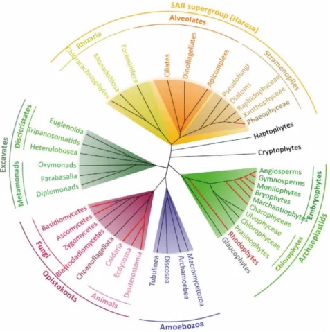
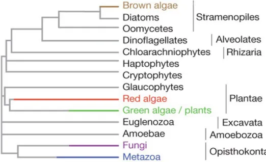
The eukaryotic tree of life has been divided into seven major lineages (Fig. 1) (Parfrey et al., 2011): the excavates (Simpson et al., 2005), the haptophytes and the cryptophytes (Burki et al., 2016), the archaeplastids (Umen, 2014), the SAR lineage (consisting of stramenopiles, alveolates and rhizarians) (Burki et al., 2007), the amoebozoans and the group that includes the most complex multicellular organisms, the opisthokonts (Cavalier-Smith et al., 2015). These diverse organisms share several basic biological features, despite the many differences between lineages and the long evolutionary times that separate them. For example, most of these organisms reproduce sexually (Fig. 2A) and have life cycles that involve an alternation between haploid and diploid phases (Fig. 2B) due to two fundamental processes: meiotic cell division (which results in the production of haploid cells from diploid cells) and gamete fusion or syngamy (which results in a doubling of the ploidy level). This alternation between ploidy phases was first recognised by Hofmeister (1851) and has been called Hofmeister alternation. The term “alternation of generations” was proposed several years later by Roe (1975). In the following sections, we will describe the life cycle diversity across the eukaryotes and discuss the relationship between life cycle structure and multicellular complexity.
Figure 1. Phylogenetic tree of the eukaryotes. There are seven main lineages in the eukaryotic tree of life: the
excavates (gray-green background), the haptophytes and the cryptophytes, the archaeplastids (green background), the SAR lineage (consisted by stramenopiles, alveolates and rhizarians; orange background), the amoebozoans (blue background) and the opisthokonts (red background). Only a few of these lineages have evolved complex multicellular organisms (branches in red). (Bourdareau, 2018)
4
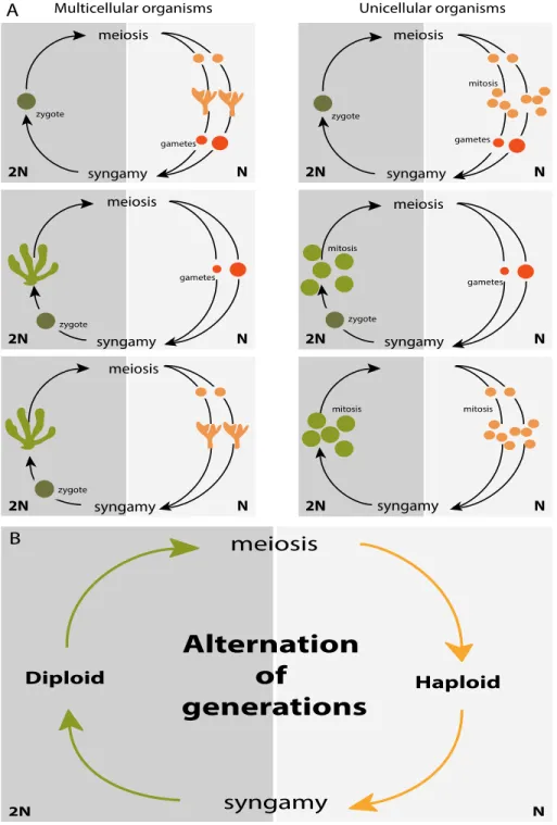
Figure 2. Sexual life cycles of eukaryotic organisms. Both multicellular (left) and unicellular (right) organisms
produce offspring via sexual reproduction (A). Multicellular development (indicated by branched structures) can occur during only the haploid phase, during only the diploid phase or during both phases. These sexual life cycles share a common feature, the alternation between haploid and diploid phases (B), due to two fundamental processes: meiotic cell division, which results in the production of haploid cells from diploid cells and gamete fusion or syngamy, which results in a doubling of the ploidy level.
1.2 Life cycle diversity in eukaryotic lineages
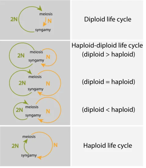
Three main types of sexual life cycle (Fig.3) can be defined, based on whether mitotic divisions occur during the haploid phase, the diploid phase or during both phases of the life cycle (Arun, 2012; Coelho et al., 2007; Perrot et al., 1991; Richerd et al., 1993; Valero et al., 1992).
Diploid (or diplontic) life cycles: in this type of life cycle, fertilization directly follows meiosis, so that mitotic cell divisions (and development in multicellular organisms) occur only in the diploid generation (e.g. Fucus). Gametes are therefore the only haploid cells. This type of life cycle is commonly observed in animals (including humans) (Mable and Otto, 1998; Trivers and Hare, 1976).
Haploid life cycles (also called haplontic life cycles or haplobiontic-haploid life cycles): in this type of cycle meiosis directly follows fertilization, so that mitotic divisions (and somatic development in multicellular organisms) only occur during the haploid generation and the zygote is the only diploid cell. This kind of life cycle is observed in a variety of green algae, such as
Volvox.
Haploid-diploid (or haplo-diplontic) life cycles: in this type of life cycle, mitosis (and somatic development in multicellular organisms) occurs during both the haploid and diploid generations so that meiosis and fertilization are temporally separated in the life cycle. The land plants, including all mosses (such as the model moss Physcomitrella patens) and ferns, some fungi (such as the model yeast Saccharomyces cerevisiae), red algae (Destombe et al., 1992), many green algae (Ulva, for example) and most brown algae have this kind of life cycle. One important consequence of this type of life cycle for multicellular organisms is that it implies the deployment of two multicellular bodyplans during the life cycle, one during the haploid phase and the other during the diploid phase. In plants and algae, these haploid and diploid phases correspond to the gametophyte and sporophyte generations, respectively, and these can occur separately or be dependent, with one generation growing on the other. The relative lengths of the haploid and diploid phases of a haploid-diploid life cycle are highly variable among multicellular eukaryotes, especially among the algae and protists, which exhibit life cycles that together correspond to the
6 complete range from complete haploid development to complete diploid development (Mable and Otto, 1998).
The following sections will discuss the life cycles of members of the major eukaryotic lineages that have evolved complex multicellularity, describing life cycle diversity and the relationship between life cycle and development.
In the green lineage (Viridiplantae), life cycles are highly variable but the majority of green algae have a haploid life cycle (e.g. Spirogyra and Zygnema) (Lewis and McCourt, 2004). The red algae (Rhodophyceae), which are an ancient sister group of the green lineage (Baldauf, 2008), exhibit both asexual and sexual life cycles. Sexual reproduction has not been reported for unicellular species (Charles, 1985; Yoon et al., 2006) but multicellular red algae have a range of different sexual life cycles. For example, the edible seaweed Porphyra has two morphologically distinct generations with different levels of ploidy (Hawkes, 1990). Some red algae of the Florideophyceae, including algae of economic interest (Gracilaria, Gelidium) and carragheenophytes (Chondrus, Eucheuma) have life cycles that involve either alternation between haploid and diploid phases, or transitions between a haploid (gametophyte) phase and two diploid (carposporophyte and tetrasporophyte) phases, the latter being referred as a “triphasic” (Graham and Wilcox, 2000) or “Polysiphonia-type” life cycle (e.g. Polysiphonia; Fig. 4) (Hoek et al., 1995). This latter class of life cycle involves two types of transition: a Steenstrupt alternation (Steenstrup, 1845) between two diploid phases (represented by the diploid carposporophyte and the diploid tetrasporophyte) and a Hofmeister alternation between haploid and diploid phases (represented by the gametophyte to carposporophyte transition) (Bell, 1994). Most animals (metazoans) have diploid life cycles, in which the haploid phase consists only of the single-celled gametes. However, some groups of metazoans have asexual life cycles.
In the fungi, somatic development does not usually occur during the diploid phase except in some groups such as certain chytrids, which exhibit an alternation between simple, filamentous haploid and diploid generations. However, syngamy and karyogamy are often delayed in the fungi resulting in an additional phase, the dikaryon, during which there is a proliferation of cells with
two unfused, haploid nuclei. This phenomenon only exists in the fungi and increases the complexity of life cycles in this group (Raper and Flexer, 1970).
Figure 3. Diversity of eukaryotic life cycles. Life cycles involve two major events of meiosis and syngamy. The
relative time spent in the haploid and diploid phase determines the organisms belong to which type of life cycle. Three main types of life cycles can be defined depending on the phase or phases that exhibit mitotic growth, they are diploid, haploid-diploid and haploid life cycles. Reproduced from Cock et al. (2014).
8
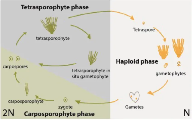
Figure 4. The tri-phasic life cycle of the red alga Polysiphonia. The diploid and haploid phases of the life cycle
are shown on the left and on the right, respectively. Note that the diploid phase includes two generations, the carposporophyte (green shading) and the tetrasporophyte generation (grey shading). Reproduced from Arun (2012).
1.3 The relationship between life cycle structure and multicellularity
The life cycles of multicellular eukaryotic organisms are complex and multiple kinds of life cycles are found in different multicellular lineages in the tree of life. However, when the distribution of the three main types of the sexual life cycle (diploid, haploid and haploid-diploid) is evaluated across the eukaryotes, a clear trend can be discerned: the organisms with the highest levels of developmental complexity tend to have diploid or diploid-dominant life cycles. Nonetheless, haploid and haploid-diploid life cycles are also distributed across multiple eukaryotic lineages and have not displayed a tendency to disappear during the process of evolution (Mable and Otto, 1998). Based on these distributions, we can conclude that all three types of the life cycle are evolutionarily stable, presumably because each has an evolutionary advantage under particular environmental conditions.
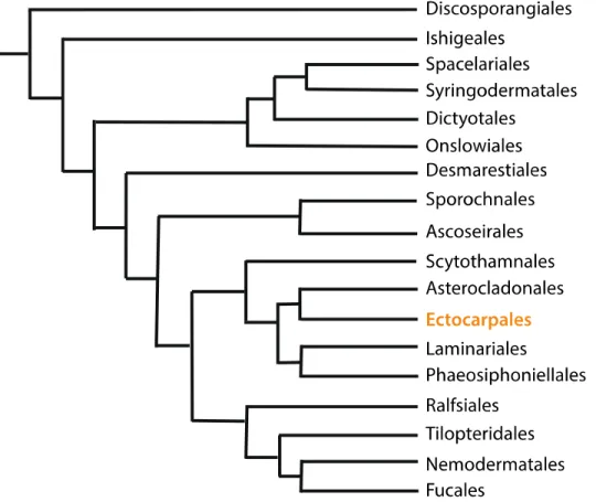
1.4 Life cycle diversity in the brown algae
The brown algal lineage is placed within the stramenopiles of the eukaryotic supergroup SAR (Hackett et al., 2007). The brown algae are multicellular, photosynthetic organisms and are one of only a few eukaryotic lineages that are considered to have evolved complex multicellularity (Fig. 5) (Cock et al., 2014; Cock et al., 2010; Knoll, 2011; Niklas and Newman, 2013). The brown algae diverged from other well-studied multicellular lineages such as land plants and animals more than a billion years ago (Bourdareau, 2018) and multicellularity then evolved independently in each lineage. Brown algae are interesting models for the study of life cycles both due to this large evolutionary distance from other eukaryotic lineages and because they exhibit a broad range of different types of life cycle ranging from dominant haploid-diploid life cycles to haploid-diploid life cycles (Fig. 6) (Cock et al., 2014; Woelkerling, 2004). Molecular phylogenetic analysis has established a robust phylogeny for the brown algae (Fig. 7). Based on this phylogeny, the ancestral life cycle of this group is thought to have consisted of two multicellular generations (gametophyte and sporophyte) (Coelho et al., 2007; Kawai et al., 2007). The order Ectocarpales, which consists principally of small, ephemeral brown algae, exhibits a remarkably wide range of different haploid-diploid life cycles. Across the different life cycles of organisms of this order, the two generations can be isomorphic (family Acinetosporaceae), slightly heteromorphic (Ectocarpaceae) or strongly heteromorphic with either a microscopic gametophyte or a microscopic sporophyte (Peters and Ramírez, 2001). Moreover, both generations may be capable of asexual reproduction, providing additional means to produce new offspring (de Reviers, 2006). The pathways of asexual reproduction include parthenogenetic development of unfertilised gametes, which can develop into either partheno-gametophytes (e.g.
Myriotrichia) or partheno-sporophytes (e.g. Ectocarpus) depending on the species, the
mito-spores produced in the plurilocular sporangia of sporophytes (Peters et al., 2008), and fragmentation of the sporophyte (e.g. Naples strain) (Müller, 1964).
10
Figure 5. Simplified representation of the evolutionary tree of complex multicellular organisms (highlighted in
colour). Five groups show complex multicellularity, i.e. they include macroscopic organisms with
defined, recognisable morphologies that are composed of multiple cell types. Colour bars indicate the approximate, relative time when complex multicellularity emerged in each lineage (Cock et al., 2010).
D
E
par theno genesis sporophyte (microscopic)A
meiosis syngamy sporophyte Male gametophyte U Female gametophyte egg sperm par theno genesis (microscopic) (microscopic) gametes egg sperm sporophyte par theno genesis sp or es unilocular sporangia plurilocular sporangiaC
gametes gametophyte meiosis syngamy sporophyte par theno genesisF
V Male gametophyte Female gametophyte Male gametophyte U Female gametophyte Male sporophyte Female sporophyte sporophyte meiosis syngamy meiosis syngamymeiosis meiosis syngamy
V V U meiosis syngamy egg sperm meiosis
B
e.g. Saccharina latissima
e.g. Ectocarpus sp.
e.g. Fucus serratus
e.g. Schytosiphon lomentaria
e.g. Fucus spiralis e.g. Halopteris congesta
female gamete male gamete
Figure 6. Life cycles of brown algae. The brown algae exhibit a wide range of life cycles, including haploid-diploid
(haploid < diploid, A and F; haploid = diploid, D; haploid > diploid, E) and diploid life cycles (B and C). Meiosis results in the formation of haploid meio-spores, these develop into either male or female gametophytes in some species (e.g. A, D, E) depending on which sex chromosome (U or V) they inherited from the parent sporophyte.
12
Figure 7. Phylogenetic tree of the major brown algal orders. The filamentous Ectocarpus (highlight in yellow)
emerged as a model organism and provides a series of genetic tools to investigate the brown algae. The length of each branch is not proportional to the relative evolutionary time. Adapted from Silberfeld et al. (2014).
13
1.5 Model systems to study life cycle regulation
Evidence of genetic control of the alternation between generations was first reported in the unicellular fungus Saccharomyces cerevisiae, where the mating-type factors α2 and a1 were shown to bind to haploid-specific genes and repress them (Goutte and Johnson, 1988). Among photosynthetic organisms, the flowering plant Arabidopsis is a major model system for the study of various phenomenon associating life cycle progression with development. This phenomena does, however, pose some limitations for life cycle analysis because the gametophyte is highly dependent on the sporophyte generation and develops surrounded by sporophyte tissues. Despite this limitation, several studies have used Arabidopsis to study the alternation of generations (e.g. Yadegari and Drews, 2004). The moss Physcomitrella patens is also being used to study life cycle regulation. Moss life cycles involve two generations that both exhibit considerable developmental complexity but in Physcomitrella patens the sporophyte grows on the macroscopic gametophyte and depends on the latter for nutrients, again making it difficult to separate the two generations and study them individually (Bowman et al., 2007). The fern
Ceratopteris (pteridophytes) has independent, multicellular gametophyte and sporophyte
generations and is considered to be a suitable system to investigate haploid-diploid life cycles, but no mutant affected in the alternation of the two generations has yet been described in this system (Banks, 1999).
Marine algae are found in most lineages of the eukaryotic tree of life and have played an important role in the evolution of life. Consequently, these organisms offer the opportunity to tackle diverse questions regarding the origin and evolution of general eukaryotic traits. As far as life cycles are concerned, as discussed above, the existence of a broad range of the life cycles in the brown algae makes them good candidates to study life cycle evolution. This is particularly the case for brown algae where, in addition, a powerful model organism has become available in recent years. Ectocarpus was the first brown alga (also the first macroalga) to be sequenced (Cock et al., 2010) and has since emerged as a genetic model for the brown algae (Peters et al., 2004b). Many genetic and genomic tools are available for Ectocarpus including a high-quality genome annotation (Cock et al., 2010; Cormier et al., 2017), transcriptomic data based on
14 microarrays (Dittami et al., 2009) and RNA-seq technologies (Ahmed et al., 2014; Luthringer et al., 2015; Macaisne et al., 2017), genetic maps based both on microsatellite (Heesch et al., 2010) and restriction-site-associated DNA sequencing (RAD-seq) markers (Avia et al., 2017) and forward genetics methodologies (Godfroy et al., 2017; Macaisne et al., 2017). These features have made Ectocarpus a good choice as a model organism providing a novel system to shed light onto developmental processes such as the regulatory mechanisms that control the life cycle at the molecular level (Mignerot and Coelho, 2016; Peters et al., 2004a).
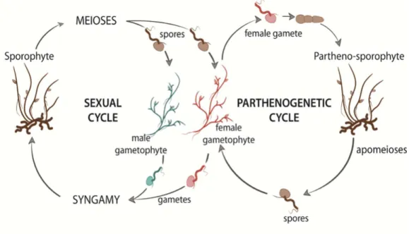
Ectocarpus is a small filamentous alga, which can be easily grown and induced to become fertile
under laboratory conditions. Wild strains can grow up to 30 cm in length but individuals in culture can become fertile when they are less than 2 cm in length. Ectocarpus species are distributed worldwide in temperate coastal areas of both hemispheres but are absent from tropical seas. This seaweed usually grows in the intertidal zone on rocks or other substrates, often as an epiphyte on other brown and red algae (Coelho et al., 2012a). Ectocarpus has a haploid-diploid sexual life cycle (Fig. 8), which involves an alternation between a diploid sporophyte and dioicous, haploid gametophytes. Sporophytes produce meio-spores, following meiotic division in unilocular sporangia. The meio-spores are released into the surrounding seawater and develop as gametophytes, which produce gametes in plurilocular gametangia. Zygotes, produced by the fusion of male and female gametes, develop as diploid sporophytes, completing the sexual cycle. Unfertilized gametes can enter an asexual, parthenogenetic cycle by germinating to produce partheno-sporophytes, which can also produce both unilocular and plurilocular sporangia. As with diploid sporophytes, meio-spores produced in the unilocular sporangia of partheno-sporophytes develop into gametophytes to complete the asexual parthenogenetic cycle.
The sexual life cycle of Ectocarpus involves an alternation between two morphologically similar but independent sporophyte and gametophyte generations. There are several indications that the developmental programs of these two generations are not absolutely linked to ploidy. For example, ploidy and life cycle generation have been shown to be uncoupled during variant life cycles (Arun et al., 2013; Bothwell et al., 2010; Coelho et al., 2011). These features, together with the technical resources described above, make Ectocarpus a good choice among eukaryotic
organisms to carry out experiments to investigate the mechanisms of life cycle regulation at the molecular level.
Figure 8. The life cycle of the brown alga Ectocarpus sp. The haploid-diploid life cycle of Ectocarpus is very complex. It consists of both sexual and asexual cycles (see text for details). Reproduced from Arun et al. (2019).
2. Alternation between gametophyte and sporophyte generations during the life cycle 2.1 The gametophyte and sporophyte generations during life cycle progression
The life cycles of multicellular photosynthetic organisms with haploid-diploid life cycles include two distinct multicellular organisms, the gametophyte and the sporophyte. In multicellular organisms, developmental processes need to be very precisely coordinated with life cycle progression and this is particularly important in organisms with haploid-diploid life cycles becaus e the appropria te processes also nee d to be deploye d duri ng th e appropria te generati on of the life cycle (Arun et al., 2019). In this context, work carried out on both land plants and brown algae has aimed at addressing several specific questions: 1) what is the extent of differential gene expressi on betwe en t he tw o generations of t he l ife cycl e? 2) Whi ch gen es a nd genet ic pathways are involved in generation-specific gene expression? 3) what are the common patterns in
16 generation-specific gene expression in different lineages of eukaryotes? 4) What general conclusions can be drawn from analyses of the evolution of life cycles across eukaryotic organisms (Szövényi et al., 2010). In the following section, we will discuss work on genes that exhibit generation-biased expression during the gametophyte and sporophyte generations.
2.2 Gene expression during the gametophyte and sporophyte generations of haploid-diploid life cycles
Differential gene expression during the life cycle results in a significant number of gametophyte- and sporophyte-biased genes (Dolan, 2009; Langdale, 2008; Mosquna et al., 2009; Okano et al., 2009). For example, in brown algal genomes (i.e. Ectocarpus sp., Scytosiphon lomentaria,
Macrocystis pyrifera, Saccharina japonica) more than 30% of genes exhibit generation-biased
expression patterns (Lipinska et al., 2019). The proportion of generation-biased genes is also high in land plants such as the moss Funaria hygrometrica (24%) or the seed plant Arabidopsis
thaliana (23%-27%) (Szövényi et al., 2010; Szövényi et al., 2013). In the Ectocarples species Ectocarpus sp. and Scytosiphon lomentaria, gametophyte-biased genes (20-23% of the genome)
are more numerous than sporophyte-biased genes (12-13% of the genome), whereas gametophyte-based genes are less numerous than sporophyte-based genes in the Laminariales (14-17% and 16-19% of the genome, respectively, in Macrocystis pyrifera and Saccharina
japonica), Funaria hygrometrica (2.5% for the gametophyte and 5% for the sporophyte) and A. thaliana (5% for the gametophyte and 25% for the sporophyte) (Haerizadeh et al., 2009; Honys and Twell, 2003; Ma et al., 2008; Pina et al., 2005). Differential gene expression during haploid-diploid life cycles (Fig. 9) is often associated with marked differences in the morphology and functions of the gametophyte and sporophyte generations and there has been considerable interest in the roles of generation-biased genes in mediating these morphological and functional differences (Shaw et al., 2011; Szövényi et al., 2010).
Figure 9. Differential gene expression during the sporophyte and gametophyte generations of the moss
Funaria hygrometrica. (A). Volcano plot showing the significance and fold change of differential expression of
genes between isogenic gametophyte and sporophyte generations. Non-differentially expressed transcripts (q value > 0.1) are in grey, whereas significant genes are in black. (B). Most specific significant (q value ≤ 0.1) GO terms (biological process ontology) and their relative expression level during the sporophyte and gametophyte generations of Funaria hygrometrica. Relative expression levels refer to the relative proportion of reads mapped to gene models with a particular GO term (black: sporophyte, gray: gametophyte). Reproduced from Szövényi et al. (2010).
2.3 Evolution of gametophyte and sporophyte developmental programs
As discussed above, various types of life cycles exist stably in nature, indicating that each type provides a unique advantage in a particular environment. Interpretation of the stability of haploid-diploid life cycles is difficult because both gametophyte and sporophyte generation possess their unique advantages and this is expected to lead to the dominance of one of the two generations under a wide range of conditions (Mable and Otto, 1998). However, it has been proposed that if the two generations are adapted to different ecological niches, a haploid-diploid life cycle could be stabilised over evolutionary time (Hughes and Otto, 1999; Stebbins and Hill, 1980; Willson, 1981).
Analysis of generation-biased gene expression can provide insights into the evolution of life cycles (Szövényi et al., 2010). In particular, such analyses can provide information about whether developmental pathways evolved independently in each generation or whether there has been the recruitment of developmental pathways from one generation to the other. For example, analysis of land plant developmental networks indicates that genetic components that played a role during the gametophyte generation, which was the dominant generation in ancestors of the land plants, were recruited to create the sporophyte generation developmental program (Dolan, 2009; Niklas and Kutschera, 2010). In addition, some aspects of the sporophyte generation program represent specific innovations associated with that generation (Sano et al., 2005; Szövényi et al., 2010). In Ectocarpus, the gametophyte and sporophyte generations exhibit several morphological differences, with a particularly marked difference during the initial cell division of each generation. Early development of the gametophyte involves an asymmetric division of the initial cell, which is immediately followed by differentiation to form an erect thallus and a basal rhizoid (formation of an apical/basal axis) (Fritsch, 1935; Godfroy et al., 2017). In contrast, early development of the sporophyte involves symmetric division of initial cell that leads to the formation of a prostrate basal structure before the development of the erect thallus. The sporophyte of the immediate upright (imm) mutant exhibits several characteristics typical of the gametophyte. The initial cell of the imm mutant sporophyte undergoes asymmetric division and the sporophyte fails to form an extensive basal system, which is replaced by a rhizoid (Peters et 18
al., 2008). Therefore, analysis of the genetic mutant imm has shown that, in principle, both symmetric (wild type) and asymmetric (mutant) initial cell divisions can be at the origin of sporophyte development. Based on this, it has been proposed that the ancestral sporophyte developmental program may have more closely resembled that of the gametophyte (Macaisne et al., 2017). In other words, the sporophyte generation probably evolved from an ancestor that resembled the gametophyte generation with an asymmetrical initial cell division that gave rise directly to apical and basal organs (Macaisne et al., 2017). In the distag (dis) mutant, the gametophyte generation lacks a rhizoid and in the sporophyte generation prostrate filaments (i.e. the basal system) fail to develop (Godfroy et al., 2017). The phenotype of this mutant, therefore, supports the hypothesis that gametophyte rhizoids and sporophyte prostrate filaments are actually developmentally equivalent, despite marked differences in terms of morphological features such as cell form and size. In summary, the basal structures of both generations seem to be homologous but only in the sporophyte has the rhizoid been modified to produce an extensive system of prostrate filaments. IMM encodes the IMM protein, which does not have any known biochemical function (Macaisne et al., 2017). The developmental program associated with the
IMM gene appears to be a specific innovation in the sporophyte generation because the imm
mutation has no phenotype in the gametophyte. In contrast, the DIS gene, which encodes the TBCCd1 protein in Ectocarpus, is required for the formation of basal structures during both the gametophyte and the sporophyte generations (Godfroy et al., 2017). This suggests that the genetic system that includes the DIS gene was shared by both gametophyte and sporophyte generations during the process of evolution.
2.4 Evolution of patterns of gametophyte and sporophyte gene expression
In nearly all metazoan taxa, multicellular development only occurs during the diploid phase of the life cycle (Couceiro et al., 2015). Diploid dominance is also observed in land plants in the sense that this lineage is thought to be derived from a haplontic ancestor, with an expansion of the sporophyte generation and a shortening of the gametophyte generation (d'Amato, 1977). There is also evidence for a tendency towards diploid dominance in the brown algae, particularly in recently evolved lineages such as the Laminariales and the Fucales. However, haploid-diploid
20 life cycles can, nonetheless, be stable over evolutionary time. This is the case for red algae, most brown algae, many green algae, some fungi, foraminiferans, mosses and ferns (Richerd et al., 1993). In these latter examples, independent gametophyte and sporophyte generations have been stably maintained over long periods of evolutionary time (Mable and Otto, 1998).
Brown algae are thought to be derived from a last common ancestor whose life cycle involved alternation between gametophyte and sporophyte generations without a clearly dominant generation. However, during evolution, there has been a tendency for most brown algae to evolve towards increased complexity of either the gametophyte or the sporophyte generation (Silberfeld et al., 2010). As mentioned above, there is a general tendency in the brown algae towards diploid dominance but, in some species, the gametophyte generation can be considered to be dominant, either because it is more complex morphologically or because the species has more gametophyte-biased than sporophyte-gametophyte-biased genes. This is the case, for example, in the Ectocarpales species
Ectocarpus sp. and Scytosiphon lomentaria (Lipinska et al., 2019). Note that, when considering
gene expression, it is important to take into account both generation-biased gene expression and the proportions of the total gene set expressed in each generation. For example, in Arabidopsis, 69% of the genes are expressed in the male gametophyte, but only 7.9% of these genes are gametophyte-specific. In most species with haploid-diploid life cycles, the majority of genes are expressed in both the gametophyte and sporophyte generation (Honys and Twell, 2004; Lipinska et al., 2019).
It is currently thought that the majority of the genetic and regulatory networks that operate during the sporophyte generation in land plants originated from equivalent components that originally functioned during the gametophyte generation (Menand et al., 2007; Nishiyama et al., 2003). However, this idea of unidirectional recruitment from gametophyte to sporophyte may be a simplification, as analysis of the evolution of generation-biased gene sets in the brown algae indicates a dynamic process with a proportion of the genes frequently changing their pattern of expression, often leading to expression during the opposite generation (Lipinska et al., 2019). Nonetheless, analysis of the extensive generation-specific gene expression across different species indicates that the genetic programs that underlie each generation evolved relatively
independently and developmental innovations were selected under natural selection for each generation (Szövényi et al., 2013). One consequence of these differences in selection is that genes associated with different generations may evolve at different rates (Orr and Otto, 1994; Otto and Gerstein, 2008). For example, in F. hygrometrica and A. thaliana, gametophyte-specific genes evolve faster than sporophyte-specific genes (Anderson et al., 2004; Orr and Otto, 1994; Otto and Gerstein, 2008; Zeyl et al., 2003). Similarly, gametophyte-biased genes are evolving rapidly compared with unbiased genes in some brown algae, e.g. Ectocarpales and Laminariales (Lipinska et al., 2019).
22
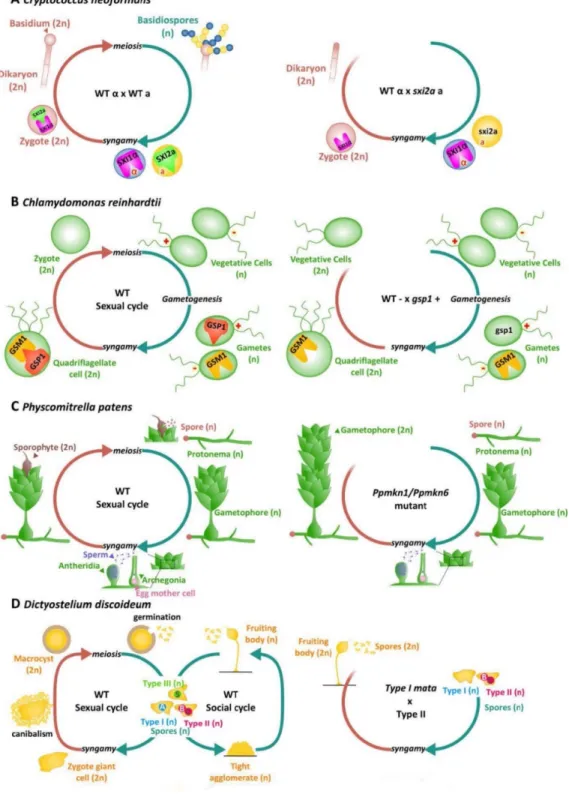
Figure 10. Homeodomains, homeodomain-like transcription factors and life cycle regulation. Reproduced
A: Life cycle of Cryptococcus neoformans. Haploid a (yellow, expressing the homeodomain transcription factor SIX2a) and α (blue, expressing the homeodomain transcription factor SIX1α) gametes fuse to form a diploid zygote. SIX1α and SIX2a interact after syngamy to initiate sexual development (Left). Zygotes that originate from a cross between a wild-type α gamete and a six2a gamete cannot initiate sexual development (right). For detailed information see Hull et al. (2005).
B: Life cycle of the green alga Chlamydomonas reinhardtii. The homeodomain transcription factors GSP1 (in plus gametes) and GSM (in minus gametes) form a heterodimer and move to the nucleus after plus and minus gametes fuse and initiate the zygote program (left). A zygote derived from a cross between a minus gamete (wild-type) and a
gsp1 gamete fails to implement the zygote program (right). For details see Lee et al. (2008b).
C: Life cycle of the moss Physcomitrella patens. Haploid spores develop to produce protonema filaments on which grow the gametophores. Male gametes swim to the archegonia from the neighbouring antheridia and fuse with egg cells to produce diploid sporophytes (left). A functional diploid gametophyte develops instead of the sporophyte in the Ppmkn1-Ppmkn6 double mutant (right). For details see (Sakakibara et al., 2013).
D: Life cycle of the amoeba Dictyostelium discoidem. Three types of haploid spores (Type Ι, Type Ⅱ and Type Ⅲ) produce three different homeodomain-like transcription factors MatA, MatB and MatC respectively. Zygotes, which are formed when each type of spore fuses with either of the other two spore mating types, form a macrocyst by attracting and engulfing nearby haploid cells. Mature macroysts produce haploid spores to complete the life cycle (Left). Zygote derived from a cross between a wild-type Type Ⅱ spore and a mata Type Ι spore develop to produce a fruiting body with diploid spores similar to haploid cells. For details see Hedgethorne et al. (2017).
24
3. Genetic control of transitions between the gametophyte and the sporophyte generation 3.1 Genetic regulators of life cycle transitions
As both the gametophyte and sporophyte generations are constructed using information from a shared genome, it follows that epigenetic regulatory processes must operate both during meiosis (at the diploid-to-haploid transition) and during syngamy (at the haploid-to-diploid transition) to trigger the initiation of the appropriate developmental program associated with each generation (Bourdareau, 2018). As discussed before, in many organisms the gametophyte and sporophyte generations are morphologically and functionally different, so the developmental switches must be precisely controlled to avoid the production of chimeric organisms.
Genetic studies have improved our understanding of the molecular basis of life cycle progression, particular the haploid-to-diploid transition (Fig. 10). The first evidence for genetic control of a transition between generations of a life cycle was reported for a unicellular fungus, the yeast
Saccharomyces cerevisiae. The mating-type genes MATα2 and MATa1 produce the α2 and a1
proteins that bind to haploid-specific genes and repress them (Goutte and Johnson, 1988). α2 shares homology with homeodomain proteins in Drosophila (Shepherd et al., 1984), and similar systems, also involving homeodomain regulators, have been described in other fungi such as
Ustilago maydis (Gillissen et al., 1992), Coprinus cinereus and the human pathogen Cryptococcus neoformans (Kües et al., 1992). The mating system of Crytococcus neoformans has
been particularly well described. Zygotes form by fusion of a and α gametes (Hull et al., 2005), which express a and α mating-type-specific factors, respectively. After gamete fusion, these two proteins interact to form a heterodimer, which initiates zygote development. The a and α factors are encoded by genes localized at the mating-type locus (SEX INDUCER α2 or SEX INDUCER
a1) and both proteins are homeodomain transcription factors (HD TFs). In the amoebozoan Dictyostelium discoideum the mating-type locus encodes two pairs of gametologs that suffice to
specify sexual compatibility: matA for type Ι, matS for type Ш, matB and matC for type Ⅱ (Hedgethorne et al., 2017). During the sexual cycle, each mating-type can fuse with either of the two other types to initiate the diploid phase. This mating-type system involves genetic
25 interactions between mating-type loci suggesting that heterodimerization (in this case of homeodomain-like mating-type proteins (i.e. genes that do not have a recognisable, conserved homeodomain motif but which possess a domain that resembles the homeodomain in terms of its three dimensional structure) may also play a role in inducing the diploid program in slime mould (Bourdareau, 2018; Hedgethorne et al., 2017).
A similar system, involving homeodomain transcription factors, has been described for the haploid life cycle of the unicellular, green alga Chlamydomonas (Fig. 10B). Homeodomain transcription factor-encoding genes have been classed into two major groups, the TALE (three-amino acid loop extension) and the non-TALE homeodomain transcription factor, because the later have no the characteristic of three amino acid loop extension of the 60 amino acid homeodomains (Nam and Nei, 2005; Nasmyth and Shore, 1987). Both classes of homeodomain transcription factor are very ancient and both are widely distributed across all the major eukaryotic lineages (Cock et al., 2014; Derelle et al., 2007). Initiation of the diploid program in
Chlamydomonas is controlled by a heterodimeric homeodomain transcription factor composed of
two TALE homeodomain proteins: Gsm1 (gamete-specific minus1), which is a knotted-like homeobox (KNOX) gene, and Gsp1 (gamete-specific plus1), which is a BEL-like (BELL) TALE homeodomain transcription factor. The major function of this system appears to be to act as a detector of the transition from haploid to diploid (when gametes fuse to form a zygote).
Recent work indicates that the multicellular streptophyte moss Physcomitrella patens may possess a similar system (Fig. 10C). Mutation of two KNOX class genes PpMKN1 and PpMKN6 results in the sporophyte generation being replaced by a fully functional diploid gametophyte (Sakakibara et al., 2013). In addition, the P. patens genome is predicted to encode four BEL-class proteins, similar to Gsp1. Several lines of evidence indicate a role for these BEL genes in controlling the life cycle. First, PpBELL1 and PpBELL2 are expressed in eggs and embryos. Second, loss or overexpression of PpBELL1 leads to a failure to build sporophytic structures or, conversely, to the production of apogamous sporophyte-like bodies on haploid caulonemal cells, respectively (Horst et al., 2016). To summarise, in the green lineage both KNOX and the BELL class TALE homeodomain transcription factors are necessary for the deployment of the
26 sporophyte generation. Together they act as master regulators of the gametophyte-to-sporophyte transition because loss of these genes leads to a block of the sporophyte developmental program leading to conversion of this generation of the life cycle into a diploid gametophyte.
Modification of chromatin by the Polycomb Repressive Complex 2 (PRC2), which regulates gene expression by tri-methylation of lysine 27 of histone H3, also appears to be involved in regulating the gametophyte-to-sporophyte transition in the green lineage. Arabidopsis mutants affected in PRC2 components such as MEDEA (MEA), MULTICOPY SUPPRESSOR OF IRA Ι (MSI1) and FERTILISATION INDEPENDENT SEED 2 (FIS2) exhibit switching from gametophyte cell fate to endosperm or embryo-like structures (Chaudhury et al., 1997; Ebel et al., 2004; Guitton and Berger, 2004; Köhler et al., 2003; Ohad et al., 1996; Ohad et al., 1999). Similar phenotypes have been observed in the moss P. patens. Two PRC2 genes
PHYSCOMITRELLA PATENS FERTILIZATION INDEPENDENT ENDOSPERM (PpFIE) and PHYSCOMITRELLA PATENS CURLY LEAF (PpCLF) induce the growth of sporophyte-like
bodies on the side branches of the gametophyte (Mosquna et al., 2009; Okano et al., 2009). Ectopic expression of other genes such as SERK, BABY BOOM (BBM), and LEC (LEC1 and
LEC2) has also been shown to affect the initiation of the sporophyte developmental program in
flowering plants. These genes may act downstream of the regulator that controls the switch from gametophyte to sporophyte (Schiefthaler et al., 1999).
Evidence that TALE homeodomain transcription factors also control life cycle transitions in brown algae has come from recent work on two Ectocarpus mutants, ouroboros (oro) and
samsara (sam). These two mutations have been shown to cause the sporophyte generation to be
converted into a fully functional gametophyte generation. The two genes therefore appear to act as master regulators of the gametophyte to sporophyte transition. The corresponding genes, ORO and SAM, both encode TALE homeodomain transcription factors and these two proteins appear to act as a heterodimer to initiate the sporophyte developmental program (Arun et al., 2019; Coelho et al., 2011). Although chromatin remodelling is probably also involved in the control of life cycle transitions in brown algae, this is unlikely to require PRC2 because genes encoding PRC2 components appear to be absent from the Ectocarpus genome (Bourdareau, 2018).
3.2 Non-cell autonomous regulation of life cycle generation identity
Genetic analysis has proved to be an important tool for understanding the molecular mechanisms underlying the life cycle but alternative, physiological approaches have also provided important insights in the model brown alga Ectocarpus. These analyses have shown that the alternation of generations in Ectocarpus is not only under the control of the TALE homeodomain transcription factors ORO and SAM (Arun et al., 2019) but is also influenced by a non-cell-autonomous factor designated the sporophyte-inducing factor that is secreted into the surrounding seawater medium by Ectocarpus sporophytes in culture (Arun et al., 2013). During the sexual life cycle of
Ectocarpus, meiotic divisions in unilocular sporangia produce haploid meio-spores that normally
develop to produce gametophytes. Occasionally, a small proportion of meio-spores develop into sporophytes instead of gametophytes, a phenomenon called heteroblasty (Müller, 1967). Arun et al. (2013) showed that the proportion of heteroblastic meio-spores was significantly increased if the spores were allowed to develop in sporophyte-conditioned medium (SCM, i.e. cell-free medium in which sporophytes had previously been cultured). Gametophyte-condition medium (GCM) did not have the same effect indicating that heteroblasty is induced by a molecule specifically produced by the sporophyte. The sporophyte identity of the heteroblastic individuals was confirmed by staining with Congo red (a water-soluble, carbohydrate-binding dye which binds to xylan fibres in algal cell walls and specifically stains Ectocarpus gametophytes but not sporophytes) (Coelho et al., 2011; Yadegari and Drews, 2004). Moreover, these individuals produced reproductive structures typical of the sporophyte generation, i.e., unilocular sporangia (a structure that is only observed during the sporophyte generation).
These experiments demonstrated that there is some plasticity with respect to the deployment of the sporophyte or gametophyte pathways, even in the absence of genetic mutations, and support the idea that life cycle generation is not strictly correlated with ploidy (Cock et al., 2014). Interestingly, additional variations of the Ectocarpus life cycle have been observed that support this idea of independent life cycle generation and ploidy. For example, unfused gametes of wild type strains can develop autonomously to produce haploid partheno-sporophytes (Müller, 1967; Mignerot et al., 2019). Therefore, the sporophyte generation can be haploid under some
28 conditions. Similarly, it is possible to construct diploid gametophytes by crossing two strains carrying the oro mutation (Coelho et al., 2011).
Meio-spores carrying either the oro or the sam mutation are resistant to the action of the diffusible sporophyte-inducing factor (Arun et al., 2019), indicating that the ORO and SAM genes are necessary for developmental reprogramming to occur and, therefore, that ORO and SAM may be part of the regulatory network triggered by the sporophyte-inducing factor (Fig. 11).
Developmental reprogramming was not observed when meio-spores were treated with SCM 48 hours after their release from the unilocular sporangium. This corresponds with the time required for the meio-spores to synthesise a cell wall, suggesting that the diffusible factor is unable to act on a cell that is surrounded by a cell wall (although it is also possible that other cellular events occur at this stage to render the developing meio-spore insensitive to the factor). The role of the cell wall was further investigated using protoplasts. Gametophyte-derived protoplasts obtained by digesting the cell walls of gametophyte filament cells normally develop as gametophytes, but a proportion regenerates into sporophytes if they are incubated with SCM (Arun et al., 2013). This experiment supported the idea that cells must lack a cell wall to respond to the diffusible factor but other processes may be involved, as the protoplast process presumably also induces some dedifferentiation of the filament cells. Note that there is evidence that the cell wall influences other developmental processes. For example, in the brown alga Fucus spiralis, the cell wall influences cell fate by inducing switching from thallus to rhizoid identity (Berger et al., 1994; Bouget et al., 1998). Similarly, the cell wall plays an important role in terrestrial plant morphogenesis (Hamant et al., 2010).
Based on this potential link between the presence or absence of the cell wall and the capacity to switch between life cycle generations and on the likely hypothesis that the sporophyte-inducing diffusible factor originates from or corresponds to a cell wall component, the following section will look more closely at the composition of brown algal cell walls.
Figure 11. The life cycle of Ectocarpus is regulated by the diffusible sporophyte-inducing factor and by the genetic factors, ORO and SAM. (A). The wild sporophyte produces meio-spores, via a meiotic division in the unilocular sporangia, which developas gametophytes and produce gametes in plurilocular gametangia. However, a proportion of meio-spores developas full functional sporophytes when incubated w ith sporophyte-inducing factor (heteroblasty). In strains carrying mutations in either ORO or SAM, the sporophyte is converted into a fully functional gametophyte. (B). oro and sam mutants are insensitive to the sporophyte-inducing factor. PS: plurilocular sporangia; US: unilocular sporangia; PG: plurilocular gametangia. Reproduced from Arun et al. (2019).
30
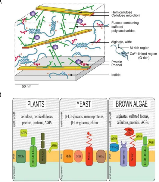
Figure 12. Cell wall model for brown algae from the order Fucales and comparison with plants and fungi. A:
The main components of brown algal cell walls are alginates and sulphated fucans. Alginates and fucose-containing sulfated polysaccharides form the greater part of cell wall polymers, the latter acting as cross-linkers between cellulose microfibrils. See details in Deniaud-Bouët et al. (2014). B: Putative cell wall sensing proteins in brown, compared with equivalent molecules found in other kingdoms. AGPs, arabinogalactan proteins; WSC, wall sensing component; ManC5-E, mannuronan C5-epimerase. Reproduced from Hervé et al. (2016).
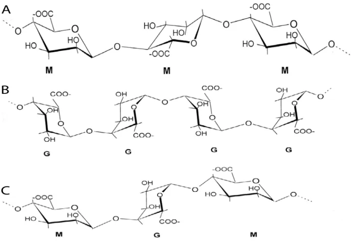
Figure 13. Chemical structures of alginates from brown algae. Alginates are linear anionic copolymers of
β-1,4-d-mannuronic acid and of its C5 epimer, α-1,4-l-guluronic acid. They consist of an alternation of homopolymeric blocks of poly-β-1,4-d-mannuronic acid, referred to here as MM blocks (A), of homopolymeric blocks of poly-α-1,4-l-guluronic acid (GG blocks; B), and of heteropolymeric blocks with random arrangements of both monomers (MG blocks; C). (Küpper et al., 2001)
3.3 Composition of brown algal cell walls
The cells of all photosynthetic, multicellular, eukaryotic organisms, including terrestrial plants and brown algae, are surrounded by a dynamic, complex, carbohydrate-rich cell wall (Popper et al., 2011b). In the tree of life, complex multicellular organisms evolved in only a small number of eukaryotic groups including notably the plants and the brown algae. Interestingly, in both the plants and the brown algae, this process was associated with the evolution of complex cell walls which play important roles in cell-to-cell recognition, adhesion and communication.
The brown algae (Phaeophyceae) are a group of multicellular algae that arose within the stramenopile lineage and which therefore obtained their plastids through a secondary endosymbiosis with a red alga (Berney and Pawlowski, 2006; Michel et al., 2010; Reyes-Prieto et al., 2007). This endosymbiosis event explains why brown algae share many features of their photosynthetic system and cell wall components with land plants because photosynthesis and carbohydrate synthesis genes were acquired during this event. Brown algal cell walls (Fig. 12A) share features with both plant and fungus cell walls, but also posses some unique characteristics (Fig. 12B). The cell walls of brown algae, like those of other marine algae and plants, can be understood as a two-phase system: a crystalline phase (the skeleton) embedded in a more amorphous phase (the matrix) (Kloareg and Quatrano, 1988). In brown algae, the skeletal phase is made up of natural, linear polymers, while in other algae the skeletal phase contains chitin-like polymers built with N-acetyl-glucosamine (Herth and Schnepf, 1982) or highly crystalline glycoproteins based with hydroxyproline (in the Volvocales) (Roberts et al., 1974).
Anionic polysaccharides, namely alginates (which also occur in some bacteria) and fucoidans (which also occur in animals), are the main components of brown algal cell walls (up to 45% of algal dry weight, e.g. alginates constitute about 60% of L. digitata cell walls) (Kloareg and Quatrano, 1988; Mabeau and Kloareg, 1987). In contrast, cellulose (which also occurs in plants) only accounts for a relatively low proportion of the cell wall (1-8% of dry weight) (Cronshaw et al., 1958). In intertidal brown algae, the average weight ratios of alginate, fucoidans and cellulose are about 3:1:1 (Mabeau and Kloareg, 1987; Kloareg and Quatrano, 1988). Alginate consists of two uronic acids (Fig. 13): β-1-4-D-mannuronate and α-1-4-L-guluronate, which initially polymerise as mannuronan. The guluronate residues are made at the polymer level by mannuronate C5-epimerases (MC5Es), which convert mannuronan to guluronate. Fucoidans are sulfated polysaccharides containing α-L fucose residues. Echinoderm fucans have regular structures composed of liner and repetitive polysaccharides (Pereira et al., 1999). Sulfated fucans may be present as either homofucans or heterofucans. These polysaccharides are referred to as fucose-containing sulfated polysaccharides (FCSPs) (Ale et al., 2011; Sakai et al., 2003). In addition, brown algal cell walls contain phlorotannins (Schmidt-Nielsen, 1997), which are composed of halogenated or sulfated phenolic compounds and about 5% protein (Quatrano and
Stevens, 1976). In general, brown algal cell walls differ markedly from those of land plants (Hervé et al., 2016).
In brown algae the alginates, sulfated fucans and phlorotannins are synthesized in the Golgi, transported in vesicles to the plasma membrane and secreted into the expanding cell wall (Callow et al., 1978; Schoenwaelder and Wiencke, 2000). In contrast, cellulose microfibrils are produced and deposited in situ by cellulose synthase complexes localized in the plasma membrane (Peng and Jaffe, 1976). Microchemical imaging analyses have indicated that apoplastic, vanadate-dependent iodoperoxidase (Colin et al., 2005) binds to large amounts of iodine in Laminariales cell walls (Verhaeghe et al., 2008). In the presence of halides, vanadate-dependent iodoperoxidase also catalyses the cross-linking of alginates with phlorotannins (Berglin et al., 2004), implying that halogenated and phenolic compounds have an important role in cell wall cohesion.
The composition of the cell wall or extracellular matrix of brown algae is variable depending on the developmental stage and season. Brown algal cell walls are thought to play an important role in innate immunity (Küpper et al., 2001), resistance to mechanical stress and protection from predators (Popper et al., 2011a). They also play crucial roles in controlling cell differentiation (development), as in land plants, and developmental processes such as growth, differentiation and morphogenesis are intimately associated with alternations in cell wall metabolism (Quatrano and Stevens, 1976). The link between cell morphogenesis and the deposition of the cell wall has been studied in Fucales zygotes, which have long served as models to study cell polarization and asymmetrical cell division in relation to the cell wall (Belanger and Quatrano, 2000; Brownlee, 2002; Deniaud-Bouët et al., 2014; Paciorek and Bergmann, 2010). During germination, tip growth is initiated at a pre-determined site on the surface of the zygote and a rhizoid emerges (Fowler and Quatrano, 1997; Kropf et al., 1988). The apical cell of the rhizoid will then elongate, whereas the thallus cell will proliferate by diffuse growth (Bisgrove and Kropf, 2001). Cell wall components (e.g. transmembrane proteins with potential roles in cell-cell communication) are thought to control cell differentiation during the sporophyte generation of Ectocarpus sp. (Le Bail et al., 2011). The important function of the major cell wall constituent, alginate, is reflected in
34 brown algal genomes, which encode a large number of MC5E enzymes (28 in Ectocarpus sp.) and many WSC (cell wall sensing components) domain proteins. The latter may be involved in interactions between alginate and proteins. Both of these large multigenic families, together with brown algae-specific receptor kinases and an expanded complement of cell wall-related genes, were described in detail as part of the analysis of the genome of the model brown alga
Ectocarpus sp. (Michel et al., 2010). Structural genomic evidence showed that MC5E genes were
associated with both WSC domains (Oide et al., 2019; Wawra et al., 2019) and arabinogalactan protein (AGP) protein core motifs (Hervé et al., 2016).
Objectives
The main objective of this study was to characterize the diffusible factor produced by sporophytes that modifies the cell fate of initial cells of the gametophyte generation, switching them to sporophyte identity. The work focused on optimizing production, storage and bioassay of this sporophyte-inducing factor to increase our understanding of its biochemical nature. The study also investigated the relationship between the sporophyte-inducing factor and two genetic regulators, ORO and SAM, to understand the developmental pathway triggered by the sporophyte-inducing factor. In addition to this work on life cycle generation identity, the thesis has also involved characterisation of the Ectocarpus baseless mutant, which exhibits a similar phenotype to the distag mutant (Godfroy et al., 2017) and is affected in developmental patterning during both the gametophyte and sporophyte generations. The specific objectives of the thesis were:
1. To set up a standardised protocol for the production and bio-assay of the diffusible factor (Chapter II).
2. To characterise the sporophyte-inducing factor that induces gametophyte-to-sporophyte developmental reprogramming in Ectocarpus (Chapter III). The work principally included the optimization of production, bioanalysis of the factor and preliminary biochemical characterisation.
3. To investigate relationships between life cycle regulators: i.e. between the sporophyte-inducing factor and two genetic regulators, ORO and SAM (Chapter IV), as part of a broader study aimed at characterising the latter two genetic regulators.
4. To characterise the baseless mutant, which is affected in developmental patterning during both generations (Chapter V).
Chapter
Ⅱ
Protocol for the production and bioassay of a diffusible
factor that induces gametophyte-to-sporophyte
developmental reprogramming in Ectocarpus
Previous studies demonstrated that alternation of generations in Ectocarpus is not determined by ploidy but is under genetic control, and can be influenced by a non-cell-autonomous factor produced by the sporophyte (Arun et al., 2013; Bothwell et al., 2010). Genetic control is mediated by two TALE HD transcription factors, ORO and SAM, which are required for the induction of the sporophyte developmental program in Ectocarpus (Arun et al., 2019; Coelho et al., 2011). The non-cell-autonomous factor produced by the sporophyte acts on gametophyte initial cells inducing a switch to the sporophyte developmental program (Arun et al., 2013; Bothwell et al., 2010). However, the exact biochemical nature of this diffusible factor is still not known. The main task of this Ph.D project was to characterise the sporophyte-inducing diffusible factor. To this end, the establishment of a standardised procedure for the production and bioassay of the diffusible factor was a key objective for the project and this protocol represents an important resource for future study of the diffusible factor. This chapter will present the optimised protocol, which has been prepared in the form of a manuscript and will be submitted for publication.