HAL Id: hal-02117021
https://hal.archives-ouvertes.fr/hal-02117021
Submitted on 1 May 2019
HAL is a multi-disciplinary open access
archive for the deposit and dissemination of
sci-entific research documents, whether they are
pub-lished or not. The documents may come from
teaching and research institutions in France or
abroad, or from public or private research centers.
L’archive ouverte pluridisciplinaire HAL, est
destinée au dépôt et à la diffusion de documents
scientifiques de niveau recherche, publiés ou non,
émanant des établissements d’enseignement et de
recherche français ou étrangers, des laboratoires
publics ou privés.
Phthalocyanine Photoregeneration for Low Power
Consumption Chemiresistors
David Tomecek, Martin Hruska, Premysl Fitl, Jan Vlcek, Eva Maresova,
Sarka Havlova, Lionel Patrone, Martin Vrnata
To cite this version:
David Tomecek, Martin Hruska, Premysl Fitl, Jan Vlcek, Eva Maresova, et al.. Phthalocyanine
Photoregeneration for Low Power Consumption Chemiresistors. ACS Sensors, American Chemical
Society, 2018, 3 (12), pp.2558-2565. �10.1021/acssensors.8b00922�. �hal-02117021�
Phthalocyanine Photoregeneration for Low Power Consumption
Chemiresistors
David Tomecek,
†Martin Hruska,
†Premysl Fitl,
†Jan Vlcek,
†Eva Maresova,
†Sarka
Havlova,
†Lionel Patrone,
‡and Martin Vrnata
*
,††University of Chemistry and Technology, Prague; Technicka 5, 166 28 Prague 6 − Dejvice, Czech Republic ‡Aix Marseille Univ., Universitéde Toulon, CNRS, IM2NP UMR 7334, Yncréa Méditerranée, ISEN Toulon, Maison du Numérique
et de l’Innovation, Place G. Pompidou, 83000 Toulon, France
*
S Supporting InformationABSTRACT: It is well-known that the applicability of phthalocyanine chemiresistors suffers from long recovery time after NO2 exposure. This circumstance enforces the necessity to operate the sensors at elevated temperatures (150−200 °C), which shortens the sensor lifetime and increases its power consumption (regardless, a typical measurement period is longer than 15 min). In this paper, we propose a new method for fast and effective recovery by UV−vis illumination at a low temperature (55 °C). The method is based on short illumination following short NO2 exposure. To support and optimize the method, we
investigated the effects of light in the wavelength and intensity
ranges of 375−850 nm and 0.2−0.8 mW/mm2, respectively, on the rate of NO2 desorption from the phthalocyanine sensitive
layer during the recovery period. This investigation was carried out for a set of phthalocyanine materials (ZnPc, CuPc, H2Pc,
PbPc, and FePc) operating at slightly elevated temperatures (55−100 °C) and was further supported by the analysis of UV−vis
and FTIR spectral changes. We found out that the light with the wavelength shorter than 550 nm significantly accelerates the
NO2 desorption from ZnPc, CuPc, and FePc, and allows bringing the measurement period under 2 min and decreasing the
sensor power consumption by 75%. Possible mechanisms of the light-stimulated desorption are discussed.
KEYWORDS: phthalocyanine chemiresistors, nitrogen dioxide detection, photoregeneration, light-stimulated NO2 desorption,
photoinduced spectral changes, low power consumption
C
onventionally, NO2 is monitored by sensitive layers of chemiresistors based on metal oxides.1 The best metaloxide-based chemiresistors are small, durable, and very sensitive electronic elements with detection limits as low as several parts per billion (ppb).2 However, the metal oxide
sensors operate at elevated temperatures (>200 °C) and suffer from low selectivity to NO2.1 To overcome these obstacles,
many researchers have focused on the investigation of organicbased sensors or organic−inorganic hybrids.
Organic materials offer a higher degree of compositional and structural variability that enables the sensor to be tailored to one selective application. Various materials such as metal− organic frameworks,3,4 graphene composites,5 or chemically
derived carbon nanotubes,6 polypyrrole,7 porphyrin,8 polytoluidine, 9 and indigo-carmine10 have already been investigated.
Phthalocyanines (Pcs), which can operate at ambient temperatures, are also highly promising materials.11−14
Phthalocyanine (H2Pc) is an organic macrocyclic molecule with a summary formula (C8H4N2)4H2. It consists of four isoindole heterocycles connected by nitrogen bridges into one macrocycle with four nitrogen atoms in the center. The cavity in the center can be occupied by dozens (more than 70) of
cation types forming phthalocyanine derivatives, metallophthalocyanines (MPc).15 They are typically intensively colored
compounds absorbing UV−vis light by four absorption bands (Q, B, N, and L). The light absorption promotes electrons across a band gap to a conduction band, generating new mobile charge carriers in phthalocyanines.15
Phthalocyanine molecules contain an extended network of conjugated double bonds that interact with NO2. For such interaction, two basic mechanisms have been proposed: (a) the partial oxidation of the macrocycle connected with the
introduction of free holes,16,17 and (b) the formation of
delocalized common molecular orbitals that significantly modify the mechanism of charge transport.18 In either case,
the interaction results in a change of resistance (and
impedance) that can be correlated with analyte concentration.
19 Regarding the gas-sensing characteristics of Pc films,
their morphology also plays a significant role.20 The most
Received: August 28, 2018 Accepted: November 15, 2018 Published: November 15, 2018 Article
Cite This: ACS Sens. 2018, 3, 2558−2565 pubs.acs.org/acssensors
© 2018 American Chemical Society 2558 DOI: 10.1021/acssensors.8b00922
ACS Sens. 2018, 3, 2558−2565
Downloaded via AIX-MARSEILLE UNIV on January 2, 2019 at 22:27:20 (UTC).
See https://pubs.acs.org/sharingguidelines for options on how to legitimately share published articles.
familiar phthalocyanine polymorphs are α, β, χ, and τ. The first two, the α and β forms, are common in phthalocyanine thin films.15,21 In particular, NO2 detection by various Pc
morphologies was reported in ref 22.
However, the strong interaction of Pcs with NO2 (bonding energy ≈ 0.4 eV) results in long recovery times, which imposes serious limits on the real applicability of the sensors.17,23 While
the effects of short-time and low-concentration exposures can usually be reversed by temperature elevation (150−200 °C) in a stream of pure air,24 longer exposures to higher
concentrations even lead to irreversible changes in sensor properties. These changes were ascribed to the gradual transformation of the original Pc+NO2
− complex to other
species with an even stronger bonding.17 The stronger bonding
“freezes” the thermal recovery mechanism as the hypothetical recovery temperature rises above the phthalocyanine degradation temperature. To mitigate the risk of sensor damage, the
exposure times and doses were shortened and decreased by many researchers.24−26 Other papers suggested nanostructuring
of Pcs or a kind of NO2 pre-exposure or preannealing to facilitate the NO2 release.27−29 Another approach that was
demonstrated on Ti(Pc)2 and Lu(Pc)2 consisted of the application of UV light to accelerate the recovery.30,31
elevated temperatures, we propose to use and to tune the last strategy: the application of intensive light in UV and visible regions to stimulate the NO2 desorption (see Video S1) at an ambient or only slightly elevated temperature (55−100 °C). So far, the illumination has been carried out either by using a UV mercury lamp with a broad spectrum with multiple wavelengths (applied at once) or only one narrow-band LED in the UV region at a single illumination intensity.30,31
Therefore, in this work, we investigate the photoregenerating effects of light with various wavelengths in the range of 375− 850 nm on various phthalocyanine derivatives (ZnPc, CuPc, H2Pc, PbPc, and FePc) at various intensities. We show that certain wavelengths in the UV−vis region (depending on the phthalocyanine derivative) are quite effective in the stimulation of NO2 desorption, thus shortening the recovery time by up to 90%.
■
EXPERIMENTAL SECTIONPreparation of the Phthalocyanine Sensitive Layers.
Phthalocyanine chemiresistors were prepared by organic molecular evaporation (OME) in high vacuum conditions (≈ 1 × 10⁻⁴ Pa) on
planar 2.5 × 2 mm2 alumina substrates equipped with two interdigital
Pt electrodes (25 μm in width, 35 μm far from each other).32
Phthalocyanine powders were bought from Sigma-Aldrich and used for the chemiresistors without further purification. The evaporation of phthalocyanines (ZnPc, CuPc, H2Pc, PbPc, and FePc) was carried out
at various temperatures and growth rates summarized in Table S1. Phthalocyanine layers (250 nm thick) grew at ambient temperature on substrates that were situated 22.5 cm from the evaporation crucible. Thicknesses of the chemiresistor layers were measured with a profilometer (Kla Tencor P-6) and microscope (Tescan FE-SEM MIRA). The layers were further characterized by optical and IR spectroscopies (HR2000+ and Nicolet iS5).
Parameters of the Photoregeneration and Sensor Operation Details. Chemiresistor sensing characteristics were measured
on pretreated sensor samples after thermal stabilization at 100 °C and double photoinitiation by light with wavelength (λ) of 441 nm and intensity (G) of 0.8 mW/mm2 for 30 s. The pretreatment was used in
order to minimize their baseline (i.e., R0 = resistance in the synthetic
air as a reference atmosphere) shift during the measurement. Sensor resistance was measured with an Agilent 34410A (R < 1.5 GΩ) or Keithley 6517B (R > 1.5 GΩ) apparatus in a constant gas flow of 25 mL/min. Sensing characteristics were investigated for each combination of phthalocyanine and illumination wavelength (i.e., 5 × 13
matrix) by the application of testing sequences described in the following protocol:
1. Stabilization = resistance stabilization in the reference atmosphere (the criterion for the stability was ΔR/R ≤ 1%/ min), 0−10 min.
2. Sensing = exposure to 0.2−1.6 ppm of NO2 in the reference
atmosphere, 30 s.
3. Rinsing = exposure to the reference atmosphere to rinse unbonded NO2, 10−30 s.
4. Photoregeneration = chemiresistor illumination by light, 30 s. 5. Evaluation = did the resistance returned to its original value (step 1)? Yes = go to step 6; No = go back to step 4, 30 s. 6. Repetition = going back to step 1 to investigate repeatability (min 3×).
The sequence protocol is also shown in Figure 1. The individual sequence steps 1−6 are marked in the figure. The chemiresistor characteristics were calculated from the testing sequences and expressed as two quantities: normalized response magnitude NRM = (Rb − Rs)/Rb and regeneration degree RD = (Rr1 − Rs)/(Rb − Rs).
The NRM is a relative change of the sensor resistance during the exposure to NO2 and represents sensor sensitivity. The RD is a
relative return of the resistance to its baseline representing the degree of sensor recovery after the NO2 exposure.
The photoregeneration of chemiresistors (step 4) was carried out by power LEDs (375−850 nm) refocused by two-lens optics to reach maximal spatial homogeneity of light intensity on the sensor (better than ±5% for the LEDs except 500 nm; for 500 nm, better than ±8%). The illumination setup is depicted in Figure 2. All diodes were spectrometrically and radiometrically characterized by using a spectrophotometer (HR2000+), a calibrated UV−vis reference source (DH-2000), and an optical fiber with a cosine corrector. Diode operating conditions were adjusted to obtain four different average light intensities (G) on the chemiresistors (G = 0.2, 0.4, 0.6, and 0.8 mW/mm2). Further details about the illumination are available in
Table S2 and Figure S1 in the Supporting Information.
Chemiresistor temperature was monitored and controlled by using an integrated Pt heater at the back side of the ceramic substrate. The chemiresistors were tempered at 55, 70, 85, or 100 °C excluding step 4 in the testing sequence. During step 4, the temperature mildly raised as a result of light absorption (ti − to = max +8 °C, see Figures 1 and
6). During that step, we kept the constant heating power equal to the
Figure 1. Testing sequence for the investigation of phthalocyanine photoregeneration. The individual sequence steps are marked with numbers: (1) stabilization, (2) sensing, (3) rinsing, (4) photoregeneration, (5) evaluation, and (6) repetition.
value from the previous step. This approach allowed to us effectively avoid the artifacts in resistance signals that originate in the PID controller’s reaction to the change of the temperature. In the end, we repeated spectroscopic characterizations of the chemiresistive layers to compare the spectra before and after illuminations (G = 0.8 mW/ mm2 for 2 h and G = 7.3 mW/mm2 for 1 h, λ = 441 nm).
■
RESULTS AND DISCUSSIONquantities, NRM and RD (defined above), obtained from the testing sequences measured for each combination of
phthalocyanine (MPc) and wavelength (λ) at various temperatures (T), illumination intensities (G), and concentrations
(c).
Photoregeneration of ZnPc Chemiresistors at Various Temperatures, Illumination Intensities, and NO2
Concentrations. Photoregeneration at Various Temperatures. ZnPc chemiresistors were photoregenerated with
certain effectivity by all the wavelengths (RD ≫ 0%).
However, the regeneration degree (RD) significantly depended on the operating temperature and sensor baseline for the measurement. For this reason, the baseline value was always indicated and kept uniform for all measurements at the
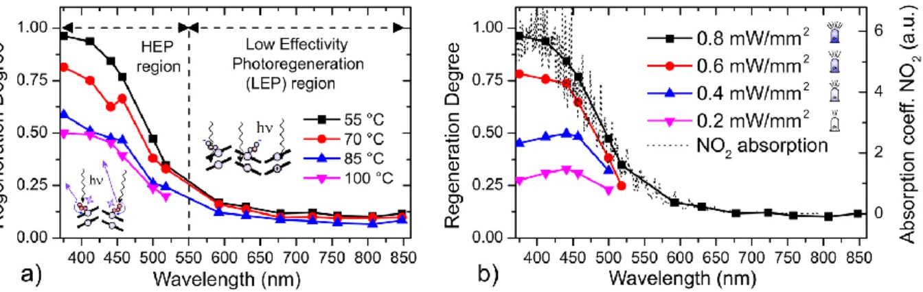
selected operating temperatures. Under these conditions, it was revealed that the regeneration degree (i.e., the effectivity of the photoregeneration process) increased with descending wavelength. For the temperature of 55 °C and baseline of (320 ± 9)
MΩ, the wavelengths longer than 550 nm had an
approximately constant RD in the range from 10% to 17%. (The intervals of the baseline values were constructed as a mean value of Rb ± corrected sample standard deviation of Rb. The same methodology was also applied for the NRM intervals construction.) We labeled this region as the low effectivity photoregeneration (LEP) region due to the small effects of the illumination on the resistance recovery to its original values. From 550 nm going down, the RD was increasing. It went from 35% for 518 nm to 96% for 375 nm (see the black curve in Figure 3a). This region is further referred to as the high effectivity photoregeneration (HEP) region. This character of the RD = RD(λ) dependence and the separation of RD values in two regions was preserved even if the operation temperature was altered. The values just slightly dropped. For the
temperatures of 70, 85 and 100 °C with the corresponding baselines (301 ± 10) MΩ, (302 ± 13) MΩ, and (252 ± 8) MΩ, the RD stayed in the intervals of (9−16%), (7−12%), and (<12%), respectively, in the LEP region. In the HEP region, the RD decreased more significantly with the increasing temperature, providing the intervals of (33−81%), (24−59%), and (20−50%), respectively (Figure 3a). At the same time, the NRM increased with temperature from 0.25 ± 0.02 for 55 °C to 0.55 ± 0.02 for 100 °C, indicating repeatable increase of sensitivity. On the contrary, for the series of measurements at one temperature, the NRM was preserved approximately constant for all sequences with no trend. This should mean that the state of the films was not significantly changing between the consecutive measurements and, in other words, that the illuminations should provide only the desorption of the captured NO2 without the undesirable drift of baseline or/ and NRM values. Regarding the influence of the temperature, it seems that the photoinitiation at higher temperatures resulted in cleaner films with lower concentration of adsorbates, thus becoming more attractive for NO2 capture. A higher amount of captured NO2 then needed a longer illumination period to get desorbed. Typical examples of raw
Figure 2. Illustration of the illumination setup. Sensors were illuminated by power LED diodes (375−850 nm) refocused by two lens optics to
reach maximal spatial homogeneity of light intensity.
Figure 3. Dependence of the regeneration degree RD on the wavelength of the incident illumination and the influence of (a) sensor operating
temperature (55, 70, 85, and 100 °C) and (b) intensity of illumination (0.2, 0.4, 0.6, and 0.8 mW/mm2). The absorption
spectrum of NO2 in the
gaseous phase at 20 °C (dotted line) was taken from the database (ref 33).
data are available in the Supporting Information (Figures S2
and S3).
Photoregeneration at Various Illumination Intensities. To further investigate the nature of the photoregeneration processes, we carried out a series of measurements at a constant temperature of 55 °C and various intensities of illumination (0.2, 0.4, 0.6, and 0.8 mW/mm2). During all these measurements, we also kept the constant baseline value of 310 ± 18 MΩ. Under these circumstances, the RD was found to be more or less linearly proportional to the intensity of the illumination (Figure 3b). This finding is particularly promising for the applicability of the chemiresistors in practice as it should allow to easily adapt the photoregenerating effect to reproducibly restore the original sensor state after the exposures to various concentrations, ideally in one short photoregenerating step. The NRM was approximately constant 0.26 ± 0.02 for all sequences with no significant trend.
Mechanism of Photoregeneration. Considering the
mechanism, two main types have been proposed for photoinduced molecular desorptions: (1) DIET (desorption or
dissociation induced by electronic transitions) and (2) LITD (laser-induced thermal desorptions).34 In our case, the
experimental evidence seems to disqualify the latter mechanism. The sensors were tempered at a selected temperature
before exposure to light. At the beginning of the exposure (step 4 of the testing sequences described in the Experimental Section), the sensor heating power was fixed and the light was allowed to further heat the phthalocyanine layer by absorption. Differences in the layer temperature were proportional to the phthalocyanine absorption coefficient and are plotted in Figure 6. For ZnPc, the maximal layer temperatures during the “55 °C measurement” were 62 °C (55 + 7 °C) for illumination by 0.8 mW/mm2 of 723 or 592 nm due to the absorption in ZnPc’s Q-band.15 Nevertheless, the regeneration degree was considerably
lower for those wavelengths than for 441 nm, for
example, where the absorption of ZnPc is negligible and hence the temperature rise is smaller (58.7 °C, i.e., 55 + 3.7 °C). This research finds a partial parallel in the class of inorganic semiconductors where UV−vis light can be used to activate the sensors to operate at ambient temperature.35 It has been
reported that the application of light can also accelerate the regeneration of such sensors based on the following model (we will refer to it as RecDes, recombinative desorption model). At first (1), the light generates a hole in a semiconductor’s valence band exciting the electron to the conduction band. Second (2), the hole reacts with negatively charged chemisorbed species to release them.36 Extending the mechanism to chemisorbed
NO2, we obtain eq 3. hυ → e + h − + (1)
h + O2 (ads) → O2(gas) + − (2)
h + NO2 (ads) → NO2(gas) + − (3)
In the end, the photoexcited electron from eq 1 is trapped in another hole because holes are major charge carriers in phthalocyanines as they are p-type semiconductors.37 This
model is consistent with the increase in the resistance of ZnPc after illumination, which was observed regardless of whether the sensor was exposed to NO2 before. This model also
predicts a threshold for the effective wavelengths as it requires sufficient energy to promote the electron across the band gap. The band gap was reported to be approximately 2 eV for ZnPc, which corresponds to the wavelength of 620 nm.38 This value
was found close to the border of the HEP region (see Figure 3a).
Nevertheless, the tight correlation between the RD(λ) and ANO2(λ) also suggests another model based on the direct
absorption of light by the chemisorbed NO2 (NLA model). It was reported that 90% of light energy absorbed by NO2 is supposed to be converted to vibrational motion that could stimulate the rate of molecular desorption.39 These effects have
been already used for photoacoustic detection of NO2 by amplitude-modulated high-power light emitting diodes.39 Both
models would expect the proportionality between the light intensity and rate of NO2 desorption.
further investigated in order to determine its NO2 detection limit. It was found out that the NRM is a linear function of NO2 concentration in the range from 0.2 to 0.9 ppm with an estimated detection limit of 20 ppb. The detection limit was calculated according to the methodology reported in ref 40.
Figure 4. (a) Illustration of the stability of the baseline 320 ± 9 MΩ, constant value of the NRM 0.25 ± 0.02, and evolution of the regeneration
degree RD. The data consists of sequence steps 1−5 recorded during the measurement of ZnPc at 55 °C illuminated by light of various
wavelengths (0.8 mW/mm2). The wavelength position (x-axis) is an auxiliary variable proportional to time (1 nm = 6
s). The beginning of each
sequence is positioned on the nominal wavelength of the LED used for the photoregeneration. (b) Dependence of the NRM on the concentration
of NO2 (0.2−1.6 ppm) for a ZnPc chemiresistor operating at 55 °C. The inset contains an example of the raw data
recorded for the concentration
of 0.55 ppm illustrating good repeatability and short measurement cycle duration. The photoregenerations were carried out by the 441 nm light.
This demonstrates the preservation of sensitivity achieved by the traditional “steady state approach” methods while the measurement cycle duration is significantly reduced (∼2 vs ∼15 min).24 For the concentrations exceeding 0.9 ppm, the
trend was still monotonic, but its slope was mildly decreasing (Figure 4b).
Impact of the Method on Sensor Power Consumption. To evaluate the impact of the proposed method on the sensor power demand, we first calculated the consumption of our chemiresistors operating according to the “standard” optimized protocol reported in ref 24. Then we compared it with the consumption when following the protocol proposed in this article. The standard 15 min protocol for one measurement consists of 2 min of exposure to NO2 at 80 °C, 12 min of cleaning at 180 °C, and 1 min of stabilization again at 80 °C. For such a regime, the average power demand of our chemiresistors was calculated as 116 mW (3/15 × 45 mW + 12/15 × 134 mW). According to the new method, the
chemiresistors operated at 55 °C, consuming 23 mW for 14.5 min and 121 mW (23 + 98 mW) for 0.5 min during the photoregeneration step with the 441 nm LED. As a result, the average consumption was ca. 26 mW, which was more than 4
times better when compared with the standard protocol. The consumption could be even better after improving the focus optics, as only ca. 4 mW (0.8 mW/mm2 × 5 mm2) is needed to promote the photoregeneration.
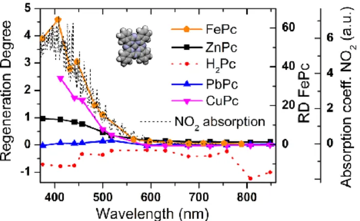
Photoregeneration of the Other Phthalocyanine Chemiresistors (CuPc, H2Pc, PbPc, and FePc) at the Temperature of 55 °C. Photoregeneration of CuPc
Chemiresistors. CuPc chemiresistors, with the baseline set at 106 ± 2 MΩ, did not photoregenerate with wavelengths greater than 550 nm (RD ∼ 0%). The photoregeneration was observed first with 518 nm (RD = 37%) and with even shorter wavelengths it was more and more effective (RD = 58−244%,
Figure 5). The trend was analogous to the one observed for ZnPc, but the difference of the photoregeneration effectivities between the LEP and HEP regions was greater. The highest achieved RD values indicate that, in some cases, it was possible to “overregenerate” the chemiresistor set at a certain baseline while fulfilling the stability criterion of ΔR/R ≤ 1%/min. The “overregeneration” was probably caused by the photoinduced desorption of some part of the chemisorbed O2 or by the photoinduced desorption of the residual traces of NO2, which were not removed during the photoinitiation procedure
described in the Experimental Section. However, the “overregeneration” is not a desirable effect, because it brings an issue
connected with the requirement of the preservation of sensor baseline.
The baseline value should be conserved for all the
measurements to ensure the preservation of a specific film state necessary for correct comparison of consecutive
detections and regenerations. In this case, the “overregeneration” was compensated by inserting an extra cycle with the
same exposition to NO2 but followed by the photoregeneration with a lower illumination intensity or shorter illumination time. This, with regard to the dependence of the RD on the
illumination intensity discussed above and a similar dependence on time, allowed return to the original baseline value
while preserving the sensor sensitivity, because the NRM was approximately constant 0.61 ± 0.04 for all sequences with no trend as in the case of ZnPc. To complete this point, it should be noted that the direct return to the original baseline after the “overregeneration” solely by the exposure to the NO2 gas (without the subsequent illumination) did not provide
comparable results as the NRM was decreasing proportionally to the dose of NO2 used for the baseline compensation.
Photoregeneration of PbPc Chemiresistors. PbPc chemiresistors, working at the baseline of 15.3 ± 0.2 MΩ, did not
significantly photoregenerate with any wavelength in the range from 375 to 850 nm (RD = −3−17%, Figure 5). As the
photoregeneration was not effective in this case, we had to use the classical thermal recovery mechanism to regenerate the sensors. After 10 steps of illumination for each wavelength, step 4 in our methodology was replaced by a temporal increase of the operating temperature. It was increased from 55 to 130 °C for 30 s, and it went back to 55 °C to check the progress of the regeneration. With this approach, we managed to get an approximately constant NRM of 0.68 ± 0.02 for all sequences
with no trend as in the case of other phthalocyanines. However, we encountered increasing instability of sensor baselines. This instability had to be compensated, so it resulted in dramatic prolongations of settling times. Nevertheless, despite these prolongations, it was problematic to fulfill the stability criterion defined in the experimental section. Due to these effects, PbPc cannot be considered as a suitable material for gas detection by our method.
Photoregeneration of FePc Chemiresistors. As opposed to PbPc, FePc chemiresistors, operating at the baseline of 1.050 ± 0.002 GΩ, photoregenerated with all the wavelengths (RD ≫ 0%) and the RD significantly increased with descending wavelength as for ZnPc and CuPc (see Figure 5). However, the regeneration was much more intensive. For the wavelengths 678 nm+, the RD varied in the range from 30% to 99%. Then it rose to 156% for 630 nm and to several thousand percent for the light of 518−375 nm. These huge “overregenerations” complicated the measurement as the sensors were returning to their baselines very slowly (one return took practically several hours or days). To compensate this, we used controlled NO2 expositions to speed up the baseline return (without the subsequent illumination). Probably as a result of this approach, the NRM was mildly decreasing in the sequence of the
experiments. At the end, it had 50% of its original magnitude. In spite of this, the application of FePc for NO2 detection seems to be prospective in combination with either longer wavelengths or significantly lower intensities (∼0.01 mW/ mm2) of the shorter wavelengths.
Photoregeneration of H2Pc Chemiresistors. H2Pc chemiresistors, with the baseline set at 176 ± 26 GΩ, did not
Figure 5. Dependence of the regeneration degree RD on the phthalocyanine material (FePc, ZnPc, H2Pc, PbPc, and CuPc). The
absorption spectrum of NO2 in the gaseous phase at 20 °C (dotted
line) was taken from the database (ref 33).
satisfactorily photoregenerate with any wavelength in the range measured. In terms of the RD, the values obtained were even negative in the range from −124% to −18%. The reason for the negative values could be that illumination causes a temporal increase in the layer conductivity and generates new free charge carriers, which seem to have much longer lifetime in H2Pc than in the other phthalocyanines. This could
be connected with H2Pc’s significantly higher resistance baseline. So, at the time of the RD evaluation, some of these photocarriers were probably still present there (see Figure S4). A similar effect has already been observed for amorphous VOPC and named as persistent photoconductivity (PPC).41 If
we waited a longer time before the evaluation of the photoregeneration (30 min, not 30 s as defined in the methodology), we obtained small positive values of the RD, mostly close to 0% (Figure S4). The inefficiency of the photoregeneration in the case of H2Pc forced us to apply thermal regeneration by temporal increases of the operating temperature to 90 °C. The NRM was approximately constant, equal to 0.28 ± 0.04 with no statistically significant trend. Comparison, Implications for the Mechanism. Extending the study to the phthalocyanine materials other than ZnPc revealed the importance of the central cation for the process of photoregeneration. The phthalocyanines (ZnPc, CuPc, H2Pc, PbPc, and FePc) divided into two groups differing in the ability to photoregenerate. ZnPc, CuPc, and FePc exhibited photoregeneration; H2Pc and PbPc did not. This division seems to correspond with the symmetry of the molecules. Zn2+, Cu2+, and Fe2+ cations are small and fit the cavity of the macrocycle, letting the phthalocyanine preserve its planar structure with the symmetry D4h. H2Pc’s hydrogens reduce its symmetry to D2h and PbPc’s large Pb2+ cation that distorts the planarity of the molecule reduces the symmetry to C4v.15
Regarding the mechanism of desorption, from the viewpoint of the models considered above, the universal shape of the RD(λ) for various phthalocyanines (it differs just by a multiplicative scaling factor) seems to favor the one based on the direct absorption of light by the chemisorbed NO2. The RecDes model would expect some shifts of the RD(λ) curve based on the variations of the band gaps (CuPc ∼ 1.7 eV).42
However, the precise value of the band gap widths may be difficult to consider as the literature contains rather a range of values for each phthalocyanine.
Stability of the Phthalocyanine Sensitive Layers
under Illumination (Spectral Study). UV−vis spectra and FTIR spectra of the layers were consistent with those in the literature. The comparison is provided in the Supporting Information.
Effects of the Illuminations on UV−Vis Spectra of the Phthalocyanine Layers. The Pc spectra did not change
significantly with the layer illuminations at working conditions for photoregeneration (441 nm, 0.8 mW/mm2, 2 h). Based on these findings, we are convinced that, under the working conditions, there are no irreversible changes in the layers caused by the illuminations. We believe that the effects of light are restricted only to (i) the photostimulated desorption of the species adsorbed on the surface of the phthalocyanine layers (NO2, O2, H2O) and (ii) the generation of photoinduced
mobile charge carriers. However, we observed some changes of FePc and PbPc UV−vis spectra after stronger illuminations (7.3 mW/mm2, 1 h). In these particular cases, the stronger illuminations led to small depreciations of the Q absorption bands (626 nm, −6%; 719 nm, −3%, respectively, Figure 6). We think that these depreciations were caused by partial temperature-stimulated recrystallizations of the FePc and PbPc
layers, as those layers exhibit some level of absorption of the 441 nm light. The temperature rose by more than several tens of degrees (°C) for the stronger illuminations and hence could provide the energy to the layers to recrystallize. PbPc, for example, is known to have two polymorphs differing in their absorption spectra: (i) monoclinic with the absorption maxima at 740 nm and (ii) triclinic with the maxima at 900 nm.43 It has
been reported that the annealing process can induce the recrystallization of the as-deposited monoclinic phases to the more thermodynamically stable triclinic form.43 On the
contrary, the absorption of the layers of the other
phthalocyanines (ZnPc, CuPc, and H2Pc) is negligible in the spectral region around 441 nm and thus their temperature rises were insufficient to alter layer morphology.
Effects of the Illuminations on FTIR Spectra of the Phthalocyanine Layers. The FTIR spectra remained
unchanged for the working intensity of the illuminations (0.8 mW/mm2), supporting the interpretation mentioned above and the claim of stability. After the stronger illuminations (7.3 mW/mm2), we again recorded the almost identical spectra containing only minor changes of very weak bands for ZnPc at 668 or 434 cm−1 and CuPc at 571 cm−1 (Figure S5). A
possible explanation of these changes may lie in the stimulated desorption of several adsorbates (NO2, O2, H2O) which are present in phthalocyanine layers at ambient conditions.
■
CONCLUSIONSZnPc, CuPc, and FePc thin layers containing certain amounts of NO2 as a result of a chemiresistive detection process were found to photoregenerate when they were exposed to light with a wavelength shorter than 550 nm. Such light
demonstrated its capability to stimulate NO2 desorption from the layer, accelerating its recovery to the original state
Figure 6. UV−vis spectra of the phthalocyanine sensitive layers before (gray area) and after (red dashed curve) illumination (7.3 mW/mm2,
1 h). The graphs contain superimposed curves (STR) illustrating temperature effects of illumination on the chemiresistors (0.8 mW/ mm2).
before the detection of NO2. This effect was ascribed either to the generation of holes in the organic semiconductor that can recombine with the chemisorbed species or to the direct desorption induced by electronic transitions originating from the light absorption in chemisorbed NO2. It was shown that photoregeneration can be conducted at ambient or slightly elevated temperatures (55−100 °C), thus opening the possibility of simple phthalocyanine based chemiresistors operating at low temperatures. Lower temperatures imply lower electrical consumption (ca. 25%) and longer lifetime of the sensors.
Moreover, we modified the kinetic operating procedure suggested in ref 25 to benefit not only from the considerable acceleration of measurement (Tmeas.cycle was ∼ 2 min, more than 7 times faster than the traditional optimized approach presented in ref 24), but also from the preservation of the limit of detection (<20 ppb) and the improved baseline stability (→ better repeatability of the measurement). The photoregeneration effects were further found to be approximately linearly
dependent on the light intensity and also proportional to the operating temperature and the phthalocyanine type.
From a practical standpoint, it has been shown that
photoregeneration could help phthalocyanine based sensors to become more than a viable alternative to conventional techniques for NO2 monitoring.
■
ASSOCIATED CONTENT*
S Supporting InformationThe Supporting Information is available free of charge on the
ACS Publications website at DOI: 10.1021/acssensors. 8b00922.
Evaporation parameters and details about the sensitive layers; details about the illumination; typical raw data of ZnPc photoregeneration; persistent photoconductivity of H2Pc; UV−vis and FTIR spectra of the sensitive
layers and their comparison with the literature; effects of the illuminations on FTIR spectra of the phthalocyanine layers (PDF)
Animated video of the photoregeneration (AVI)
■
AUTHOR INFORMATION Corresponding Author *E-mail: [email protected]. ORCIDMartin Vrnata:
0000-0002-1199-1458 Notes■
ACKNOWLEDGMENTSThis work was supported by Czech Science Foundation (GACR), Projects No. 17-13427S and No. 18-09347S and also by Ministry of Education, Youth and Sports within the projects LTC17058 and 8J18FR011 and by COST Action CA15107 MultiComp. Financial support from specific university research (MSMT No 21-SVV/2018) and PHC Barrande 2018
(40672RL) is also acknowledged.
■
REFERENCES(1) Kim, I.-D.; Rothschild, A.; Lee, B. H.; Kim, D. Y.; Jo, S. M.; Tuller, H. L. Ultrasensitive Chemiresistors Based on Electrospun TiO2 Nanofibers. Nano Lett. 2006, 6 (9), 2009−2013.
(2) Zhang, D.; Liu, Z.; Li, C.; Tang, T.; Liu, X.; Han, S.; Lei, B.; Zhou, C. Detection of NO2 down to ppb Levels Using Individual and
Multiple In2O3 Nanowire Devices. Nano Lett. 2004, 4 (10), 1919−
1924.
(3) Campbell, M. G.; Liu, S. F.; Swager, T. M.; Dinca, M.
Chemiresistive Sensor Arrays from Conductive 2D Metal-Organic Frameworks. J. Am. Chem. Soc. 2015, 137 (43), 13780−13783. (4) Assen, A. H.; Yassine, O.; Shekhah, O.; Eddaoudi, M.; Salama, K. N. MOFs for the Sensitive Detection of Ammonia: Deployment of fcu-MOF Thin Films as Effective Chemical Capacitive Sensors. ACS Sensors 2017, 2 (9), 1294−1301.
(5) Yuan, W.; Huang, L.; Zhou, Q.; Shi, G. Ultrasensitive and Selective Nitrogen Dioxide Sensor Based on Self-Assembled Graphene/Polymer Composite Nanofibers. ACS Appl. Mater. Interfaces 2014, 6 (19), 17003−17008.
(6) Cho, S.-Y.; Cho, K. M.; Chong, S.; Park, K.; Kim, S.; Kang, H.; Kim, S. J.; Kwak, G.; Kim, J.; Jung, H.-T. Rational Design of
Aminopolymer for Selective Discrimination of Acidic Air Pollutants. ACS Sensors 2018, 3 (7), 1329−1337.
(7) Pavluchenko, A. S.; Mamykin, A. V.; Kukla, A. L.; Konoshchuk, N. V.; Posudievsky, O. Y.; Koshechko, V. G. Estimation of
multicomponent organic solvent vapor mixture composition with electroconducting polymer chemiresistors. Sens. Actuators, B 2016, 232, 203−218.
(8) Evyapan, M.; Dunbar, A. D. F. Improving the selectivity of a free base tetraphenylporphyrin based gas sensor for NO2 and carboxylic
acid vapors. Sens. Actuators, B 2015, 206, 74−83. (9) Surwade, S. P.; Agnihotra, S. R.; Dua, V.; Manohar, S. K. Nitrogen dioxide vapor detection using poly-o-toluidine. Sens. Actuators, B 2009, 143 (1), 454−457.
(10) Othman, M.; Théron, C.; Bendahan, M.; Caillat, L.; Rivron, C.; Bernardini, S.; Le Chevallier, G.; Chevallier, E.; Som, M. P.; Aguir, K.; Tran-Thi, T. H. Efficiency of new ozone filters for NO2 sensing and
air depollution. Sens. Actuators, B 2018, 265, 591−599.
(11) Fitl, P.; Vrnata, M.; Kopecky, D.; Vlcek, J.; Skodova, J.; Bulir, J.; Novotny, M.; Pokorny, P. Laser deposition of sulfonated
phthalocyanines for gas sensors. Appl. Surf. Sci. 2014, 302, 37−41. (12) Lee, Y.-L.; Sheu, C.-Y.; Hsiao, R.-H. Gas sensing characteristics of copper phthalocyanine films: effects of film thickness and sensing temperature. Sens. Actuators, B 2004, 99 (2), 281−287.
(13) Tomeček, D.; Fitl, P.; Marešová, E.; Vlček, J.; Hofmann, J.; Vrňata, M. Silver phthalocyanine thin films carrying gold, palladium
and silver nanoparticles for detection of taggants in explosives. Thin Solid Films 2017, 630, 31−37.
(14) Zhou, R.; Josse, F.; Göpel, W.; Öztürk, Z. Z.; Bekaroǧlu, Ö. Phthalocyanines as sensitive materials for chemical sensors. Appl. Organomet. Chem. 1996, 10 (8), 557−577.
(15) Kadish, K. M.; Smith, K. M.; Guilard, R. The Porphyrin
Handbook: Phthalocyanines: properties and materials; Academic Press: 2000.
(16) Guillaud, G.; Simon, J.; Germain, J. P. Metallophthalocyanines Gas sensors, resistors and field effect transistors. Coord. Chem. Rev. 1998, 178−180, 1433−1484.
(17) Park, J. H.; Royer, J. E.; Chagarov, E.; Kaufman-Osborn, T.; Edmonds, M.; Kent, T.; Lee, S.; Trogler, W. C.; Kummel, A. C. Atomic imaging of the irreversible sensing mechanism of NO2
adsorption on copper phthalocyanine. J. Am. Chem. Soc. 2013, 135 (39), 14600−14609.
(18) Šebera, J.; Fitl, P.; Vlček, J.; Vrňata, M.; Fendrych, F.; Kopeček, J.; Kratochvílová, I. Interaction of selected gases with zinc
phthalocyanine thin films: Theoretical and experimental studies. Eur. Phys. J.: Appl. Phys. 2013, 64 (1), 10202.
(19) Słota, R.; Dyrda, G.; Hnatejko, Z.; Karolczak, J.; Stryła, Z. Effect of air-absorbed oxygen and moisture on the chemical stability of photoexcited Mg, Zn and Eu phthalocyanines in dimethylformamide. J. Porphyrins Phthalocyanines 2006, 10 (1), 43−54.
(20) Gadenne, V.; Porte, L.; Patrone, L. Structure and growth mechanism of self-assembled monolayers of metal protoporphyrins and octacarboxylphthalocyanine on silicon dioxide. RSC Adv. 2014, 4 (110), 64506−64513.
(21) Novotný, M.; Šebera, J.; Bensalah-Ledoux, A.; Guy, S.; Bulíř, J.; Fitl, P.; Vlček, J.; Zákutná, D.; Marešová, E.; Hubík, P.; Kratochvílová, I.; Vrňata, M.; Lančok, J. The growth of zinc phthalocyanine thin films by pulsed laser deposition. J. Mater. Res. 2016, 31 (1), 163−172. (22) Pizzini, S.; Timo, G. L.; Beghi, M.; Butta, N.; Mari, C. M.; Faltenmaier, J. Influence of the structure and morphology on the sensitivity to nitrogen oxides of phthalocyanine thin-film resistivity sensors. Sens. Actuators 1989, 17, 481−491.
(23) Lee, Y. L.; Sheu, C. Y.; Hsiao, R. H. Gas sensing characteristics of copper phthalocyanine films: Effects of film thickness and sensing temperature. Sens. Actuators, B 2004, 99 (2−3), 281−287.
(24) Brunet, J.; Garcia, V. P.; Pauly, A.; Varenne, C.; Lauron, B. An optimized gas sensor microsystem for accurate and real-time measurement of nitrogen dioxide at ppb level. Sens. Actuators, B 2008, 134 (2), 632−639.
(25) Wilson, A.; Wright, J. D.; Chadwick, A. V. A microprocessorcontrolled nitrogen dioxide sensing system. Sens. Actuators, B 1991, 4
(3), 499−504.
(26) Zhou, Q.; Gould, R. D. A study of the response rate to nitrogen dioxide exposure in metal phthalocyanine thin film sensors. Thin Solid Films 1998, 317 (1), 436−439.
(27) Jalil, A. R.; Chang, H.; Bandari, V. K.; Robaschik, P.; Zhang, J.; Siles, P. F.; Li, G.; Burger, D.; Grimm, D.; Liu, X.; Salvan, G.; Zahn, D. R. T.; Zhu, F.; Wang, H.; Yan, D.; Schmidt, O. G. Fully Integrated Organic Nanocrystal Diode as High Performance Room Temperature NO2 Sensor. Adv. Mater. 2016, 28 (15), 2971−2977.
(28) Newton, M. I.; Starke, T. K. H.; Willis, M. R.; McHale, G. NO2
detection at room temperature with copper phthalocyanine thin film devices. Sens. Actuators, B 2000, 67 (3), 307−311.
(29) Sadaoka, Y.; Jones, T. A.; Göpel, W. Fast NO2 detection at
room temperature with optimized lead phthalocyanine thin-film structures. Sens. Actuators, B 1990, 1 (1), 148−153.
Pennesi, G. Reversible and selective detection of NO2 by means of
optical fibres. Sens. Actuators, B 2001, 74 (1), 12−17.
(31) Bueno, A.; Lahem, D.; Caucheteur, C.; Debliquy, M. Reversible NO2 Optical Fiber Chemical Sensor Based on LuPc2 Using
Simultaneous Transmission of UV and Visible Light. Sensors 2015, 15 (5), 9870.
(32) Tomeček, D.; Fitl, P.; Vlček, J.; Marešová, E.; Vrňata, M. Detection of taggants in explosives on nanostructured metal/silver phthalocyanine chemiresistors: Influence of analyte photoactivation. Sens. Actuators, B 2017, 239, 147−156.
(33) Keller-Rudek, H.; Moortgat, G. K.; Sander, R.; Sörensen, R. The MPI-Mainz UV/VIS Spectral Atlas of Gaseous Molecules of Atmospheric Interest. Earth Syst. Sci. Data 2013, 5 (2), 365−373. (34) Kawasaki, T.; Mochida, T.; Katada, J. I.; Okahata, Y. Laser
response of a quartz crystal microbalance: Frequency changes induced by light irradiation in the air phase. Anal. Sci. 2009, 25 (9), 1069− 1075.
(35) Park, S.; An, S.; Mun, Y.; Lee, C. UV-enhanced NO2 gas
sensing properties of SnO2-Core/ZnO-shell nanowires at room
temperature. ACS Appl. Mater. Interfaces 2013, 5 (10), 4285−4292. (36) Zhang, C.; Boudiba, A.; De Marco, P.; Snyders, R.; Olivier, M.- G.; Debliquy, M. Room temperature responses of visible-light illuminated WO3 sensors to NO2 in sub-ppm range. Sens. Actuators,
B 2013, 181, 395−401.
(37) Bassoul, P.; Toupance, T.; Simon, J. Semiconductivity and gassensing properties of crown-ether-substituted lutetium bisphthalocyanines. Sens. Actuators, B 1995, 26 (1), 150−152.
(38) Senthilarasu, S.; Velumani, S.; Sathyamoorthy, R.; Subbarayan, A.; Ascencio, J. A.; Canizal, G.; Sebastian, P. J.; Chavez, J. A.; Perez, R. Characterization of zinc phthalocyanine (ZnPc) for photovoltaic applications. Appl. Phys. A: Mater. Sci. Process. 2003, 77 (3), 383−389. (39) Saarela, J.; Sorvajärvi, T.; Laurila, T.; Toivonen, J. Phasesensitive method for background-compensated photoacoustic detection of NO2 using high-power LEDs. Opt. Express 2011, 19 (S4),
A725−A732.
(40) Loock, H.-P.; Wentzell, P. D. Detection limits of chemical sensors: Applications and misapplications. Sens. Actuators, B 2012, 173, 157−163.
(41) Santos, L. F.; Faria, R. M.; Del Caño, T.; de Saja, J. A.; Constantino, C. J. L.; Amorim, C. A.; Mergulhão, S. Observation of persistent photoconductivity in vanadyl phthalocyanine. J. Phys. D: Appl. Phys. 2008, 41 (12), 125107.
(42) So, F. Organic Electronics: Materials, Processing, Devices and Applications; CRC Press: 2009.
(43) Vasseur, K.; Rand, B. P.; Cheyns, D.; Froyen, L.; Heremans, P. Structural evolution of evaporated lead phthalocyanine thin films for near-infrared sensitive solar cells. Chem. Mater. 2011, 23 (3), 886− 895.