NEWSLETTER
Volume 24, No. 1, 1-24
Circulation 84,122
Spring 2009
www.apsf.org
The Official Journal of the Anesthesia Patient Safety Foundation
In the Summer 2007 issue of the APSF Newsletter, Cullen and Kirby reported on 2 patients in whom a catastrophic, new-onset brain injury was discovered after surgery in the beach chair (barbershop) posi-tion.1The authors presented views on the effect that blood pressure monitoring and management may have had on neurologic injury and provided a for-mula for correcting hydrostatic blood pressure gra-dients from the site of measurement to the site of vulnerable brain tissues. This publication generated a series of letters to the Newsletter, either supporting or challenging the need for the blood pressure cor-rections suggested by Cullen and Kirby. Notable among those letters was that of Munis who argued that a correction for hydrostatic gradients was not needed because, in the head-up position, the circula-tion above the heart funccircula-tions as a siphon.2Cucchiara took another approach and chided practitioners to place an arterial catheter in head-up patients and measure blood pressure at the level of the head to avoid the need for arithmetically corrected measure-ments altogether.3This debate continues in the cur-rent issue of the Newsletter with letters from Drummond et al. who argue that clinical manage-ment of head-up patients must account for hydrosta-tic gradients,4and Kirby and Cullen5who expand on concepts raised in their earlier publication.1
This debate about blood pressure monitoring and management in head-up patients is unavoidable because of inadequate empirical data involving anes-thetized, head-up patients who are at risk for rare, but debilitating, postoperative neurologic deficits.1,6 Various forms of head-up positioning are used not only for neurosurgical procedures (e.g., posterior fossa craniectomy and cervical laminectomies) where the effects on hemodynamics have been more intensely pondered, but also for surgery to the thy-roid gland, shoulder, and other non-neurosurgical sites where debate about blood pressure manage-ment has been less common. Placing the patient supine or prone to avoid physiologic challenges imposed by a head-up position is not always an option, as the sitting position for posterior fossa cran-iotomy is reported to diminish operative blood loss and significantly improve postoperative cranial nerve function.7With cervical spine surgery or pos-terior fossa intracranial surgery, converting from the sitting to prone position may potentially worsen pul-monary gas exchange in patients having medically complicated obesity, or may contribute to the risk of postoperative visual impairment in rare instances. Other surgeries (e.g., thyroid and shoulder surgery) are simply made more technically difficult by vary-ing from an ideal head-up position. As such, it
appears that the head-up position during anesthesia and surgery is here to stay, even though ideal blood pressure monitoring and management in these patients is controversial.
One of the core features of the current debate about blood pressure management in the head-up position revolves around whether the circulation above the heart functions as a siphon system2or as a waterfall system.1,4,5Based on the available evidence, either scenario is probably an oversimplification in anesthetized, surgically positioned patients. The siphon concept is very appealing when speaking of the physiology of unanesthetized healthy humans or giraffes; however, anesthetized surgical patients placed head up—often with the head position devi-ating considerably from neutral—may introduce more complex physiology. As we will see later, these head-position variations, independent of a gravity effect, have a bearing on cerebral circulation. Further, the siphon analogy assumes that vessels will function in series, when in fact the vessels connecting the heart to the most remote areas of the brain tissues and spinal cord have some elements in series and
Inside:
Cerebral Autoregulation...Page 5 Hydrostatic Gradient is Important—Blood Pressure Should be Corrected ...Page 6 Cerebral Oximetry ...Page 7 EEG Monitoring ...Page 10 Dear SIRS: Fluid Warmers Interfere with ECG...Page 12 Q&A: Cross-Contamination Via Anesthesia Equipment ...Page 14 Perioperative Stent Thrombosis...Page 17 Intraoperative Hypercapnea During Thoracoscopy—A Case Report ...Page 18 Intralipid Treatment of Bupicavaine Toxicity ...Page 20
o
SPECIAL EDITORIAL
o
Cerebral Perfusion: Err on the Side of Caution
See “Head Up,” Page 3
As controversy continues regarding the hemodynamic management of patients in the head-up or beach chair
position, the APSF Newsletter turns to Dr. William Lanier for editorial perspective. Dr. Lanier is Editor-in-Chief of
Mayo Clinic Proceedings as well as a highly regarded neuroanesthesiologist and neurophysiology investigator.
APSF GOES ALL GREEN
Important Notice!
Beginning with the Spring 2010 issue (our 25th anniversary), the APSF Newsletter will be all electronic. Regular hard copy distribution will cease. Be sure your respective organization (ASA, AANA, AAAA, ASATT, etc.) has your correct and current email to ensure you receive your electronic copy. The APSF will provide hard-copies of the APSF Newsletter beginning with the Spring 2010 for an annual subscription of $100. Please contact Deanna Walker at [email protected] if you wish to subscribe.
NEWSLETTER
The Official Journal of the Anesthesia Patient Safety Foundation
The Anesthesia Patient Safety Foundation
Newsletteris the official publication of the nonprofit Anesthesia Patient Safety Foundation and is pub-lished quarterly in Wilmington, Delaware. Annual contributor cost: Individual–$100, Corporate–$500. This and any additional contributions to the Founda-tion are tax deductible. © Copyright, Anesthesia Patient Safety Foundation, 2009.
The opinions expressed in this Newsletter are not necessarily those of the Anesthesia Patient Safety Foundation. The APSF neither writes nor promulgates standards, and the opinions expressed herein should not be construed to constitute practice standards or practice parameters. Validity of opinions presented, drug dosages, accuracy, and completeness of content are not guaranteed by the APSF.
APSF Executive Committee:
Robert K. Stoelting, MD, President; Nassib G. Chamoun, Vice President; Jeffrey B. Cooper, PhD, Executive Vice President; George A. Schapiro, Executive Vice President; Matthew B. Weinger, MD, Secretary; Casey D. Blitt, MD, Treasurer; Sorin J. Brull, MD; Robert A. Caplan, MD; David M. Gaba, MD; Lorri A. Lee, MD; Robert C. Morell, MD; Michael A. Olympio, MD; Richard C. Prielipp, MD; Steven R. Sanford, JD; Mark A. Warner, MD. Consultants to the Executive Committee: John H. Eichhorn, MD; Patricia A. Kapur, MD.
Newsletter Editorial Board:
Robert C. Morell, MD, Editor; Lorri A. Lee, MD, Associate Editor; Sorin J. Brull, MD; Joan Christie, MD; Jan Ehrenwerth, MD; John H. Eichhorn, MD; Steven B. Greenberg, MD; Rodney C. Lester, PhD, CRNA; Glenn S. Murphy, MD; Karen Posner, PhD; Andrew F. Smith, MRCP, FRCA; Wilson Somerville, PhD; Jeffery Vender, MD.
Address all general, contributor, and subscription correspondence to:
Administrator, Deanna Walker Anesthesia Patient Safety Foundation Building One, Suite Two
8007 South Meridian Street Indianapolis, IN 46217-2922 e-mail address: [email protected] FAX: (317) 888-1482
Address Newsletter editorial comments, questions, letters, and suggestions to:
Robert C. Morell, MD Editor, APSF Newsletter
c/o Addie Larimore, Editorial Assistant
Department of Anesthesiology
Wake Forest University School of Medicine 9th Floor CSB
Medical Center Boulevard Winston-Salem, NC 27157-1009 e-mail: [email protected]
www.apsf.org
®
Supports APSF Research
APSF gratefully acknowledges
the generous contribution of $100,000 from Covidien
toward the funding ($150,000) of a
2009 APSF Research Grant that will be designated the
APSF/Covidien Research Award
www.covidien.com
Correction:
The Winter 2008-209 issue of the APSF Newsletter lead article “Does Anesthetic Management Affect Cancer Outcome?” was authored by Anije Gottschalke, MD, Research Fellow; Marcel E. Durleux, MD, PhD, Professor of Anesthesiology and Neurolgoical Surgery; Mohamed Tiouririne, MD, Assistant Professor of Anesthesiology at the University of Virginia Health System, Charlottesville, VA. Dr. Gottschalk and Dr. Tiouririne were omitted from the original credits.
PLEASE NOTE
Effective with the Spring of 2010 issue, the
APSF Newsletter will become an
all-electronic publication
.
Routine hard copy publication will cease as of
the Winter 2009-2010 issue.
Please be sure your affiliated organization
(ASA, AANA, AAAA, ASATT, etc.)
has your accurate email to ensure your
continued receipt of the Newsletter.
The APSF will provide hardcopies of
the APSF Newsletter beginning with the Spring
2010 issue for an annual subscription of $100.
Please contact Deanna Walker at
[email protected] if you wish to subscribe.
the other patient had simultaneous EEG changes in both cerebral hemispheres, though more prominent in the right. Angiography revealed that, because of widespread atherosclerosis, the left carotid artery con-tributed nothing to the circulation of either cerebral hemisphere; however, the right carotid artery sup-plied blood for both hemispheres. Clearly these col-lective observations of Toole and Tucker8,9 and Perkins et al.10speak to the fact that the plumbing of the human brain can be variable, dependent on changes in head positioning, and conceptually quite different from household plumbing.
Parallel Plumbing Important
If this is the case, one should examine the extremes of blood pressure required to prevent permanent neu-rologic injury. At the lower end of this range, we could assume a young, healthy, normotensive patient, with classic vessel anatomy, and an intracranial pressure never deviating from 0 mmHg or regional cerebral blood flow distribution never deviating from parity. Assuming a siphon based physiology, then it should be possible to measure blood pressure at the level of the heart, and maintain blood pressure at the lower limit of autoregulation without causing ischemic neu-rologic injury. Any small errors created by deviations from a pure siphon system, and some uncertainty as to whether there is a precise lower limit of autoregulation and where it might occur in this patient,11would be somewhat offset by the fact that, even as perfusion pressure declines below the lower limits of autoregu-lation, blood flow does not fall into the abyss but instead declines gradually, perhaps still leaving enough circulation to prevent permanent neurologic injury. At the other extreme, if we assume a waterfall-based physiology, we must not only account for a hydrostatic gradient imposed by the sitting position, but we must also take into account the parallel plumb-ing feedplumb-ing the waterfall, and the effects that regional variations in intracranial pressure, surgical retractor pressure, head positioning, atherosclerosis, geographic variants of blood vessel distribution, and other factors may have on the flow through contributing vessels, some of which may be critical to patient well-being. Clearly there is a considerable difference between the physiologies described by these 2 extremes.Simple Study May Not Yield
Simple Answer
It is tempting to rush to the animal laboratory to try to mimic and study the exact patterns of physiol-ogy during anesthesia and patient positioning. How-ever, such studies will likely reflect the physiology of healthy animals in which the various combinations of heart and head positioning, species-related anatomic variations, and other factors, will not accurately reproduce the conditions of the rare, highest-risk humans. If such studies are eventually performed in
animals to better explore the issue of monitoring site versus cerebral well-being as related to siphon versus waterfall hemodynamic models, it must be remem-bered that measurements of well-being must take into account the watershed regions of brain, eyes, and spinal cord, using techniques such as microspheres, laser Doppler flowmetry, or multidimensional radio-logic imaging to quantify regional blood flows, and multiple-lead electrical recordings to assess electrical well-being. Crude assessments of well-being, using transcranial Doppler sonography of conducting ves-sels, and processed or geographically non-discrimi-nating eletrophysiologic measurements, will simply not address the root of the problem. Unfortunately, attempting to monitor and assess individual patients will be problematic, if for no other reason than that the patients at greatest risk of injury during the head-up position are probably those with some atypical anatomy or baseline physiology. Such patients will be hard to identify, the influence of variations in patient positioning may be impossible to explore in the clini-cal environment, and data from these patients will be hard to generalize to other high-risk patients.
Absent such evidence, it is tempting to instead analyze and rationalize blood pressure monitoring and management in individual patients, based on core principles. However, we anesthesiologists should be reluctant to choose this approach, recog-nizing how such a process has ill served us in the past. We need not be reminded that for a period of 3 or more decades, this type analysis of a possible intracra-nial pressure increase in response to intravenous suc-cinylcholine,12,13or to “bucking” and coughing in tracheally intubated subjects,14,15erroneously ascribed increases in intrathoracic pressure and central venous pressure as the operant mechanisms. However, when such concepts were first tested experimentally in the 1980s and ‘90s, neither clinical condition was even remotely related to the long-touted operant mecha-nism.12-15Instead, other altogether different mecha-nisms appeared to be responsible, and the onset, magnitude, and duration of the intracranial pressure increases were not at all what anesthesiologists had long envisioned. There are a sufficient number of sim-ilar, faulty analyses in the history of anesthesiology to make us fearful of introducing new errors in manage-ment, based on core-principle analysis absent empiri-cal support. However, unlike previous examples involving transient increases in intracranial pressure, the end result of the current discussion of blood pres-sure management in head-up patients is not to declare a winner of some innocuous academic pillow fight, but instead to optimize patient management for the purpose of avoiding irreversible neurologic injury.
Without the data we need to definitively identify ideal blood pressure monitoring and management in
Too High Likely Safer Than Too Low
some in parallel. These parallel aspects of the circu-lation may place tissues within remote watershed regions at risk for ischemic injury coincident with global cerebral and spinal cord blood flow remain-ing adequate. It is not so simple to model the cere-bral circulation as a waterfall either, because a waterfall analogy dictates that the hydrostatic gradi-ent of the column of blood in vessels meaningfully influences the relationship between the pressure at the aortic root and the remote regions of the brain. This analysis, too, overlooks the input of vessels in parallel, some of which may be occluded at baseline (e.g., from atherosclerosis) or as a result of surgical positioning. Some examples are in order:
Toole and Tucker8,9reviewed the literature con-cerning awake patients who acquire new-onset neu-rologic symptoms related to changes in head position, and they identified multiple contributing factors such as: 1) intraluminal atherosclerosis, 2) deviations from classic vessel configurations within the neck (most commonly involving a diminutive or non-functioning vertebral artery unilaterally), 3) changing relationships between the geography of the brainstem and vertebral vessels during head flex-ion, and 4) external compression of the carotid and vertebral arteries by osteophytes or normal vertebral anatomy. In a prospective study,9they examined the effect of head flexion/extension, rotation, and tilt on blood flow through the carotid and vertebral arter-ies in 20 fresh cadavers. They determined that, if a change in flow was to occur at all, it occurred at flex-ion/extension of <45°, rotation of <45°, or tilt of <30°. A positive response was manifested as simul-taneous cessation of blood flow in both vertebral arteries in 30% of cadavers, and in both internal carotid arteries (but not simultaneously) in 45% of cadavers. This research also determined that the diminution or ablation of blood flow in these vessels was not linear with head movement, but instead developed precipitously over an incremental 5-10° change. Additionally, they determined that it was not possible to predict in which vessel, or even on which side of the body, vessel occlusion would occur during head rotation. Elsewhere Perkins et al.10 reported on 2 patients who underwent right carotid endarterectomy while the patients were supine with the head rotated to the left. Inadvertent lidocaine injection into the right carotid arteries (during attempted local anesthesia of the carotid sinus baroreceptors) produced electroencephalographic (EEG) changes in both patients, but the EEG patterns varied greatly for reasons made clear by the preop-erative angiogram. In 1 patient, atherosclerotic changes limited the contributions of the right carotid artery to the right side of the brain. Not surprisingly, EEG changes in this patient were unilateral and ipsi-lateral to the site of lidocaine injection. In contrast,
“Head Up,” From Page 1
head-up, anesthetized patients, what should we do for contemporary blood pressure measurement and management? It would seem appropriate that our practices should err on the side of providing exces-sive blood pressure to non-critical tissues, and ade-quate blood pressure to critical tissues. Such an approach has merit not because we have proven that a modified watershed model of cerebral circulation is operant in head-up patients or that core principles have led us to an unimpeachable conclusion, but instead because such an approach moves us in a management direction away from hypoperfusion (whatever the cause). This approach also has merit because experience tells us that small reductions from normal blood pressure are statistically more likely to produce long-term injury (e.g., from ischemia) than are small elevations in blood pressure (e.g., from hemorrhage or edema formation). Risk of cerebral aneurysm rupture is a notable exception.
In the face of inadequate information, pursuing good outcomes primarily by avoiding bad outcomes is not new to anesthesiologists and nurse anesthetists. Indeed, with an ongoing, decades-long debate about alpha-stat versus pH-stat management of blood gases and pH during clinically induced hypothermia,16the most commonly accepted management philosophy is directed toward avoiding harm, not pursuing perfec-tion.
It should be remembered that invoking a siphon-related analysis of cerebral perfusion is basically an exploration of the minimal blood pressure required to provide adequate blood flow from the heart, through the brain, and back to the heart, and does not adequately account for the distribution of that blood flow within the brain. It is an analysis of extremes, to determine how far we can push our management approach yet not do harm. Indeed, we are sometimes called upon to transiently push the extremes of systemic blood pressure, to permit the clipping of a cerebral aneurysm, allow the place-ment of a suture in a critical cardiovascular struc-ture, or ensure adequate perfusion and oxygenation of a fetus. However, these infrequent instances are different from the discussion of blood pressure man-agement in head-up patients. Here, we are not exploring the transient, extreme manipulation of physiology to permit benefit (as in the aforemen-tioned examples), but the prolonged management of blood pressure to avoid harm (e.g., watershed cere-bral ischemia).
As such, until we have definitive data proving otherwise, it seems prudent to direct our blood pres-sure management in head-up patients in a manner that will accommodate for hydrostatic gradients, patient’s baseline blood pressure (with its implica-tions for cerebral autoregulation), and the impact of atherosclerotic and other vascular anomalies,
regional intracranial pressure, and head positioning. Such an analysis dictates measuring blood pressure at the level of the most vulnerable tissue (i.e., the brain), and maintaining blood pressure well within the patient’s normal range of blood pressures observed while unanesthetized. This management philosophy is consistent with our historic role as the vulnerable patient’s last homeostatic defense for avoiding injury during anesthesia and surgery.
William L. Lanier, MD Professor of Anesthesiology Mayo Clinic
Editor-in-Chief Mayo Clinic Proceedings Rochester, MN
References
1. Cullen DJ, Kirby RR. Beach chair position may decrease cerebral perfusion: Catastrophic outcomes have occurred.
APSF Newsletter 2007;22(2):25,27.
2. Munis J. The problems of posture, pressure, and perfu-sion. APSF Newsletter 2008; 22(4):82-83.
3. Cucchiara RF. Hazards of beach chair position explored.
APSF Newsletter 2008;22(4):81.
4. Drummond JC, Hargens AR, Patel PM. Hydrostatic gra-dient is important—blood pressure should be corrected.
APSF Newsletter 2009;24(1):6.
5. Kirby RR, Cullen DJ. Lower Limit of Cerebral Autoregu-lation Questioned. APSF Newsletter 2009;24(1):5. 6. Wilder BL. Hypothesis: The etiology of midcervical
quad-riplegia after operation with the patient in the sitting posi-tion. Neurosurgery 1982;11:530-1.
7. Black S, Ockert DB, Oliver WC Jr, Cucchiara RF. Outcome following posterior fossa craniectomy in patients in the sit-ting or horizontal positions. Anesthesiology 1988;69:49-56. 8. Toole JF. Effects of change of head, limb, and body position
on cephalic circulation. New Engl J Med 1968;279:307-11. 9. Toole JF, Tucker SH. Influence of head position upon
cere-bral circulation. Studies on blood flow in cadavers. Arch
Neurol 1960;2:42-9.
10. Perkins WJ Jr, Lanier WL, Sharbrough FM. Cerebral and hemodynamic effects of lidocaine accidentally injected into the carotid arteries of patients having carotid endarterectomy. Anesthesiology 1988;69:787-90. 11. Drummond JC. The lower limit of autoregulation: Time
to revise our thinking? Anesthesiology 1997;86:1431-3. 12. Lanier WL, Milde JH, Michenfelder JD. Cerebral
stimula-tion following succinylcholine in dogs. Anesthesiology 1986;64:551-9.
13. Lanier WL, Iaizzo PA, Milde JH. Cerebral function and muscle afferent activity following intravenous succinyl-choline in dogs anesthetized with halothane: the effects of pretreatment with a defasciculating dose of pancuro-nium. Anesthesiology 1989;71:87-95. Erratum in: Anesthe-siology 1989;71:482.
14. Lanier WL, Iaizzo PA, Milde JH, Sharbrough FW. The cerebral and systemic effects of movement in response to a noxious stimulus in lightly anesthetized dogs. Possible modulation of cerebral function by muscle afferents.
Anesthesiology 1994;80:392-401.
15. Lanier WL, Albrecht RF 2nd, Iaizzo PA. Divergence of intracranial and central venous pressures in lightly anes-thetized, tracheally intubated dogs that move in response to a noxious stimulus. Anesthesiology 1996;84:605-13. 16. Kern FH, Greeley WJ. Pro: pH-stat management of blood
gases is not preferable to alpha-stat in patients undergo-ing brain coolundergo-ing for cardiac surgery. J Cardiothorac Vasc Anesth 1995;9:215-8.
Goal Should Be to Avoid Harm
“Head Up,” From Preceding Page
George A. Schapiro, Chair
APSF Executive Vice President
Dan Brennan . . . Abbott Laboratories
Sean Lynch . . . Anesthesia Healthcare
Partners
Cliff Rapp. . . Anesthesiologists
Professional Insurance
Company
Nassib G. Chamoun . . . . Aspect Medical System
Charles H.
McLeskey, MD . . . Baxter Healthcare
Michael S. Garrison . . . . Becton Dickinson
Timothy W.
Vanderveen, PharmD . . Cardinal Healthcare
Roger S. Mecca, MD . . . . Covidien
Thomas W. Barford . . . . Datascope Corporation
Robert Clark . . . Dräger Medical
Kevin L.
Zacharoff, MD. . . EKR Therapeutics
Bert deJong, MD . . . Ethicon Endo-Surgery
Mike Gustafson. . . Endo Pharmaceuticals
Thierry Leclercq . . . GE Healthcare
Tim Hagerty . . . Hospira
Barbara “Be” Stark . . . Linde LifeGas
Steven R. Block . . . LMA of North America
Joe Kiana . . . Masimo
Deborah
Lange-Kuitse, PhD . . . McKesson Provider
Technologies
Brian Eckley. . . Eisai Inc.
Kirk D. Kalmser . . . Minrad
Kathy Hart. . . Nihon Kohden America
Dominic Corsale . . . Oridion
Mark Wagner . . . PharMEDium
Walter Huehn . . . Philips Medical
Systems
Steven R. Sanford, JD . . Preferred Physicians
Medical Risk Retention
Group
J. C. Kyrillos. . . ResMed
Shane Varughese, MD . . Schering-Plough
Tom Ulseth . . . Smiths Medical
Dominic Spadafore. . . Somanetics
Joseph Davin . . . Spacelabs
Susan K. Palmer, MD . . . The Doctors Company
Casey D. Blitt, MD
Robert K. Stoelting, MD
A N E S T H E S I A P A T I E N T
S A F E T Y F O U N D A T I O N
CORPORATE
ADVISORY COUNCIL
To the Editor:
In the Summer 2007 issue of the APSF Newsletter, the editors published our communication concerning catastrophic neurologic outcomes in patients having shoulder surgery in the beach chair position.1We were gratified that the publication generated interest in some readers and several letters to the editor. While a couple of letters2,3seemed to agree with our thesis (corrections for blood pressure should be made to account for different values in the brain compared to the usual sites of measurement in the arm: “open” model) and provided additional insights,4others sub-sequently took us to task because of the authors’ hypothesis that the site of pressure measurement is irrelevant because gravity has little or no effect on blood flow to and from the brain (“closed model”).5-7
Although the original thrust of our article did not dwell on cerebral blood flow (CBF) according to the open or closed models,5that is a topic of immense interest to us and to those who have commented on our publication.3,5,7-9
The presence or absence of a siphon effect is the key point of differentiation in closed and open sys-tems. A vascular siphon depends on the presence of a continuous column of blood in both the arterial and venous limbs of the loop. With respect to brain perfu-sion, this loop includes the thoracic aorta, brain arter-ies and arterioles, cerebral and jugular veins, vertebral venous plexus, the superior vena cava, and the right atrium. According to the siphon concept, no work is done by gravity against blood flow to the brain, and none is performed in the return of blood from the brain, because gravitational effects are identical on the ascending and descending limbs of the vascular loop.5,8,9The proponents of this system state that no correction is needed for blood pressure in the brain versus that in the arm, because the afferent and effer-ent effects of gravity cancel each other.5-9
The siphon concept is not accepted by all investi-gators. Opponents state that collapsible veins prevent gravitational pressure gradients from being matched on the arterial and venous sides of the vascular loop above the heart, thus preventing the siphon from operating. Fluid in the descending limb "falls" (water-fall concept) and as a result does not aid the ascending limb. If the siphon concept is invalid, the heart alone is responsible for pumping blood to the brain and overcoming viscous resistance to blood flow through the brain, and in the upright patient the descending limb does not aid ascending flow. In this case, a pres-sure gradient will exist from the heart to the brain, and mean arterial pressure (MAP) in the brain will be lower than that in the arm according to the difference in height of the brain above the arm (and the heart).1
Blood flow through the brain is determined by the driving pressure from the left ventricle to overcome
cerebrovascular resistance, intracranial pressure, cere-bral autoregulation, arterial PCO2, and venous out-flow resistance. If the internal jugular veins are collapsed, a parallel route, the vertebral venous plexus, still exists and can be a conduit to maintain the descending limb of the siphon. This system is thought to be protected from collapse, because of its attach-ments to rigid structures. However, regardless of which outflow tract is operational, the vessels of the brain are likely to act as a "baffle" and to prevent a siphon effect from being operational at all times in upright patients.
Independent of the siphon or waterfall concepts, the lower limit of autoregulation (LLA) also is criti-cally important. For as long as we can remember, arti-cles and textbooks almost uniformly have quoted this value as a cerebral perfusion pressure (CPP) of 50 mm Hg, where CPP=MAP-ICP (normal ICP should be assumed to be 5-15 mm Hg). Most anesthesiologists and anesthetists have been taught this value, and many have employed it clinically, reasoning that as long as they keep the lower value for CPP at 50 mmHg, CBF will remain constant, and hypoxic ischemic encephalopathy (HIE) will not occur. How-ever, most work over the past 35 years has demon-strated significantly higher values for the LLA, perhaps as high as 80 ± 8 mmHg.10-18Assuming this is correct, and current evidence supports the view that it is, an anesthesiologist or nurse anesthetist who per-sists in adhering to the 50 mmHg value (particularly in the beach chair position) runs the risk of inducing the potentially catastrophic complication of HIE.
Drummond18noted that values lower than the LLA do not necessarily mean that patients will develop HIE, but some of them do. Unfortunately, which of these individuals will is unknown preoper-atively. Since publication of the initial article by Pohl and Cullen,19several additional cases of severe brain damage occurring in healthy patients undergoing shoulder surgery in the beach chair position have come to our attention. Why then should we assume that the closed concept of CBF is always correct and, therefore, not bother to correct for MAP at the brain level? If, in the future, this concept is validated as absolute and always true, so be it, and such correc-tions will be unnecessary. However, because we are dealing with hypotheses rather than established facts regarding open versus closed (siphon) concepts for CBF, our feeling is that we shouldn’t bet on patients’ well-being by adhering to an unproven hypothesis and an antiquated value of the LLA that should have been retired years ago.
If we have to undergo shoulder surgery in the beach chair position, we’ll make every effort to ensure that our anesthesiologist maintains a safe and appro-priate CPP well above 50 mm Hg. In so doing, we
would rather be safe than sorry. Other patients deserve no less.
Robert R. Kirby, MD David J. Cullen, MD
References
1. Cullen DJ, Kirby RR. Beach chair position may decrease cerebral perfusion; catastrophic outcomes have occurred.
APSF Newsletter 2007;22(2):25,27.
2. Lofsky AS. Labetalol may decrease cerebral perfusion in beach chair position. APSF Newsletter 2007;22(3):52. 3. Cucchiara RF. Hazards of beach chair position explored.
APSF Newsletter 2007-08;22(4):81.
4. Budnyk S. Hazards of beach chair position explored.
APSF Newsletter 2007-08;22(4):81.
5. Munis J. The problems of posture, pressure, and perfu-sion. APSF Newsletter 2008;22(4):82,83.
6. Kleinman B. Reader calls attention to change from base-line pressure. APSF Newsletter 2008;22(1):19.
7. Pranevicius M, Pranevicius O. Modified calculation of the cerebral perfusion pressure in a sitting position: jugular Starling resistor and related clinical implications. APSF
Newsletter 2008;23(2):32,33.
8. Gisolf J, Gisolf A, van Lieshout JJ, Karemaker JM. The siphon controversy: an integration of concepts and the brain as baffle. Am J Physiol Regul Integr Comp Physiol 2005;289:R627-9.
9. Badeer HS. Is the water-tower analogy justified? Adv
Physiol Educ 2001;25:187-8.
10. Strandgaard S. Autoregulation of brain circulation in severe arterial hypertension. Br Med J 1973;1:507-10. 11. Strandgaard S. Autoregulation of cerebral blood flow in
hypertensive patients. The modifying influence of pro-longed antihypertensive treatment on the tolerance to acute drug-induced hypertension. Circulation 1976; 53:720-7.
12. Ohsumi H, Furuya H, Kishi Y, et al. Preoperative estima-tion of cerebral blood flow autoregulaestima-tion curve for con-trol of cerebral circulation after cerebral revascularization.
Resuscitation 1985; 13:41-5.
13. Waldemar G, Schmidt JF, Andersen AR, et al. Angiotensin converting enzyme inhibition and cerebral blood flow autoregulation in normotensive and hyper-tensive man. J Hypertens 1989; 7:229-35.
14. Schmidt JF, Waldemar G, Vorstrup S, et al. Computerized analysis of cerebral blood flow autoregulation in humans: validation of a method for pharmacologic studies. J
Car-diovasc Pharmacol 1990; 15:983-8.
15. Larsen FS, Olsen KS, Hansen BA, et al. Transcranial Doppler is valid for determination of the lower limit of cerebral blood flow autoregulation. Stroke 1994;25:1985-8. 16. Olsen KS, Svendsen LB, Larsen FS, et al. Effect of labetalol on cerebral blood flow, oxygen metabolism and autoreg-ulation in healthy humans. Br J Anaesth 1995; 75:51-4. 17. Olsen KS, Svendsen LB, Larsen FS. Validation of
tran-scranial near-infrared spectroscopy for evaluation of cerebral blood flow autoregulation. J Neurosurg Anesth 1996; 8:280-5.
18. Drummond JC. The lower limit of autoregulation: time to revise our thinking? Anesthesiology 1997; 86:1431-3. 19. Pohl A, Cullen DJ. Cerebral ischemia during shoulder
surgery in the upright position: a case series. J Clin Anesth 2005;17:463-9.
Letter to the Editor
To the Editor:
In the Summer 2007 issue of the APSF Newsletter, Cullen and Kirby described 4 instances of catastrophic neurologic outcomes after surgical procedures performed in the beach chair position.1They surmised that the com-bination of the failure to make allowance for the hydro-static mean arterial pressure (MAP) gradient between the heart and the head with the clinicians' acceptance of rela-tively low MAPs at heart level had resulted in cerebral hypoperfusion and ischemic injury. They advocated that clinicians manage patients undergoing procedures in the beach chair position on the basis of MAP measured at the level of the head or corrected to head level by imposing an arithmetic adjustment to MAPs recorded at other sites (*see footnote for illustration).
Dr. James Munis responded with a commentary in which he disputed the necessity to measure MAP at (or correct it to) head level.2The essence of his argument (in very brief summary of a lengthy submission) is that the cerebral circulation is a closed system that functions like a siphon. While agreeing that intra-luminal pressures decrease above heart level, he argued that the same reduction in pressure occurs on both the venous and arte-rial sides of the circulation with, therefore, no net change in the driving (perfusion) pressure across the cerebral vascular bed and, therefore, no change in cerebral blood flow (CBF). The author of a subsequent letter to the editor of the APSF Newsletter applauded Dr. Munis's dismissal of "the nonissues of transmural pressures, altering trans-ducer height and 'correction' formulas."3
We disagree with Dr. Munis. Furthermore, the possi-bility that clinicians might broadly accept this dismissal of the significance of the hydrostatic pressure gradient is of substantial concern to us. We believe that corrections for hydrostatic pressure gradients (also referred to as "gravi-tational pressure gradients") in head-up positions are nec-essary and appropriate. One approach to convincing the readers of the APSF Newsletter of our position might be to take issue with some or all of the many arguments in favor of the closed model of the cerebral circulation that Dr. Munis has laid out in various publications.2,4,5 How-ever, out of the concern that readers will be unconvinced (or simply confused) by such a complicated discussion, and because these arguments have been set forth in detail previously,6,7it seems preferable to argue first that even if the cerebral circulation is a closed system in some circum-stances (functioning in the manner of a siphon with bal-anced hydrostatic pressures in the ascending and descending limbs of closed loop), it will not be so in all sit-uations. The closed circulation/siphon theory posits that CBF is a function of the arterial to venous pressure differ-ence across the brain (the "perfusion pressure") and that head elevation leads to equivalent hydrostatic pressure changes in arterial and venous pressure with, therefore, no net change in perfusion pressure or in CBF. However, the argument for parallel (and therefore compensating) changes in venous and arterial hydrostatic pressure becomes irrelevant in a circumstance in which there is
direct compression of nervous tissue. In those circum-stances, which arise when brain parenchyma is com-pressed by retractors or in the context of cervical spinal stenosis when the spinal cord is compressed by protrud-ing discs or a hypertrophied posterior longitudinal liga-ment, it will be the transmural pressure, i.e., the gradient between the intraluminal and extraluminal (tissue) pres-sures that will be the principal determinant of flow. The siphon model would be similarly invalid when intralu-minal pressure on the arterial side of the circulation has decreased more than venous pressure because of a steno-sis in the arterial tree. We take the position that even if the closed (siphon) model of the circulation is in effect some, or even most of the time (which we do not accept), the "rules" are likely to be different in the context of the com-pression or arterial stenoses just mentioned. It would be a slim consolation to the 4 patients who sustained ischemic injuries in the beach chair position (or to their families) to know that hydrostatically reduced intraluminal pressures at the level of the head are sometimes not a matter of con-sequence.
Furthermore, we believe that the non-applicability of the closed circulation/siphon model goes beyond cir-cumstances of CNS compression or vascular stenoses. In a thought experiment used by Dr. Munis to illustrate the non-importance of absolute intraluminal pressure,2,4he asks that we imagine an intravenous (IV) infusion system with a fluid bag in its typical position some distance higher than the venous access site. Would the IV continue to flow, he asks rhetorically, if a loop of the IV tubing were raised to the level of the top of the IV bag, and what would the intraluminal pressure be at the apex of that loop of tubing? The answer to the former, from experi-ence, is, "Yes, it would flow" and to the latter, that the pressure would be very close to atmospheric. Voila! Q.E.D! The theorem is proven. The hydrostatic gradient is unimportant! And, those who have used a siphon might accept this. Flow will continue even in the presence of the hypothetical loop.
But the shortcoming of this analogy is that the siphons that the readers have used were invariably com-posed of relatively rigid tubing. What if some portion of that loop at the apex of the system was composed of col-lapsible tubing with a consistency similar to that of a Pen-rose drain? As previously argued in detail,6the tubing would collapse; flow would cease; and the siphon would not function. The cerebral vessels are not all rigid like the tubing in the IV analogy. At least some of the cerebral veins, venules, and capillaries are non-rigid. Munis and Lozado acknowledge this, but argue that at least some vessels within the nervous system will be patent at any given moment in time and that at least a portion of flow will continue.4Perhaps, but we cannot take reassurance from the notion that at any given time "some" of the brain is not ischemic. Again, it would be a slim consolation to the 4 devastated patients (or to their families) to know that blood flow had continued to some portions of their nervous systems while disabling damage was evolving in others.
We cannot assert that Munis and associates' belief in a closed model of the cerebral circulation in which hydro-static pressure gradients are of no physiologic conse-quence has yet been refuted definitively. However, we take the position that such a model is highly improbable and at best unproven. We are concerned that there is a substantial potential for the occurrence of additional neu-rologic injuries if clinicians accept Kleinman's opinion that "transmural pressures, altering transducer height and 'correction' formulas" are "nonissues."3We hold the view that clinicians managing patients in significantly head-up postures should continue to measure blood pressure at (or correct it to) head level. We think that if unbiased observers with no prior knowledge of the ele-ments of this debate were to visit and examine the exist-ing body of information, they would be intrigued by the intricacies of the physiologic discussions. But we suspect that they would conclude, in the absence of definitive proof that hydrostatic pressure gradients were never of consequence in determining blood flow to the nervous system, that blood pressure should always be measured at, or corrected to, head level.
* As blood flows vertically from the heart, there will be a reduction in arterial pressure that is related to the weight of a column of blood. That reduction will be approximately 2 mm Hg for each inch (2.54 cm) of vertical displacement. For illustration, consider a patient in a semi-recumbent position such that the external auditory canal (EAC) is 12 inches above the mid-point of a blood pressure cuff on the upper arm. If MAP as measured by the cuff were 65 mmHg, the MAP at the EAC would not be greater than 41 mm Hg.
John C. Drummond, MD, FRCPC Professor of Anesthesiology University of California, San Diego Staff Anesthesiologist, VAMC San Diego Alan R. Hargens, PhD
Professor of Orthopaedic Surgery University of California, San Diego Piyush M. Patel, MD, FRCPC Professor of Anesthesiology University of California, San Diego Staff Anesthesiologist, VAMC San Diego
References
1. Cullen DJ, Kirby RR, Beach chair position may decrease cerebral perfusion; catastrophic outcomes have occurred.
APSF Newsletter 2007;22(2):25,27.
2. Munis J. The problems of posture, pressure, and perfusion: cerebral perfusion pressure defined. APSF Newsletter 2008;22(4):82-3.
3. Kleinman B. Reader calls attention to change from baseline pressure. APSF Newsletter 2008;23(1):19.
4. Munis JR, Lozado, LJ. Giraffes, siphons, and Starling resis-tors: cerebral perfusion pressure revisited [Editorial]. J
Neu-rosurg Anesthesiol 2000;12:290-6.
5. Hicks JW, Munis JR. The siphon controversy counterpoint: the brain need not be "baffling." Am J Physiol-Regul Integr
Comp Physiol 2005;289:R629-632
6. Seymour RS, Hargens AR, Pedley TJ. The heart works against gravity. Am J Physiol-Regul Integr Comp Physiol 1993;265:R715–R720.
7. Seymour RS. Model analogues in the study of cephalic cir-culation. Comp Biochem Physiol A Mol Integr Physiol. 2000;125:517-24.
Letter to the Editor
Hydrostatic Gradient is Important—
Blood Pressure Should be Corrected
Figure 1: Diagram of cerebral oximetry illustrating a deep and shallow photo detector paired with each light source. by Christopher A. Troianos, MD
Perioperative neurologic injury is a devastating complication that is not always predictable, but tributes to significant morbidity, mortality, and con-sumption of health care dollars.1The etiologies for stroke and neurocognitive dysfunction following car-diac and non-carcar-diac surgeries are broadly divided into embolic and perfusion related insults. Epiaortic ultrasound, transcranial Doppler, and screening carotid ultrasonography may reduce the incidence of perioperative neurologic injury by targeting larger arterial vessels (aorta, middle cerebral, and carotid arteries, respectively) for embolic sources of cerebral injury using ultrasound technology. Diffuse insults and subtle neuro-cognitive deficits suggest an etiol-ogy related to regional cerebral microcirculation per-fusion imbalance. In their prospective study of over 11,000 patients, Likosky and colleagues found that 75% of strokes occur among the 90% of patients with low to intermediate risk undergoing coronary artery bypass grafting (CABG) surgery.2These neurologic insults often occur despite well-maintained global cir-culatory and perfusion parameters of blood pressure and cardiac output.
The adequacy of cerebral hemispheric oxygena-tion can be estimated by sampling oxygen content of blood in the internal jugular vein, but requires the invasive placement of a jugular venous catheter for repeated measurements. Additionally, these data merely reflect global hemispheric oxygenation, expos-ing the risk of unrecognized regional malperfusion despite adequate global cerebral oxygenation. Com-mercially available cerebral oximeters estimate regional tissue oxygenation by transcutaneous mea-surement of areas most vulnerable to changes in oxygen supply and demand (frontal cerebral cortex). These technologies exploit the ability of light to pene-trate the skull and determine hemoglobin oxygena-tion according to the amount of light absorbed by hemoglobin. Cerebral oximetry differs from pulse oximetry by utilizing 2 photo-detectors with each light source, thereby allowing selective sampling of tissue beyond a specified depth beneath the skin. Near-field photo-detection is subtracted from far-field photo-detection to provide selective tissue oxygena-tion measurement beyond a pre-defined depth (Figure 1).
Cerebral oximetry differs from pulse oximetry in that tissue sampling represents primarily (70-75%) venous, and less (20-25%) arterial blood. Cerebral oxi-metric monitoring is also not dependent upon pul-satile flow. Regional estimates of cerebral oxygenation in the vulnerable watershed region of the frontal cere-bral cortex provide a sensitive method of detecting changes in oxygen delivery due to the limited oxygen reserve of this area. Cerebral oximetric monitoring thus may serve as an “early warning” of decreased oxygen delivery to the rest of the brain and other major organs. These estimates of regional cerebral oximetry may be used to reverse decreasing cerebral
perfusion and avert prolonged ischemia of the brain and other major organs by instituting a strategy that optimizes factors that affect cerebral oxygen supply and demand.3Several recent articles have demon-strated the association between decreased cerebral oximetric measurements and neurocognitive decline, increased major organ morbidity, and increased hos-pital length of stay (LOS). Interventions utilized to improve regional cerebral oximetry depend on the clinical situation and generally follow less invasive manipulations of physiologic parameters before transfusion of packed red blood cells. These interven-tions include correction of patient or cannula malpo-sitioning, increasing blood pressure, increasing cardiac output (or cardiopulmonary bypass (CPB) flow) above 2.5 L/m2/min, increasing FiO2, increas-ing PaCO2to >40 mmHg (if <40 mmHg) by decreas-ing minute ventilation (or decreasdecreas-ing oxygenator fresh gas sweep flows during CPB), administering anesthesia and/or muscle relaxants as indicated, and finally administering a red blood cell transfusion if the hematocrit is <20%.
Clinical Applications
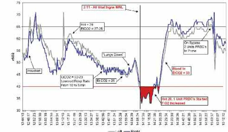
The value of measuring cerebral oximetry during surgery is illustrated in Figure 2 (which shows cerebral oximetry changes during a proximal humerus repair in a 94-year-old female with multiple co-morbidities) and in Figure 3 (which represents changes in cerebral oximetry during cardiopulmonary bypass events).Indeed, a number of recent articles have demon-strated the benefits of cerebral oximetry among patients undergoing both cardiac and non-cardiac surgery. Slater et al. demonstrated the association between intraoperative cerebral oxygen desaturation and an increased risk of cognitive decline and pro-longed hospital stay by assessing cognitive function in adult patients undergoing CABG surgery preoper-atively, postoperpreoper-atively, and 3 months after surgery, using a battery of neurocognitive tests. Patients were prospectively randomized to a blinded control group or to an unblinded interventional group. The area under the curve indicating the degree of desaturation below a 50% threshold over time accounted for both
Cerebral Oximetry May Provide Helpful Information
Light Source Shallow Detector Deep Detector Deep Detector Shallow Detector Light Source
See “Oximetry, ” Next Page
The Anesthesia Patient Safety Foundation
is pleased to announce the
BOARD OF DIRECTORS WORKSHOP
How Low (Cerebral Perfusion Pressure) Can You Go?
Friday, October 16, 2009
1300-1700 (Riverside Ballroom I-II)
Hampton Inn Suites Convention Center, New Orleans, LA
Registration is not required for attendance.
www.apsf.org
“Oximetry,” From Preceding Page
80 70 60 50 40 30 20 10 0 1 2 :3 4 :1 5 1 2 :3 7 :4 7 1 2 :4 1 :2 0 1 2 :4 4 :4 8 1 2 :4 8 :1 7 1 2 :5 1 :4 6 1 2 :5 5 :1 4 1 2 :5 8 :4 3 1 3 :0 2 :1 2 1 3 :0 5 :4 1 1 3 :0 9 :1 0 1 3 :1 2 :3 9 1 3 :1 6 :0 8 1 3 :1 9 :3 7 1 3 :2 3 :0 6 1 3 :2 6 :3 5 1 3 :3 0 :0 4 1 3 :3 3 :3 3 1 3 :3 7 :0 3 1 3 :4 0 :3 2 1 3 :4 4 :0 1 1 3 :4 7 :3 0 1 3 :5 0 :5 9 1 3 :5 4 :2 9 1 3 :5 7 :5 8 1 4 :0 1 :2 8 1 4 :0 4 :5 7 1 4 :0 8 :2 6 1 4 :1 1 :5 5 1 4 :1 5 :2 4 1 4 :1 8 :5 3 1 4 :2 2 :2 3 1 4 :2 5 :5 2 1 4 :2 9 :2 1 1 4 :3 2 :5 0 1 4 :3 6 :1 8 rS O2Right Cerebral rSO2 Left Cerebral rSO2
S et b as el in e Critical Cerebral Desaturation Threshold Induction Intubation NIBP 176/96 etCO2 32 etCO2 33 etCO2 38 etCO2 43 etCO2 41 NIBP 180/98 NIBP 91/53 NIBP 134/60 NIBP 175/83 HOB up to “beach chair” position Incision Vent changes to increase CO2 Nitro-Paste On and Off 1u PRBCs infusing slowly dressing wound Fluid Bolus for
relative hypotension Vent changes to increase CO2
Figure 2. Time line of events and cerebral oximetry changes during a proximal humerus fracture repair in a 94-year-old female who presented with a history of hypertension, atrial fibrillation, COPD, and TIAs.
See “Oximetry,” Next Page
the depth and duration of desaturation below this50% threshold. Patients with rSO2desaturation score >3,000%-second had a significantly higher risk of early postoperative cognitive decline [p=0.024]. Patients with rSO2desaturation score >3,000%-second also had a near 3-fold increased risk of pro-longed hospital stay (>6 days) [p=0.007].4Similarly, Yao et al. found a significant correlation between the degree and duration of cerebral oximetry desatura-tion and early postoperative neuropsychological dysfunction among patients undergoing elective car-diac surgery with CPB.5
Patients with cerebral desaturation during non-cardiac surgery have also been shown to exhibit declines in their Mini Mental State Examination (MMSE). Casati and colleagues studied 122 patients older than 65 years, scheduled for major abdominal, nonvascular surgery under general anesthesia (with an expected duration >2 hr). Surgical procedures represented by major abdominal surgery with a xiphopubic skin incision included gastric resection, colonic resection, hepatic resection, and duodeno-cephalo-pancreatectomy. Patients were randomly assigned to an intervention group (the monitor was visible and rSO2maintained at >75% of pre-induc-tion values) or a control group (the monitor was blinded and anesthesia was managed routinely). There was a significant correlation between the area under the curve of rSO2<75% of baseline and post-operative decrease in MMSE score from preopera-tive values. Control group patients with intraopera-tive cerebral desaturation also experienced a longer
time to post-anesthesia care unit (PACU) discharge (47 min [13–56 min]) and hospital LOS (24 days [7–53] days) compared with patients of the treat-ment group (25 min [15–35 min] and 10 days [7–23 days], respectively; p = 0.01 and p = 0.007).6
Cullen and Kirby reported an unexpected and devastating complication of neurologic injury in this
Newsletter, occurring in a healthy 47-year-old woman
undergoing shoulder surgery in the “beach chair” position.7They identified the lack of appreciation of cerebral hypoperfusion that occurs with blood pres-sure meapres-surement in the non-operative arm that is positioned well below the level of the brain, and the physiologic and anatomic changes that occur with the beach chair position. These can include decreased venous return, vasodilation, and head flexion, which may impede jugular venous flow and thus decrease cerebral perfusion. Deliberate hypotension coinci-dent with these physiologic changes, requires enhanced vigilance on the part of the clinicians caring for these patients. Use of a cerebral oximeter in this setting provides an additional tool in assessing ade-quate oxygen delivery to vulnerable cerebral tissue.
Cerebral oximetry has been shown to reduce major organ dysfunction following cardiac surgery. Murkin et al. prospectively randomized 200 patients undergoing CABG surgery to intraoperative cerebral regional oxygen saturation monitoring with active display and treatment intervention protocol, or blinded rSO2monitoring.8Significantly more patients in the blinded group had prolonged cerebral
desatu-ration (p=0.014) and a longer ICU LOS (p=0.029) versus intervention patients. Significantly more patients in the blinded group had major organ mor-bidity or mortality (death, ventilation >48 hr, stroke, myocardial infarction, return for re-exploration) versus patients in the intervention group (p=0.048). Patients experiencing major organ morbidity or mor-tality had lower baseline and mean rSO2 measure-ments, more cerebral desaturation, longer ICU LOSs, and longer postoperative hospitalization than patients without major organ complications. There was a significant inverse correlation between intra-operative rSO2measurements and duration of post-operative hospitalization among patients requiring more than 10 postoperative days.
Discussion
The clinical studies described above demonstrate the potential benefits of cerebral oximetric monitor-ing in a variety of clinical situations. Although the vast majority of clinical studies have been conducted among cardiac surgical patients, the application of cerebral oximetry to non-cardiac surgical patients is compelling in certain clinical situations. The previ-ously described work by Casati demonstrated the benefits of using cerebral oximetry among elderly patients undergoing major abdominal surgery.6The use of intraoperative cerebral regional oxygen satu-ration monitoring for patients undergoing carotid artery surgery can guide surgical and physiologic
intervention in terms of shunt use and blood pressure management, as bilateral measurements are com-pared with each other and with baseline measure-ments. An emerging area of cerebral oxygen saturation monitoring is the patient undergoing shoulder surgery in the beach chair position for the reasons described above. Cerebral malperfusion may be unappreciated in this setting, where blood pres-sure monitoring may not be optimal, head position may impede cerebral venous drainage, and positive pressure ventilation impedes an already compro-mised decreased venous return to the heart because of the beach chair positioning.
The reduction of major organ morbidity (death, ventilation >48 hr, stroke, myocardial infarction, return for re-exploration) associated with intraopera-tive cerebral oximetric monitoring in patients under-going CABG surgery is another very important aspect of cerebral oximetric monitoring. Maintaining an ade-quate oxygen balance in the most vulnerable water-shed tissue of the frontal cerebral cortex apparently provides an “early warning” of decreased oxygen delivery to the rest of the brain and other major organs. Interventions that reverse decreasing cerebral perfusion may avert prolonged ischemia of the brain and thus minimize oxygen desaturation in other major organs (Figure 3). The early warning aspect of cere-bral oximetry was further demonstrated by a pediatric study that examined the time to a 5% and 10%
reduc-tion in baseline oxygen saturareduc-tion measurements in 10 children subjected to apnea during laser surgery of their airway. The average time for their pulse oxime-ter to exhibit a 5% and 10% reduction from pre-apnea levels was 146 ± 49 and 189 ± 64 seconds, respectively, while the cerebral oximeter exhibited an earlier warn-ing of 5% and 10% cerebral desaturation at 94 ± 8 and 138 ± 89 sec, respectively. Cerebral desaturation thus occurred 1 min before the pulse oximeter indicated desaturation among these children.9
The benefits of cerebral oximetric monitoring are continually emerging as more work is published demonstrating improved outcome and enhanced patient safety. Although the majority of published data have demonstrated improved outcomes among cardiac surgical patients, the studies performed thus far among non-cardiac surgical patients are begin-ning to identify its utility in other clinical scenarios. Future research to identify and validate the benefits of cerebral oximetry monitoring in improving patient outcomes among non-cardiac surgical patients (as well as cardiac surgical patients) represents an excit-ing and important opportunity to explore and utilize this recent technology.
Dr. Troianos is Chair and Residency Program Direc-tor of the Department of Anesthesiology, Western Penn-sylvania Hospital, and Professor of Anesthesiology, Western Campus of Temple University School of Medicine in Pittsburgh, PA.
Disclosures: Dr. Troianos has had no financial rela-tionship with any manufacturer of cerebral oximetry tech-nology. Somanetics, a manufacturer of this technology, is a corporate donor to the APSF.
References
1. Goldman SM, Sutter FP, Wertan MA, et al. Outcome improvement and cost reduction in an increasingly morbid cardiac surgery population. Semin Cardiothorac
Vasc Anesth 2006;10:171-5.
2. Likosky DS, Leavitt BJ, Marrin CA, et al. Intra- and post-operative predictors of stroke after coronary artery bypass grafting. Ann Thorac Surg 2003;76:428-35.
3. Denault A, Deschamps A, Murkin JM. A proposed algo-rithm for the intraoperative use of cerebral near-infrared spectroscopy. Semin Cardiothorac Vasc Anesth 2007;11:274-81. 4. Slater JP, Guarino T, Stack J, et al. Cerebral oxygen desat-uration predicts cognitive decline and longer hospital stay after cardiac surgery. Ann Thorac Surg 2009;87:36-45. 5. Yao FS, Tseng CC, Ho CY, et al. Cerebral oxygen
desatu-ration is associated with early postoperative neuropsy-chological dysfunction in patients undergoing cardiac surgery. J Cardiothorac Vasc Anesth 2004;18:552-8. 6. Casati A, Fanelli G, Pietropaoli P, et al. Continuous
moni-toring of cerebral oxygen saturation in elderly patients undergoing major abdominal surgery minimizes brain exposure to potential hypoxia. Anesth Analg 2005;101:740-7. 7. Cullen DJ, Kirby RR. Beach chair position may decrease cerebral perfusion: catastrophic outcomes have occurred.
APSF Newsletter 2007;22:25-7.
8. Murkin JM, Adams SJ, Novick RJ, et al. Monitoring brain oxygen saturation during coronary bypass surgery: a ran-domized, prospective study. Anesth Analg 2007;104:51-8. 9. Tobias JD. Cerebral oximetry monitoring with near infrared spectroscopy detects alterations in oxygenation before pulse oximetry. J Intensive Care Med 2008;23:384-8.
Oximetry May Be Useful in Non-Cardiac Surgery As Well
“Oximetry” From Preceding Page
Figure 3. Cerebral oximetry values and time-line of events during a cardiac surgical procedure. First Alert - Unknown Blood Loss
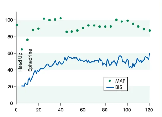
Figure 1. 120 min BIS and MAP trend during orthostatic hypotension treated with ephedrine
H
e
a
d
U
p
E
p
h
e
d
ri
n
e
100
80
60
40
20
0
0
20
40
60
80
100
120
MAP
BIS
by Mindaugas Pranevicius, MD, and Osvaldas Pranevicius, MD
Recent reports about unrecognized cerebral ischemia under general anesthesia are alarming,1,2 and reveal that it may start insidiously, progress covertly, and result in devastating outcomes. Although much of the emphasis for anesthesia man-agement is focused on maintenance of adequate cere-bral perfusion pressure (CPP), does this ensure sufficient perfusion of the brain? Perfusion pressure by itself is the propulsion force only; it does not deter-mine distribution of cerebral blood flow (CBF), ensure adequate collateral circulation, nor account for variations in the venous outflow path. Here we pre-sent a case that demonstrates the utility as well as lim-itations of processed EEG monitoring to assess adequacy of the CBF during an episode of postural cerebral venous steal.
To measure CPP, an arterial catheter is placed and zeroed at the level of the external acoustic meatus to approximate the circle of Willis. Most anesthesiolo-gists and nurse anesthetists aim to maintain CPP within 15-20% of the baseline value. To calculate CPP in the sitting position one needs to know both arterial (inflow) and venous (effective outflow) pressures.3 We can easily measure the arterial (inflow) pressure, but we usually only estimate the effective outflow pressure. When outflow through the vertebral venous plexus is not adequate, atmospheric pressure
assumes the role of effective backpressure due to jugular vein compression at the skull base.3,4In this context pressure measurement at the external acoustic meatus estimates the CPP, assuming that the effective outflow pressure is zero (atmospheric) at the scull base.3
We also can only assume that the effective out-flow pressure is uniform throughout the brain. Regional differences in the effective outflow pressure in the brain can cause a cerebral venous steal phe-nomenon diverting blood flow to the pathway of least resistance.5During head-up tilt, atmospheric pressure hinders outflow from the upper body with zero venous pressure and diverts the flow to the lower body where venous pressure exceeds atmos-pheric (postural “steal”). Similarly, alveolar pressure diverts pulmonary blood flow into dependent parts of pulmonary circulation.6,7Thus the adequacy of cerebral perfusion can not be determined solely from the arterial pressure, but requires consideration of the effects of vascular anatomy, autoregulation, PaCO2, anesthetic, viscosity, vascular tone, and regional vari-ation of these factors on the CBF. More direct assess-ment of cerebral perfusion would be desirable.
Ideally, a sensitive neurological exam can assess potential compromise of cerebral function occurring during periods of inadequate cerebral perfusion. However, under general anesthesia assessment of cerebral function is more challenging. Although
Can Processed EEG Monitoring Detect Postural
Cerebral Ischemia or Cerebral Hypoperfusion?
many options are available, no single monitoring modality has been demonstrated as superior.8Neurophysiologic monitoring with evoked potentials requires dedicated trained staff and equipment and is typically limited to select cases. Transcranial Doppler monitoring of middle cerebral artery flow velocity is operator-dependent, very sen-sitive to the acoustic contact, and fails when the acoustic “window” cannot be found. Jugular vein oximetry is invasive. Near infrared oximetry moni-tors tissue oxygenation in hair-free areas of the head but is not broadly available. In contrast, processed EEG monitoring devices are widespread, and although typically utilized to help manage intraop-erative anesthetic dosing, they could help to detect cerebral hypoperfusion.
Case Report
A 34-year-old female, 5'8", 76.2 kg underwent laparoscopic cholecystectomy under general endo-tracheal anesthesia. Medical history was significant for hypertension, controlled with metoprolol. She had previously undergone gastric bypass surgery for morbid obesity. Following induction, anesthesia was maintained with sevoflurane 2% inspired in air/oxygen. The patient’s baseline blood pressure was 143/97 mmHg; the immediate preinduction value was 167/113 mmHg; and following induction was 130/80 mmHg. Approximately 40 min after induction, with pneumoperitoneum and reverse Trendelenburg position, blood pressure was noted to decrease acutely to 95/50. At this point, a BIS monitor was applied and the initial BIS value was 20 with sevoflurane end-tidal value at 1 MAC. Two doses of ephedrine (5 mg + 10 mg) were adminis-tered. Blood pressure promptly returned to 130/80 and BIS simultaneousely increased to 40-50 without decreasing the MAC (see Figure 1). The case was completed and the patient recovered uneventfully.
We submitted the BIS trend data for review by the manufacturer (Aspect Medical Systems) who confirmed that BIS was functioning as designed. Interrogation of the trend recording revealed the BIS system was detecting delta wave activity at the beginning of the record and the BIS values were appropriately low during the initial readings.
Discussion
Although the BIS Index was originally devel-oped to measure the effects of anesthetic and seda-tive agents, multiple case reports have described the ability of BIS monitoring to detect episodes of cere-bral ischemia.9-17In the absence of larger validating studies, sensitivity and specificity of BIS for this
See “EEG, ” Next Page
application has been questioned.13,18,19Although we believe our case demonstrates the ability of processed EEG monitoring to detect cerebral hypoperfusion, it also highlights important limitations. Without a base-line record prior to and following induction of anes-thesia, a low BIS value during orthostatic hypotension may reflect the effect of the anesthetic on the EEG with or without the additive effects of hypoperfusion. In our patient, we observed rapid resolution of the low BIS values following ephedrine administration. Although ephedrine increases blood pressure and CBF, it has also been reported to increase BIS values directly.20Similar to cerebral oximetry, BIS and other processed EEG monitors analyze EEG signals from the frontal lobe only, and consequently may miss regional CBF abnormalities. Although our patient exhibited low BIS values during the period of pos-tural hypotension, it should be noted that a variety of artifact conditions may cause spuriously elevated BIS values.13
Conclusion
Processed EEG monitoring systems are operator-independent, widely available, relatively inexpensive technologies to use during the intraoperative period. Although these technologies are not specifically developed as monitors for cerebral perfusion, they may help detect otherwise unrecognized global cere-bral ischemia. If anesthetic dosing and surgical stim-ulation are stable, and postural changes or acute hypotension result in a precipitous decrease in the brain function values, clinicians may consider cor-recting the hypotension till brain function values improve.
The recent case reports should remind us that the brain is a critical and fragile organ that we rarely monitor directly during general anesthesia. We do not believe that the absence of the “perfect” cerebral monitor should discourage us from obtaining the most information from available modalities. We believe that available processed EEG monitoring devices could help in this regard. Larger scale valida-tion studies to determine processed EEG and CBF correlates could advance functional cerebral moni-toring and improve patient safety.
The authors acknowledge and appreciate the technical help from Aspect Medical Systems, Inc. for the data extrac-tion from the BIS monitor, and the editorial review and comments from Dr. Scott Kelley.
Disclosure: Drs. Mindaugas and Osvaldas have no financial relationship with Aspect Medical Systems, Inc. Aspect Medical Systems is a corporate donor to the APSF.
BIS and MAP Trend Up After Treatment
“EEG,” From Preceding Page
Dr. Mindaugas Pranevicius is an assistant professor at the Albert Einstein College of Medicine, Jacobi Medical Center, Bronx, NY, and Dr. Osvaldas Pranevicius is an attending anesthesiologist at the New York Hospital Queens, Flushing, NY.
References
1. Pohl A, Cullen DJ. Cerebral ischemia during shoulder surgery in the upright position: a case series. J Clin
Anesth 2005;17:463-9.
2. Cullen DJ, Kirby RR. Beach chair position may decrease cerebral perfusion; catastrophic outcomes have occurred.
APSF Newsletter 2007;22(2):25-7.
3. Pranevicius M, Pranevicius O. Modified calculation of the cerebral perfusion pressure in a sitting position: jugu-lar Starling resistor and related clinical implications.
APSF Newsletter. 2008;23(2):32-3.
4. Valdueza JM, von Münster T, Hoffman O, et al. Postural dependency of the cerebral venous outflow. Lancet 2000;355:200-1.
5. Pranevicius M, Pranevicius O. Cerebral venous steal: blood flow diversion with increased tissue pressure.
Neurosurgery 2002;51:1267-74.
6. West JB, Dollery CT, Naimark A. Distribution of blood flow in isolated lung: relation to vascular and alveolar pressures. J Appl Physiol 1964;19:713-24.
7. Lopez-Muniz R, Stephens NL, Bromberger-Barnea B, et al. Critical closure of pulmonary vessels analyzed in terms of Starling resistor model. J Appl Physiol 1968;24:625-35.
8. Moritz S, Kasprzak P, Arlt M, et al. Accuracy of cerebral monitoring in detecting cerebral ischemia during carotid endarterectomy: a comparison of transcranial Doppler sonography, near-infrared spectroscopy, stump pres-sure, and somatosensory evoked potentials.
Anesthesiol-ogy 2007;107:563-9.
9. Adam N, Sebel PS. BIS monitoring: awareness and cata-strophic events. Semin Cardiothorac Vasc Anesth 2004;8:9-12.
10. Bonhomme V, Desiron Q, Lemineur T, et al. Bispectral index profile during carotid cross clamping. J Neurosurg
Anesthesiol 2007;19:49-55.
11. el-Dawlatly AA. EEG bispectral index during carotid endarterectomy. Middle East J Anesthesiol 2003;17:287-93. 12. Mérat S, Lévecque JP, Le Gulluche Y, et al. BIS monitor-ing may allow the detection of severe cerebral ischemia.
Can J Anaesth 2001;48:1066-9.
13. Myles PS, Cairo S. Artifact in the bispectral index in a patient with severe ischemic brain injury. Anesth Analg 2004;98:706-7.
14. Umegaki N, Hirota K, Kitayama M, et al. A marked decrease in bispectral index with elevation of suppres-sion ratio by cervical haematoma reducing cerebral per-fusion pressure. J Clin Neurosci 2003;10:694-6. 15. Azim N, Wang CY. The use of bispectral index during a
cardiopulmonary arrest: a potential predictor of cerebral perfusion. Anaesthesia 2004;59:610-2.
16. Morimoto Y, Monden Y, Ohtake K, et al. The detection of cerebral hypoperfusion with bispectral index monitoring during general anesthesia. Anesth Analg 2005;100:158-61. 17. Prabhakar H, Ali Z, Rath GP, Singh D. Zero bispectral index during coil embolization of an intracranial aneurysm. Anesth Analg 2007;105:887-8.
18. Dahaba AA. Different conditions that could result in the bispectral index indicating an incorrect hypnotic state.
Anesth Analg 2005;101:765-73.
19. Deogaonkar A, Vivar R, Bullock RE, et al. Bispectral index monitoring may not reliably indicate cerebral ischaemia during awake carotid endarterectomy. Br J
Anaesth 2005;94:800-4.
20. Ishiyama T, Oguchi T, Iijima T, et al. Ephedrine, but not phenylephrine, increases bispectral index values during combined general and epidural anesthesia. Anesth Analg 2003;97:780-4.