HAL Id: tel-01148799
https://tel.archives-ouvertes.fr/tel-01148799
Submitted on 5 May 2015HAL is a multi-disciplinary open access archive for the deposit and dissemination of sci-entific research documents, whether they are pub-lished or not. The documents may come from teaching and research institutions in France or abroad, or from public or private research centers.
L’archive ouverte pluridisciplinaire HAL, est destinée au dépôt et à la diffusion de documents scientifiques de niveau recherche, publiés ou non, émanant des établissements d’enseignement et de recherche français ou étrangers, des laboratoires publics ou privés.
Role of salivary gland epithelial cells in the
differentiation and activation of T lymphocytes in
primary Sjögren’s syndrome
Ya-Zhuo Gong
To cite this version:
Ya-Zhuo Gong. Role of salivary gland epithelial cells in the differentiation and activation of T lympho-cytes in primary Sjögren’s syndrome. Immunology. Université de Strasbourg, 2013. English. �NNT : 2013STRAJ100�. �tel-01148799�
1 UNIVERSITE DE STRASBOURG
ECOLE DOCTORALE DES SCIENCES DE LA VIE ET DE LA SANTE
THESE
Presented to obtain the grade of
Doctor of Strasbourg University
Discipline: life sciences - molecular and cell biology
Specialty: immunology
by
Ya-Zhuo GONG
September 13, 2013
Role of salivary gland epithelial cells in the differentiation and
activation of T lymphocytes in primary Sjögren's syndrome
Member of the jury:
External reporter: Dr. Jérémie SELLAM External reporter: Pr. Vincent GOËB Internal examinator: Dr. Fanny MONNEAUX
2 Remerciements
Je tiens à remercier tout d’abord mon directeur de thèse, le Pr. Jacques-Eric Gottenberg, pour m’avoir accueillie au sein de son laboratoire et pour m’avoir proposé ce sujet de thèse et avoir dirigé mon travail. Sa disponibilité, ses conseils m’ont beaucoup apporté.
Je remercie également les membres du jury qui ont accepté de juger ce travail : Dr. Jérémie Sellam, Pr. Vincent Goëb et Dr. Fanny Monneaux.
Je voudrais exprimer mes remerciements sincères à Pr. Dominique Wachsmann et au Dr. Ghada Alsaleh, qui m’ont aidé au quotidien. Leurs conseils et leurs commentaires précieux m'ont permis de surmonter mes difficultés et de progresser dans mes études.
Je remercie Pr. Jean Siblia pour son soutien et sa gentillesse, ainsi que de m’avoir accueillie au sein de son laboratoire.
Un grand merci également à tous les membres de notre équipe : pour leur aide scientifique et leur amitié : Lucas Philipe, Antoine François, Angélique Pichot et particulièrement Nawal Rahal pour la très bonne ambiance de travail ainsi que tous les repas et toutes les soirées. Mes remerciements vont également aux membres passés et présents de l’équipe EA4438, Christelle Sordet, Manon Lasseaux, Etienne Dahan, Alain Meyer, Arnaud Theulin pour leur aide et surtout de m’avoir aidé à collecter toutes les biopsies.
Je souhaite également remercier les Pr. Siamak Bahram et Philippe Georgel pour leur accueil au sein de l’équipe ’ImmunoRhumatologie Moléculaire’, ainsi que tous les membres de l’équipe : Cécile, Eléonore, Raphaël C, Pilar, Véronique, Mirjana, Raphaël
3 D, Gaëlle, Aurore, Irina, Louise, Wassila, Pierre, Nicodème, Meiggie, Marion, Laure, Alice, Sandra, Laurent, Cédric, Amélie.
Je tiens à remercier tout particulièrement les membres des équipes qui ont contribués à ce travail : l’équipe Chambon avec Dr. Mei Li et Jiagui Li, ainsi que l’équipe ‘CNRS UPR3572’ avec Dr. Pauline Soulas-Sprauel pour ses conseils et ses encouragements.
Et pour finir, je tiens à remercier tout particulièrement mes parents, mon mari pour leur soutient et mon amie Ying Li qui a corrigé l’anglais de mon rapport.
4 Table of Contents
ABBREVIATION LIST 8
FIGURE LIST 11
TABLE LIST 13
PUBLICATION AND COMMUNICATION LIST 14
I. INTRODUCTION 16
1 SJÖGREN'S SYNDROME (SS) 17
1.1 Definition 17
1.1.1 Definition and history 17
1.1.2 Epidemiology 17
1.1.3 Symptoms and complications 17
1.1.4 Diagnosis 19
1.1.5 Genetic and environmental aspects 21
1.1.5.1 Genetic aspects 21
1.1.5.2 Environmental aspects 22
1.1.6 Treatment 23
1.2 Physiopathology 24
1.2.1 Mice models of SS 24
1.2.2 Scenario for the pathogenesis of pSS 27
5
1.2.3.1 SGEC activation by PAMPs and DAMPs 31
1.2.3.2 ECM remodeling and homeostatic changes in epithelial tissues 32
1.2.3.3 Increased apoptosis and NF-κB activity in epithelial cells 33
1.2.3.4 Autoantigens presentation by SGECs 34
1.2.3.5 In-situ activation and immunologic functions of SGECs in SS 36
1.2.4 Salivary gland infiltrates. 38
1.2.5 T-lymphocyte contribution to the pathophysiology of pSS 38 1.2.5.1 T-lymphocyte migration to exocrine tissue 38
1.2.5.2 In situ antigen-driven stimulation of T lymphocytes 39
1.2.5.3 T- cell- mediated immunopathogenesis 41
1.2.5.4 T helper cell subsets and cytokine profile in pSS. 42
1.2.5.5 Th1/Th2 balance 43
1.2.5.6 Th17 cells, IL-17 and IL-23 44
1.2.5.7 Regulatory T cells 44
1.2.5.8 Tfh and IL-21 45
1.2.6 Ectopic germinal center-like structures 58 1.2.7 Salivary gland epithelial cells and lymphoma 59 1.2.8 Role of T-cell dependent B-cell activation in pSS 60
6
1.2.8.2 B-cell polarization 61
1.2.9 Autoantibodies 61
1.2.10 The role of cytokines in the physiopathology of pSS 64 1.2.10.1 Activation of the type I interferon (IFN) pathway in pSS 65
1.2.10.2 IFN-γ 67
1.2.10.3 TNF-α 68
1.2.10.4 BAFF 68
1.2.10.5 IL-6 69
1.2.10.6 IL-7 69
1.2.10.7IL-12 and IL-18 70
1.2.10.8 IL-22 70 1.2.11 Chemokines 71 1.2.12 Costimulatory molecules 74 1.2.12.1 CD80/CD86 - CD28 interaction 75 1.2.12.2 ICOS/ICOSL interaction 75 1.2.12.3 PD-1/PD-L1 interaction 77 1.2.12.4 CD40/CD40L interaction: 77
1.2.12.5 Main immunological consequences of the interaction between OX40/OX40L 78
7
1. OBJECTIVE 1 84
2. PUBLICATION NO 1: 85
3. Supplementary data that were not included in the submitted manuscript 130
3.1 Analysis of cell purification state 130
3.2 Using IL-12-induced Tfh as positive control 130 3.3 Epithelial cell line HSG cannot induce the Tfh differentiation 131
III. OBJECTIVE 2 AND RESULT 2 133
1. OBJECTIVE 2 134
2. PUBLICATION NO 2: 135
3. SUPPLEMENTARY DATA THAT WERE NOT INCLUDED IN THE
SUBMITTED MANUSCRIPT 169
3.1 OX40 induction by SGECs is dependent on soluble factors 169
3.2 The blockade of OX40L fails to inhibit Tfh differentiation induced by SGECs 171
IV. CONCLUSIONS AND PERSPECTIVES 172
V. REFERENCES 185
8
ABBREVIATION LIST
AID enzyme activation-induced deaminase ANA antinuclear antibody
APC antigen-presenting cell
APRIL a proliferation-inducing ligand autoAb autoantibody
autoAg autoantigen
BAFF B cell-activating factor Bax BCL2-associated X protein Bcl-2 B-cell leukemia/lymphoma-2 BR3 BAFF receptor 3
CCL chemokine (C-C motif) ligand CCR chemokine receptor
CHRM3 muscarinic receptor 3 gene CIA collagen-induced arthritis CTL cytotoxic T lymphocytes
CTLA-4 Cytotoxic T-lymphocyte antigen 4 CXCL chemokine (C-X-C motif) ligand CXCR CXC chemokine receptor
DAMP damage-associated molecular pattern DC dendritic cells
EAE experimental autoimmune encephalomyelitis EBV Epstein–Barr virus
EC epithelial cell
9 FAS cell surface death receptor
FDC follicular dendritic cells FoxP3 forkhead box protein 3 GC germinal centres
HLA histocompatibility leukocyte antigen HSG human salivary gland
ICAM-1 intercellular Adhesion Molecule-1 ICOS inducible costimulatory
IFN interferon Ig Immunoglobulin
IRF5 interferon regulatory factor 5 iTreg induced Treg cells
LFA-1 lymphocyte function-associated antigen 1 LT lymphotoxin
M3R Muscarinic acetylcholine receptor MCP-1 Monocyte chemotactic protein-1 MHC major histocompatibility complex MZ B cells marginal zone B cells
nTreg natural Treg OX40L OX40 ligand
PAMP pathogen-associated molecular pattern PD-1 programmed death-1
pDC plasmacytoid dendritic cells pSS primary Sjögren's syndrome RA rheumatoid arthritis
RF rheumatoid factor
SAP SLAM-associated protein SG salivary glands
10 SHM somatic hypermutation
SLE systemic lupus erythematosus SNP single-nucleotide polymorphism SS Sjögren's syndrome
SSA Anti-Sjogren’s syndrome antigen A SSB Anti-Sjogren’s syndrome antigen B
STAT4 signal transducer and activator of transcription 4 T0 B cells transitional type 0 B cells
T1D type I diabetes TCR T cell receptor
Tfh Follicular helper T cells TGF-β transforming growth factor-β TGF- transforming growth factor-TLR Toll-like receptor
TNF tumor necrosis factor
TNFRSF tumor necrosis factor receptor superfamily TNFSF tumor necrosis factor (ligand) superfamily Tregs regulatory T cells
VCAM-1 vascular cell adhesion molecule 1 VEGF-A vascular endothelial growth factor A VEGFR2 vascular endothelial growth factor receptor 2 VLA-4 very late antigen-4
11
FIGURE LIST
Figure Title Page
Figure 1 Manifestations and complications of SS. 18 Figure 2 A proposed scenario for the pathogenesis of pSS. 27 Figure 3 A schema of acinar SGECs and ductal SGECs in salivary glands
and human non-neoplastic SGEC lines.
28
Figure 4 A schematic model for early events in the pathogenesis of lymphoepithelial lesions in the salivary glands of SS patients.
35
Figure 5 Molecular mechanisms involved in T cell migration into exocrine glands and in the immunological synapse between CD4 T cells and SGECs.
39
Figure 6 T helper cell lineage development and function. 42 Figure 7 Anatomical localization as well as molecules and cellular
requirements for Tfh differentiation.
47
Figure 8 B cell devellpoment and lymph node stucture. 48 Figure 9 Initial T cell priming by DC and T cell differentiation. 51 Figure 10 Cognate interaction of primed T cells and Ag-specific B cells. 52 Figure 11 Molecules implicated in GC response 54
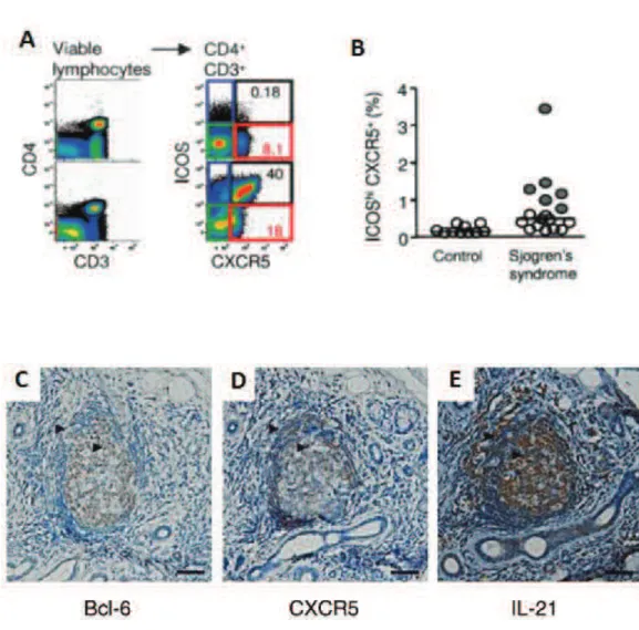
Figure 12 Tfh cells in patients with SS. 57
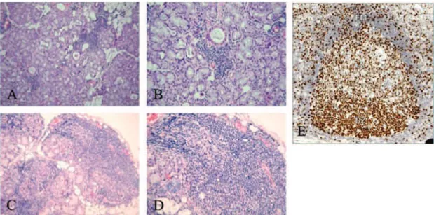
Figure 13 Periductal focal mononuclear cell infiltrates in the minor salivary gland biopsies of patients with pSS and salivary gland tissue GC-like structures
59
Figure 14 Main markers of mature B-cells 60
Figure 15 Expression of CXCL13 and CXCR5 chemokines in patients with Sjögren's syndrome.
73
Figure 16 Main roles of OX40/OX40L pathway 79
Figure 17 Immunohistochemical staining of OX40L in submandibular glands of MRL-Faslpr and MRL/+ mice
12 Figure 18 Purity analysis of freshly isolated naïve CD4+ T cells by flow
cytometry.
130
Figure 19 Tfh induction and IL-21 secretion by naïve CD4+ T cells activated by IL-12 or cocultived with SGECs.
131
Figure 20 Expression of ICOS and CXCR5, as well as IL-21 production by naïve CD4+ T cells cocultured with HSG.
132
Figure 21 OX40 induction by SGECs is dependent on soluble factors 170 Figure 22 The blockade of OX40L fails to inhibit Tfh differentiation
induced by SGECs.
172
Figure 23 SGECs lead to B-cell activation via different approaches in pSS 182 Figure 24 New pathogenic roles of salivary gland epithelial cells in pSS 183
13
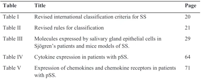
TABLE LIST
Table Title Page
Table I Revised international classification criteria for SS 20
Table II Revised rules for classification 21
Table III Molecules expressed by salivary gland epithelial cells in Sjögren’s patients and mice models of SS.
29
Table IV Cytokine expression in patients with pSS. 64 Table V Expression of chemokines and chemokine receptors in patients
with pSS.
14
PUBLICATION AND COMMUNICATION LIST
Publications
GONG YZ., Nititham J., Taylor K., Miceli C, Sordet C., Wachsmann D., Bahram S., Georgel P., Criswell L. A., Sibilia J., Mariette X., Alsaleh G and Gottenberg J.E., Differentiation of follicular helper T cells by salivary gland epithelial cells and increase of IL-21 correlate with disease activity in primary Sjögren’s syndrome. (Manuscript submitted)
GONG YZ., LI J.G., LI M., Wachsmann D., Bahram S., Georgel P., Sibilia J., Alsaleh G and Gottenberg J.E., Pathogenic role of the OX40 ligand-OX40 costimulatory pathway in primary Sjögren's syndrome. (Manuscript submitted)
Communications
GONG Y., Alsaleh G., Sibilia J., Wachsmann D. and Gottenberg J.E., Salivary gland epithelial cells are capable to directly induce the differentiation of IL-21-secreting follicular helper CD4 T cells in Primary Sjögren’s Syndrome. International Symposium on Sjogren Symposia (ISSS). Athens, Greece. September 2011. (Oral communication)
GONG Y., Alsaleh G., Sibilia J., Wachsmann D. and Gottenberg J.E., Salivary gland epithelial cells are capable of inducing the differentiation of follicular helper T cells in primary Sjögren's syndrome. American College of Rheumatology (ACR). Chicago, America. November 2011. (Oral communication)
GONG Y., Alsaleh G., Sibilia J., Wachsmann D. and Gottenberg J.E., Les cellules épithéliales des glandes salivaires sont capables d’induire la différenciation des lymphocytes T folliculaire au cours du syndrome de Sjögren primitif. Société
15 Française de Rhumatologie (SFR). Paris, France. December 2011. (Poster)
GONG Y., Alsaleh G., Sibilia J. and Gottenberg J.E., A new pathogenic role of salivary gland epithelial cells in the costimulation of T lymphocytes in primary sjögren’s syndrome: OX40 ligand expression, T-cell induction of OX40 and promotion of T-cell survival, proliferation and activation. American College of Rheumatology (ACR). Washington, D.C., America. November 2012. (Poster)
GONG Y., Alsaleh G., Sibilia J. and Gottenberg J.E., Rôle des cellules épithéliales des glandes salivaires dans l’induction d’OX40, la survie et la prolifération des lymphocytes T au cours du syndrome de Sjögren primitif. Société Française de Rhumatologie (SFR). Paris, France. December 2012. (Poster)
16
I. INTRODUCTION
17
1 Sjögren's syndrome (SS)
1.1 Definition
1.1.1 Definition and history
Sjögren's syndrome (SS) is a chronic inflammatory systemic autoimmune disease. It was first described by Swedish ophthalmologist Henrik Sjögren in 1933. It is characterized by inflammation and dysfunction of the exocrine glands which are associated with lymphocytic infiltrates and autoantibody secretion. Sjögren's syndrome can arise as a primary disease entity (primary Sjögren's syndrome (pSS)) or be associated with other autoimmune diseases (secondary Sjögren's syndrome), such as rheumatoid arthritis (RA), systemic lupus erythematosus (SLE), scleroderma, primary biliary cirrhosis etc. About 5-20% of the patients with RA, SLE and scleroderma present a secondary SS [1].
1.1.2 Epidemiology
SS is the second most common systemic autoimmune disease after RA. It has been estimated that SS affects 0.1% of the general population (~0.03% for pSS). Approximately 90% of patients are female. The disease most commonly develops in the fourth and the fifth decade of life, the average age of onset is late 40s. However, males and younger or older subjects can be affected.
1.1.3 Symptoms and complications
Manifestations of SS are very diverse, but three types of signs are found in most patients: dryness, fatigue and pain. The clinical syndrome is caused by progressive infiltration of the exocrine glands, especially the salivary and lacrimal glands, resulting in dry mouth and dry eyes (Figure 1A and B). Other exocrine glands are also
18 affected, which often causes skin, nose and vaginal dryness. Asthenia and pain are most frequent. Usually, pain is related to arthromyalgia. The most frequent glandular complications of SS include caris, tooth loss, mycosis of the mouth and keratoconjunctivitis favored by dryness. Approximately one third of the patients with pSS show evidence of extraglandular involvement. Arthritis, myositis, Raynaud's phenomenon, skin vasculitis, lymphadenopathy, renal, pulmonary and central or peripheral nervous system involvement might occur (Figure 1 C-G). Lymphoma represents the most serious systemic complication of pSS.
The development of non-Hodgkin lymphoma is observed in approximately 5% of patients with SS (Figure 1 H). The risk of lymphoma is 16-18 fold higher than the general population. The mucosa associated lymphoid tissue (MALT) type and diffuse large B-cell lymphoma (DLBCL) represent the most common types of pSS-related lymphomas. Some factors have been identified as predictors for lymphoma development in SS. These factors include recurrent or persistent swelling of major salivary glands (SG), lymphadenopathy, cryoglobulinemia, splenomegaly, skin vasculitis or palpable purpura, low levels of complement factor C3 and C4, Monoclonal-component, peripheral neuropathy, glomerulonephritis, CD4+ T-lymphopenia, a low CD4+/CD8+ T cell ratio and ectopic germinal center-like structures in salivary glands [2, 3].
Figure 1. Manifestations and complications of SS. Patients with SS hace dry mouth and dry eyes (A and B), purpura (C), Raynaud's phenomenon (D), interstitial nephritis (E), pulmonary (F), central nervous system involvement (G) and non-Hodgkin lymphoma (H). Figures downloaded on the Club Rhumatismes et Inflammation (CRI) website (www.cri-net.com)
19 1.1.4 Diagnosis
The diagnosis of Sjögren's syndrome is complicated by the wide range of symptoms that a patient might manifest. Complaints of dry eyes and dry mouth are common and nonspecific. Diagnosis often requires a biopsy of lip minor salivary glands to confirm the lymphocytic infiltration. The detection of rheumatoid factor (RF) and antinuclear antibodies (ANA) is very frequent. About 90% of Sjögren’s patients have a positive ANA test result and 60% of patients have a positive RF [4]. Anti-Sjogren’s syndrome antigen A (SSA)/Ro and anti-SSB/La are part of the diagnostic criteria. They are present in 60-80% of cases. Cytopenias (especially lymphopenia) can be observed in 20-30% of patients. A monoclonal immunoglobulin (Ig) can be detected in 10-15% of patients. Cryoglobulinemia is observed in 5% of patients. Tear flow reduction may be assessed by the Schirmer's test. The saliva production is mostly measured by the unstimulated salivary flow rate. There is often a several-year delay from the start of symptoms to diagnosis. The American–European Consensus Group criteria (AECC) for Sjögren's syndrome published in 2002 are widely used for the clinical diagnosis of Sjögren's syndrome [5] (Table I, II). Less specific criteria were recently proposed [6].
20 Table I. Revised international classification criteria for Sjögren's syndrome.
I. Ocular symptoms: a positive response to at least one of the following questions 1. Have you had daily, persistent, troublesome dry eyes for more than 3 months? 2. Do you have a recurrent sensation of sand or gravel in the eyes?
3. Do you use tear substitutes more than 3 times a day?
II. Oral symptoms: a positive response to at least one of the following questions: 1. Have you had a daily feeling of dry mouth for more than 3 months?
2. Have you had recurrently or persistently swollen salivary glands as an adult? 3. Do you frequently drink liquids to aid in swallowing dry food?
III. Ocular signs—that is, objective evidence of ocular involvement defined as a positive result for at least one of the following two tests:
1. Schirmer's I test, performed without anaesthesia (≤5 mm in 5 minutes)
2. Rose bengal score or other ocular dye score (≥4 according to van Bijsterveld's scoring system)
IV. Histopathology: In minor salivary glands (obtained through normal-appearing mucosa) focal lymphocytic sialoadenitis, evaluated by an expert histopathologist, with a focus score ≥1, defined as a number of lymphocytic foci (which are adjacent to normal-appearing mucous acini and contain more than 50 lymphocytes) per 4 mm2 of glandular tissue18
V. Salivary gland involvement: objective evidence of salivary gland involvement defined by a positive result for at least one of the following diagnostic tests: 1. Unstimulated whole salivary flow (≤1.5 ml in 15 minutes)
2. Parotid sialography showing the presence of diffuse sialectasias (punctate, cavitary or destructive pattern), without evidence of obstruction in the major ducts19
3. Salivary scintigraphy showing delayed uptake, reduced concentration and/or delayed excretion of tracer20
VI. Autoantibodies: presence in the serum of the following autoantibodies: 1. Antibodies to Ro(SSA) or La(SSB) antigens, or both
21 Table II. Revised rules for classification
For primary SS
In patients without any potentially associated disease, primary SS may be defined as follows:
a. The presence of any 4 of the 6 items is indicative of primary SS, as long as either item IV (Histopathology) or VI (Serology) is positive
b. The presence of any 3 of the 4 objective criteria items (that is, items III, IV, V, VI) c. The classification tree procedure represents a valid alternative method for
classification, although it should be more properly used in clinical-epidemiological survey
For secondary SS
In patients with a potentially associated disease (for instance, another well defined connective tissue disease), the presence of item I or item II plus any 2 from among items III, IV, and V may be considered as indicative of secondary SS
Exclusion criteria:
Past head and neck radiation treatment Hepatitis C infection
Acquired immunodeficiency disease (AIDS) Pre-existing lymphoma
Sarcoidosis
Graft versus host disease
Use of anticholinergic drugs (since a time shorter than 4-fold the half life of the drug)
1.1.5 Genetic and environmental aspects
1.1.5.1 Genetic aspects
As in many other autoimmune diseases, genetic factors contribute to Sjögren's syndrome. A strong association to specific major histocompatibility complex (MHC)
22 alleles has been shown. Among the histocompatibility leukocyte antigen (HLA) haplotypes, HLA-DRB1*03, -DQA1*05, and -DQB1*02 have been consistently associated with primary SS [7]. The interferon regulatory factor 5 (IRF5) gene on chromosome 7q32.1 and the signal transducer and activator of transcription 4 (STAT4) gene on chromosome 2q32,3 are two genes in the type I interferon (IFN) system. Recent replicated studies revealed that they contribute to the genetic predisposition of pSS. The lymphotoxin (LT) system also belongs to the tumor necrosis factor (TNF) superfamily. LT-β is essential for lymphoid organogenesis and the maintenance of tertiary lymphoid tissues. Several single-nucleotide polymorphism (SNP) in the LT-α/LT-β/TNF-α locus have been found associated with pSS [8]. Monocyte chemotactic protein-1 (MCP-1, chemokine (C-C motif) ligand (CCL) 2) is associated with pSS in the Japanese population [9]. Early B-cell factor 1 (EBF1), B lymphoid tyrosine kinase (BLK), genes involved in B-cell development and activation, are associated with pSS [7]. TNFSF4 (OX40 ligand (OX40L), CD252) is also involved in the genetic predisposition to pSS. A NF-κB inhibitory protein, IκB-α, promoter polymorphism is associated with susceptibility to pSS [10]. Polymorphisms of muscarinic receptor 3 Gene (CHRM3) are also associated with pSS.
1.1.5.2 Environmental aspects
Virus, hormones and environmental pollutants are suspected to contribute to the pathogenesis of pSS [11, 12]. Glandular viral infection could promote epithelial cells (ECs) to activate the innate immune system through Toll-like receptor (TLRs). ECs of SS patients show high constitutive expression of TLR3. Some retrovirus such as human T cell leukemia virus type 1 (HTLV1) and human immunodeficiency virus (HIV), or hepatitis C virus (HCV) can give manifestations close to pSS, increasing sicca syndrome and lymphoid infiltrates.
23 A high incidence of EBV infection was reported in SS patients. EBV is a ubiquitous herpes virus that infects >90% of the general population. EBV mainly infects the salivary gland epithelial cells (SGECs) and B cells. EBV antigen and its DNA have been found in salivary gland tissues of patients with SS. Infectious EBV is present in both the saliva and in the supernatants of B cell lines of patients [13]. EBV reactivation is thought to contribute to the initiation or perpetuation of tissue destruction in SG and lacrimal glands in SS. There is a cross-reactivity between EBV-derived antigens (Eber1 and Eber2) and anti-SSB. These results suggest that EBV might play a role in the pathogenesis of SS. Recently, Inoue and colleagues showed binding of the aryl hydrocarbon receptor (AhR) to environmental pollutants stimulate BZLF1 transactivation which mediates the switch from the latent to the lytic form of EBV infection in the saliva of SS patients [14].
The fact that SS is predominant in postmenopausal women suggests that the lack of estrogen may involve in the etiology of pSS. Functional estrogen receptors (ER) α and β are detected in SGECs. Studies of several estrogen deficiency mouse models shown the development autoimmune exocrinopathy resembling SS. Ishimaru et al., found that estrogen deficiency activates SGECs and CD4+ T cells and induces the release of IFN-γ [15].
1.1.6 Treatment
Up to now, there is no specific treatment which has demonstrated its efficacy in reducing the severity of clinical signs of the disease or in restoring gland secretion. The therapeutic approach is based on symptomatic treatment of glandular manifestations. Artificial tears and saliva substitutes might ease the oral and ocular dryness. Punctual occlusion can help to retain tears on the ocular surface [16]. Pilocarpine is often used to increase salivary secretion. Non-steroidal anti-inflammatory drugs (NSAIDs) may be used to treat musculoskeletal symptoms.
24 Corticosteroids or immunosuppressive drugs might be used for systemic involvement.
Anti-TNF-α therapy is used to treat other autoimmune disease like RA. A randomized, double-blind, placebo-controlled study of infliximab (Remicade ®), a chimeric monoclonal anti-TNF-α did not show any evidence of efficacy in pSS [17]. Another TNF inhibitor, etanercept, was also ineffective. Actually, TNF blockade results in increased plasma levels of IFN-α and B cell-activating factor (BAFF), which might explain the inefficacy of anti-TNF in pSS [18].
Given the importance of B cells in the pathophysiology of the disease, a therapeutic approach inhibiting B lymphocytes is very attractive. The randomized trial of Rituximab showed a significant improvement of dryness whereas another randomized trial was negative (Saraux A et al., unpublished data). Registry data suggested the interest of Rituximab in patients with systemic involvement [19]. A recent open trial suggested the potential interest of belimumab, a monoclonal antibody targeting BAFF in pSS (Mariette X, et al., unpublished data).
1.2 Physiopathology
The etiology of Sjögren's syndrome remains unclear. Several factors such as genetic predisposition and environmental factors (mainly infectious, hormonal) influence the development of Sjögren's syndrome. The pathogenetic mechanisms of Sjögren's syndrome have not been fully elucidated. A widely accepted model is that in genetically predisposed individuals various environmental factors might drive glandular epithelial cell activation and apoptosis, which triggers autoimmune - mediated tissue injury.
1.2.1 Mice models of SS
25 pSS. Typically, these mouse models show lymphocyte infiltration of the exocrine glands, increased expressions of pro-inflammatory cytokines, present autoantibodies and impair secretory function.
The MRL/lpr mouse
MRL/lpr mouse is originally a model of SLE. The mice carry a mutated lpr gene encoding a defective tumor necrosis factor superfamily, member 6 (TNFSF6) (Fas) protein which results in failure of apoptosis and clonal deletion of lymphocytes in peripheral lymphoid organs. MRL/lpr mice also develop inflammation in the lacrimal glands and SG associated with autoantibodies such as anti-SSA, anti-SSB and anti-M3R. MRL/lpr mouse is considered as a model of secondary SS.
The NOD mice
The NOD mice is also a model of type I diabetes (T1D). They have the similar manifestations as human SS including the presence of lymphocytic infiltration of the exocrine glands, autoantibodies and decreased secretory function. Its congenic strain NOD.B10-H2b, is also a model for primary SS, without T1D. [20]
The IQI/Jic mouse
IQI/Jic mice develop focal lymphocytic infiltration in both SG and lacrimal glands. Infiltrating lymphocytes consist of T and B cells. Autoantibodies are detected as well. The ductal SGECs have a MCH class II-restricted capacity to present autoantigens [21].
The C57BL/6.NOD-Aec1Aec2 mouse
The C57BL/6.NOD-Aec1Aec2 mouse is a C57BL/6 mouse carrying both the
26 maintains the NOD SS-like disease profile in the absence of T1D susceptibility [22].
The Baff gene knock-in mouse
Transgenic mice for BAFF develop autoimmune-like manifestations, such as increased numbers of mature B cells and effector T cells, the presence of RF, circulating immune complexes, anti-DNA autoantibodies, immunoglobulin deposition in the kidneys [23].
The AdV5-infected C57BL/6 mouse
Bombardieri and his colleagues have recently developed a novel model of pSS. They have delivered a replication-deficient adenoviral vectors (AdV5) in submandibular glands of wild-type (WT) C57B1/6 mice [24]. The mice rapidly developed sialoadenitis characterized by immune cell infiltration evolving into ectopic GC and secrete autoantibodies. The secretion of lymphoid chemokines precedes the development of ectopic GC (CXCL13, CCL19, LT-β).
Poly I:C treated NZB/W F1 mouse
Polyinosinic:polycytidylic acid (Poly I:C) treated NZB/W F1 mouse is one of the inducible mice models of pSS. Poly I:C treatment rapidly up-regulates type I IFN and inflammatory cytokines in the submandibular glands, which results in dryness [25].
MCMV infected mouse
Murine CMV (MCMV) infected autoimmune-prone NZM2328 mice have acute and chronic glandular disease which resembles human SS. These mice develop severe chronic periductal inflammation in both submandibular and lacrimal glands and have decreased secretory function [26].
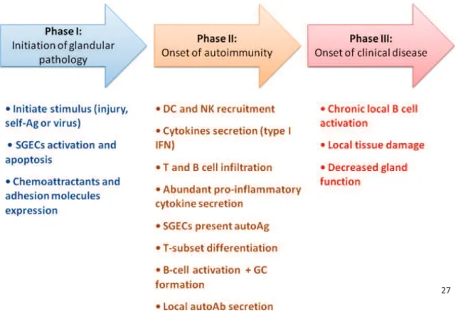
27 1.2.2 Scenario for the pathogenesis of pSS
Based on the results obtained from mouse models of SS or findings observed in patients, it is proposed that SS progresses through three phases (Figure 2). In phase I, in a genetically predisposed individual, a primary infection trigger of SGECs is thought to initiate an immune response. Some chemotactic signals or cytokines contribute to acinar cell apoptosis. In phase II, NK and dendritic cells (DCs) firstly infiltrate the SG and lacrimal glands, secrete IFNs, followed by CD4+ T cells and B cells. Both innate and acquire immune system are then activated. At the same time, epithelial cells function as antigen-presenting cells (APC) to activate T cells. Then, abundant pro-inflammatory cytokines are secreted in the microenvironment. In some patients, ectopic germinal centers develop inside salivary glands. In phase III, B lymphocytes are activated to generate autoantibodies (autoAb). Autoantibodies contribute to local tissue damage and impairment of salivary and lacrimal gland secretory functions [22, 27, 28].
Figure 2. A proposed scenario for the pathogenesis of pSS.
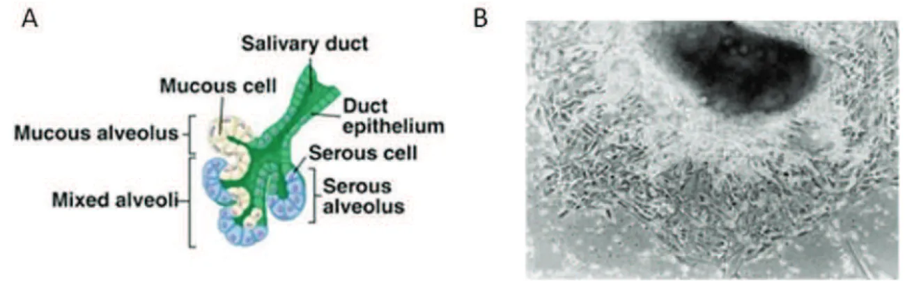
28 1.2.3 The pathogenic role of epithelial cells
Numerous studies have emphasized the key pathogenic role of SGECs in pSS. The salivary glands are composed of two subsets of epithelial cells: acinar and ductal cells (Figure 3A and B). These two cell types play very different roles in the formation of saliva. Acinar cells are water permeable and salt secreting, whereas ductal cells are relatively water impermeable and salt absorbing. In 2002, Dimitriou et
al., have published a protocol for the establishment of human non-neoplastic SGEC
lines (of ductal type) from a single lobule of labial minor salivary glands for the study of the physiology and pathophysiology of these cells [29] (Figure 3C).
Figure 3. A schema of acinar SGECs and ductal SGECs in salivary glands and human non-neoplastic SGEC lines. Figures downloaded on Quizlet website (quizlet.com). SGECs outgrowing from a small fragment of labial minor salivary glands after 5 days of culture (B) [29].
pSS is considered as an autoimmune epithelitis in which salivary gland epithelial cells play a crucial pathogenic role. The immunohistochemical analysis of inflamed salivary gland tissues of patients has indicated that ductal and acinar SGECs display a wide range of MHC molecules, TLRs, costimulatory molecules. These
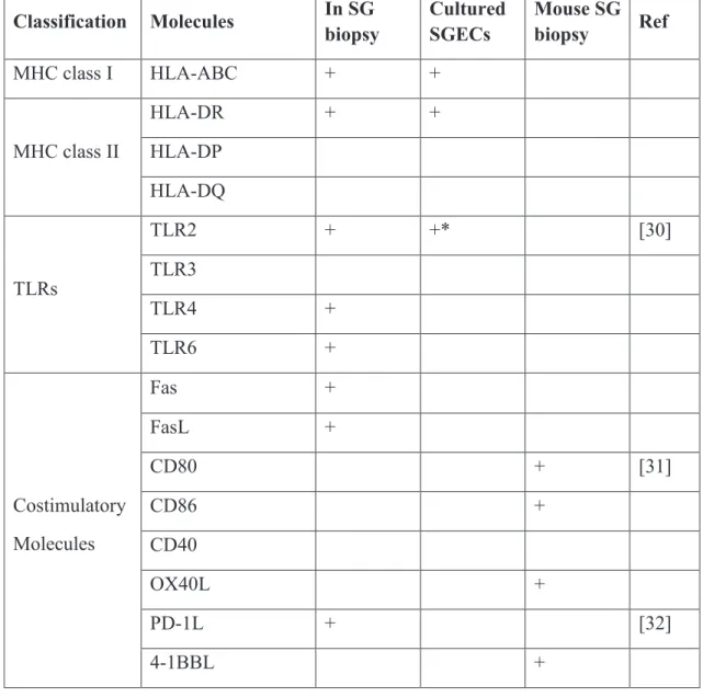
29 molecules are known to mediate lymphoid cell homing, antigen presentation, neovascularization and the amplification of epithelial–immune cells interactions. Molecules expressed by salivary gland epithelia cells in SG tissues or in primary culture of SGECs in SS patients, as well as the results obtained from mouse model of pSS, are summarized in table III. This indicates that SGECs are not only victims but also initiators or amplifiers of the autoimmune process.
Table III: Molecules expressed by salivary gland epithelial cells in Sjögren’s patients and mice models of SS.
Classification Molecules In SG biopsy Cultured SGECs Mouse SG biopsy Ref MHC class I HLA-ABC + + MHC class II HLA-DR + + HLA-DP HLA-DQ TLRs TLR2 + +* [30] TLR3 TLR4 + TLR6 + Costimulatory Molecules Fas + FasL + CD80 + [31] CD86 + CD40 OX40L + PD-1L + [32] 4-1BBL +
30 GITRL + Cytokines BAFF + IL-6 +* [33] TGF-β -* TNF-α + + IL-18 + IL-12 + + IL-1β + Chemokines CCL3 (MIP-1α) + CCL4 (MIP-1β) + CCL5 (RANTES) + CXCL1 (GRO-α) + +* [34] CCL17 (TARC) + [35] CCL12 (MDC) + CXCL9 (Mig) + [36] CXCL10 (IP-10) + CXCL13 + [37] CCL21 + CXCR3 + [38] CCL19 + CXCL8 (IL-8) + CXCL11 + [39] CXCL12 + Angiogenesis VEGF-A + +* [40] VEGFR2 + +* Adhesion α6β4 integrin - [41] ICAM-1 + +
31
VCAM-1 + +
E-cadherin + E-selectin +
+/- : up- or downregulated in patients with pSS compared with controls in basal conditions.
+/-*: up- or downregulated in patients with pSS compared with controls after various stimulations
FAS, cell surface death receptor; 4-1BBL, cell surface death receptor 4-1BB ligand; GITRL, glucocorticoid-induced TNF receptor-related protein ligand; TGF-β, transforming growth factor-β; CXCL, chemokine (C-X-C motif) ligand; MIP-1α/CCL3, macrophage inflammatory protein-1α; MIP-1β/CCL4, macrophage inflammatory protein-1β; RANTES/CCL5, regulated upon activation normal T expressed and secreted; GRO-α/CXCL1, growth related oncogene-alpha; TARC/CCL17, thymus and activation-regulated chemokine; MDC/CCL22, macrophage-derived chemokine; MIG/CXCL19, monokine induced by IFN-γ; IP-10/CXCL10, IFN-γ-inducible 10-kd protein; VEGF-A, vascular endothelial growth factor A; VEGFR2, vascular endothelial growth factor receptor 2; ICAM-1, Intercellular Adhesion Molecule-1; VCAM-1; vascular cell adhesion molecule 1.
1.2.3.1 SGEC activation by PAMPs and DAMPs
The expression by SGECs of several TLRs indicates their role in the induction of innate immune responses upon the recognition of pathogen-associated molecular patterns (PAMPs). TLR signaling has been shown to result in the production of proinflammatory cytokines and in the upregulation of costimulatory and adhesion molecules resulting in the activation of adaptive immune response. TLRs are considered as a critical link between innate and adaptive immunity. In SG of patients
32 with pSS, ductal SGECs and infiltrating mononuclear cells express TLR2, TLR4 and TLR6. Cultured SGECs express TLR2, TLR3 and TLR4 when stimulated with ligand of TLR2 (peptidoglycan (PGN)) and TLR3 (poly-inosinic cytidylic acid (poly I:C)) respectively. Stimulations by TLR2, TLR3 and TLR4 ligands up-regulate ICAM-1, CD40, CD86 and MHC-I expressions in cultured SGECs [42]. Treatment of NZB/W F1 mice with poly I:C increases type I IFN and multiple chemokine production, then accelerates sialoadenitis [43]. In pSS, TLR signal also induces apoptosis of SGECs. Only the ligand of TLR3, but not TLR2 or TLR4, induces apoptosis of SGECs from both patients with pSS and normal subjects.
Extracellular ATP can act as a damage-associated molecular pattern (DAMP) under many pathological conditions. High level of extracellular ATP is associated with inflammation and cell apoptosis. Woods et al., suggest that treatment of mouse submandibular gland cell aggregates with ATP or its high affinity agonist BzATP induces membrane blebbing, enhances caspase activity and releases α-fodrin via P2X7R in vitro. In addition, administration in vivo of BzATP in mouse SG enhances immune cell infiltrations and initiates apoptosis of SGECs. These results suggest that DAMPs such as ATP could play a role pathogenic in SS [44].
1.2.3.2 ECM remodeling and homeostatic changes in epithelial tissues
Epithelial tissues rely heavily on the extracellular matrix (ECM) to maintain structure and function. Some studies described that changes in ECM molecules is the first signs of homeostatic changes in epithelial tissues. In human SG biopsies, major ECM laminin 1 and 5 increase their expression before lymphocytic infiltration. With C57BL/6.NOD-Aec1Aec2 Mice, Peck et al., showed that during the pre-autoimmune phase, molecules associated with interepithelial tight junctions complexes are differently expressed [45]. These complexes are important not only for mechanical adhesions but also for the growth, the differentiation, the morphogenesis, the
33 migration and the extrusion of apoptotic cells. In patients with ECM alterations, downregulation of adhesion molecule α6β4 integrin makes acinar cells fail to maintain their survival [41]. The homeostasis of epithelial tissues could be altered by some proinflammatory cytokines. TNF-α and IFN-γ could alter the tight junction structure and the function in the rat parotid gland cell line [46].
1.2.3.3 Increased apoptosis and NF-κB activity in epithelial cells
Apoptosis has been proposed as a possible mechanism responsible for the impairment of exocrine gland secretory function associated with SS (Figure 4). Apoptosis is important for tissue remodeling because it is necessary in cell proliferation in the regenerating tissues. A significant reduction of acinar SGECs is observed in patients with pSS. Histopathologic lesions are associated with increased apoptosis. Both ductal and acinar SGECs display elevated in situ apoptosis and the ductal SGECs display elevated proliferation. Moreover, cultured SGECs undergo apoptotic process after stimulation with TNF-α and IFN-γ.
Both the Fas/FasL (CD95/CD95L) pathway and the substances (perforin, granzymes) released by cytotoxic T lymphocytes (CTL) are expressed in the SG of patients with pSS. Ductal and acinar SGECs express the Fas and FASL whereas infiltrating T cells express FasL [47]. So apoptotic death of ECs may be caused by autocrine Fas/FasL interaction at the epithelial cell level or by paracrine interaction with infiltrating T cells. The imbalance between the pro-apoptotic proteins BCL2-associated X protein (Bax) and anti-apoptotic B-cell leukemia/lymphoma-2 (Bcl-2) observed in SGECs of patients with pSS might cause increased apoptosis. Converse to SGECs, the infiltrating mononuclear cells present a high Bcl-2/Bax expression ratio. This imbalance may explain their resistance of lymphocytes to apoptosis despite Fas expression. CD4 or CD8 CTL can also induce EC apoptosis by perforin and granzyme B release and the secretion of cytokines, such as TNF-α, IFN-γ
34 and TGF-β1. At last, direct B cells contact can induce apoptosis of EC line (HSG) in vitro. This B cells-mediated cell death is not dependent on Fas/FasL interactions but requires translocation of protein kinase C delta (PKC δ) into the nucleus of epithelial cells [48].
It has been noted that in SS, aberrant apoptosis is not only the consequence but also the trigger of lymphocytic infiltrates. In NOD-scid mice, a glandular EC apoptosis occurs in the absence of lymphocytic infiltration. Increased apoptosis of acinar SGECs as well as increased IFN-γ expression occur before infiltration by lymphocytes in NOD mice. In C57BL/6.NOD-Aec1Aec2 mice, altered glandular homeostasis and elevated apoptotic epithelial cells are observed in the submandibular glands on the 8th week before disease onset [49]. Using neonatal NOD mice, Cha et al., revealed that acinar cell proliferation was reduced while expression of Fas, FasL and bcl-2 were increased in submandibular glands on the 1st day postpartum [50]. Recently, Okuma et al., demonstrated that IκB-ζ-deficient epithelial cells exhibited increased apoptosis. This is sufficient to elicit SS-like pathology in mice, such as lymphocytic inflammatory infiltrates of the lacrimal glands with reduced tear secretion and secretion of anti-SSA and anti-SSB. IκB-ζ regulates NF-κB transcriptional activity, thus being implicated in both cell survival and apoptosis [51]. The expression of another NF-κB inhibitory protein IκB-α is downregulated in SS SGECs [52].
1.2.3.4 Autoantigens presentation by SGECs
Activated SGECs in SS lesions appear to be suitably equipped for the presentation of antigens to T cells and the subsequent crosstalk between T and B cells leading to autoantibody secretion. SGECs express HLA-DR, CD80 and CD86 molecules in close proximity to the lymphocytic infiltrates [53]. Moreover, IFN-γ can induce or upregulate the expression of those molecules in cultured controls or SS
35 SGECs [54]. The Ro52, Ro60 and La autoAgs are components of human Ro/La ribonucleoprotein (RNP) complex. Normally, the localization of these proteins is mainly nuclear. Under certain physiopathological conditions (e.g. stress, UV radiation or viral infection), these proteins can be found on cell surface. Acinar ECs express autoantigens on their membrane. More specifically, antigen La has been observed in conjunctival epithelial cells of SS patients. In fact, two mechanisms are implicated in the antigen presenting process. Epithelial cells could either express autoantigens on their membrane or release exosomes containing autoantigens (Ro, La and α-fodrin) [4] (Figure 4). The expression of HLA-DR+ by epithelial cells in SG of patients is closely associated with T-cell infiltrates [55]. In addition, autoAg La-derived peptide contains T cell epitopes and can trigger T-cell proliferation [56]. These data in literature strongly suggest that SGECs are capable to present autoAg to T cells.
Figure 4: A schematic model for early events in the pathogenesis of lymphoepithelial lesions in the salivary glands of SS patients. Environmental and hormonal factors in an appropriate genetic background activate glandular ECs. ECs undergo apoptosis and recruit DCs and NK following T cells via chemokines and specific adhesion molecules productions. Altered extracellular interactions facilitate immune cell migration and amplify the apoptosis of ECs. ECs activate T cells by direct presentation of autoAgs and MHC II and costimulatory molecule expression as well as by the release of autoAg-containing exosomes. The early recruitment of plasmacytoid dendritic cells (pDC) and NK produce high levels of type I and II IFNs that increase the antigen presentation capability of ECs. Activated T cells further activate ECs by secreting proinflammatory cytokines. At last, B cells are recruited into SG leading to plasma cells differentiation and autoAbs production. All the previous step result in local B cells differentiation into autoAb secreting plasma cells.
36 1.2.3.5 In-situ activation and immunologic functions of SGECs in SS
In patients with pSS, the capacity of SGECs to secrete cytokines and chemokines has been previously reported. Cultured SGECs from SS patients also showed an abnormal cytokine production. Cultured SS SGECs secret more IL-6 and less TGF-β upon IFN-γ stimulation compared with normal controls. This may affect the local balance between Tregs and Th17 cells. The pro-inflammatory cytokine IL-18 is detected in the acinar SGECs of SS patients. IL-18 could amplify the production of IL-6 and IL-8 induced by IL-17 in salivary gland cells (human parotid gland cell line HSY) [57]. Using the autoimmune regulator (Aire)-knockout (KO) mice, Chen et al., have demonstrated that the induction of SS-like lacrimal exocrinopathy mediated by autoreactive CD4+ T cells depends on IL-1 receptor type 1 (IL-1R1) signaling. IL-1R1 is detected only on resident ductal lacrimal gland epithelial cells but not on resident
37 APCs or infiltrating immune cells. Moreover, IL-1 expression in ocular epithelial cells is significantly correlated with the development of ocular pathological changes. These results indicate that in targeted tissues, the interplay between resident epithelial cells and CD4+ T cells plays a central role in the pathogenesis of SS [58]. SGECs are also capable to secrete BAFF after type I/II IFN stimulations, TLR activation or viral infection [59].
Although T cells proliferate locally in SGs, it seems that much of the infiltration is due to the migration of peripheral blood T cells. In the SG of patients, the ductal SGECs express various chemokines and the adjacent infiltrating T lymphocytes express their cognate receptors (see 1.2.9). This suggests that SGECs are involved in the migration of T cells into salivary glands. CXCL10 (IP-10) antagonist decreases the infiltration of Th1 type CXCR3+ T cells and improves sialoadenitis in MRL/lpr mice [60].
SGECs also express costimulatory molecules and adhesion molecules to act as nonprofessional APCs. All these signals as well as molecules mediating APC-T cell adhesion are essential in the formation and stabilization of the immunological synapse which is requested for an effective activation of T cells. In pSS patients with severe sialoadenitis, CD80 and CD86 are strongly expressed on ductal epithelial cells and their ligand CD28 is expressed on some infiltrating cells. In SG biopsies, CD40 is constitutively expressed by lymphocytes, ductal epithelial cells and endothelial cells. CD40 expression is significantly higher in cultured SS SGECs, which could be further enhanced by IFN-γ and IL-1β. CD40L staining is detected in 30-50% of the infiltrating lymphocytes in the biopsies of SS patients [61]. Adhesion molecule ICAM-1, VCAM-1 and E-selectin are detected on duct cells from all patients. These findings indicate SGECs may be involved in the induction and the maintenance of lymphocytic infiltrates.
38 1.2.4 Salivary gland infiltrates.
Salivary glands are the best-studied organs because they are affected in almost all patients and are readily accessible. The diagnostic of SS often needs a minor salivary gland biopsy. In minor salivary gland, lymphocytic infiltrate mainly consists of activated T and B cells and antigen presenting cells. Poly I:C-treated NZB/W F1 mice showed that dendritic cells and NK cells dominate early cell infiltrates, followed by CD4+ T cells [43]. T lymphocytes (composed of 33% of CD4+ T cells, 15% of CD8+ T cells, 3% of CD4+CD8+ T cells and 2% of Foxp3+ Tregs) and B lymphocytes represent 90% of infiltrating cells and have variable frequency. T cells predominate in mild lesions, whereas B cells do in advanced lesions. T cells decrease, whereas B cells increase with the extent of infiltration. Macrophages and dendritic cells are also found in up to 5% of infiltrates, mainly in glands with ectopic GCs. NK cells are very rare but were less studied until recently [62]. The presence of macrophages varies according to the severity of lesion. The proportion of major regulators of immune response, Foxp3+ Tregs, is found to decrease with lesion severity. This suggests an inability of regulatory mechanisms to control local immune activation [63].
1.2.5 T-lymphocyte contribution to the pathophysiology of pSS
The predominant cells in SG infiltrates are T cells with a CD4/CD8 ratio of 4:1. Almost all infiltrating T cells express αβ T-cell receptor (TCR), only 1-5% express γδ receptor. These CD4 T cells present an activated (HLA class II+, CD25+) and primed (CD45RO+) phenotype. On the contrary, T cells in peripheral blood do not to have the same characteristics (absence of expression of HLA class II molecules) [64].
1.2.5.1 T-lymphocyte migration to exocrine tissue
39 adhesion molecules expressed by vascular endothelium and epithelial tissues. Various chemokines play a role in recruiting T cells. CCL21, CXCL9, CXCL10, CXCR3, CXCL11 and CXCL12 are expressed by SGECs while CXCR4 (ligand of CXCL12) and CXCR3 (ligand of CXCL9, CXCL10) are expressed by activated T cells. SGECs also express ICAM-1 and VCAM-1. These adhesion molecules bind to their ligands expressed on T cells, lymphocyte function-associated antigen 1 (LFA-1) and very late antigen-4 (VLA-4). These interactions first play a role in T cell-recruitment into salivary gland (Figure 5A) and are also involved in the antigen presentation process and the formation of the immunological synapse (Figure 5B).
1.2.5.2 In situ antigen-driven stimulation of T lymphocytes
Assessment of the T cell receptor repertoire of infiltrating T cells reveals that TCR Vβ gene usage is relatively restricted. This fact suggests an antigen-driven stimulation rather than a superantigen-induced proliferation. In fact, autoantigens (Ro, La and α-fodrin)-specific T cells are detected in patients with SS. DCs, macrophages and activated SGECs function as APCs to activate antigen-specific T cell. Activated SGECs express all the molecules (MHC I and II, CD80, CD86, CD40) needed for antigen presentation and T cell activation. TCR interacts with the MHC–peptide complex in the immunological synapse between SGECs and T cells. The co-stimulatory signals are provided by different pairs of molecules including CD80/CD86 - CD28 and CD40-CD40L. Adhesion molecules stabilize the immunological synapse. After that, engaged T cells undergo proliferation and secrete cytokines. T cells infiltrated in SG are oligoclonal and some proliferating T cells can be detected in the SG of patients [65].
Figure 5. Molecular mechanisms involved in T cell migration into exocrine glands and in the immunological synapse between T cells and SGECs. A. Environment triggers lead to the upregulation of adhesion molecules on
40 endothelial and epithelial cells and to the expression of chemokines. T cells are recruited by chemokines, and interact with adhesion molecules found on vascular endothelium. T cells come across the endothelial vessels and interact with SGECs. B. Two signals are required for T-cell activation that leads to clonal expansion and cytokine secretion. TCR interacts with MHC II-peptide complex as well as CD4 or CD8 to provide the signal-1. Costimulatory molecules CD80/CD86-CD28 and CD40-CD40L are delivered as signal-2. Adhesion molecule interactions such as ICAM-1-LFA-1 and VCAM-1-VLA-4 stabilize the immunological synapse. This interaction leads to clonal expansion of T cells, activation and induction of apoptosis of SGECs and release of T-cell cytokines.
41 1.2.5.3 T- cell- mediated immunopathogenesis
In pSS, both CD4 helper T cells and CD8 cytotoxic T cells drive tissue lesions in the exocrine glands via different mechanisms. CD8+ T cells are located around the acinar ECs. CD103 (αEβ7 integrin) expressed by CD8 T cells interact with E-cadherin expressed by adjacent SS acinar ECs, and mediates the adhesion between CD8 T cells and acinar ECs. CD8 T cells induce EC apoptosis via both the perforin/granzyme B and Fas/FasL pathways [66]. CD4+ T cells with cytotoxic activity are detected in immunopathologic lesions of minor SG of SS patients. They comprise 20% of the infiltrating lymphocytes and utilize perforin-mediated cell lysis.
42
1.2.5.4 T helper cell subsets and cytokine profile in pSS.
During TCR activation, naive CD4 T cells differentiate into different lineages of T helper cells, including Th1, Th2, Th17, Tfh and Treg. IL-12 and IFN-γ activates STAT4 and induces T box transcription factor (T-bet) which is specific to Th1 cells. Th1 cells produce Il-2, TNF-α and IFN-γ to promote cellular immune system. Similarly IL-2, IL-7 and IL-4 induces Th2 cells through STAT6, WGATAR nucleotide consensus sequence (Gata-3) and c-maf and promote humoral immune system. The signature cytokines of Th2 are IL-4, IL-5, IL-6, IL-10 and IL-13. IFN-γ inhibits the production of Th2 type cytokines such as IL-4, while IL-10 inhibits a variety of Th1 cytokines including IL-2, IFN-γ and IL-12. Recently, some new cytokine and lineage of CD4 T cells were distinguished. In 2003, Th17 cells were characterized by the production of IL-17A, IL-17F and IL-22 as signature cytokines, as well as retinoid orphan receptor γt (RORγt) as a specific transcription factor. Th17 can be induced by IL-6 and TGF-β. Th17 cells also produce IL-21. It was shown that cells with regulatory function could be induced from naïve CD4 by IL-2 and TGF-β. Distinct from natural regulatory T cells (nTreg), which are not derived in the periphery from naïve CD4 T cells, these cells were designated induced Tregs (iTregs). Induced Treg also express the FoxP3 (Figure 6).
Figure 6. T helper cell lineage development and function. Figure adapted from [67]
43 In SG from patients with pSS, the expression of cytokines and transcription factors of T helper subsets such as Th1, Th2, Th17 and follicular helper T cells are significantly increased in comparison with controls. The localization of these T helper subsets is associated with disease severity. Th2 and Tfh are closely associated with strong lymphocytic infiltrations, converse to Th1, Th17 and Tregs. Th1 and Th17-related molecules are mostly detected in SG without GC, while Th2 and Tfh-related molecules are more frequently detected in SG with GC. It has been proposed that SS could be initiated by Th1 and Th17 cells, and then be replaced by Th2 and Tfh cells in patients with GC [68].
1.2.5.5 Th1/Th2 balance
44 Th2 cytokines were also found elevated. Thus, both Th1 (IFN-γ) and Th2 cytokines (IL-4, IL-13) are produced by lymphocytes infiltrating SG of patients [69]. In another report, infiltrating CD4+ T cells mainly secreted IL-2, IFN-γ and IL-10, but neither IL-4 nor IL-5 [70]. Th1 cytokines were associated with the importance of T-cell infiltration whereas Th2 cytokine, such as IL-4 and IL-5, were rather associated with B cell infiltrates [71]. Th1/Th2 balance in salivary glands might also depend on the duration of symptoms. Chemokines involved in the Th1/Th2 balance in pSS are described in chapter 1.2.9.
1.2.5.6 Th17 cells, IL-17 and IL-23
Some studies suggested pathogenic role for Th17 cells in pSS. Cytokines required for the differentiation and the maintenance of Th17, TGF-β, IL-6 and IL-23 are all detected in SGs or in the serum of patients with pSS. High level of IL-17 is found in serum and saliva of pSS patients. IL-17 is predominantly expressed by infiltrating CD4+ T cells rather than by CD8+ T cells [57]. Th17 memory cells are detected within the exocrine glands of C57BL/6.NOD-Aec1Aec2 mice and human pSS patients. Blockage of IL-17 expression in C57BL/6.NOD-Aec1Aec2 mice results in decreased SG lymphocytic infiltration and increased saliva secretion.
1.2.5.7 Regulatory T cells
There are two main populations of Tregs: thymus-derived natural Treg (nTreg) cells and peripherally generated induced Treg (iTreg) cells. Both are forkhead/winged helix transcription factor (Foxp3) positive cells. nTreg are critical for the control of immune response, including autoimmunity. Following engagement of their TCR, Tregs suppress the proliferation and the activity of conventional effector CD4+ T cells as well as that of CD8+ T cells.
45 with importance of the inflammatory infiltrate. In the peripheral blood, the incidence of Tregs is higher in SS patients than in healthy individuals [72]. The migration of Tregs is inversely correlated in minor SG lesions and in peripheral blood [73]. The functionality of Treg in pSS remains unclear. It is reported that Foxp3 positive Tregs can be induced from naive T cells by IL-2 and TGF-β whereas the combination of IL-6 and TGF-β induces Th17. Moreover, IL-6 can convert nTregs to Th17 cells in the presence of TGF-β whereas iTregs are resistant to Th17 conversion by IL-6 [74]. Thus, the imbalance of Tregs and Th17 in pSS patients may be due to the local conversion effect of IL-6 which turns iTreg into Th17 cells.
1.2.5.8 Tfh and IL-21
Characteristics of Tfh
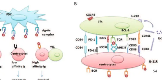
The fifth lineage of CD4 T cells, follicular helper T cells were firstly described by Schaerli P and Breitfeld D in 2000 [75]. They found that a human memory CD4 T cell subpopulation (CD45RO+) expressed CXCR5 in tonsils and blood. These CXCR5+ T cells migrate in response to CXCL13, which is selectively expressed by reticular cells and blood vessels within B cell follicles. Tfh are localized in the mantle and light zone of germinal centers in the B cell follicles. Ma et al., identified human tonsillar CD4+CXCR5high T cells as Tfh cells. They uniformly displayed the highest levels of ICOS, CD95, PD-1, CD200, cytoplasmic adaptor protein SLAM-associated protein (SAP) and CD57. Tfh cells also express CD126, BTLA (ligand of B7-H4), OX40, CXCR4, CD40L, CD69 and CD126. The transcriptional factor B-cell lymphoma 6 protein (BCL6) but not T-bet, GATA3, Foxp3 or RORγt is highly expressed by Tfh. Tfh also produce high level of B-cell activity cytokines, particularly IL-21, IL-4 and IL-10 [76]. The ICOS+CXCR5+ IL-21 producing-Tfh can be induced
in vitro from naïve T cell by IL-12. Schmitt et al., also described that naive CD4+ T cells primed with IL-12 induced B cells to produce Igs in a fashion dependent on
46 IL-21 and ICOS. There are two different types of IL-21-producing T Cells induced by IL-12: IFN-γ+IL-21+ and IFN-γ−IL-21+ cells. Furthermore, IFN-γ+IL-21+ T cells expressed T-bet, whereas IFN-γ−IL-21+ cells did not. Bacteria or CD40L-activated DCs can induce IL-21-producing CD4+ Tfh cells through IL-12 [77]. Other studies showed that Tfh also express molecular critical for their development and function, including HLA-DR, CD84 as well as the transcription factors c-Maf. Cytokines such as IL-6, IL-21 and IL-27 are necessary but insufficient for Tfh cell generation and maintenance in vivo.
Generation of Tfh
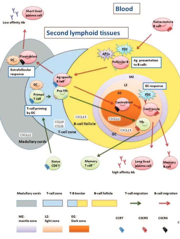
Blood naïve CD4 T cells express CCR7. In respond to CCL19 and CCL21 produced in the T-zone of secondary lymphoid organs: spleen, lymph nodes and mucosal-associated secondary lymphoid tissues, they migrate from the circulation to the T cell zone. T cells recognize Ag and costimulatory signals provided by DCs in the T cell zone. Depending on the costimulatory and cytokine signal provided, T cells differentiate into Th1 or Th2 cells. They downregulate CCR7 and exit lymph node to participate the effector responses. Some primed T cells downregulate CCR7 and upregulate CXCR5 and express BCL-6. The ligand of CXCR5, CXCL13 is expressed by cells in B cell follicle and GC. This results a gradient of CXCL13. In response to CXCL13, these CXCR5+BCL-6+ pre-Tfh cells migrate from T cell zone towards B cell follicle. At the interface between the T and B zones (T-B border), pre-Tfh cells interact with Ag-specific B cells. During this interaction, the help provided by pre-Tfh is essential for the initiation of both germinal center and extrafollicular response of B cells. In turn, depending on the signal provided by B cells, this cognate interaction is important for the complete downregulation of CCR7 which results in pre-Tfh entering the GC and terminally differentiate into Tfh. After this interaction, pre-Tfh cells maintain BCL-6 expression and enter the light zone of GC. There, they fully differentiate into Tfh express PD-1, IL-21, CD84 and ICOS and provide survival
47 signal to centrocytes. After GC responses, Tfh decrease their expression of signature Tfh molecules. These cells contribute the CD4+CXCR5- memory T cell pool [78] (Figure 7).
Figure 7. Anatomical localization as well as molecules and cellular requirements for Tfh differentiation.
48 Tfh-cell driven B-cell differentiation in secondary lymphoid tissues.
CD4 helper T cells provide a second signal to drive B-cell maturation, terminal differentiation and isotype switching. Circulating naïve mature B cells migrate into the primary B-cell follicle of the secondary lymphoid tissue of lymph nodes (Figure 8). B-cell follicle is composed of follicular B cells and follicular dendritic cells (FDC). This migration is mediated by the expression of CXCR5 on follicular B cells. At this step, follicular B cells collect the Ag presented by APCs including FDC, DC, macrophages as well as other stromal cells. Ag specific B cells become activated and upregulate CCR7 which allow them to migrate towards T cell zone by a gradient of CCL21. At the T-B border, they present Ag peptide by CMH II to cognate DC-primed T cells. Activated B cells have two distinct fates: an extrafollicular process or affinity maturation in GC (Figure 7).
Figure 8. B cell devellpoment and lymph node stucture. Immature B cells exit the bone marrow. They circulate as naïve B cells. These circulating B cells then migrate through the circulation to the red pulp of the spleen (transitional type 0 (T0) B cells). These T0 B cells migrate into the white pulp and mature into T1 and then T2 B cells. Finally they differentiate into either follicular or marginal zone (MZ) B cells. Follicular B cells recirculate as mature naïve B cells among secondary lymphoid organs. Once activated, they enter the primary follicles of secondary lymphoid tissues. After being challenged by T-dependent antigen, they move to the T-B border and make cognate interaction with primed T helper cells. Some activated B cells migrate to rapid extrafollicular foci and differentiate into plasma cells with a short lift span which rapidly secrete low-affinity antibodies. Otherwise, a few of activated B cells migrate into the primary lymphoid follicles and form the germinal center (secondary lymphoid follicles). They firstly divide
49 as centroblasts and then undergo a SHM. The centroblasts then migrate to the light zone as centrocytes. They come into contact with the FDC following interaction with cognate Tfh which trigger immunoglobulin class switching. B cells which gained improved affinity are positively selected to proliferate whereas the low affinity clone and self-reactive clone undergo a cell death or apoptosis process. Centrocytes then become either plasma cells with a long life span or memory B cells. HEV, high endothelial venules. Figures adapted from the 8th edition of the Janeway’s immunobiology.
50 Extrafollicular process
Some of Ag specific B cells downregulate CCR7 expression and upregulate CXCR4. This leads them to differentiate into plasmablasts and remigrate from B cell follicle to the medullary cords to form extrafollicular foci in response to CXCL12. DC support plasmablasts to survive and differentiate into plasma cells with a short life span (3 days) and produce low and modest affinity Abs. These antibodies may be either switched (e.g. IgG1) or unswitched (IgM). This initial burst of Ig is important in the early control of infection (Figure 7).
Affinity maturation in GC
T-cell activated Ag-specific B cells migrate into the B cell follicle in response of CXCL12. There, they continue to divide as centroblasts. The centroblasts form the dark zone of the GC. Thanks to their activation-induced deaminase (AID) expression, they undergo a somatic hypermutation (SHM), on the variable regions of the BCR. The centroblasts stop to proliferate and migrate to the light zone of GC as centrocytes. Centrocytes with mutated BCR come into contact with the FDC. Through interaction with antigen held on FDC, they acquire survival signals. Centrocytes present their Ag to cognate Tfh. At this step, B cells which gained improved BCR affinity during SHM receive positive selection signals from Tfh. They proliferate and either differentiate to plasma cells with a long life span and produce high affinity Ab or become memory B cells. Tfh also trigger Ig class switch recombination to IgG in the GC. At the same time, the low affinity clone and self-reactive clone undergo cell death or apoptosis. [79, 80] (Figure 7).
T-cell priming in the T-cell zone
Depending on the microenvironment and on costimulatory signals, T cells can differentiate into Th1, Th2 or Tfh. Th1 and Th2 will help B cell differentiation in the
![Figure 9. Initial T cell priming by DC and T cell differentiation. Adapted from [82]](https://thumb-eu.123doks.com/thumbv2/123doknet/14516544.721855/52.892.141.774.919.1195/figure-initial-cell-priming-dc-cell-differentiation-adapted.webp)
![Figure 14. Main markers of mature B-cells. Adapted from [100].](https://thumb-eu.123doks.com/thumbv2/123doknet/14516544.721855/61.892.144.745.838.1139/figure-main-markers-mature-b-cells-adapted.webp)