Combining immunotherapy and anticancer agents: the
right path to achieve cancer cure?
L. Apetoh
1,2,3, S. Ladoire
1,2,3, G. Coukos
4& F. Ghiringhelli
1,2,3*
1Lipids, Nutrition, Cancer, INSERM, U866, Dijon;2
Department of Medicine, Université de Bourgogne, Dijon;3
Department of Oncology, Centre Georges François Leclerc, Dijon, France;4
Department of Oncology, University of Lausanne, Lausanne, Switzerland
Received 23 February 2015; revised 13 April 2015; accepted 17 April 2015
Recent clinical trials revealed the impressive efficacy of immunological checkpoint blockade in different types of metastatic cancers. Such data underscore that immunotherapy is one of the most promising strategies for cancer treatment. In add-ition, preclinical studies provide evidence that some cytotoxic drugs have the ability to stimulate the immune system, resulting in anti-tumor immune responses that contribute to clinical efficacy of these agents. These observations raise the hypothesis that the next step for cancer treatment is the combination of cytotoxic agents and immunotherapies. The present review aims to summarize the immune-mediated effects of chemotherapeutic agents and their clinical relevance, the biological and clinical features of immune checkpoint blockers andfinally, the preclinical and clinical rationale for novel therapeutic strategies combining anticancer agents and immune checkpoint blockers.
Key words:chemotherapy, radiotherapy, anticancer immunity, immunomodulation, CD4 T cells, CD8 T cells
introduction
The involvement of the immune system in tumor control is now
accepted. The ability of innate and adaptive immune cells to
detect and eliminate tumor cells was termed
‘cancer
immuno-surveillance’. However, subsequent studies have shown that
immune cells could also facilitate cancer progression by
promot-ing the growth of tumor clones resistant to anticancer
immun-ity. The term
‘cancer immunoediting’ encapsulates the dual
activity of the immune system on tumors [
1
]. The positive effect
of the immune system on the control of tumor growth is
under-lined by the observation that HIV infection or
immunosuppres-sive states induced by genetic de
ficiency or immunosuppression
increase the frequency of solid cancers and hematological
malig-nancies in mice and humans [
2
–
5
]. Recent data show that
growing tumors are frequently in
filtrated by immune cells,
notably CD8 T cells and these cells probably contribute to the
control of tumor growth in humans because their presence is
associated with better outcomes [
6
–
8
]. This anti-tumor immune
response can be manipulated to enhance tumor immune attack,
leading to clinical bene
fits for cancer patients. Challenging the
presiding view that chemotherapeutic agents were
immunosup-pressive [
9
–
14
], we and others have shown that some
che-motherapeutic agents could elicit an immunogenic form of
tumor cell death that enhances anticancer immune responses
and contributes to the clinical efficacy of these chemotherapies
[
15
–
18
]. Although the demonstration of immunogenic cell
death (ICD) relies on mouse models of intratumor injection of
chemotherapy or only one systemic injection of chemotherapy
which do not mimic the clinical setting of repetitive systemic
injections of high doses of chemotherapy, the
findings that
patients with genetic de
ficiencies in molecules involved in the
detection of ICD have poorer prognosis under
chemotherapeu-tic treatment underscore the clinical relevance of this concept
[
15
–
17
]. Recently, the use of monoclonal antibodies (mAbs)
blocking key inhibitory receptors of T cells such as cytotoxic
T-lymphocyte-associated protein 4 (CTLA-4) and programmed
cell death 1 (PD-1) has led to robust anti-tumor immune responses
and has yielded clinical benefits across multiple tumor types
[
19
]. In addition, impressive clinical responses were observed
upon adoptive transfer of tumor-speci
fic autologous T cells
using the generation of T cells that expressed cloned T-cell
receptors (TCRs) or chimeric antigen receptors (CAR) [
20
].
However, despite these recent successes, many patients are not
cured. Emerging evidence suggest that combination strategies
may be important to achieve deeper tumor responses. Although
combination of immune therapy with some conventional
cyto-toxic chemotherapies can be envisioned, the important question
of how to integrate these novel immunotherapy treatments with
the current clinical strategy still remains. In this review, we will
summarize our knowledge on the immune-mediated effects of
chemotherapies and on the mechanisms of action of novel
im-munotherapies and propose a rationale for the design of
syner-gistic anticancer combinations.
*Correspondence to: Prof. François Ghiringhelli, Centre de Recherche, INSERM U866, Facultés de Médecine et de Pharmacie, 7 Bd Jeanne d’Arc, 21079 Dijon, France. Tel: +33-3-80-39-33-53; Fax: +33-3-80-39-34-34; E-mail: fghiringhelli@cgfl.fr
revie
ws
© The Author 2015. Published by Oxford University Press on behalf of the European Society for Medical Oncology. All rights reserved. For permissions, please email: [email protected].
the emergence of novel
immunotherapeutic strategies
against cancer
dendritic cells
Owing to their strong ability to initiate and control T-cell
responses, dendritic cells (DCs) have long been regarded as
at-tractive candidates for the design of immunotherapy strategies
[
21
]. Initially, DC cancer vaccines consisted of ex vivo generated
DCs that were loaded with tumor antigens. Although this
vaccin-ation strategy has successfully elicited anticancer immune
responses in cancer patients, no or very few clinical bene
fits were
noted (reviewed in [
22
]). This was, for instance, illustrated by the
results of a phase III randomized clinical trial comparing the
clin-ical ef
ficacy of DC therapy to dacarbazine in metastatic melanoma
patients. The objective response rate for DC therapy was 3.8%,
explaining the early discontinuation of the study because of lack
of ef
ficacy [
23
]. Proposed reasons explaining the lack of ef
ficacy of
DC vaccines include the use of inappropriate DC maturation
cocktail, thus compromising the ability of DC to induce
antican-cer responses. To circumvent these issues, different strategies are
being implemented. Cytokine combinations to favor DC
matur-ation and antigen presentmatur-ation properties to design more effective
DC-based immunization strategies are being tested (reviewed in
[
22
,
24
]). A recent study demonstrated that the ability of a DC
vaccine to increase, without any associated side effects, the
breadth and diversity of melanoma neoantigen-speci
fic T cells
further enhances the potential of this approach [
25
].
adoptive T-cell therapy
Adoptive T-cell therapy for cancer aims to eliminate cancer cells
upon administration of T cells into tumor-bearing hosts. In 1955,
Mitchison [
26
] initially demonstrated the feasibility of this
ap-proach in mice. In 1966, Southam et al. studied the anticancer
activity of leucocytes from tumor-bearing patients against their
re-spective tumor and raised the hypothesis that lymphocytes from
cancer patients have a speci
fic inhibitory effect against cancer cells.
This provided impetus to exploit T cells from cancer patients to
design effective adoptive transfer strategies. Subsequent studies
focused on the isolation, expansion and reinfusion of
tumor-in
filtrating lymphocytes (TILs) into cancer patients. The studies
pioneered by Rosenberg and colleagues relied on the culture of
tumor-derived lymphocytes in the presence of high doses of IL-2
that were then transferred into tumor-bearing patients. Although
encouraging results were noted, side effects impeded the large
clin-ical implementation of this strategy. The safety and ef
ficacy of TIL
therapy was improved by implementing preconditioning regimens
driving lymphodepletion. The use of a preconditioning regimen
relying on cyclophosphamide and
fludarabine was shown to
eliminate the endogenous lymphocyte repertoire and favor
growth and long-lasting persistence of the transferred TILs. This
has led to improved responses rate up to 40% [
27
]. Research in
T-cell biology has also improved TIL culture conditions, leading
to shorter TIL expansion phase, thereby reducing the time from
TIL collection to their reinfusion into cancer patients.
An alternative strategy to target tumor cells using T cells is
the engineering of CAR T cells, which are endowed with a
spe-ci
fic ability to recognize and kill cancer cells. CARs contain a
fusion protein of light and heavy chains from an antibody,
linked to the signaling machinery of the TCR. Such structure
enables T-cell activation upon CAR recognition of its target. As
CARs are not Major Histocompatibility complex
(MHC)-restricted, they are insensitive to tumor-driven
immunosuppres-sion mediated through downregulation of MHC molecules.
Another advantage of CAR T cells is the possibility of
transdu-cing genes that will enhance further T-cell functions upon
acti-vation or chemokine receptors that will favor T-cell homing.
Although the initial trials in ovarian cancer and renal cell
carcinoma using CAR T cells were disappointing because of
tox-icity and limited T-cell persistence in the tumor
microenviron-ment [
28
,
29
], the use of second-generation CAR T cells in
leukemic patients has resulted in remarkable anticancer effects
[
30
,
31
]. The success of CAR T-cell therapy in these diseases
was associated with a high level of CAR T-cell proliferation
fol-lowing infusion into patients. The feasibility of this approach
was further established in 30 patients suffering from relapsed or
refractory acute lymphoblastic leukemia. CAR T-cell therapy
targeting CD19 led to complete remission in 27 patients and
sustained remission was achieved with a 6-month overall
sur-vival rate of 78% [
32
]. This notable ef
ficacy has led to the United
States Food and Drug Administration to designate the
anti-CD19 CAR T-cell therapy as a
‘breakthrough therapy’.
cytokines
The efficacy of IL-2 as an anticancer agent has been investigated
in multiple cancer types. It has been shown that high doses of
IL-2 could be effective in eliciting anticancer responses in renal
cell carcinoma and melanoma. Nevertheless, the overall
re-sponse rates were low and the associated toxicities were severe
(reviewed in [
22
]). IL-2 was further shown to drive the
expan-sion of regulatory T cells which in turn suppress anticancer
immune responses. This has prompted the test of additional
cytokines for their anticancer potential upon in vivo
administra-tion. IL-15 was later identi
fied as an interesting candidate
mol-ecule. The anticancer effects of IL-15 have been demonstrated in
several preclinical tumor models. The underlying mechanisms
have been subsequently identified. IL-15 has been shown to
enhance NK cell effector functions. In addition, IL-15 was shown
to support the proliferation and effector functions of CD8 T cells
in the presence of regulatory T cells, suggesting that IL-15 could
preserve the persistence and anticancer functions of T cells in
the tumor microenvironment [
33
]. The ability of IL-15 to
acti-vate cell functions was further exploited in the context of
T-cell therapy. Culture of TILs with IL-15 was indeed shown to
improve the quality of CD8 T cells for adoptive therapy [
34
].
CAR T cells expressing the IL-15 gene featured greater
expan-sion in vitro and reduced the cell death rate over control CAR T
cells. Upon adoptive transfer in mice, IL-15 expressing CARs
showed enhanced anticancer effects in vivo [
35
]. The clinical
im-plementation of IL-15 began in 2009. The safety and efficacy of
IL-15 is currently being tested in patients with lymphoma,
melanoma, or renal cell carcinoma ([
36
] and NCT01572493,
NCT01385423).
In addition to IL-15, IL-21 is also an immunomodulatory
cytokine that is currently being tested for its anticancer activity
in humans. Preclinical studies using IL-21-overexpressing
tumors revealed that IL-21 prevented B16 melanoma and
MCA205 carcinoma growth and increased mouse survival [
37
].
In addition, administration of IL-21 was found to control CD8
T-cell expansion and effector functions and to synergize with
IL-15, leading to the rejection of large melanoma tumors in
mice [
38
]. The ability of IL-21 to enhance T-cell functionality
was shown in the context of adoptive transfer. CD8 T cells
cul-tured with IL-21 enhanced their anticancer activity, leading to
rejection of large tumors upon transfer [
39
]. Similarly, IL-21
enhances CAR T-cell anticancer functions for effective
im-munotherapy against B-cell malignancies [
40
]. We have also
re-cently reported that naïve CD4 T cells differentiated into
effector Th9 cells in the presence of TGF-β, IL-4 and IL-1β,
secreted high levels of IL-21 and mediated IL-21-dependent
anticancer effects against melanoma tumors upon adoptive
transfer [
41
]. IL-21 mediated its anticancer activity through
acti-vation of NK and CD8 T cells which in turn controlled tumor
progression through IFN
γ. The clinical efficacy of IL-21 was
investigated in phase I and II trials involving melanoma, renal
cell carcinoma and metastatic colorectal cancer patients. In the
phase II melanoma trial including 40 patients, the overall
re-sponse rate to IL-21 was 22.5%, with 9 patients exhibiting
partial responses and with 16 who had stable disease [
42
].
checkpoint inhibitors
A balance between co-stimulatory and inhibitory signals regulates
the amplitude and the quality of T-cell responses driven by TCR
signaling. T cells require CD28-mediated co-stimulation (also
known as signal 2) for the full acquisition of effector functions.
However, excessive T-cell activation can result in the loss of
self-tolerance, underscoring the importance of immune inhibitory
pathways, or immune checkpoints, that regulate T-cell activity.
The immunosuppressive tumor microenvironment directly affects
the expression of immune checkpoint proteins, thereby favoring
resistance to anti-tumor immune response. T cells are essential
effectors for cancer immune surveillance, and inhibition of
T-cell-dependent anti-tumor response can promote tumor progression
[
43
]. Engagement of the CD28 homologue receptor cytotoxic
T-lymphocyte-associated protein 4 (CTLA-4) on T cells by
co-stimu-latory molecules negatively regulates T-cell activation [
44
]. Leach
et al. [
45
] have exploited this modulation of T-cell function
thera-peutically. They showed that administration of neutralizing
CTLA-4 antibody into tumor-bearing mice resulted in tumor rejection
[
45
]. In addition, mice that had rejected their tumors following
anti-CTLA-4 treatment were protected against subsequent tumor
rechallenge, indicating the establishment of immunological memory
[
45
]. Additional mouse and human studies have validated these
results and shown that CTLA-4 blockade triggers anticancer
immune responses. Importantly, inhibition of CTLA-4 signaling
not only enhances effector T-cell functions, but it also renders
effector T cells insensitive to regulatory T-cell-driven
suppres-sion. Infusion of anti-CTLA-4 antibodies after vaccination with
irradiated, autologous tumor cells secreting GM-CSF
(GVAX)-induced anti-tumor immunity but no toxicity in metastatic
mel-anoma patients [
46
]. The clinical ef
ficacy of anti-CTLA-4
therapy was further confirmed in a phase III clinical trial where
ipi-limumab, a human mAb against CTLA-4, was shown to enhance
the overall survival of metastatic melanoma patients [
47
]. The
demonstrated anticancer activity of ipilimumab (Yervoy) led to its
approval by the FDA for the treatment of metastatic melanoma.
Other key inhibitory checkpoints that are relevant in cancer
immunotherapy include 1 and Tim-3. Expression of the
PD-1 receptor is induced in T cells upon activation [
48
]. Tumor cells
can drive T-cell dysfunction because of their expression of PD-1
receptor ligands, PD-L1 and PD-L2 [
49
–
52
]. Iwai et al. [
52
] have
shown that transgenic expression of PD-L1 in mastocytoma
tumor cells prevented their elimination by CTL and enhanced
their invasiveness in vivo. Thus, cancer tissues limit the host
immune response through 1 ligands and their ligation to
PD-1 on antigen-speci
fic CD8 T cells, a phenomenon termed
adap-tive immune resistance. The molecular bases accounting for
adaptive immune resistance remain elusive. However, it has been
suggested that the therapeutic ef
ficacy of PD-1 blockade is due to
the restoration of CD8 T-cell effector function in the tumor
microenvironment [
53
]. Preclinical models have demonstrated
that blockade of PD-L1/PD-1 interactions could reinforce
antic-ancer immune responses and promote tumor control [
51
,
52
]. In
2014, pembrolizumab and nivolumab, two anti-PD-1 antibodies,
were approved by the FDA for the treatment of advanced
melan-oma patients (Table
1
). Tim-3 is another T-cell inhibitory
recep-tor that was initially identified on fully differentiated Th1 cells.
The Tim-3 ligand, galectin-9, induces T-cell death [
64
]. In the
tumor microenvironment, dysfunctional CD8 T cells could be
identified by the co-expression of Tim-3 and PD-1. Importantly,
the concomitant administration of neutralizing Tim-3 and PD-1
antibodies showed synergistic effects in preventing tumor
out-growth [
65
]. As Tim-3 and PD-1 expression are associated with
tumor antigen-speci
fic CD8+ T-cell dysfunction in melanoma
patients and prevent the expansion of tumor antigen-specific
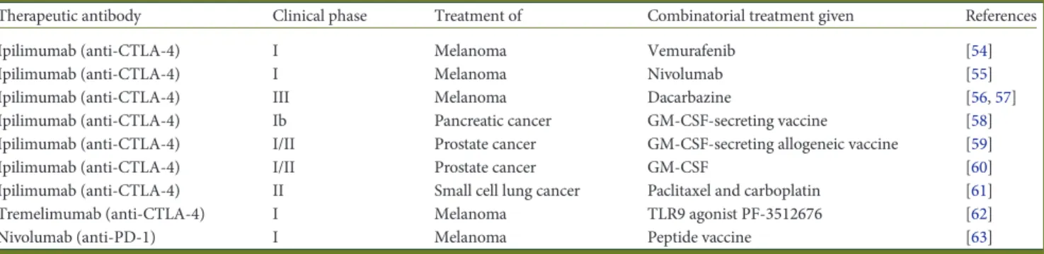
Table 1. Published studies having investigated the effect of combination therapies with checkpoint inhibitors
Therapeutic antibody Clinical phase Treatment of Combinatorial treatment given References
Ipilimumab (anti-CTLA-4) I Melanoma Vemurafenib [54]
Ipilimumab (anti-CTLA-4) I Melanoma Nivolumab [55]
Ipilimumab (anti-CTLA-4) III Melanoma Dacarbazine [56,57]
Ipilimumab (anti-CTLA-4) Ib Pancreatic cancer GM-CSF-secreting vaccine [58]
Ipilimumab (anti-CTLA-4) I/II Prostate cancer GM-CSF-secreting allogeneic vaccine [59]
Ipilimumab (anti-CTLA-4) I/II Prostate cancer GM-CSF [60]
Ipilimumab (anti-CTLA-4) II Small cell lung cancer Paclitaxel and carboplatin [61]
Tremelimumab (anti-CTLA-4) I Melanoma TLR9 agonist PF-3512676 [62]
Nivolumab (anti-PD-1) I Melanoma Peptide vaccine [63]
CD8 T cells induced by vaccination [
66
,
67
], evaluating the
clinic-al ef
ficacy of anti-Tim-3 antibodies in a clinical setting will be of
high interest. Other therapies targeting immune checkpoints are
currently in development such as agonist antibodies targeting
molecules which activate T cells such as CD137 (BMS-663513)
[
68
], OX40 (MEDI6383) NCT02221960, CD40 (CP870,893) [
69
]
or GITR (TRAX518) NCT01239134 as well as drugs favoring DC
activation such as LAG3-Fusion protein (IMP321) [
70
].
One of the challenging problems with the use of checkpoint
inhibitors is the management of autoimmune side effects called
immune-related adverse events (irAEs) (for detailed review see
[
71
,
72
]. Mild to severe irAEs are observed with ipilimumab in
about 60% of patients and in about 15% with anti-PD-1 drugs.
irAEs include dermatologic, gastrointestinal, hepatic, endocrine,
and other less common in
flammatory events. IrAEs are believed
to arise from general immunologic enhancement, and
tempor-ary immunosuppression with corticosteroids, tumor necrosis
factor-alpha antagonists, mycophenolate mofetil or other agents
can be an effective treatment in most cases. Interestingly, an
as-sociation between irAEs and clinical outcome was observed for
anti-CTLA-4 therapy [
73
]. The recent approval of anti-CTLA-4
and anti-PD1 mAbs in the clinic opens a new
field in cancer
im-munotherapy. The discovery that anti-PD-1 mAb treatment
could be effective in many types of cancers like melanoma, renal
carcinoma, lung cancer, bladder cancer gastric cancer
under-scores the possible development of immune checkpoint
inhibi-tors in many clinical contexts of solid tumors.
In addition to antibodies targeting checkpoint inhibitors,
bis-pecific antibodies are being developed. These antibodies are
artifi-cial proteins that are composed of fragments of two different
mAbs and consequently bind to two different types of antigens.
This approach is used for cancer immunotherapy, where these
proteins simultaneously bind to cytotoxic T cells using CD3
and a tumor cell target. Two drugs are currently available.
Catumaxomab consists of one heavy chain and one light chain
of an anti-EpCAM antibody and one heavy chain and one light
chain of an anti-CD3 antibody as a consequence of which the
chi-meric protein can bind both EpCAM and CD3. In addition, the
Fc-region can bind to an Fc receptor on accessory cells like other
antibodies, which has led to calling the drug a trifunctional
anti-body. This structure allows both T cell and macrophage or DC
ac-tivation to favor adaptive immune response and tumor cell lysis
by immune effectors. This drug could be used to treat patients
with ascites with EpCam + tumor cells. Blinatumomab is a
bi-specific T-cell engager that combines two binding sites: a CD3 site
for T cells and a CD19 site to target B cells. The drug works by
linking these two cell types and activating the T cell to exert
cyto-toxic activity on the target cells. Blinatumomab could be used to
target malignant B-cell lymphoma/leukemia and make
blinatu-momab a potential therapeutic option for pediatric and adult
B-cell lymphoma or acute B-cell lymphoblastic leukemia.
rationale to combine conventional
cancer treatments with immunotherapy
combining radiotherapy with immunotherapy
Accumulating data identifying molecular changes in the tumor
microenvironment induced by tumor irradiation have recently
contributed to better understand the contribution of the
immune system in the response of the irradiated tumor [
74
–
76
]
(and reviewed in [
77
] and [
78
]). Tumor irradiation can induce
the priming of immune response after induction of ICD [
15
,
79
,
80
], which could explain the observation of regression of
unirra-diated distant tumor sites (the so-called abscopal effect) [
81
,
82
].
In addition, irradiation of tumor cells contributes to the effector
phase by inducing expression of numerous molecules (MHC I,
NKG2D ligands, death receptors, adhesion molecules) able to
activate effector immune cells [
83
–
87
]. Thus, combining
radi-ation with immunotherapy appears to provide an optimal
thera-peutic partnership to achieve immune-mediated systemic tumor
control [
88
]. In preclinical models, tumor irradiation induces
Fas upregulation by tumor cells, thereby enhancing Fas-dependent
CTL killing [
89
], and the effectiveness of cancer vaccines [
87
,
90
,
91
]. This was often accompanied by important tumor influx
by CD8+ and/or abscopal effect [
90
]. Similarly, upregulation of
MHC class I molecules by tumor cells following irradiation
enhance the anti-tumor effect of adoptive cell therapy (ACT)
[
92
,
93
]. Moreover, combining mAbs targeting important
immune checkpoints (CD137, CD40, PD-1, CTLA-4) with tumor
irradiation has shown promising synergistic activity [
94
–
96
]. In
humans, localized radiotherapy combined with
immunothera-peutic interventions has been shown to increase tumor-specific
T-cell number, and encouraging clinical results have been
reported in patients with hepatocellular carcinoma or prostate
cancer [
97
–
99
]. Clinical trials combining radiotherapy with
imi-quimod (a TLR7 agonist) (NCT01421017), fresolimumab (a mAb
that neutralizes TGF-β) (NCT01401062) or ipilimumab (a mAb
directed against CTLA-4) (NCT01689974) are actually ongoing,
paving the way for the use of radiation as a partner for
immuno-therapy (Table
2
).
combining chemotherapy with immunotherapy
Because most chemotherapeutic agents were regarded as
immu-nosuppressants, combinations between immunotherapy and
chemotherapy were long considered as incompatible. However,
the emergence of the concept of ICD (discussed above), the
observations that some chemotherapies such as
cyclophospha-mide and 5-fluorouracil can eliminate regulatory immune cell
subsets (reviewed in [
111
,
112
]) and some clinical trials results
showing that patients treated
first with immunotherapy,
fol-lowed by chemotherapy demonstrated better clinical outcomes
than patients that have received chemotherapy alone [
113
,
114
],
have prompted scientists and physicians to reassess the potential
of combination therapies between chemotherapy and
immuno-therapy. Subsequent preclinical and clinical investigations have
revealed that chemotherapy could enhance the efficacy of
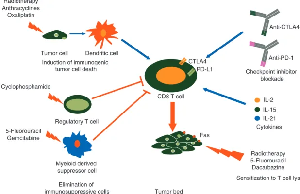
immuno-therapy through various mechanisms (Table
3
and Figure
1
).
Chemotherapy can not only improve anti-tumor effects of
im-munotherapy by overcoming parts of immunosuppression, but
also by enhancing cross-presentation of tumor antigens and by
supporting better penetration of immune cells in tumor core
(Table
3
and Figure
1
).
combining targeted therapies with immunotherapy
small molecules. Among targeted therapies, number of tyrosine
kinase inhibitors (TKIs), proteasome inhibitors or mTOR
Table 2. Non-exhaustive list of ongoing clinical trials investigating the effect of combination therapies (also reviewed in [100–110])
Therapeutic agent Cancer Clinical
phase
Combined with Reference
Imiquimod Breast cancer with skin metastases II Radiotherapy + cyclophosphamide NCT01421017
Fresolimumab Metastatic breast cancer I/II Radiotherapy NCT01401062
Ipilimumab Metastatic melanoma II Radiotherapy NCT01689974
Multipeptide cancer vaccine
Advanced/mRCC III Sunitinib NCT01265901
Peptide-based vaccine Advanced stage HER2-overexpressing breast cancer
I/II Adoptive T-cell therapy NCT00791037
Trastuzumab High-risk/metastatic HER-2/Neu-overexpressing breast cancer with no evidence of disease
II Cyclophosphamide, and an allogeneic GM-CSF-secreting breast tumor vaccine
NCT00847171
Trastuzumab Metastatic breast cancer overexpressing HER-2/NEU
II DC vaccine given with and vinorelbine NCT00266110
Trastuzumab HER2 positive breast cancer II Peptide-based vaccine NCT00343109
Nivolumab or ipilimumab with nivolumab
Resected stages IIIC/ IV melanoma I Vaccine combining multiple class I peptides and montanide ISA 51VG
NCT01176474
Anti-PD-1 antibody BMS-936558
Unresectable stages III/IV melanoma I Multiple class I peptides and montanide ISA 51 VG
NCT01176461
MPDL3280A Metastatic melanoma Ib Vemurafenib and cobimetinib NCT01656642
Ipilimumab Cervical carcinoma I Combined with cisplatin and radiation therapy NCT01711515
Ipilimumab CrC lymphoma melanoma I/II Combined with radiation therapy NCT01769222
Ipilimumab Head and neck cancer I Combined with cetuximab and radiation therapy NCT01860430
Ipilimumab Head and neck cancer I Combined with cetuximab and radiation therapy NCT01935921
Ipilimumab Hodgkin’s lymphoma I Combined with brentuximab vedotin NCT01896999
Ipilimumab Leukemia lymphoma n.a. Combined with lenalidomide NCT01919619
Ipilimumab Lymphoma I Combined with rituximab NCT01729806
Ipilimumab Melanoma n.a. Followed by lymphodepletion, TIL infusion
and IL-2
NCT01701674
Ipilimumab Melanoma 0 Combined with radioembolization NCT01730157
Ipilimumab Melanoma I Combined with SrS or wBrT NCT01703507
Ipilimumab Melanoma I Combined with dabrafenib ± trametinib NCT01767454
Ipilimumab Melanoma I Combined with Ny-eSO-1-targeting
vaccine ± montanide
NCT01810016
Ipilimumab Melanoma I Combined with BCG NCT01838200
Ipilimumab Melanoma I/II As single agent or combined with oncolytic
virotherapy
NCT01740297
Ipilimumab Melanoma II Combined with cyclophosphamide NCT01740401
Ipilimumab Melanoma II As single agent or combined with IFNα-2b NCT01708941
Ipilimumab Melanoma II Combined with nivolumab NCT01783938
Ipilimumab Melanoma II Combined with paclitaxel NCT01827111
Ipilimumab Melanoma III Combined with nivolumab NCT01844505
Ipilimumab Melanoma III Combined with lambrolizumab NCT01866319
Ipilimumab Melanoma Iv Combined with high-dose IL-2 NCT01856023
Ipilimumab NSCLC I Combined with carboplatin, cisplatin and
paclitaxel
NCT01820754
Ipilimumab Pancreatic cancer II As single agent or combined with a
GM-CSF-secreting vaccine
NCT01896869
Ipilimumab Prostate cancer I Combined with sipuleucel-T NCT01832870
Ipilimumab Prostate cancer II After sipuleucel-T treatment NCT01804465
Ipilimumab Advanced solid tumors I Combined with imatinib NCT01738139
Ipilimumab Advanced solid tumors I Combined with lirilumab NCT01750580
Ipilimumab Advanced solid tumors I Combined with lenalidomide NCT01750983
Ipilimumab Advanced solid tumors I/II Combined with nivolumab NCT01928394
Lambrolizumab Melanoma II Combined with ipilimumab NCT01866319
Lambrolizumab Advanced solid tumors II/III As single agent or combined with conventional chemotherapy
NCT01840579
Lirilumab Advanced solid tumors I Combined with nivolumab NCT01714739
Continued
inhibitors have been shown to in
fluence immune response
against cancer cells, mostly by affecting T-cell or DC functions
[
133
–
139
], but also by depleting Tregs or myeloid derived
suppressor cells (MDSCs) as discussed above [
140
–
142
]. Thus, a
randomized phase III clinical trial is presently testing IMA901, a
multipeptide cancer vaccine ( preceded by a single low dose of
cyclophosphamide), in combination with sunitinib in
first-line
metastatic renal cell carcinoma (mRCC; NCT01265901). This
constitutes one of the examples of this new strategy of
chemo-immunotherapy combining targeted therapy, chemo-immunotherapy
and immunogenic chemotherapy schedule. TKIs may disrupt
signal transducer and activator of transcription (STAT) signaling
pathways, thus potentially decreasing immunosuppression by
Tregs, MDSCs or DCs, making combinations with mAbs
blocking immune checkpoints also quite attractive [
115
,
143
,
144
].
STAT activation can also control the expression of several
immunosuppressive molecules (like PD-L1), providing further
rationale for combinations [
145
]. Numerous clinical trials are
actually testing anti-PD-1/PD-L1 mAbs with TKIs, especially in
mRCC patients, with encouraging preliminary results [
146
]. Of
note, emerging data demonstrate that the normalization of
tumor neovasculature by anti-angiogenic agents could improve
endogenous and vaccination-induced anti-tumor immune
responses [
147
–
149
].
tumor-targeting mAbs. Contribution of the immune response,
especially through antibody-dependent-cellular cytotoxicity, has
been demonstrated for the clinical efficacy of therapeutic mAbs,
like rituximab [
150
], cetuximab [
151
] and trastuzumab [
152
].
Preclinical studies also have shown that trastuzumab is able to
stimulate adaptative anti-tumor immunity [
153
,
154
] and that
combination of trastuzumab with anti-PD-1 and anti-CD137
can synergize [
154
,
155
]. Some of these studies also suggest that
the synergy between anthracyclines and trastuzumab could be
Table 2. Continued
Therapeutic agent Cancer Clinical
phase
Combined with Reference
Lirilumab Advanced solid tumors I Combined with ipilimumab NCT01750580
MeDI4736 Advanced solid tumors I Combined with tremelimumab NCT01975831
Nivolumab Melanoma II Combined with ipilimumab NCT01783938
Nivolumab Melanoma III Combined with ipilimumab NCT01927419
Nivolumab Melanoma III Combined with ipilimumab NCT01844505
Nivolumab NSCLC II Combined with azacitidine ± entinostat NCT01928576
Nivolumab Advanced solid tumors I Combined with lirilumab NCT01714739
Nivolumab Advanced solid tumors II As single agent or combined with
immunotherapy
NCT01968109
Nivolumab Advanced solid tumors I/II As single agent or combined with ipilimumab NCT01928394
Tremelimumab Hepatocellular carcinoma I Combined with rFa and TaCe NCT01853618
Tremelimumab Advanced solid tumors I Combined with MeDI4736 NCT01975831
Urelumab Non-Hodgkin’s lymphoma, chronic lymphocytic leukemia
I Combined with rituximab NCT01775631
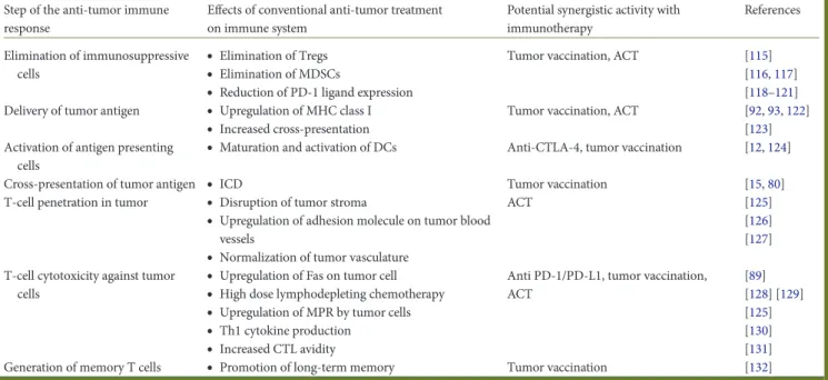
Table 3. Main mechanisms by which conventional anticancer therapies could synergize with immunotherapy
Step of the anti-tumor immune response
Effects of conventional anti-tumor treatment on immune system
Potential synergistic activity with immunotherapy References Elimination of immunosuppressive cells
•
Elimination of Tregs•
Elimination of MDSCs•
Reduction of PD-1 ligand expressionTumor vaccination, ACT [115]
[116,117] [118–121] Delivery of tumor antigen
•
Upregulation of MHC class I•
Increased cross-presentationTumor vaccination, ACT [92,93,122] [123] Activation of antigen presenting
cells
•
Maturation and activation of DCs Anti-CTLA-4, tumor vaccination [12,124]Cross-presentation of tumor antigen
•
ICD Tumor vaccination [15,80]T-cell penetration in tumor
•
Disruption of tumor stroma•
Upregulation of adhesion molecule on tumor blood vessels•
Normalization of tumor vasculatureACT [125]
[126] [127] T-cell cytotoxicity against tumor
cells
•
Upregulation of Fas on tumor cell•
High dose lymphodepleting chemotherapy•
Upregulation of MPR by tumor cells•
Th1 cytokine production•
Increased CTL avidityAnti PD-1/PD-L1, tumor vaccination, ACT [89] [128] [129] [125] [130] [131]
Generation of memory T cells
•
Promotion of long-term memory Tumor vaccination [132]explained in part by increased anti-tumor immune response
[
154
]. Combinatorial approaches of tumor vaccines with passive
immunotherapy have been developed in HER2-overexpressing
breast cancer. In metastatic breast cancer patients previously
treated with trastuzumab, association of HER2-based peptide
vaccine and trastuzumab resulted in boosting and prolongation
of T-cell response against HER2, with an estimated progression
free survival of 33% at 3 years [
156
]. Preliminary results on the
clinical ef
ficacy of this combination [
157
–
160
], and its superiority
compared with vaccination alone [
157
], have also been reported
by other groups. Ongoing clinical trials incorporating
immu-nization with trastuzumab, with or without chemotherapy, are
currently ongoing (NCT00791037, NCT00847171, NCT00266
110, NCT00343109) [
161
].
immune checkpoint inhibitors. Preclinical, but also recent
clinical evidences suggest that mAbs targeting inhibitory immune
checkpoints can be used in combination. Concurrent PD-1
blockade with mAb blocking CTLA-4, LAG3 or TIM-3 has
shown preclinical signs for anti-tumor synergy without signi
ficant
toxicity [
65
,
162
,
163
]. A recent study tested the combination of
nivolumab (anti-PD-1 mAb) with ipilimumab (anti-CTLA-4
mAb) in patients with advanced melanoma, at a concomitant or
sequential schedule (ipilimumab followed by nivolumab). A total
of 53 patients received concurrent treatment. The objective
–
response rate for all patients in the concurrent group was 40%,
and at the maximum doses that were associated with an
acceptable level of severe adverse events (nivolumab at 1 mg/kg
and ipilimumab at 3 mg/kg) 53% of patients had an objective
response, all with tumor reduction of 80% or more. However,
grade 3 or 4 adverse events occurred in over half of the patients
but were generally reversible [
55
]. Checkpoint blockade has also
been combined with standard doses and regimens of cytotoxic
chemotherapy. Signs of potential ef
ficacy have been reported
with a combination of ipilimumab with sequential chemotherapy
in non-small cell lung cancer (NSCLC) patients [
164
], or in
metastatic melanoma [
165
]. A recent clinical study conducted in 30
metastatic breast cancer patients showed that the combination of
the immune checkpoint modulator IMP321 (recombinant soluble
LAG3/Ig fusion), preceded by standard dose weekly paclitaxel is
feasible, and is followed by objective response rates of 50%, and
immune activation of NK cells, as well as durable effector memory
CD8+ T-cell responses [
166
]. Moreover, synergy of anti-CTLA-4
associated with chemotherapy and radiotherapy has recently been
reported [
167
], and the combination of mAbs blocking
PD-1/PD-L1 with therapeutic vaccines or targeted anticancer agents (BRAF
inhibitor vemurafenib) is actually being explored in melanoma
(NCT01176474 and NCT01176461) and advanced metastatic
cancer patients (NCT01656642), respectively (Table
2
).
conclusions
For immunologists, it has become clear that immunotherapeutic
strategies should engage multiple effector mechanisms to overcome
the immunosuppressive mechanisms of cancer. For patients
with metastatic cancer or larger burden of disease, a single
therapeutic agent is unlikely to be effective, and immunotherapy
should be combined with conventional cancer treatments, with
IL-2 IL-15 IL-21 PD-L1 Anti-PD-1 Anti-CTLA4 CTLA4 Fas CD8 T cell Tumor bed Checkpoint inhibitor blockade Cytokines
Sensitization to T cell lysis Radiotherapy 5-Fluorouracil Dacarbazine Induction of immunogenic
tumor cell death
Regulatory T cell
Myeloid derived suppressor cell
Elimination of immunosuppressive cells Tumor cell Dendritic cell Radiotherapy Anthracyclines Oxaliplatin Cyclophosphamide 5-Fluorouracil Gemcitabine
Figure 1. Molecular bases for the rationale to combine immunotherapy with anticancer agents. Conventional chemotherapeutic agents can (1) induce an immunogenic form of tumor cell death, (2) eliminate immunosuppressive cells and (3) sensitize tumor cells to immune effector cells. Immunostimulatory cytokines and checkpoint inhibitor blockers promote CD8 T-cell activation and prevent their subsequent dysfunction in the tumor microenvironment. Combination therapies thus not only target tumor cells, but also enhance CD8 T-cell activation, ultimately resulting in enhanced anticancer effects.
the aim of not only to reduce tumor load, but also to abrogate
immune tolerance and to enhance anticancer immune responses
(Table
3
and Figure
1
). This opens a
field for fundamental and
clinical research to better develop the concept of
chemoimmu-notherapy, especially to design the optimal choice, schedule and
dose of therapeutic associations. As the success of these combined
approaches may rely on the crosstalk between cancer cells, tumor
stroma and the patient’s immune system, three major
considera-tions appear crucial for implementation of
chemoimmunother-apy ef
ficient combinations. First, cancer-bearing patients who
may bene
fit from such combinations must be properly selected
using appropriate biomarkers, which underscores the crucial
need for predictive biomarkers that can be introduced in the
clin-ical routine. Second, the optimal choice of combinations along
with the schedule and dosage of administration of each
compo-nent of the chemoimmunotherapy treatment remain to be
deter-mined, which highlights the need to develop clear immune
biological and clinical parameters that allow for rapid go/no-go
decisions. Finally, it is important to keep in mind that cancer
patients are often heavily co-medicated with a number of drugs
for symptom management as well as over-the-counter products
and supplements, which can also affect the immune system.
Future research and clinical trials that will rationally sequence
immunomodulators, cancer vaccines and conventional
treat-ments of cancer will benefit from taking into account these
im-portant aspects.
funding
The authors were supported by grants from the Ligue Nationale
contre le Cancer (F.G.), the Institut National du Cancer (F.G.),
the Association pour la recherche sur le cancer (F.G. and L.A.),
the Conseil Régional de Bourgogne (F.G. and L.A.), the FEDER
(F.G. and L.A.), the Fondation de France (L.A.), the Agence
Nationale de la Recherche [ANR-13-JSV3-0001-01] (L.A.) and
[ANR-11-LABX-0021], the Cancéropôle du Grand-Est (L.A.),
the ARSEP foundation (L.A.), the Ligue Régionale contre le
cancer Comité Grand-Est (L.A.) and the European Community
(Marie Curie Fellowship PCIG10-GA-2011-303719) (L.A.).
disclosure
The authors have declared no conflicts of interest.
references
1. Dunn GP, Old LJ, Schreiber RD. The three Es of cancer immunoediting. Annu Rev Immunol 2004; 22: 329–360.
2. Wimmer CD, Angele MK, Schwarz B et al. Impact of cyclosporine versus tacrolimus on the incidence of de novo malignancy following liver transplantation: a single center experience with 609 patients. Transpl Int 2013; 26: 999–1006. 3. Robbins HA, Pfeiffer RM, Shiels MS et al. Excess Cancers Among HIV-Infected
People in the United States. J Natl Cancer Inst 2015; 107.
4. Shin M, Moon HH, Kim JM et al. Comparison of the incidence of de novo malignancy in liver or kidney transplant recipients: analysis of 2673 consecutive cases in a single center. Transplant Proc 2013; 45: 3019–3023.
5. Salavoura K, Kolialexi A, Tsangaris G, Mavrou A. Development of cancer in patients with primary immunodeficiencies. Anticancer Res 2008; 28: 1263–1269. 6. Fridman WH, Pages F, Sautes-Fridman C, Galon J. The immune contexture in
human tumours: impact on clinical outcome. Nat Rev Cancer 2012; 12: 298–306.
7. Zhang L, Conejo-Garcia JR, Katsaros D et al. Intratumoral T cells, recurrence, and survival in epithelial ovarian cancer. N Engl J Med 2003; 348: 203–213. 8. Apetoh L, Smyth MJ, Drake CG et al. Consensus nomenclature for CD8 T cell
phenotypes in cancer. Oncoimmunology 2015; 4, doi: 10.1080/2162402X.2014. 998538.
9. Andersen MH, Sorensen RB, Schrama D et al. Cancer treatment: the combination of vaccination with other therapies. Cancer Immunol Immunother 2008; 57: 1735–1743.
10. Menard C, Martin F, Apetoh L et al. Cancer chemotherapy: not only a direct cytotoxic effect, but also an adjuvant for antitumor immunity. Cancer Immunol Immunother 2008; 57: 1579–1587.
11. Zitvogel L, Apetoh L, Ghiringhelli F, Kroemer G. Immunological aspects of cancer chemotherapy. Nat Rev Immunol 2008; 8: 59–73.
12. Yu B, Kusmartsev S, Cheng F et al. Effective combination of chemotherapy and dendritic cell administration for the treatment of advanced-stage experimental breast cancer. Clin Cancer Res 2003; 9: 285–294.
13. Weiner HL, Cohen JA. Treatment of multiple sclerosis with cyclophosphamide: critical review of clinical and immunologic effects. Mult Scler 2002; 8: 142–154. 14. Javeed A, Ashraf M, Riaz A et al. Paclitaxel and immune system. Eur J Pharm Sci
2009; 38: 283–290.
15. Apetoh L, Ghiringhelli F, Tesniere A et al. Toll-like receptor 4-dependent contribution of the immune system to anticancer chemotherapy and radiotherapy. Nat Med 2007; 13: 1050–1059.
16. Ghiringhelli F, Apetoh L, Tesniere A et al. Activation of the NLRP3 inflammasome in dendritic cells induces IL-1beta-dependent adaptive immunity against tumors. Nat Med 2009; 15: 1170–1178.
17. Tesniere A, Schlemmer F, Boige V et al. Immunogenic death of colon cancer cells treated with oxaliplatin. Oncogene 2010; 29: 482–491.
18. Lake RA, Robinson BW. Immunotherapy and chemotherapy—a practical partnership. Nat Rev Cancer 2005; 5: 397–405.
19. Melero I, Grimaldi AM, Perez-Gracia JL, Ascierto PA. Clinical development of immunostimulatory monoclonal antibodies and opportunities for combination. Clin Cancer Res 2013; 19: 997–1008.
20. Kalos M, June CH. Adoptive T cell transfer for cancer immunotherapy in the era of synthetic biology. Immunity 2013; 39: 49–60.
21. Banchereau J, Steinman RM. Dendritic cells and the control of immunity. Nature 1998; 392: 245–252.
22. Kirkwood JM, Butterfield LH, Tarhini AA et al. Immunotherapy of cancer in 2012. CA Cancer J Clin 2012; 62: 309–335.
23. Schadendorf D, Ugurel S, Schuler-Thurner B et al. Dacarbazine (DTIC) versus vaccination with autologous peptide-pulsed dendritic cells (DC) in first-line treatment of patients with metastatic melanoma: a randomized phase III trial of the DC study group of the DeCOG. Ann Oncol 2006; 17: 563–570.
24. Wimmers F, Schreibelt G, Skold AE et al. Paradigm shift in dendritic cell-based immunotherapy: from in vitro generated monocyte-derived DCs to naturally circulating DC subsets. Front Immunol 2014; 5: 165.
25. Carreno BM, Magrini V, Becker-Hapak M et al. A dendritic cell vaccine increases the breadth and diversity of melanoma neoantigen-specific T cells. Science 2015; 348: 803–808.
26. Mitchison NA. Studies on the immunological response to foreign tumor transplants in the mouse. I. The role of lymph node cells in conferring immunity by adoptive transfer. J Exp Med 1955; 102: 157–177.
27. Dudley ME, Wunderlich JR, Robbins PF et al. Cancer regression and autoimmunity in patients after clonal repopulation with antitumor lymphocytes. Science 2002; 298: 850–854.
28. Kershaw MH, Westwood JA, Parker LL et al. A phase I study on adoptive immunotherapy using gene-modified T cells for ovarian cancer. Clin Cancer Res 2006; 12: 6106–6115.
29. Lamers CH, Sleijfer S, Vulto AG et al. Treatment of metastatic renal cell carcinoma with autologous T-lymphocytes genetically retargeted against carbonic anhydrase IX: first clinical experience. J Clin Oncol 2006; 24: e20–e22.
30. Kalos M, Levine BL, Porter DL et al. T cells with chimeric antigen receptors have potent antitumor effects and can establish memory in patients with advanced leukemia. Sci Transl Med 2011; 3: 95ra73.
31. Porter DL, Levine BL, Kalos M et al. Chimeric antigen receptor-modified T cells in chronic lymphoid leukemia. N Engl J Med 2011; 365: 725–733.
32. Maude SL, Frey N, Shaw PA et al. Chimeric antigen receptor T cells for sustained remissions in leukemia. N Engl J Med 2014; 371: 1507–1517.
33. Perna SK, De Angelis B, Pagliara D et al. Interleukin 15 provides relief to CTLs from regulatory T cell-mediated inhibition: implications for adoptive T cell-based therapies for lymphoma. Clin Cancer Res 2013; 19: 106–117.
34. Klebanoff CA, Finkelstein SE, Surman DR et al. IL-15 enhances the in vivo antitumor activity of tumor-reactive CD8+ T cells. Proc Natl Acad Sci USA 2004; 101: 1969–1974.
35. Hoyos V, Savoldo B, Quintarelli C et al. Engineering CD19-specific T lymphocytes with interleukin-15 and a suicide gene to enhance their anti-lymphoma/leukemia effects and safety. Leukemia 2010; 24: 1160–1170.
36. Conlon KC, Lugli E, Welles HC et al. Redistribution, hyperproliferation, activation of natural killer cells and CD8 T cells, and cytokine production during first-in-human clinical trial of recombinant first-in-human interleukin-15 in patients with cancer. J Clin Oncol 2015; 33: 74–82.
37. Wang G, Tschoi M, Spolski R et al. In vivo antitumor activity of interleukin 21 mediated by natural killer cells. Cancer Res 2003; 63: 9016–9022.
38. Zeng R, Spolski R, Finkelstein SE et al. Synergy of IL-21 and IL-15 in regulating CD8+ T cell expansion and function. J Exp Med 2005; 201: 139–148. 39. Hinrichs CS, Spolski R, Paulos CM et al. IL-2 and IL-21 confer opposing
differentiation programs to CD8+ T cells for adoptive immunotherapy. Blood 2008; 111: 5326–5333.
40. Singh H, Figliola MJ, Dawson MJ et al. Reprogramming CD19-specific T cells with IL-21 signaling can improve adoptive immunotherapy of B-lineage malignancies. Cancer Res 2011; 71: 3516–3527.
41. Vegran F, Berger H, Boidot R et al. The transcription factor IRF1 dictates the IL-21-dependent anticancer functions of TH9 cells. Nat Immunol 2014; 15: 758–766.
42. Petrella TM, Tozer R, Belanger K et al. Interleukin-21 has activity in patients with metastatic melanoma: a phase II study. J Clin Oncol 2012; 30: 3396–3401. 43. Shankaran V, Ikeda H, Bruce AT et al. IFNgamma and lymphocytes prevent
primary tumour development and shape tumour immunogenicity. Nature 2001; 410: 1107–1111.
44. Allison JP, Krummel MF. The Yin and Yang of T cell costimulation. Science 1995; 270: 932–933.
45. Leach DR, Krummel MF, Allison JP. Enhancement of antitumor immunity by CTLA-4 blockade. Science 1996; 271: 1734–1736.
46. Hodi FS, Butler M, Oble DA et al. Immunologic and clinical effects of antibody blockade of cytotoxic T lymphocyte-associated antigen 4 in previously vaccinated cancer patients. Proc Natl Acad Sci USA 2008; 105: 3005–3010.
47. Hodi FS, O’Day SJ, McDermott DF et al. Improved survival with ipilimumab in patients with metastatic melanoma. N Engl J Med 2010; 363: 711–723. 48. Zha Y, Blank C, Gajewski TF. Negative regulation of T-cell function by PD-1. Crit
Rev Immunol 2004; 24: 229–237.
49. Latchman Y, Wood CR, Chernova T et al. PD-L2 is a second ligand for PD-1 and inhibits T cell activation. Nat Immunol 2001; 2: 261–268.
50. Freeman GJ, Long AJ, Iwai Y et al. Engagement of the PD-1 immunoinhibitory receptor by a novel B7 family member leads to negative regulation of lymphocyte activation. J Exp Med 2000; 192: 1027–1034.
51. Dong H, Strome SE, Salomao DR et al. Tumor-associated B7-H1 promotes T-cell apoptosis: a potential mechanism of immune evasion. Nat Med 2002; 8: 793–800.
52. Iwai Y, Ishida M, Tanaka Y et al. Involvement of PD-L1 on tumor cells in the escape from host immune system and tumor immunotherapy by PD-L1 blockade. Proc Natl Acad Sci USA 2002; 99: 12293–12297.
53. Tumeh PC, Harview CL, Yearley JH et al. PD-1 blockade induces responses by inhibiting adaptive immune resistance. Nature 2014; 515: 568–571. 54. Ribas A, Hodi FS, Callahan M et al. Hepatotoxicity with combination of
vemurafenib and ipilimumab. N Engl J Med 2013; 368: 1365–1366. 55. Wolchok JD, Kluger H, Callahan MK et al. Nivolumab plus ipilimumab in
advanced melanoma. N Engl J Med 2013; 369: 122–133.
56. Robert C, Thomas L, Bondarenko I et al. Ipilimumab plus dacarbazine for previously untreated metastatic melanoma. N Engl J Med 2011; 364: 2517–2526.
57. Sherrill B, Wang J, Kotapati S, Chin K. Q-TWiST analysis comparing ipilimumab/ dacarbazine vs placebo/dacarbazine for patients with stage III/IV melanoma. Br J Cancer 2013; 109: 8–13.
58. Le DT, Lutz E, Uram JN et al. Evaluation of ipilimumab in combination with allogeneic pancreatic tumor cells transfected with a GM-CSF gene in previously treated pancreatic cancer. J Immunother 2013; 36: 382–389.
59. Santegoets SJ, Stam AG, Lougheed SM et al. T cell profiling reveals high CD4+ CTLA-4 + T cell frequency as dominant predictor for survival after prostate GVAX/ipilimumab treatment. Cancer Immunol Immunother 2013; 62: 245–256.
60. Kwek SS, Dao V, Roy R et al. Diversity of antigen-specific responses induced in vivo with CTLA-4 blockade in prostate cancer patients. J Immunol 2012; 189: 3759–3766.
61. Reck M, Bondarenko I, Luft A et al. Ipilimumab in combination with paclitaxel and carboplatin as first-line therapy in extensive-disease-small-cell lung cancer: results from a randomized, double-blind, multicenter phase 2 trial. Ann Oncol 2013; 24: 75–83.
62. Millward M, Underhill C, Lobb S et al. Phase I study of tremelimumab (CP-675 206) plus PF-3512676 (CPG 7909) in patients with melanoma or advanced solid tumours. Br J Cancer 2013; 108: 1998–2004.
63. Weber JS, Kudchadkar RR, Yu B et al. Safety, efficacy, and biomarkers of nivolumab with vaccine in ipilimumab-refractory or -naive melanoma. J Clin Oncol 2013; 31: 4311–4318.
64. Zhu C, Anderson AC, Schubart A et al. The Tim-3 ligand galectin-9 negatively regulates T helper type 1 immunity. Nat Immunol 2005; 6: 1245–1252. 65. Sakuishi K, Apetoh L, Sullivan JM et al. Targeting Tim-3 and PD-1 pathways to
reverse T cell exhaustion and restore anti-tumor immunity. J Exp Med 2010; 207: 2187–2194.
66. Fourcade J, Sun Z, Benallaoua M et al. Upregulation of Tim-3 and PD-1 expression is associated with tumor antigen-specific CD8+ T cell dysfunction in melanoma patients. J Exp Med 2010; 207: 2175–2186.
67. Fourcade J, Sun Z, Pagliano O et al. PD-1 and Tim-3 regulate the expansion of tumor antigen-specific CD8(+) T cells induced by melanoma vaccines. Cancer Res 2014; 74: 1045–1055.
68. Li SY, Liu Y. Immunotherapy of melanoma with the immune costimulatory monoclonal antibodies targeting CD137. Clin Pharmacol 2013; 5: 47–53. 69. Vonderheide RH, Burg JM, Mick R et al. Phase I study of the CD40 agonist
antibody CP-870,893 combined with carboplatin and paclitaxel in patients with advanced solid tumors. Oncoimmunology 2013; 2: e23033.
70. Brignone C, Escudier B, Grygar C et al. A phase I pharmacokinetic and biological correlative study of IMP321, a novel MHC class II agonist, in patients with advanced renal cell carcinoma. Clin Cancer Res 2009; 15: 6225–6231. 71. Postow MA, Callahan MK, Wolchok JD. Immune Checkpoint Blockade in Cancer
Therapy. J Clin Oncol 2015.
72. Kong YC, Flynn JC. Opportunistic autoimmune disorders potentiated by immune-checkpoint inhibitors anti-CTLA-4 and anti-PD-1. Front Immunol 2014; 5: 206.
73. Downey SG, Klapper JA, Smith FO et al. Prognostic factors related to clinical response in patients with metastatic melanoma treated by CTL-associated antigen-4 blockade. Clin Cancer Res 2007; 13: 6681–6688.
74. Stone HB, Peters LJ, Milas L. Effect of host immune capability on radiocurability and subsequent transplantability of a murinefibrosarcoma. J Natl Cancer Inst 1979; 63: 1229–1235.
75. Lee Y, Auh SL, Wang Y et al. Therapeutic effects of ablative radiation on local tumor require CD8+ T cells: changing strategies for cancer treatment. Blood 2009; 114: 589–595.
76. Demaria S, Formenti SC. Role of T lymphocytes in tumor response to radiotherapy. Front Oncol 2012; 2: 95.
77. Formenti SC, Demaria S. Combining radiotherapy and cancer immunotherapy: a paradigm shift. J Natl Cancer Inst 2013; 105: 256–265.
78. Demaria S, Pilones KA, Vanpouille-Box C et al. The optimal partnership of radiation and immunotherapy: from preclinical studies to clinical translation. Radiat Res 2014; 182: 170–181.
79. Obeid M, Tesniere A, Ghiringhelli F et al. Calreticulin exposure dictates the immunogenicity of cancer cell death. Nat Med 2007; 13: 54–61.
80. Golden EB, Pellicciotta I, Demaria S et al. The convergence of radiation and immunogenic cell death signaling pathways. Front Oncol 2012; 2: 88. 81. Demaria S, Ng B, Devitt ML et al. Ionizing radiation inhibition of distant untreated
tumors (abscopal effect) is immune mediated. Int J Radiat Oncol Biol Phys 2004; 58: 862–870.
82. Postow MA, Callahan MK, Barker CA et al. Immunologic correlates of the abscopal effect in a patient with melanoma. N Engl J Med 2012; 366: 925–931. 83. Matsumura S, Wang B, Kawashima N et al. Radiation-induced CXCL16 release by breast cancer cells attracts effector T cells. J Immunol 2008; 181: 3099–3107.
84. Lugade AA, Sorensen EW, Gerber SA et al. Radiation-induced IFN-gamma production within the tumor microenvironment influences antitumor immunity. J Immunol 2008; 180: 3132–3139.
85. Reits EA, Hodge JW, Herberts CA et al. Radiation modulates the peptide repertoire, enhances MHC class I expression, and induces successful antitumor immunotherapy. J Exp Med 2006; 203: 1259–1271.
86. Ruocco MG, Pilones KA, Kawashima N et al. Suppressing T cell motility induced by anti-CTLA-4 monotherapy improves antitumor effects. J Clin Invest 2012; 122: 3718–3730.
87. Chakraborty M, Abrams SI, Coleman CN et al. External beam radiation of tumors alters phenotype of tumor cells to render them susceptible to vaccine-mediated T-cell killing. Cancer Res 2004; 64: 4328–4337.
88. Demaria S, Bhardwaj N, McBride WH, Formenti SC. Combining radiotherapy and immunotherapy: a revived partnership. Int J Radiat Oncol Biol Phys 2005; 63: 655–666.
89. Chakraborty M, Abrams SI, Camphausen K et al. Irradiation of tumor cells up-regulates Fas and enhances CTL lytic activity and CTL adoptive immunotherapy. J Immunol 2003; 170: 6338–6347.
90. Hodge JW, Sharp HJ, Gameiro SR. Abscopal regression of antigen disparate tumors by antigen cascade after systemic tumor vaccination in combination with local tumor radiation. Cancer Biother Radiopharm 2012; 27: 12–22.
91. Newcomb EW, Demaria S, Lukyanov Y et al. The combination of ionizing radiation and peripheral vaccination produces long-term survival of mice bearing established invasive GL261 gliomas. Clin Cancer Res 2006; 12: 4730–4737. 92. Medina-Echeverz J, Berraondo P. Colon cancer eradication after
chemoimmuno-therapy is associated with intratumoral emergence of proinflammatory myeloid cells. Oncoimmunology 2012; 1: 118–120.
93. Zhang B, Bowerman NA, Salama JK et al. Induced sensitization of tumor stroma leads to eradication of established cancer by T cells. J Exp Med 2007; 204: 49–55.
94. Demaria S, Kawashima N, Yang AM et al. Immune-mediated inhibition of metastases after treatment with local radiation and CTLA-4 blockade in a mouse model of breast cancer. Clin Cancer Res 2005; 11: 728–734.
95. Dewan MZ, Galloway AE, Kawashima N et al. Fractionated but not single-dose radiotherapy induces an immune-mediated abscopal effect when combined with anti-CTLA-4 antibody. Clin Cancer Res 2009; 15: 5379–5388.
96. Verbrugge I, Hagekyriakou J, Sharp LL et al. Radiotherapy increases the permissiveness of established mammary tumors to rejection by immunomodula-tory antibodies. Cancer Res 2012; 72: 3163–3174.
97. Chi KH, Liu SJ, Li CP et al. Combination of conformal radiotherapy and intratumoral injection of adoptive dendritic cell immunotherapy in refractory hepatoma. J Immunother 2005; 28: 129–135.
98. Lechleider RJ, Arlen PM, Tsang KY et al. Safety and immunologic response of a viral vaccine to prostate-specific antigen in combination with radiation therapy when metronomic-dose interleukin 2 is used as an adjuvant. Clin Cancer Res 2008; 14: 5284–5291.
99. Slovin SF, Higano CS, Hamid O et al. Ipilimumab alone or in combination with radiotherapy in metastatic castration-resistant prostate cancer: results from an open-label, multicenter phase I/II study. Ann Oncol 2013; 24: 1813–1821. 100. Vacchelli E, Eggermont A, Fridman WH et al. Trial watch: immunostimulatory
cytokines. Oncoimmunology 2013; 2: e24850.
101. Vacchelli E, Vitale I, Tartour E et al. Trial watch: anticancer radioimmunotherapy. Oncoimmunology 2013; 2: e25595.
102. Vacchelli E, Vitale I, Eggermont A et al. Trial watch: dendritic cell-based interventions for cancer therapy. Oncoimmunology 2013; 2: e25771.
103. Semeraro M, Vacchelli E, Eggermont A et al. Trial watch: lenalidomide-based immunochemotherapy. Oncoimmunology 2013; 2: e26494.
104. Aranda F, Vacchelli E, Eggermont A et al. Trial watch: peptide vaccines in cancer therapy. Oncoimmunology 2013; 2: e26621.
105. Vacchelli E, Aranda F, Eggermont A et al. Trial watch: tumor-targeting monoclonal antibodies in cancer therapy. Oncoimmunology 2014; 3: e27048. 106. Aranda F, Vacchelli E, Eggermont A et al. Trial watch: immunostimulatory
monoclonal antibodies in cancer therapy. Oncoimmunology 2014; 3: e27297. 107. Vacchelli E, Aranda F, Eggermont A et al. Trial watch: chemotherapy with
immunogenic cell death inducers. Oncoimmunology 2014; 3: e27878. 108. Aranda F, Vacchelli E, Obrist F et al. Trial watch: toll-like receptor agonists in
oncological indications. Oncoimmunology 2014; 3: e29179.
109. Vacchelli E, Aranda F, Obrist F et al. Trial watch: immunostimulatory cytokines in cancer therapy. Oncoimmunology 2014; 3: e29030.
110. Vacchelli E, Eggermont A, Sautes-Fridman C et al. Trial watch: oncolytic viruses for cancer therapy. Oncoimmunology 2013; 2: e24612.
111. Apetoh L, Vegran F, Ladoire S, Ghiringhelli F. Restoration of antitumor immunity through selective inhibition of myeloid derived suppressor cells by anticancer therapies. Curr Mol Med 2011; 11: 365–372.
112. Ghiringhelli F, Apetoh L. Enhancing the anticancer effects of 5-fluorouracil: current challenges and future perspectives. Biomed J 2014.
113. Schlom J, Arlen PM, Gulley JL. Cancer vaccines: moving beyond current paradigms. Clin Cancer Res 2007; 13: 3776–3782.
114. Wheeler CJ, Das A, Liu G et al. Clinical responsiveness of glioblastoma multiforme to chemotherapy after vaccination. Clin Cancer Res 2004; 10: 5316–5326.
115. Lesterhuis WJ, Punt CJ, Hato SV et al. Platinum-based drugs disrupt STAT6-mediated suppression of immune responses against cancer in humans and mice. J Clin Invest 2011; 121: 3100–3108.
116. Ghiringhelli F, Menard C, Puig PE et al. Metronomic cyclophosphamide regimen selectively depletes CD4+CD25+ regulatory T cells and restores T and NK effector functions in end stage cancer patients. Cancer Immunol Immunother 2007; 56: 641–648.
117. Rettig L, Seidenberg S, Parvanova I et al. Gemcitabine depletes regulatory T-cells in human and mice and enhances triggering of vaccine-specific cytotoxic T-cells. Int J Cancer 2011; 129: 832–838.
118. Bolitho P, Street SEA, Westwood JA et al. Perforin-mediated suppression of B-cell lymphoma. Proc Natl Acad Sci USA 2009; 106: 2723–2728.
119. Vincent J, Mignot G, Chalmin F et al. 5-Fluorouracil selectively kills tumor-associated myeloid-derived suppressor cells resulting in enhanced T cell-dependent antitumor immunity. Cancer Res 2010; 70: 3052–3061.
120. Ko HJ, Kim YJ, Kim YS et al. A combination of chemoimmunotherapies can efficiently break self-tolerance and induce antitumor immunity in a tolerogenic murine tumor model. Cancer Res 2007; 67: 7477–7486.
121. Suzuki E, Kapoor V, Jassar AS et al. Gemcitabine selectively eliminates splenic Gr-1+/CD11b+ myeloid suppressor cells in tumor-bearing animals and enhances antitumor immune activity. Clin Cancer Res 2005; 11: 6713–6721. 122. Serrano A, Tanzarella S, Lionello I et al. Rexpression of HLA class I antigens and
restoration of antigen-specific CTL response in melanoma cells following 5-aza-20-deoxycytidine treatment. Int J Cancer 2001; 94: 243–251.
123. Nowak AK, Lake RA, Marzo AL et al. Induction of tumor cell apoptosis in vivo increases tumor antigen presentation, priming rather than cross-tolerizing host tumor-specific CD8 T cells. J Immunol 2003; 170: 4905–4913. 124. Tong Y, Song W, Crystal RG. Combined intratumoral injection of bone
marrow-derived dendritic cells and systemic chemotherapy to treat pre-existing murine tumors. Cancer Res 2001; 61: 7530–7535.
125. Ramakrishnan R, Assudani D, Nagaraj S et al. Chemotherapy enhances tumor cell susceptibility to CTL-mediated killing during cancer immunotherapy in mice. J Clin Invest 2010; 120: 1111–1124.
126. Ganss R, Ryschich E, Klar E et al. Combination of T-cell therapy and trigger of inflammation induces remodeling of the vasculature and tumor eradication. Cancer Res 2002; 62: 1462–1470.
127. Shrimali RK, Yu Z, Theoret MR et al. Antiangiogenic agents can increase lymphocyte infiltration into tumor and enhance the effectiveness of adoptive immunotherapy of cancer. Cancer Res 2010; 70: 6171–6180.
![Table 2. Non-exhaustive list of ongoing clinical trials investigating the effect of combination therapies (also reviewed in [100–110])](https://thumb-eu.123doks.com/thumbv2/123doknet/14901854.654040/5.918.97.837.106.1090/table-exhaustive-ongoing-clinical-investigating-combination-therapies-reviewed.webp)