Extracellular proteases and their inhibitors in
genetic diseases of the central nervous system
Florence Molinari
1, Virginia Meskanaite
2, Arnold Munnich
1, Peter Sonderegger
2and
Laurence Colleaux
1,*
1
De´partement de Ge´ne´tique et Unite´ de Recherche sur les Handicaps Ge´ne´tiques de l’Enfant, INSERM U393, Hoˆpital Necker-Enfants Malades, 149 rue de Se`vres, 75743 Paris Cedex 15, France and 2Institute of
Biochemistry, University of Zurich, CH-8057 Zurich, Switzerland
Received June 27, 2003; Accepted August 7, 2003
Cumulative evidence has shown that a delicate balance between serine proteases and their inhibitors is crucial for normal functioning of several biological pathways. The importance of proteases and their inhibitors is well documented in several human diseases. Among them, the best documented are hemophilia B, a genetic deficiency of the serine protease coagulation factor IX and serpinophathies. Alpha-1-antitrypsin deficiency (MIM 107400), is associated with early-onset emphysema and liver disease, while hereditary angioedema (HANE; MIM 106100) is caused by mutations in the C1 inhibitor, a serpin involved in the regulation of the complement cascade. Recently, two human genetic diseases of the central nervous system have been related to mutations in components of extracellular proteolytic systems. Here, we review the recent advances in this field.
INTRODUCTION
Serine proteases is a group of enzymes that catalyzes hydrolysis of peptide bonds via a nucleophilic attack triggered by their active site serine. Serine proteases act sequence-specifically and are usually synthesized and secreted as inactive proenzymes (called zymogens) further activated by proteolysis. The best characterized members of this family play a role in intestinal digestion, blood coagulation and fibrinolysis (i.e. trypsin, thrombin or plasmin). In addition, several studies have pointed out the role of serine proteases in the central nervous system (CNS) (1). Indeed, during neural development, serine proteases contribute to cell migration, axon outgrowth and synapse elimination. In adult, they play a role in neuropeptide processing, regulation of neuronal survival and structural plasticity associated with learning and memory processes. Their mechanism of action probably involves degradation of extracellular matrix components, activation/inhibition of protease activated receptors (PARs) or activation/inhibition of other serine proteases. For instance, urokinase-plasminogen activator (uPA) and tissue type-plasminogen activator (tPA) convert plasminogen into active plasmin which in turn remodels synaptic connections via the degradation of an extracellular matrix component, laminin (2). In addition, tPA has been repor-ted to cleave N-methyl-D-aspartate (NMDA) receptors and, thereby, regulate the function of glutamatergic synapses (3).
Similarly, thrombin induces a proteolytic activation of PAR-1, resulting in the activation of protein kinase C, which reduces the voltage-dependent Mg2þblockade of NMDA channels. Finally, neuropsin, a serine protease exclusively expressed in the central nervous system, cleaves the Ig superfamily cell adhesion molecule L1, which is located in the presynaptic membrane and plays an important role in the maintenance of long-term potentiation (LTP) (4).
A DELICATE PROTEASE/ANTIPROTEASE BALANCE
Extracellular proteolysis by serine proteases often involves regulatory antiproteases called serpins (5–7). The serine protease inhibitors of the serpin family act by binding to the active site of their target protease(s) (8). This physiological inhibitory mechanism involves docking of the serpin to the target protease, cleavage of the reactive center loop, and rapid translocation of the protease to the opposite pole of the serpin (8). Protease nexin-1 (PN-1), the first identified neural serpin, is expressed in glia and neurons (9–11). It is a potent inhibitor of thrombin, but it can also inhibit tPA. Mice overexpressing PN-1 in neurons show increased LTP in the CA1 field of hippocampal slices and develop progressive disturbances of motor behavior and sensorimotor integration, while
PN-1-deficient mice have decreased hippocampal LTP (12,13). Both overexpression and lack of PN-1 cause epileptic activity in vivo and in vitro.
Neuroserpin, the second neural serpin, has been recognized as an axonally secreted member of the serpin superfamily (14). Searches for target proteases (15,16) revealed strong inhibition of tPA and uPA and to a somewhat lesser degree of plasmin, but no action against thrombin was noted. The half-life of neuroserpin/tPA complexes may be shorter than usually found in cognate protease–serpin pairs (17). This raises the question of whether tPA is really the cognate target protease of neuroserpin. Plasminogen activator inhibitor 1 (PAI-1), the cognate inhibitor of tPA in plasma, is not normally expressed in the brain, although it may be induced after exitotoxic injury (18). Therefore, it is possible that the cognate target of neuroserpin may be another, as yet unidentified, serine protease. Likewise, the activity of tPA activity in the brain might be controlled by an inhibitor other than neuroserpin.
NEUROTRYPSIN MUTATION IN AUTOSOMAL RECESSIVE MENTAL RETARDATION
Mental retardation (MR) is a frequent disorder affecting about 3% of the population (19). The severity of mental retardation is commonly classified on the basis of intelligence quotient (IQ) although other criteria have also been used. Assuming a population mean of 100 and a standard deviation of 15, MR is usually classified as ‘mild’ when the IQ ranges between 50 and 70 and as ‘severe’ when the IQ value is below 50 (20). The causes of MR are diverse and include environmental factors, teratogens, chromosomal anomalies and metabolic diseases impairing neuronal function. Genetically determined forms of severe MR average largely 25–50% (21,22). MRs of genetic origin include metabolic diseases impairing neuronal function in a non-specific manner and conditions which alter the normal patterning of the brain. However, the most common form of MR is not linked to metabolic disorders or abnormal brain development, nor any other clinical features and, therefore, is termed non-syndromic MR. An autosomal recessive mode of inheritance may account for nearly a quarter of mentally retarded individuals with non-syndromic MR. However, while 11 genes for non-syndromic X-linked MR have been found, none of the genes causing non-syndromic autosomal recessive MR had been identified until recently.
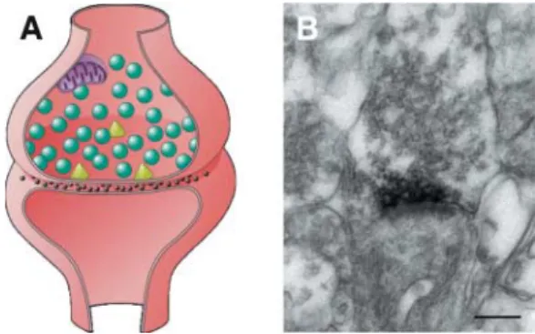
We analyzed a large consanguineous Algerian family, in which four out of eight children (three girls and one boy) were affected by a severe impairment of cognitive functions with an IQ below 50 (23). Because the parents were first-degree cousins, we suspected an autosomal-recessive pattern of inheritance. By means of a genome-wide screen with 400 microsatellite markers, we identified a single region of shared homozygosity on chromosome 4q24–q25 in the four affected individuals. A detailed sequence analysis of selected genes of this region revealed a 4 bp deletion in the neurotrypsin gene. Neurotrypsin is a neuronal serine protease predominantly expressed in neurons of the cerebral cortex, the hippocampus and the amygdala (24). By immuno-electronmicroscopy, neurotrypsin was localized in the presynaptic membrane and the presynaptic active zone of both asymmetrical (excitatory) and symmetrical (inhibitory) synapses (Fig. 1). In vitro studies
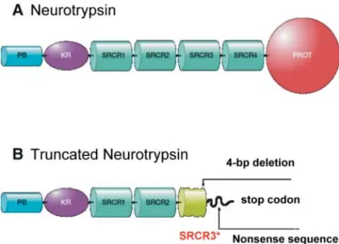
have demonstrated that it is a secreted protein which remains associated with the presynaptic membrane after its secretion. Neurotrypsin shows a unique domain composition (25). It consists of a proline-rich basic segment, one kringle domain, four scavenger receptor cysteine-rich (SRCR) domains, and a protease domain (Fig. 2). The 4 bp deletion, located in the 7th exon, is most likely a null allele as it is predicted to result in a shortened protein lacking the catalytic domain (Fig. 2). These findings, therefore, indicate neurotrypsin as the first gene to be identified as a cause of a nonsyndromic autosomal recessive form of MR.
A neurotrypsin defect does not appear to be a common cause of MR. We did not find any evidence for neurotrypsin mutations in individuals affected with MR in 17 non-syndromic inbread families. However, we found the same 4 bp deletion in another child born to first-cousin Algerian parents. The two families appear unrelated, but originate from the same area of Eastern Algeria. In both families, the mutation was carried on the background of the same haplotype across the neurotrypsin locus, suggesting a founder effect in the Algerian population.
The pathophysiological phenotype and the age of onset of the disease in affected individuals is consistent with the function of neurotrypsin as a regulator of adaptive synaptic functions, such as synapse reorganization during later stages of neurodevelop-ment and adult synaptic plasticity. In all affected children the course of the disease was similar. They reached the milestones of normal psychomotor development in the first 18 months. Signs of mental retardation were first observed by their parents when they were around 2 years of age. This suggests that neurotrypsin function is crucial in later stages of brain development. Normal cerebral MRI indicates the absence of gross neurodevelopmental abnormalities and a normal ratio between gray and white matter. This suggests that neurotrypsin Figure 1. Immuno-electronmicroscopic localization of neurotrypsin in the pre-synaptic active zone of a CNS synapse. (A) Schematic drawing of a CNS synapse. The presynaptic terminal contains numerous synaptic vesicles (green) containing neurotransmitter. Mitochondria deliver energy required for the various presynaptic processes, such as transmitter synthesis and transport, the exocytosis of synaptic vesicles, and their recycling by endocytosis.The triangles (yellow) indicate the presynaptic active zones, i.e. the sites where synaptic vesicles dock and release their content by fusion of their membrane with the presynaptic plasma membrane lining the synaptic cleft. (B) Electron-microscopic image of a CNS synapse showing the localization of neurotrypsin in the presynaptic membrane and the presynaptic active zone. Neurotrypsin was visualized by an affinity-purified antibody against the proteolytic domain, followed by a secondary antibody conjugated with peroxidase, in order to generate an electron-dense precipitate at the location of the antigen.
does not play a critical role in formation of synapses, but rather may be crucial for adaptive synaptic functions, such as those required to establish and/or maintain higher cognitive functions. Taken together, these results provide the first evidence for an association between cognitive impairment and defects in extracellular proteolytic activity at the synapse, opening a novel field in the pathophysiology of mental retardation. The recent generation of mice deficient in the catalytic domain of neurotrypsin in CNS neurons and of mice over-expressing neurotrypsin will provide further insight into the function of this protein.
MUTATIONS OF NEUROSERPIN IN FAMILIAL ENCEPHALOPATHY WITH NEUROSERPIN INCLUSION BODIES (FENIB)
Familial encephalopathy with neuroserpin inclusion bodies (FENIB, MIM 604218) is an autosomal dominant disorder causing dementia and epilepsy (26). This novel familial disease has first been reported in two Caucasian families living in the USA. In the larger family, affected individuals presented clinically around the fifth decade of life with cognitive decline, including deficits in attention and concentration, difficulties in response regulation, and impaired visuospatial skills. Memory was also impaired, but to a lesser degree than is typically seen in patients with Alzheimer disease. After several years of disease progression, most affected individuals were unable to work and eventually required nursing-home care. The second family showed an earlier clinical onset, during the second or third decade of life. Affected individuals presented with both
epilepsy and progressive cognitive decline that eventually required hospitalization. Cognitive changes in mildly to moderately affected subjects were characteristic for deficits in processes dependent on frontal areas. The main neuropatholo-gical finding in the two families was the presence of eosino-philic neuronal inclusions distributed throughout the deeper layers of the cerebral cortex and in many subcortical nuclei, especially in the substantia nigra. These inclusions, called Collins bodies, are round, 5–50 mm in diameter, and strongly positive for periodic acid Schiff (PAS) but diastase resistant. Biochemical analysis of the inclusion bodies purified from a postmortem brain revealed neuroserpin as the single major protein. Molecular analysis of the neuroserpin gene allowed Davis and colleagues to identify a single point mutation in each family (a ser49-to-pro and a ser52-to-arg mutation, respectively) (26). Three other families with neuroser-pin mutations have been described since. In one of these families with two affected family members, the same ser52-to-arg mutation was found, although the two families were unrelated. The two other families bear different neuroserpin mutations (gly392-to-glu and his338-to-arg, respectively) (27). Interestingly, all disease-causing mutations known so far are clustered in the so-called shutter region (Fig. 3). The shutter region is a small area found in all inhibitory serpin species that controls the shutter-like opening and closure of the superficial four-stranded b-sheet, into which a segment of the reactive site loop is inserted during the inhibitory interaction of the serpin with its target serine protease (8).
Neuroserpin is a serine protease inhibitor of the serpin family that has been identified as an axonally secreted glycoprotein in neuronal cultures of chicken dorsal root ganglia (28). In vivo, it is first expressed during late stages of neurogenesis and it has been postulated that neuroserpin plays a role during later stages of synaptogenesis. Persistence of neuroserpin expression during adulthood also suggests a role in adult synaptic plasticity. In vitro studies with cultured hippocampal neurons demonstrated that transcription of neuroserpin mRNA is regulated by electrical activity and increased by depolarization with elevated extracellular KCl. Neuroserpin may act as an activity-regulated modulator of proteolytic activity supporting the hypothesis that proteolytic processes at CNS synapses are involved in mechanisms regulating or realizing structural changes associated with synaptic plasticity (29). In addition, mice overexpressing neuroserpin in CNS neurons under the control of the Thy-1 promoter and mice deficient in neuro-serpin have been generated (30). Both deviations from normal neuronal neuroserpin expression result in a marked disturbance of the explorative behavior and severe neophobia (30).
The mutated forms of neuroserpin resulting in FENIB deviate from wild-type neuroserpin both in enzymological and physicochemical characteristics. All four mutations found in FENIB are thought to induce neuroserpin polymerization by loop–sheet interaction followed by precipitation. For the ser49-to-pro mutation, a pronounced reduction of the proteinase inhibitor function compared to wild-type neuroserpin has been determined (31). Although for mutated neuroserpin no functional studies are available at the cellular and the systemic level, it is generally accepted that intracellular precipitation and formation of Collins bodies (formed by intracellular precipitation of neuroserpin) underlies the clinical manifestation. Figure 2. Domain composition of wild-type neurotrypsin and the truncated
form of neurotrypsin generated by the 4 bp deletion in exon 7. (A) Wild-type neurotrypsin is composed of a proline-rich basic segment at the aminoterminus (PB), a Kringle domain (KR), four scavenger receptor cysteine-rich domains (SRCR1-4), and a serine protease domain (PROT). Molecular modeling revealed a specificity pocket wide and deep enough to accommodate a basic amino acid (arginine or lysine). The PB domain may mediate cell surface or ECM attachment. (B) Truncated neurotrypsin generated by the 4 bp deletion in exon 7. The frameshift caused by the 4 bp deletion results in a nonsense sequence leading to a shortened ill-folded third SRCR domain. The fourth SRCR and the protease domain are missing due to the premature termination of translation.
This is best supported by the strong link between the extent of the conformational changes associated with the tendency to precipitate and the onset and the severity of dementia (27). Based on molecular modeling, a functional role of these mutations for the stability of the molecule was predicted. The extent of the conformational changes with regards to the propensity of the mutated protein to aggregate that were predicted for the four mutations showed a clear correlation with the age of onset and the severity of dementia in the affected individuals of the five families.
The progressive myoclonus epilepsy that dominates the clinical picture during the early stages of the more severe forms of FENIB is thought to be due to a preferential degeneration of inhibitory neurons. As highest expression of neuroserpin is found in GABA-ergic neurons (C. Cen, V. Meskanaite, and P. Sonderegger, unpublished data), degeneration due to intracellular neuroserpin precipitation could affect inhibitory neurons prior to excitatory neurons, resulting in a disrupted balance between excitatory and inhibitory activites in the affected regions. A dysfunction of inhibitory neurons could alternatively be explained by a reduced proteinase inhibitory function of mutated neuroserpin (31). However, recently generated mice deficient in neuroserpin exhibit a pronounced difference in their exploratory behavior and an abnormal reaction to novel objects, but no seizures or any other signs of altered excitability (30). In summary, FENIB shows a clear
genotype-phenotype correlation with the severity of the disease correlating with the propensity of the mutated neuroserpin to form polymers, demonstrating that intracellular protein aggregation is responsible for neurodegeneration (27).
MANY OTHER SUSPECTS . . .
Further pieces of evidence suggesting that the balance between serine proteases and their inhibitors should be further examined in relation to diseases of the CNS come from analyses of animal models. tPA-deficient mice perform poorly in two-way avoidance tests and exhibit a perturbance in long-lasting LTP (32). By contrast, transgenic mice with neuronal overexpres-sion of tPA exhibit enhanced LTP (33). Both overexpresoverexpres-sion and inactivation of neural protease nexin-1, a serine protease inhibitor of the serpin family, altered hippocampal LTP (12). Finally, abnormalities of both neurons and synapses have been observed in neuropsin-deficient mice (34). Interestingly, neuropsin is able to cleave the cell adhesion molecule L1 (L1CAM) at the synapse (4), while molecular defects in the L1CAM gene are responsible for X-linked hydrocephalus associated with MR (35). These genes are therefore good candidates for being involved in other mental retardation syndromes.
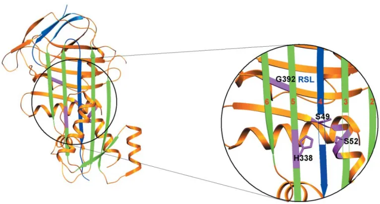
Figure 3. Localization of the precipitation-causing mutations of neuroserpin in the ‘shutter region’. The spatial structure of neuroserpin is shown as a ribbon diagram based on its crystal structure (38). The inhibitory interaction between a serpin and a target protease induces a shutter-like opening between strands 3 and 5 of the superficial b-sheet that allows part of the reactive site loop (RSL, blue) to be inserted as the fourth strand. The b-strands of the superficial b-sheet are presented in green and numbered 2, 3, 5 and 6 in the magnified window. The inserted RSL is drawn in blue and numbered as strand 4. Strands 3 and 4 are made partially trans-parent in order to allow sight on the underlying structures. The accommodation of the RSL requires a sliding movement of the b-strands of the superficial b-sheet on the underlying peptide scaffold. All four disease-causing mutations (colored in pink) affect amino acids located in the ‘shutter region’ and are thought to perturb the function of the shutter. This in turn results in the insertion of the reactive site loop of one neuroserpin molecule into the superficial b-sheet of another, whose reactive site loop inserts into the superficial b-sheet of a next neuroserpin molecule, etc, resulting in polymerization and precipitation of mutated neuroserpin.
Another interesting point is that alterations of the human Reelin gene is associated with an autosomal recessive form of lissencephaly (36). Lissencephaly is a severe developmental disorder in which neuronal migration is impaired, leading to a thickened cerebral cortex whose normally folded contour is simplified and smooth. The Reelin protein is essential for correct cytoarchitectonic organization during CNS develop-ment. Mutations of the Reelin gene in mouse disrupts neuronal migration in several brain regions and gives rise to functional deficits, such as ataxic gait and trembling. Thus, reelin is thought to control cell–cell interactions critical for cell positioning in the brain. Its function in adult brain is far less well understood, but altered brain and blood reelin levels have been reported in some psychiatric disorders. An involvement of the reelin signaling pathway in neurodegeneration has been suggested. Recently, it was reported that reelin has proteolytic activity. By in vitro studies, the capacity of reelin to degrade the adhesion molecules fibronectin and laminin was convincingly demonstrated (37). The effect of reelin on neuronal migration may therefore be mediated by its ability to modulate cell adhesion through its proteolytic activity.
CONCLUSION
Impaired proteolysis appears to be associated with a broad range of defects and the increasing number of studies describing the effects of serine proteases on glial and neuronal function suggests that these proteins may be involved in many others diseases of the nervous system. The association of neurotrypsin defect with isolated mental retardation is of particular interest, as it confirms that synaptic proteolysis plays a key role in cognitive function. Unraveling the molecular bases of other forms of neurological diseases not associated with mutations in neurotrypsin or neuroserpin will possibly involve other components of the extracellular proteolysis signaling pathway. The careful description of these phenotypes, combined with the study of disease causing mutations in animal models will help elucidate the targets of these proteins. Hopefully, identifying these targets will in turn contribute to a better management of these conditions.
REFERENCES
1. Yoshida, S. and Shiosaka, S. (1999) Plasticity-related serine proteases in the brain (review). Int. J. Mol. Med., 3, 405–409.
2. Saksela, O. and Rifkin, D.B. (1988) Cell-associated plasminogen activation: regulation and physiological functions. A. Rev. Cell Biol., 4, 93–126. 3. Gingrich, M.B. and Traynelis, S.F. (2000) Serine proteases and brain
damage—is there a link? Trends Neurosci., 23, 399–407. 4. Matsumoto-Miyai, K., Ninomiya, A., Yamasaki. H., Tamura, K.,
Nakamura, Y., and Shiosaka, S. (2003) NMDA-dependent proteolysis of presynaptic adhesion molecule L1 in the hippocampus by neuropsin. J. Neurosci. (in press).
5. Gettins, P.G. (2002) Serpin structure, mechanism, and function. Chem. Rev., 102, 4751–4804.
6. Janciauskiene, S. (2001) Conformational properties of serine proteinase inhibitors (serpins) confer multiple pathophysiological roles. Biochim. Biophys. Acta, 1535, 221–235.
7. Ye, S. and Goldsmith, E.J. (2001) Serpins and other covalent protease inhibitors. Curr. Opin. Struct. Biol., 11, 740–745.
8. Silverman, G.A., Bird, P.I., Carrell, R.W., Church, F.C., Coughlin, P.B., Gettins, P.G., Irving, J.A., Lomas, D.A., Luke, C.J., Moyer, R.W. et al. (2001) The serpins are an expanding superfamily of structurally similar but functionally diverse proteins. Evolution, mechanism of inhibition, novel functions, and a revised nomenclature. J. Biol. Chem., 276, 33293–33296. 9. Gloor, S., Odink, K., Guenther, J., Nick, H. and Monard, D. (1986)
A glia-derived neurite promoting factor with protease inhibitory activity belongs to the protease nexins. Cell, 47, 687–693.
10. Reinhard, E., Meier, R., Halfter, W., Rovelli, G. and Monard, D. (1988) Detection of glia-derived nexin in the olfactory system of the rat. Neuron, 1, 387–394.
11. Mansuy, I.M., van der Putten, H., Schmid, P., Meins, M., Botteri, F.M. and Monard, D. (1993) Variable and multiple expression of protease nexin-1 during mouse organogenesis and nervous system development. Development, 119, 1119–1134.
12. Luthi, A., Van der Putten, H., Botteri, F.M., Mansuy, I.M., Meins, M., Frey, U., Sansig, G., Portet, C., Schmutz, M., Schroder, M. et al. (1997) Endogenous serine protease inhibitor modulates epileptic activity and hippocampal long-term potentiation. J. Neurosci., 17, 4688–4699. 13. Meins, M., Piosik, P., Schaeren-Wiemers, N., Franzoni, S., Troncoso, E.,
Kiss, J.Z., Brosamle, C., Schwab, M.E., Molnar, Z. and Monard, D. (2001) Progressive neuronal and motor dysfunction in mice overexpressing the serine protease inhibitor protease nexin-1 in postmitotic neurons. J. Neurosci., 21, 8830–8841.
14. Osterwalder, T., Contartese, J., Stoeckli, E.T., Kuhn, T.B. and
Sonderegger, P. (1996) Neuroserpin, an axonally secreted serine protease inhibitor. EMBO J., 15, 2944–2953.
15. Hastings, G.A., Coleman, T.A., Haudenschild, C.C., Stefansson, S., Smith, E.P., Barthlow, R., Cherry, S., Sandkvist, M. and Lawrence, D.A. (1997) Neuroserpin, a brain-associated inhibitor of tissue plasminogen activator is localized primarily in neurons. Implications for the regulation of motor learning and neuronal survival. J. Biol. Chem., 272, 33062–33067. 16. Osterwalder, T., Cinelli, P., Baici, A., Pennella, A., Krueger, S.R.,
Schrimpf, S.P., Meins, M. and Sonderegger, P. (1998) The axonally secreted serine proteinase inhibitor, neuroserpin, inhibits plasminogen activators and plasmin but not thrombin. J. Biol. Chem., 273, 2312–2321. 17. Barker-Carlson, K., Lawrence, D.A. and Schwartz, B.S. (2002)
Acyl-enzyme complexes between tissue-type plasminogen activator and neuroserpin are short-lived in vitro. J. Biol. Chem., 277, 46852–46857. 18. Salles, F.J. and Strickland, S. (2002) Localization and regulation of
the tissue plasminogen activator-plasmin system in the hippocampus. J. Neurosci., 22, 2125–2134.
19. Roeleveld, N., Zielhuis, G.A. and Gabreels, F. (1997) The prevalence of mental retardation: a critical review of recent literature. Dev. Med. Child Neurol., 39, 125–132.
20. Organization of services for the mentally retarded. (1968) Fifteenth Report of the WHO Expert Committee on Mental Health. WHO Technical Report Series, no. 392. WHO, Geneva, pp. 5–55.
21. Lamont, M.A. and Dennis, N.R. (1988) Aetiology of mild mental retardation. Arch. Dis. Child., 63, 1032–1038.
22. Turner, G. (1975) An aetiological study of 1,000 patients with an I.Q. assessment below 51. Med. J. Aust., 2, 927–931.
23. Molinari, F., Rio, M., Meskenaite, V., Encha-Razavi, F., Auge, J., Bacq, D., Briault, S., Vekemans, M., Munnich, A., Attie-Bitach, T. et al. (2002) Truncating neurotrypsin mutation in autosomal recessive nonsyndromic mental retardation. Science, 298, 1779–1781.
24. Wolfer, D.P., Lang, R., Cinelli, P., Madani, R. and Sonderegger, P. (2001) Multiple roles of neurotrypsin in tissue morphogenesis and nervous system development suggested by the mRNA expression pattern. Mol. Cell. Neurosci., 18, 407–433.
25. Proba, K., Gschwend, T.P. and Sonderegger, P. (1998) Cloning and sequencing of the cDNA encoding human neurotrypsin. Biochim. Biophys. Acta, 1396, 143–147. 26. Davis, R.L., Shrimpton, A.E., Holohan, P.D., Bradshaw, C., Feiglin, D.,
Collins, G.H., Sonderegger, P., Kinter, J., Becker, L.M., Lacbawan, F. et al. (1999) Familial dementia caused by polymerization of mutant neuroserpin. Nature, 401, 376–379.
27. Davis, R.L., Shrimpton, A.E., Carrell, R.W., Lomas, D.A., Gerhard, L., Baumann, B., Lawrence, D.A., Yepes, M., Kim, T.S., Ghetti, B. et al. (2002) Association between conformational mutations in neuroserpin and onset and severity of dementia. Lancet, 359, 2242–2247.
28. Stoeckli, E.T., Lemkin, P.F., Kuhn, T.B., Ruegg, M.A., Heller, M. and Sonderegger, P. (1989) Identification of proteins secreted from axons of embryonic dorsal-root-ganglia neurons. Eur. J. Biochem., 180, 249–258.
29. Berger, P., Kozlov, S.V., Cinelli, P., Kruger, S.R., Vogt, L. and
Sonderegger, P. (1999) Neuronal depolarization enhances the transcription of the neuronal serine protease inhibitor neuroserpin. Mol. Cell. Neurosci., 14, 455–467.
30. Madani, R., Kozlov, S., Akhmedov, A., Cinelli, P., Kinter, J., Lipp, H.-P., Sonderegger, P., and Wolfer, D.P. (2003). Impaired explorative behaviour and neophobia in genetically modified mice lacking or overexpressing the extracellular serine protease inhibitor neuroserpin. Mol. Cell. Neurosci., 23, 473–494.
31. Belorgey, D., Crowther, D.C., Mahadeva, R. and Lomas, D.A. (2002) Mutant Neuroserpin (S49P) that causes familial encephalopathy with neuroserpin inclusion bodies is a poor proteinase inhibitor and readily forms polymers in vitro. J. Biol. Chem., 277, 17367–17373.
32. Huang, Y.Y., Bach, M.E., Lipp, H.P., Zhuo, M., Wolfer, D.P., Hawkins, R.D., Schoonjans, L., Kandel, E.R., Godfraind, J.M., Mulligan, R. et al. (1996) Mice lacking the gene encoding tissue-type plasminogen activator show a selective interference with late-phase long-term potentiation in both Schaffer collateral and mossy fiber pathways. Proc. Natl Acad. Sci. USA, 93, 8699–8704.
33. Madani, R., Hulo, S., Toni, N., Madani, H., Steimer, T., Muller, D. and Vassalli, J.D. (1999) Enhanced hippocampal long-term potentiation and learning by increased neuronal expression of tissue-type plasminogen activator in transgenic mice. EMBO J., 18, 3007–3012.
34. Davies, B., Kearns, I.R., Ure, J., Davies, C.H. and Lathe, R. (2001) Loss of hippocampal serine protease BSP1/neuropsin predisposes to global seizure activity. J. Neurosci., 21, 6993–7000.
35. Weller, S. and Ga¨rtner, J. (2001) Genetic and clinical aspects of X-linked hydrocephalus (L1 disease): mutations in the L1CAM gene. Hum. Mut., 18, 1–12. 36. Hong, S.E., Shugart, Y.Y., Huang, D.T., Shahwan, S.A., Grant, P.E.,
Hourihane, J.O., Martin, N.D. and Walsh, C.A. (2000) Autosomal recessive lissencephaly with cerebellar hypoplasia is associated with human RELN mutations. Nat. Genet., 26, 93–96.
37. Quattrocchi, C.C., Wannenes, F., Persico, A.M., Ciafre, S.A., D’Arcangelo, G., Farace, M.G. and Keller, F. (2002) Reelin is a serine protease of the extracellular matrix. J. Biol. Chem., 277, 303–309. 38. Briand, C., Kozlov, S.V., Sonderegger, P. and Grutter, M.G. (2001) Crystal
structure of neuroserpin: a neuronal serpin involved in a conformational disease. FEBS Lett., 505, 18–22.